vitreoretinal interface routinely. - MIMH...
29
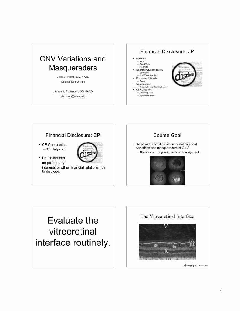
1 CNV Variations and Masqueraders Carlo J. Pelino, OD, FAAO [email protected] Joseph J. Pizzimenti, OD, FAAO [email protected] Financial Disclosure: JP • Honoraria – Alcon – Notal Vision – Reichert • Scientific Advisory Boards – Zeavision – Carl Zeiss Meditec • Proprietary Interests – None • CEO/Founder – Optometryboardcertified.com • CE Companies – CEinItaly.com – EyeSkiUtah.com Financial Disclosure: CP • CE Companies – CEinItaly.com • Dr. Pelino has no proprietary interests or other financial relationships to disclose. Course Goal • To provide useful clinical information about variations and masqueraders of CNV. – Classification, diagnosis, treatment/management Evaluate the vitreoretinal interface routinely. The Vitreoretinal Interface retinalphysician.com
Transcript of vitreoretinal interface routinely. - MIMH...
1
CNV Variations andMasqueraders
Carlo J. Pelino, OD, FAAO
Joseph J. Pizzimenti, OD, FAAO
Financial Disclosure: JP• Honoraria
– Alcon– Notal Vision– Reichert
• Scientific Advisory Boards– Zeavision– Carl Zeiss Meditec
• Proprietary Interests– None
• CEO/Founder– Optometryboardcertified.com
• CE Companies– CEinItaly.com– EyeSkiUtah.com
Financial Disclosure: CP
• CE Companies– CEinItaly.com
• Dr. Pelino hasno proprietaryinterests or other financial relationshipsto disclose.
Course Goal• To provide useful clinical information about
variations and masqueraders of CNV.– Classification, diagnosis, treatment/management
Evaluate thevitreoretinal
interface routinely.
The Vitreoretinal Interface
retinalphysician.com

2
PersistentVitreomacular
Adhesions (VMA)
Anomalous PVD
Anomalous PVD• May hasten the Wet
AMD process. ---->
The Retina
• RPE• Neurosensory• 6 million Cones
• Detailed vision• Color vision
• 120 million Rods• Peripheral retinal
receptors• Great sensitivity to
light
OSIS/OSELM RPEIS
NFL: Nerve Fiber Layer OPL: Outer Plexiform Layer IS/OS: Junction of inner and outerILM: Inner Limiting Membrane ONL: Outer Nuclear Layer photoreceptor segmentsGCL: Ganglion Cell Layer ELM: External limiting membrane OS: Photoreceptor Outer SegmentIPL: Inner Plexiform Layer IS: Photoreceptor Inner Segment RPE: Retinal Pigment EpitheliumINL: Inner Nuclear Layer
ILM GCLNFL
Choroid
IPL INL OPL ONL
SD-OCT Healthy Macula
Jetrea (ocriplasmin)

3
Retina
• The Pigment Epithelium– Monolayer– Cuboidal cells– Function of RPE– Tight junctions form outer blood-retina
barrier
RPE
Retinal Pigment Epithelium
• 120 million cells inmonolayer
• Functions of RPE– Phagocytosis of
renewable discs ofPRs
– O-2 diffusion to PRs– Provision of nutrients
to PRs
Early AMD: Accumulation of Lipofuscinand Vitamin A Metabolites
Reduced degradation of cellular debrisleads to the accumulation of lipofuscin,toxic vitamin A metabolites
Drusen Retina
• The Neurosensory Retina– The Photoreceptors
• Structure and function of cones and rods
– Inner and outer segment junction• Importance of structural integrity to visual function
– Outer limiting membrane– Outer nuclei– Synaptic layer (plexiform)

4
Photoreceptors Neuro-sensory Retina
• Inner nuclei• Synaptic layer
(plexiform)• Ganglion cells• Nerve fiber layer• Internal limiting
membrane
Retinal Vasculature• 2 main sources of blood
supply:• Choroidal BV
– Supplies outer retinallayers, including PRs
• CRA– 4 branches nourish inner
retina– Run radially toward fovea
Retinal Capillaries• Pericytes surround
each endothelial cell– provide support
• Tight junctions betweenendothelial cells
• Pericytes + tightjunctions form innerblood-retinal barrier.
Pericytes marked by ng2 staining(blue) and endothelial cells aremarked by PECAM (red).
Retina
• Photransduction– conversion of light into an electrical
impulse• The retina is damaged by it’s own
operation.• Autoregulation of blood flow

5
Functional Anatomy: The FoveaThe Choroid
• Vascular layers• Melanocytes• Bruch’s membrane• Sympathetic regulation of blood flow• Function of choriocapillaris
– Supply of nutrients– Absorption of light
Diagnostic Dilemma
ChoroidalNeovascularization
Angiogenesis
Environmentalfactors1
(hypoxia,2 pH)Growth factors,
hormones1
(EGF, bFGF,PDGF,
IGF-1, IL-1!, IL-6, estrogen)
VEGF-A bindingand activation ofVEGF receptor3
Endothelial cellactivation3
VEGF-A = vascular endothelial growth factor A; EGF = epidermal growth factor; bFGF = basic fibroblast growth factor; PDGF = platelet-derived growthfactor; lGF = insulin-like growth factor; IL= interleukin.1. Dvorak HF. J Clin Oncol. 2002;20:4368. 2. Aiello LP, et al. Arch Ophthalmol. 1995;113:1538.3. Ferrara N, et al. Nat Med. 2003;9:669. 4. Griffioen AW and Molema G. Pharmacol Rev. 2000;52:237.
CNV ---> FV Scar

6
CNV has several variations,causes, and masqueraders.
Choroidal Neovascularization
• Subjective symptoms• Objective data• Diagnostic Workup• Making the diagnosis
Common Causes of CNV
• Exudative AMD• Ocular Histoplasmosis• High Myopia• Angioid Streaks
Fundus Autofluorescence
Fundus Autofluorescence Fundus AutofluorescenceWet AMD

7
Fluorescein Angiography Indocyanine Green Angiography (ICGA)• Uses digital imaging systems• Dye properties• “Sees” through blood• Delineates choroidal circulation better than
fluorescein angiography• Boundaries of occult membranes imaged
Occult CNV Classic CNV
Type I CNVM
Retina
RPE
Bruch’s
CC
CNV beneath RPE (AMD) *
Fovea
Retina
RPE
Bruch’s
Type II CNVM
CNV in subsensory space(POHS)

8
ANGIOID STREAKS
• Note Angioid Streaks radiating from the optic discs and macularlaser scarring
Differential Dx. of AngioidStreaks: PEPSI Causes of CNV
• High Myopia in a 52y/o WM
• CNV w/heme

9
48 y/o WM-12.00D
Concave fundus, CNV, schisis
Causes of CNV
• OHS
Case
! 81 Year old Female with a history ofarthritis.
! 7 year history of injections withAvastin or Lucentis
! PMH: AMD OU, Cataracts OU! OcHx: Injections for AMD.
Ophthalmic Exam
! VA:- OD: 20/400 OS: 20/80
! IOP- OD: 11 OS: 12
! SLE:-OD: NS +1 OS: NS + 1
- DFE:-PED OD and Geo Atrophy OS
OCT After Switching to Eyelea

10
Comparison!"#$%&'"()"*+%',-"%-./01$-,'
234533
2"6-7$1,(-'"()"89$:$%
234;3
Another PED example
Variations and Masqueraders of CNV
! Polypoidal Choroidal Vasculopathy (PCV)! Retinal Angiomatous Proliferation (RAP)! Masqueraders of CNV
• Choroidal Neoplastic Disease• Primary Tumors of the Choroid
! Nevus vs. melanoma
• Metastatic Tumors to the Choroid! Common primary sites! Breast! Lung
• Central Serous Chorioretinopathy (CSC)
CNV Variants
R
C
ICG Angiography AMD vs PCV
AMD -SUBRETINAAL HEMORRHAGE
SUSPICIOUS POLYPOIDAL

11
Retinal Angiomatous Proliferation
! Sub retinal neovascularization ensues.
Retinal Angiomatous Proliferation
! First described by Yannuzzi in 1991,RAP is a retino-choridal anastamosis.
! Intraretinal capillary proliferation,which extends throughout thesensory retina and then into the subretinal space.
Retinal Angiomatous Proliferation
! 10-20 % of neovascular AMDpatients start with RAP.
! The age group is thought to beslightly older.
! ICGA aids in confirming diagnosis,identifying “hot spots” of ICG dyepools in the sub retinal space.
Retinal Angiomatous Proliferation“Hot Spot”
Retinal Angiomatous Proliferation 82 y/o WM w/drusen

12
RAP Stage I: intraretinalneovascularization.
RAP Stage II: subretinal NVw/retinal-retinal anastomosis.
RAP Stage III: subretinal NVw/vascularized RPED andretina-choroid anastomosis.

13
RAP: Current Treatment Options
! Thermal Laser
! Photodynamic Therapy
! Anti VEGF Therapy
CNV Masquerador:
Neoplastic Disease
CNV or Mass?
CNV Masquerador:
Central SerousChorioretinopathy
Mystery Macula! Subjective
• 35 y/o WM• sudden, unilateral blur OD• no pain or trauma• “Type A”
! Objective• VA
! OD 20/60! OS 20/20
• Hyperopic shift

14
Describe That Fundus!
! DFE shows large, serous elevation! Focal detachment of sensory retina
What other tests would you liketo perform?
OCT

15
(Idiopathic) Central SerousChorioretinopathy
(ICSC)
Patient Outcome
• VA recovered to 20/25at week 12
• Reduction of fluid,20/40 VA at week 5
Central Serous Chorioretinopathy! 36 y/o WM! CC: Sudden
central blur OS! VA OD 20/20! VA OS 20/200
ICSC
! Objective• Breakdown of outer blood-retina barrier• FA shows classic “smoke-stack”
! Pooling beneath RPE detachment! Dye ascends vertically, then laterally in SRS
! Differential Diagnosis• Tumor• RPE detachment/CNVM• Steroid-induced CSC

16
ICSC Plan
! Observation• 60% regain 20/20 w/no intervention• monitor q4wks for 6 mon
! Focal Laser• Unresolved after 4-6 mon• Recurrent
•Focal, direct treatment
•Leak must be outside FAZ (500 um)
Treatments for CSC! Thermal laser
! Photodynamic Therapy• Visudyne (Verteporfin)• A light-activated drug
Photodynamic Therapy for CSC
! Serousdetachmentbefore PDT.
Resolution ofdetachment with residualRPE mottling after PDT.
What’s new in CSC Treatment?
! Intravitrealbevacizumab(Avastin) hasshown somebenefit in smallcase series.
Low-fluence PDT
ICGA-guided, lower flow, lighter dosage resultedin less hypoperfusion of the choriocapillaris

17
Current and FutureTreatment of CNV
Laser
! Now reserved for extrafoveal disease
! Ectopic disciform
! Poylpoidal demarcation
Limitations of Laser
PDT
! Poylpoidal choroidal vasculopathy
! Used in conjunction with anti VEGF
! Cental Serous Chorioretinopathy
Photodynamic (Visudyne) Therapy: A2-Step Process
Step 1
Step 2
10 Min Infusion
83 Sec ActivationA treatment odyssey

18
Vascular endothelial growth factor(VEGF)
! VEGF was found to be essential in normal and pathologicalangiogenesis.
! Hypoxia (ischemia) and inflammation induce secretion ofVEGF.
! VEGF binds to its receptors, promoting endothelial cellmigration and proliferation, which are required to developnew vessels.
! VEGF breaks down the blood retina barrier whichincreases vascular permeability (edema).
! Maximum expression of VEGF at border of vascular andavascular tissue.
Antiangiogenic Drugs: VEGFInhibitors
VEGF binds to receptor
Anti VEGF
! The mainstay of CNV treatment atthis point
! Requires intravitreal injection
! Post operative care
Pathogenesis of CNVM! Breaks in Bruch’s
Theory• Diffuse thickening
of Bruch’s w/softdrusen
• Predisposes Bruch’sto breaks
• New BV’s from CCgrow andproliferate
Wet AMD Pathology

19
Anti VEGF Treatment! Concept of antiangiogenesis was first proposed
by Judah Folkman as a cancer treatment! The concept has been extended to ocular
proliferative retinopathies! Three anti-VEGF treatments currently being used
(Avastin, Lucentis, Eyelea)
VEGF-A
! VEGF-A has 3 isoforms which differin their solubility and receptorbinding properties
! VEGF 121! VEGF 188! VEGF 165
VEGF Inhibition for Wet AMD VEGF Inhibitors! Pegaptanib sodium-Macugen (Pfizer/Eyetech)
• FDA Approved• Aptamer (decoy): inhibits protein activity
! Ranibizumab- Lucentis (Genentech) $2,000.00• FDA Approved• Antibody-based• Compared favorably to PDT in ANCHOR study
! Bevacizumab- Avastin (Genentech) $40.00• Off label• Anti-neoplastic• Intravitreal injection• 1 injection/mon x 3 mon
Pegaptanib
! Macugen (Pfizer)! RNA aptamer (decoy) that binds the
isoform VEGF 165! Received FDA approval in December
2004 for treatment of neovascularmacular degeneration
! First ocular VEGF therapy• Seldom used today
Ranibizumab! Lucentis (Genetech, Inc.)! Recombinant humanized monoclonal antibody
fragment! Low molecular weight for better retinal
penetration! Binds and inactivates all 3 isoforms of VEGF! FDA approved in June 2006 for treatment of
neovascular macular degeneration

20
Bevacizumab! Avastin (Genetech, Inc.)! Full length humanized monoclonal antibody! Binds all 3 isoforms of VEGF and inhibits its
interaction with receptors on endothelial cells! FDA approved for IV treatment of metastatic
colorectal cancer in 2004! Used off label for ocular neovascularization! Less expensive than Lucentis! Longer half life than Lucentis due to larger
molecule weight
VA 55 L
VA 78 L
Pre and Post Avastin Treatment
Treatment Studies! VISION => VEGF Inhibition Study in Ocular
Neovascularization! MARINA => Minimally Classic/Occult Trial of he Anti VEGF
Antibody Ranibizumab in the Treatment of NeovascularAMD
! ANCHOR => Anti VEGF Antibody for the Treatment ofPredominantly Classic Choroidal Neovascularization in AMDStudy
! PrONTO; CATT (ongoing), SAILOR, and more
Intravitreal Injections for Wet AMDAnti-VEGF Agents
Antiangiogenic therapy! Pegaptanib (Macugen)
• Dec 2004, for neovascular (wet) AMD
! Bevacizumab (Avastin)• For metastatic colorectal cancer
! Ranibizumab (Lucentis)• June 2006, for neovascular (wet) AMD
AMD = age-related macular degeneration; VEGF = vascular endothelial growth factor.
! Eylea (Regeneron)• Aflibercept intravitreal injection• Approved for CNV/AMD.• Binds all forms of Vascular
Endothelial Growth Factor-A (VEGF-A)and Placental Growth Factor (PlGF).
• Binds tightly to VEGF receptors• Rapid decrease in foveal thickening,
improved visual function.

21
Wet AMD Treatmentson the Horizon
VEGF Inhibitors! Squalamine lactate- Envizon (Genaera): Phase
II• Isloated from dogfish shark tissue• Originally developed for oncology• Aminosterol
! Inhibits plasma membrane ion channels! Blocks proliferation of endothelial cells
• Administered Intravenously! Weekly x 4 wks
• Small sample showed improved or stabilized VA• Low systemic toxicity• Topical drug delivery??
Squalamine lactate- Envizon(Genaera)
Squalamine works INSIDE endothelial cells to blockmultiple intracellular pathways generated by the bindingof VEGF and PDGF! to receptors.
Fovista (Ophthotech)! Anti-PDGF
• Platelet derived GF
! To be used with Anti-VEGF
! Decreases size of CNVwhen used w/Lucentis
! Better efficacy thanLucentis alone
! No adverse events at6 mon
! Phase 3 under way
Conversion to Exudative AMD
Can we prevent this?

22
Dry AMD Treatmentson the Horizon
Future Dry AMD Treatments?
! Neuroprotection
! Prevention of oxidativestress/damage
! Inflammation inhibition
Future Dry AMD Treatments?
! Ciliary neurotrophicfactor (CNTF)delivered viaintraocularencapsulated celltechnology implant.
! CNTF retards PR loss.! Right, an 84 y/o
subject with GA.
Future Dry AMD Treatments?
! Neuroprotection
! Prevention of oxidativestress/damage
! Inflammation inhibition
OT-551 (Othera)
! Topical Anti-• -oxidant• -inflammatory• -angiogenic
! Tested incombo w/L & Z
Future Dry AMD Treatments?! Fenretinide po
• Synthetic Vitamin A derivative
• Reduces amount of lipofuscinaccumulation

23
Future AMD Treatments! Visual Cycle Modulation:
! ACU - 4429 (Acucela and Otsuka) - canmodulate, slow down, the visual cycle. Oral drugtargets the rod system by inhibiting a keyenzyme. Does not affect the cones.
! Reduces the amount of A2E and lipofuscinaccumulating in the RPE.
! Fenretinide aka RT-101 (Sirion Therapeutics) -reduces the amount of A2E and lipofuscin bymodifying the visual cycle. Oral administration.
Future Dry AMD Treatments?
! Neuroprotection
! Prevention of oxidativestress/damage
! Inflammation inhibition
Future Dry AMD Treatments?
! Complement inhibition• Intravitreal (ARC 1905)• Sub-conj (Soliris)
! Toxic RNA (Alu RNA) inhibition! Increase “Dicer” enzyme
Future AMD Treatments! Complement Inhibition:! POT-4 (Potentia Pharmaceuticals)! Inhibits complement component C3. Single
intravitreal injection
! Neuroprotection:! NT-501 intraocular implant (Neurotech)! Delivers human cells that have been genetically
modified to secrete ciliary neurotrohic factor(CNTF).• CNTF is a neuroprotectant cytokine under investigation
for neurodegenerative diseases like ALS.
A Nutritional Approach AMD Risk Factors! Age
• Gender - F > M
! AMD Family History
! Smoking
! Iris Color - lighter iris
! Obesity
! CV Disease
! Poor nutrition
! Dietary and Serum Levels- Complex analyses (most,but not all) show arelationship.
! Low Macular Pigment
! MPOD- Most (but not all)studies have shown reducedMPOD in AMD (by multiplemeasurement techniques).

24
Macular Pigment OpticalDensity (MPOD)
HFP
Risk assessment, early detectionand monitoring of AMD
! Macular Pigment Optical Density• MPOD
Xanthophylls andAMD
! Lutein and zeaxanthinform the macularpigment
! Dietary sourcesinclude green leafyvegetables andorange-yellow fruits
! Act as antioxidants orlight screeningcompounds
Macular Pigment Optical Density (du)
Low Average High
0.1- 0.25 0.25- 0.45 > 0.45
Lutein Zeaxanthin

25
Dietary Lutein and Zeaxanthin:Eggs AREDS 1 and 2
Daily Dosage in AREDS 1
Supplement DosageAntioxidants
Beta-carotene 15 mgVitamin C 500 mgVitamin E 400 IU
Essential Trace ElementsCopper 2 mg
Zinc 80 mg
Supplements were manufactured to have the following minimum contents: AREDS 1
AREDS Grading Scale
1. No drusen or a few small drusen.2. Pigment abnormalities or non-extensive small or
intermediate drusen.3. Extensive intermediate drusen or any large drusen
or non-central atrophy.4. Good acuity and no advanced AMD in the study
eye. Advanced AMD in the fellow eye (choroidalneovacularization or geographic atrophy).
"AREDS 1 resulted in a formulation ofvitamin C, beta carotene, zinc, andvitamin E that reduced the risk of
progression of advanced disease by25%" at 5 years.”
Emily Chew, MD, from the National EyeInstitute in Bethesda, Maryland,

26
AREDS 2: NEI Trial Overview
Feature DescriptionObjective To evaluate the effect of high-dose vitamin
supplementation on age-related macular degeneration
(AMD) progression and visual acuity.
Design Double-masked, randomized, placebo-controlled trial
Population 3640 high risk patients (55-80 years)
Duration 6.3 years supplementation and follow up
A AREDS report no. 8. Arch Ophthalmol 2001.119(10): 1417-36.
AREDS 2
"Lutein (10mg)
"Zeaxanthin (2mg)
"Omega-3 fatty acids (350 mg DHA, 650 mg EPA)
"With and without !-carotene (15 mg vs 0 mg)
"High vs low zinc levels (80mg vs 25mg)
" Antioxidant activity• Prevent free radical damage in the retina• More effective than Beta-carotene
" Filter blue light• Most damaging type of light due to it short
wavelength
" Selectively binds to tubulin• Improves structure integrity• Maintains eye health and quality of vision
Lutein/Zeaxanthin AREDS 2
Multi-center, multi-factorial,randomized, control-group trial.
AREDS 2
# Lutein/Zeaxanthin and Omega-3 Fatty Acids forAge-Related Macular Degeneration. The Age-Related Eye Disease Study 2 (AREDS2) ControlledRandomized Clinical Trial. AREDS2 ResearchGroup. JAMA, May 5, 2013 Online.
# Lutein/Zeaxanthin for the Treatment of Age-Related Cataract. AREDS2 Research Group. JAMAOphthalmology, May 5, 2013 Online.
4203 participants aged 50 to 85with bilateral large drusen
orlarge drusen in 1 eye and
advanced AMD in the fellow eye.
2006-2012AREDS 2

27
AREDS 2: Purpose
#To determine if adding lutein/zeaxanthin,omega-3s, or a combination could improveupon the positive results found in theAREDS 1.
#To evaluate the effect of eliminating betacarotene, lowering zinc, or both.
AREDS 2 Design
#4203 participants were randomized to placebowith no additional supplementation or to 1 of 3treatment groups:#Group 1 tablet w/10 mg L + 2 mg Z#Group 2 gel cap w/350 mg DHA + 650 mg EPA#Group 3 both the tablet and gel cap
#On a daily basis
AREDS 2: Primary Study Outcome
#An additional 25% decrease in the risk ofprogression to advanced AMD in the threetreatment groups over the study subjectstaking the original AREDS1 supplement.
Study Subjects:AREDS 1 vs AREDS 2
# All stages of AMD# Average age = 69# 67% took Centrum
(no L)# Varied diets# Varied serum L and Z
# More advanced stage# average age = 74# 89% taking Centrum
Silver (w/minimal L)# diet high in carotenoids
and vegetables# higher serum L and Z
These differences could impact the abilityto detect a more significant reduction inprogression!
AREDS 2 First Results
"In the overall analysis, using 3treatment groups, we found nosignificant difference in rates of
macular degeneration," Dr. Chew said.

28
AREDS 2 Sub-group Analysis
# 10% reduction in progression to advanced AMD w/L &Z compared to no L&Z
# 18% reduction in progression in subjects whoreceived L&Z + AREDS 1 supplement (without betacarotene) compared to those who took the originalAREDS 1 supplement with beta carotene
# 26% reduction in progression in the participantstaking L&Z that were in the lowest quintile ofdietary L&Z intake
AREDS 2 Conclusions:First, the Bad News
#Overall, the addition of 10 mg L and 2 mgZ, 1g DHA + EPA, or both to the AREDSformulation did not further reduce risk ofprogression to advanced AMD.
AREDS 2 Conclusions
#Results reaffirm previous epidemiological datathat high dietary intakes of L&Z reduce the riskof AMD.
#Results support the safety and treatmentbenefits of substituting 10 mg L and 2 mg Z forbeta carotene in AREDS formulations.
What about omega-3 EFAs?
# Fish oil supplement didnot significantly alter theprogression of AMD inAREDS 2.
AREDS 2 Limitations
#A greater reduction in AMD progression may havebeen demonstrated if the subject’s diet hadbeen more representative to that of the generalUS population.
#Inability to determine if the null findings areattributable to lack of efficacy of thesupplements, inadequate dosing, inadequate Tx.duration, or a combination of these.
Conclusions! Choroidal Neovascularization (CNV) is a
leading cause of vision impairmentworldwide.
! An understanding of the functionalanatomy of the posterior segment isessential in understanding CNV.
! CNV has several causes, variants, andmasqueraders.
! Early diagnosis of CNV enables earlytreatment with today’s effective therapies,thereby preserving visual function.