Vitamins D and A can be successfully measured by LC–MS/MS ... D and A can be... · Vitamins D and...
8
Vitamins D and A can be successfully measured by LC–MS/MS in cord blood diluted plasma Ali A. Albarhani a,b , Fiona Collier c , Ronda F. Greaves a,d , Anne-Louise Ponsonby d,e , Katrina J. Allen d,e , Peter J. Vuillermin c,f , Peter Roche a , Michael W. Clarke g, ⁎, BIS Steering Committee a School of Medical Sciences, RMIT University, Victoria, Australia b Security Forces Hospital, Dammam, Saudi Arabia c Deakin University, Victoria, Australia d Murdoch Children's Research Institute, Melbourne, Australia e Department of Paediatrics, University of Melbourne, Victoria, Australia f Child Health Research Unit, Barwon Health, Victoria Australia g University of Western Australia, Western Australia, Australia abstract article info Article history: Received 16 January 2015 Received in revised form 4 April 2015 Accepted 17 April 2015 Available online 25 April 2015 Keywords: Cholecalciferol Retinol Neonates Mass spectrometry Umbilical cord blood diluted plasma Objectives: In widely used protocols for the collection and isolation of cord blood mononuclear cells, inves- tigators are left with substantial volumes of diluted plasma which could be used for other measurements. The aim of this study was to ascertain the validity of umbilical cord blood (UCB) diluted plasma samples for vitamin D, A and E analysis compared to UCB serum samples. Design & methods: Twenty UCB matched samples of diluted plasma and serum were collected. The samples were analysed by two liquid chromatography–tandem mass spectrometry (LC–MS/MS) methods on two separate occasions. Results: The results of 25(OH)D3 obtained by the two laboratories demonstrated close agreement with a mean difference of 0.14 nmol/L [95% confidence interval (95% CI), -6.8 to 7.1]. Both methods demonstrate close agree- ment for 25(OH)D3 in UCB serum versus diluted UCB plasma; mean difference 2.2 nmol/L [95% CI, -9.5 to 13.9] and 4.1 nmol/L [95% CI, -14.5 to 6.1] for the results from Lab A and Lab B, respectively. Vitamin A was quantified by Lab A in UCB serum and diluted UCB plasma; mean difference 0.07 μmol/L [95% CI, -0.41 to 0.28]. Results of 25(OH)D3 epimer and vitamin E in the diluted UCB plasma were below the limit of quantification, and could not be compared with UCB serum. Conclusions: Diluted UCB plasma can be used for the quantification of retinol and 25(OH)D3 by LC–MS/MS. By contrast, quantification of 25(OH)D3 epimer and vitamin E in diluted UCB plasma is not supported by this study due to limitations in analytical sensitivity. © 2015 The Canadian Society of Clinical Chemists. Published by Elsevier Inc. All rights reserved. 1. Introduction Fat soluble vitamin deficiency is classically associated with com- plications of diseases presenting in neonates [1]. Of the four vitamins in this group, vitamins A, D and also K have pleiotropic actions whilst vitamin E has important anti-oxidant activity. Of these, vitamin D has received a lot of attention recently as a result of the meteoric rise in the number of publications showing that this secosteroid plays a crucial role in a plethora of physiological functions and is associated with many acute and chronic illnesses. In particular, there is mounting interest in the potential importance of vitamin D status, and to a lesser extent vita- min A, during early life for a wide range of health outcomes [2]. Liquid chromatography coupled with tandem mass spectrometry (LC–MS/MS) quantification of each of these fat soluble vitamins, includ- ing separation of epi-25(OH)D3, is now established [3–5]. Serum, and also undiluted plasma, are the validated matrixes for analysis of vitamins A (retinol), D (25(OH)D3) and E (α-tocopherol). However the diluted plasma matrix, which is widely used in protocols for the collection and isolation of viable mononuclear cells, has not been validated for use in the LC–MS/MS analysis of small molecules. Given the limited volumes of blood available in birth cohort studies, and the implicit value of these in the context of a research intensive large-scale epidemiological projects, it is of interest to determine whether vitamins D, A and E may be ade- quately measured in diluted plasma from umbilical cord blood (UCB). Clinical Biochemistry 48 (2015) 1105–1112 Abbreviations: 25(OH)D3, 25-hydroxy vitamin D3; BIS, Barwon Infant Study; LoQ, limit of quantification; LC–MS/MS, liquid chromatography coupled with mass selective detec- tion; MNC, mononuclear cell; MRM, multiple reaction monitoring; PFP, pentafluorophenyl; RCPAQAP, Royal College of Pathologists of Australasia Quality Assurance Programs; TM, transport medium; 25(OH)D3-d3, tri-deuterated 25-hydroxy vitamin D3; UCB, umbilical cord blood. ⁎ Corresponding author at: Room 3.42, Level 3, Bayliss Building, Plant Energy Biology, ARC Centre of Excellence, School of Chemistry and Biochemistry, The University of Western Australia, 35 Stirling Highway, Crawley, Perth, Western Australia, 6009, MBDP: M316, Australia. E-mail address: [email protected] (M.W. Clarke). http://dx.doi.org/10.1016/j.clinbiochem.2015.04.014 0009-9120/© 2015 The Canadian Society of Clinical Chemists. Published by Elsevier Inc. All rights reserved. Contents lists available at ScienceDirect Clinical Biochemistry journal homepage: www.elsevier.com/locate/clinbiochem
Transcript of Vitamins D and A can be successfully measured by LC–MS/MS ... D and A can be... · Vitamins D and...

Clinical Biochemistry 48 (2015) 1105–1112
Contents lists available at ScienceDirect
Clinical Biochemistry
j ourna l homepage: www.e lsev ie r .com/ locate /c l inb iochem
Vitamins D and A can be successfully measured by LC–MS/MS in cordblood diluted plasma
Ali A. Albarhani a,b, Fiona Collier c, Ronda F. Greaves a,d, Anne-Louise Ponsonby d,e, Katrina J. Allen d,e,Peter J. Vuillermin c,f, Peter Roche a, Michael W. Clarke g,⁎, BIS Steering Committeea School of Medical Sciences, RMIT University, Victoria, Australiab Security Forces Hospital, Dammam, Saudi Arabiac Deakin University, Victoria, Australiad Murdoch Children's Research Institute, Melbourne, Australiae Department of Paediatrics, University of Melbourne, Victoria, Australiaf Child Health Research Unit, Barwon Health, Victoria Australiag University of Western Australia, Western Australia, Australia
Abbreviations:25(OH)D3, 25-hydroxyvitaminD3; BISof quantification; LC–MS/MS, liquid chromatography coution;MNC,mononuclear cell;MRM,multiple reactionmonRCPAQAP, Royal College of Pathologists of Australasia Qutransportmedium; 25(OH)D3-d3, tri-deuterated 25-hydrcord blood.⁎ Corresponding author at: Room 3.42, Level 3, Bayliss Bu
Centre of Excellence, School of Chemistry and BiochemisAustralia, 35 Stirling Highway, Crawley, Perth, WesternAustralia.
E-mail address: [email protected] (M.W. Cla
http://dx.doi.org/10.1016/j.clinbiochem.2015.04.0140009-9120/© 2015 The Canadian Society of Clinical Chem
a b s t r a c t
a r t i c l e i n f oArticle history:
Received 16 January 2015Received in revised form 4 April 2015Accepted 17 April 2015Available online 25 April 2015Keywords:CholecalciferolRetinolNeonatesMass spectrometryUmbilical cord blood diluted plasma
Objectives: In widely used protocols for the collection and isolation of cord blood mononuclear cells, inves-tigators are leftwith substantial volumes of dilutedplasmawhich could beused for othermeasurements. The aimof this study was to ascertain the validity of umbilical cord blood (UCB) diluted plasma samples for vitamin D, Aand E analysis compared to UCB serum samples.
Design & methods: Twenty UCB matched samples of diluted plasma and serum were collected. The sampleswere analysed by two liquid chromatography–tandemmass spectrometry (LC–MS/MS) methods on two separateoccasions.
Results: The results of 25(OH)D3 obtained by the two laboratories demonstrated close agreement with ameandifference of 0.14 nmol/L [95% confidence interval (95% CI),−6.8 to 7.1]. Both methods demonstrate close agree-ment for 25(OH)D3 in UCB serum versus diluted UCB plasma; mean difference 2.2 nmol/L [95% CI, −9.5 to 13.9]and 4.1 nmol/L [95% CI,−14.5 to 6.1] for the results from Lab A and Lab B, respectively. Vitamin A was quantified
by Lab A in UCB serum and diluted UCB plasma; mean difference 0.07 μmol/L [95% CI, −0.41 to 0.28]. Results of25(OH)D3 epimer and vitamin E in the diluted UCB plasma were below the limit of quantification, and could notbe compared with UCB serum.Conclusions: Diluted UCB plasma can be used for the quantification of retinol and 25(OH)D3 by LC–MS/MS. Bycontrast, quantification of 25(OH)D3 epimer and vitamin E in dilutedUCB plasma is not supported by this study dueto limitations in analytical sensitivity.
© 2015 The Canadian Society of Clinical Chemists. Published by Elsevier Inc. All rights reserved.
1. Introduction
Fat soluble vitamin deficiency is classically associated with com-plications of diseases presenting in neonates [1]. Of the four vitaminsin this group, vitamins A, D and also K have pleiotropic actions whilstvitamin E has important anti-oxidant activity. Of these, vitamin D hasreceived a lot of attention recently as a result of the meteoric rise in
, Barwon Infant Study; LoQ, limitpledwithmass selective detec-itoring; PFP, pentafluorophenyl;ality Assurance Programs; TM,oxy vitamin D3; UCB, umbilical
ilding, Plant Energy Biology, ARCtry, The University of WesternAustralia, 6009, MBDP: M316,
rke).
ists. Published by Elsevier Inc. All rig
the number of publications showing that this secosteroid plays a crucialrole in a plethora of physiological functions and is associatedwithmanyacute and chronic illnesses. In particular, there is mounting interest inthe potential importance of vitamin D status, and to a lesser extent vita-min A, during early life for a wide range of health outcomes [2].
Liquid chromatography coupled with tandem mass spectrometry(LC–MS/MS) quantification of each of these fat soluble vitamins, includ-ing separation of epi-25(OH)D3, is now established [3–5]. Serum, andalso undiluted plasma, are the validated matrixes for analysis of vitaminsA (retinol), D (25(OH)D3) and E (α-tocopherol). However the dilutedplasma matrix, which is widely used in protocols for the collection andisolation of viable mononuclear cells, has not been validated for use inthe LC–MS/MS analysis of small molecules. Given the limited volumesof blood available in birth cohort studies, and the implicit value of thesein the context of a research intensive large-scale epidemiological projects,it is of interest to determine whether vitamins D, A and E may be ade-quately measured in diluted plasma from umbilical cord blood (UCB).
hts reserved.

1106 A.A. Albarhani et al. / Clinical Biochemistry 48 (2015) 1105–1112
The aim of this studywas to validate themeasurement of vitamins Dplus vitamins A and E using LC–MS/MS in diluted UCB plasma versusUCB serum.
2. Methods
2.1. Subjects
Twenty participants, recruited as part of the Barwon Infant Study(BIS),were randomly selected for comparison ofmatched serumand di-luted plasma of UCB samples. BIS is a population derived birth cohortstudy conducted in south-eastern Australia that has been designed toinvestigate the early life origins of immune dysregulation. UCB was col-lected and stored as part of the BIS protocol. The project was approvedby the Barwon Health Human Research Ethics Committee (10/24) andwritten informed consent was obtained prior to collection.
2.2. Sample collection
The primary aim in collection of UCB was to isolate a large num-ber of viable mononuclear cells (MNC) that could be cryopreservedfor future immune studies. To this end, two separate samples of UCBwere collected using a 50 mL syringe inserted into the umbilicalcord vein. Where there was an adequate volume of UCB, the major-ity of the sample was added to a sterile tube containing exactly20 mL of sterile Transport Medium (RPMI-1640) with 10 IU/mLpreservative-free heparin (DBL Heparin Injection BP (porcine mu-cous) 5000 IU/5 mL), and the remaining blood added directly to aserum collection tube. Samples of serum were collected and aliquotedafter the tube was centrifuged (2700 g, 10 min at 20 °C). In addition, thevolume of anti-coagulated diluted UCB was accurately measured, andthe tube centrifuged (2700 g, 10 min at 20 °C). The diluted plasmasamples were aliquoted and stored with the matched serum sam-ples at −80 °C.
2.3. Dilution of UCB plasma
Once the blood cells were pelleted, the diluted plasma volume wasestimated (=total volume anti-coagulated diluted UCB − volume ofpelleted blood cells), and then the dilution factor was calculated (=(di-luted plasma volume− 20)mL/diluted plasma volume (mL)). Depend-ing on the volume of UCB collected, samples ranged in dilution from0.26 to 0.43 (mean ± SEM 0.32 ± 0.01) of neat plasma.
2.4. Experimental
Twenty UCB sera and 20 diluted plasma de-identified samples werethawed and 150 μL aliquots delivered in a Styrofoam container to labo-ratory A (Lab A) [LC–MS/MS laboratory, Clinical Biochemistry MassSpectrometry Laboratory, RMIT University, VIC, Australia] and to labora-tory B (Lab B) [UWA Centre for Metabolomics, Metabolomics Australia,University of Western Australia, WA, Australia]. Both laboratories wereblinded to the sample pairs for analysis and results were returned to theBIS coordinator (FC) for pair identification. Samples were analysed intwo non-consecutive runs (R1 and R2) in random order to consider be-tween run effects in the two laboratories.
The two LC–MS/MS methods are briefly described below:
2.4.1. Laboratory AThis LC–MS/MSmethod was established for the simultaneous quan-
tification of fat soluble vitamins [25(OH)D3, vitamin A (retinol) and E(α-tocopherol)] and utilised an Agilent-1200 LC coupled with anAgilent-6410 Triple Quadrupole Mass Spectrometer (Agilent Technolo-gy Inc., VIC, Australia).
Samples (100 μL) were prepared using a routine liquid–liquid hexaneextraction which incorporated tri-deuterated 25-hydroxy vitamin D3
(25(OH)D3-d3) and hexa-deuterated α-tocopherol as the internalstandards (IsoSciences LLC, PA, USA). A pursuit pentafluorophenyl(PFP) column (150 mm × 2 mm, 3 μm) (Agilent Technology Inc., VIC,Australia), with matching guard column was used to separate the fatsoluble vitamins; this included clear separation of 25(OH)D3 from itsepimer (epi-25(OH)D3).
Electrospray ionisation (positive-mode) in association with mul-tiple reaction monitoring (MRM) was utilised to quantify 25(OH)D3and its isomer (401 → 383), retinol (269 → 93) and α-tocopherol(431 → 165). The 25(OH)D3-d3 (404 → 386) was used as the internalstandard for 25(OH)D3 and retinol (its match stable internal standardwas unavailable, thus, and 25-(OH)D3-d3 was used as the closest reten-tion time to retinol) whilst hexa-deuterated α-tocopherol (437 → 171)was the internal standard for α-tocopherol [6]. Vitamin D was calibratedusing a Recipe Calibrator set (Recipe, Munich, Germany) which is report-ed to be traceable to NIST-SRM972. Vitamins A and E were calibratedusing the Bio-Rad Calibrator (Bio-Rad Laboratories, Munich, Germany)which is traceable to NIST-SRM968e [7].
Method imprecision for 25(OH)D3 is 2.6%, 3.1% and 4.7% at 150, 68and 25 nmol/L, respectively; for vitamin A 2.9%, 3.8% and 4.7% at 3.4,1.7 and 0.5 μmol/L, respectively; and for vitamin E is 4.4%, 4.0% and5.5% at 54, 22 and 6 μmol/L, respectively. The LOQ was 3.5 nmol/L for25(OH)D3 and its epimer, 0.16 μmol/L and 3 μmol/L for vitamins Aand E, respectively [8]. Independent ongoing peer reviewof thismethodis conducted through participation in the Royal College of Pathologistsof Australasia Quality Assurance Programs (RCPAQAP) (Fig. 1a) [9].
2.4.2. Laboratory BThis LC–MS/MS method is for the analysis of 25(OH)D3 and it
epimer. Analysis was performed on an Agilent-6460 coupled to a2-dimensional 1290 UPLC system. The method uses 50 μL of serumand has a run time of 8 min. Vitamin D was calibrated using aChromsystems Calibrator set (Chromsystems, Munich, Germany)which is reported to be traceable to NIST-SRM972. The imprecisionof the method for 25(OH)D3 at 75 nmol/L and 18 nmol/L is 0.5%and 2.2%, respectively. The LOQ for 25(OH)D3 is 2 nmol/L [10]. Inde-pendent ongoing peer review of this method is conducted throughparticipation in the Vitamin D standardisation programme run bythe CDC and NIH [11] (Fig. 1b).
2.5. Statistical analysis
Passing–Bablok regression and Bland–Altman difference plots wereused to compare the results of vitamin measurements in UCB serumand diluted UCB plasma. Spearman correlation was used to examine thegroup of results. A p-value was calculated using the Mann–Whitneytwo-tailed test, and p b 0.05 was considered statistically significant. Per-centage mean differences were calculated based on the average percent-age differences of the overall peer results. All statistical calculations andcomparison plots were conducted using XLSTAT software [12].
Allowable total error (TEa) for vitamins A and E was taken from theRicos Biological Variation database [13]. TEa for vitamin D was calculat-ed as follows [14]:
TEa% = Z × X × CVw + BWhere: Z=1.65; X=0.5; B=desirable specification for inaccu-racy (bias).Bias can be calculated from: B = 0.25 × [CVw2 + CVg2]1/2.From reference [14]: Within subject biological variation(CVw) = 8%; and between subject biological variation(CVg) = 20%.Then B = 0.25 × [8^2 + 20^2]^1/2 = 5.4%.Hence the allowable total error for 25(OH)D3 is TEa% = 1.65 ×0.5 × 8 ± 5.4 = 12%.

y = 1.0153x + 2.6236R² = 0.99322
0.0
50.0
100.0
150.0
200.0
250.0
0.0 50.0 100.0 150.0 200.0 250.0
UW
A n
M
CDC nM
25(OH)D3 CDC vs UWA
a
b
Fig. 1. a: Laboratory A external quality assurance results. RCPAQuality Assurance Programs end of cycle performance for vitaminD, A and E for the secondhalf of 2013 [9]. Reproducedwithpermission. b. Laboratory B, external quality assurance results. Comparison of the CDC reference assay vs. UWA for 25(OH)D3. Data from May 2013; n = 40 [11]. Reproduced withpermission.
1107A.A. Albarhani et al. / Clinical Biochemistry 48 (2015) 1105–1112
3. Results
Twenty matched samples of UCB serum and diluted UCB plasmawere analysed by two LC–MS/MS laboratories (Lab Ameasured vitaminD and its epimer, plus vitamins A and E; Lab B measured vitamin D andits epimer) on two consecutive occasions; Fig. 2. As part of the contin-ued monitoring of ion suppression by Lab A, two transition ions forphospholipids (104 → 104 and 184 → 184) were monitored for eachsample that was analysed. These phospholipids could be a source ofion suppression due to their effects on the efficiency of chromatographicseparation and the ionisation process. Inspection of the chromatograms
from Lab A demonstrated that both serum and diluted plasma displaythe chromatographic separation of the target analytes as well as theintensity of the phospholipids detected in the samples. Lab A foundthat there were no co-eluted phospholipids with the target analytesacross the UCB serum and diluted plasma with RPMI. An example chro-matogram is provided in the Supplement.
3.1. Vitamin D
The results of 25(OH)D3 obtained by the two laboratories demon-strate close agreement as demonstrated by the Passing–Bablok regression

1108 A.A. Albarhani et al. / Clinical Biochemistry 48 (2015) 1105–1112
and Bland–Altman plots in Fig. 3a. Results obtained by Lab A comparedwith Lab B (r = 0.983, p = 0.703) with a mean difference of 0.14 nmol/L (−4.42%) [95% confidence interval (95% CI),−6.8 to 7.1]. Bothmethodsdemonstrate a close relationship between serum compared to the dilutedplasma (r = 0.914, p = 0.532 for Lab A; r = 0.904, p = 0.205 for LabB) with a mean differences of 2.2 nmol/L (6.6%) [95% CI, −9.5 to13.9]and 4.1 nmol/L (−8.5%) [95% CI, −14.5 to 6.1] for the results of Lab Aand Lab B, respectively; Figs. 3b and 4a.
a) i
a) ii
a) iii
Retinol
Vitamin A in serum
25(OH)D3 Epi-25(OH)D3
Vitamin D in serum
Epi-25(OH)D3
Retinol
25(OH)D3-d3
25(OH)D3
Fig. 2. a. Laboratory Amethod chromatograms demonstrating separation of i) fat soluble vitaminUCB serum and diluted UCB plasma samples. All figures displayed are from the one subject. i) ThUCB serum and diluted plasma; and iii) chromatogram of vitamin A in UCB serum and dilutedfrom epi-25(OH)D3 in UCB serum and diluted UCB plasma from the same subject; dilution fac
3.2. Epi-vitamin D
Epi-25(OH)D3 in UCB serum and diluted plasma samples werequantified by both laboratories. Epi-25(OH)D3 was detected in allserum samples, however, 40% (Lab A) and 30% (Lab B) of serum resultsand all diluted plasma results were below the limit of quantification(LoQ); Lab A LoQ is 3.5 nmol/L and Lab B LoQ is 2.0 nmol/L. Serumepi-25(OH)D3 results above the LOQ obtained by the two laboratories
25(OH)D3 Epi-25(OH)D3
Vitamin D in plasma (diluted)
Retinol
Vitamin A in plasma (diluted)
-Tocopherol-
-Tocopherol
α
α
s (D, A and E), ii) vitamin D inUCB serum and diluted UCB plasma sample, iii) vitamin A inree fat soluble vitamin chromatogram of serum sample; ii) chromatogram of vitamin D inplasma. b. Laboratory B method chromatograms demonstrating separation of 25(OH)D3tor is 0.37. Note y-axis scaled for comparison purposes.

25(OH)D3
Epi-25(OH)D3
25(OH)D3
Epi-25(OH)D3
b
Fig. 2 (continued).
1109A.A. Albarhani et al. / Clinical Biochemistry 48 (2015) 1105–1112
were correlated (r = 0.869) with a mean difference −0.76 nmol/L(−16.5%) [95% CI,−2.3 to 0.77].
3.3. Vitamin A
Retinolwasmeasured in UCB serum anddiluted samples using Lab Amethod. Vitamin Awas quantifiable in all serum samples and 65% of di-luted plasma samples; with 35% of diluted plasma results below themethod LoQ. Quantified results of vitamin A in serum and diluted plas-ma samples demonstrated amediumcorrelation andmeandifference of−9.9% across the analytical runs; Figs. 3c and 4b.
3.4. Vitamin E
α-Tocopherol levels were quantified in both serum and dilutedplasma; however, all of the diluted plasma results were below the LoQ(3 μmol/L) and hence a reliable comparison could not be made.
4. Discussion
This study examined the suitability of dilutedUCBplasma, comparedwith undiluted UCB serum, and provides the first report on its utility forthe quantification of vitamins A and D by LC–MS/MS. This study alsodemonstrates the agreement between results obtained across two LC–MS/MS laboratories for vitamin D, plus the continued challenges facedin the quantitation of epi-25(OH)D3.
The appropriate selection of sample matrix is an important issue inthe assessment of a number of blood analytes in clinical laboratories. Al-though serum and plasma are commonly used as blood specimen type,they are not equivalent biological matrices; for example, serum has lessprotein concentration than plasma as a result of blood clotting process[15]. Whilst, evidence based recommendations are in place supportingserum and plasma (undiluted) for the quantification of vitamins A andE [6], similar recommendations are not currently in place for vitaminD. The results presented here support the reliable use of serum andRPMI 1640 diluted plasma for the quantification of vitamins A (retinol)and D (25(OH)D3), with a wide dilution range of 26–43%.
Accuracy of results generated is important for clinical decision mak-ing, especially when clinical practice guidelines quote absolute finitenumbers for interpretation. This point has been hotly debated in recentyears in relation to vitamin D results. Reassuringly, the 25(OH)D3 re-sults obtained by Lab A and Lab B demonstrate close agreement, evenwith the use of different commercial calibrators; with amean differencebetween labs of 4.4%. This further supports the important efforts ofstandardisation of methods to improve clinical utility of results, withboth commercial calibrators being traceable to the one standard refer-ence material; NIST 972. In addition, this standardisation is further sup-ported by the ongoing peer review of both laboratories through theirparticipation in an external quality assurance programme.
Although the clinical role of epi-25(OH)D3 is still unclear, epi-25(OH)D3 is reportedly detectable serum levels in approximately90% of adults and 93% of children [16]. Consequently, chromatographicseparation and detection of the epimer are important for accurate

1110 A.A. Albarhani et al. / Clinical Biochemistry 48 (2015) 1105–1112
quantification of 25(OH)D3 to avoid over-estimation. In the currentstudy, epi-25(OH)D3 was detected in all samples of UCB serum, ofwhich 60% (Lab A) and 70% (Lab B) of results were higher than the
Fig. 3. a: Passing–Bablok regression plots and Bland–Altman plot demonstrating the agreementi.e., both serumand diluted plasma. Results obtained by Lab A comparedwith Lab B (r=0.983, pCI),−6.8 to 7.1]. b: Passing–Bablok regression plots and Bland–Altman plot demonstrating themethods demonstrate a close relationship between serum compared to the diluted plasma (r2.2 nmol/L (6.6%) [95% CI,−9.5 to13.9] and 4.1 nmol/L (−8.5%) [95% CI,−14.5 to 6.1] for the rthe limit for the desirable specification for allowable total error (TE%) for 25(OH)D3 (12.0%). c:vitamin A results obtained fromUCB serum and diluted UCB plasma. Results abovemethod LoQdiluted plasma samples demonstrated themeandifference of a−0.07 μmol/L [95% CI,−0.41 toversus diluted UCB plasma showed a medium correlation (r = 0.45, p = 0.224).
LoQ of themethods. Previously, we have found, from studies conductedin LabB, that the diluted epimer value can be reported typicallywhere theinitial serum value is greater than 6 nmol/L [unpublished data]. Of note
in 25(OH)D3 results obtained from LabA comparedwith Lab B for all the samples analysed=0.703)with ameandifferences of 0.14 nmol/L (−4.42%) [95% confidence interval (95%agreement in 25(OH)D3 results obtained from UCB serum and diluted UCB plasma. Both
= 0.914, p = 0.532 for Lab A; r = 0.904, p = 0.205 for Lab B) with a mean differences ofesults of Lab A and Lab B, respectively. Both these percentage mean differences are withinPassing–Bablok regression plots and Bland–Altman plot demonstrating the agreement in(0.16 μmol/L)were only plotted in the graphs. Quantified results of vitamin A in serum and0.28] representing amean change of−9.9% across the analytical runs. Results ofUCB serum
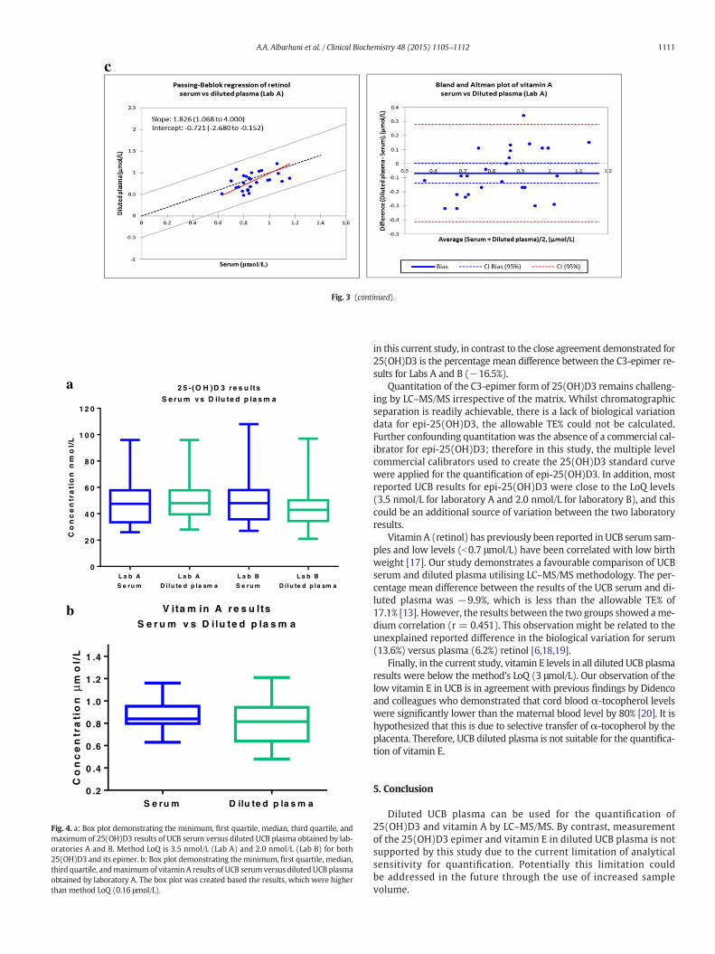
Fig. 3 (continued).
L a b A
S e ru m
L a b A
D i lu te d p la sm a
L a b B
S e ru m
L a b B
D i lu te d p la sm a
0
2 0
4 0
6 0
8 0
1 0 0
1 2 0
2 5 -(O H )D 3 re s u lts
S e ru m v s D ilu te d p la s m a
Co
nc
en
tra
tio
nn
mo
l/L
S e ru m D ilu te d p la s m a0 .2
0 .4
0 .6
0 .8
1 .0
1 .2
1 .4
V ita m in A r e s u lts
S e r u m v s D ilu te d p la s m a
Co
nc
en
tra
tio
nm
ol/
Lμ
a
b
Fig. 4. a: Box plot demonstrating the minimum, first quartile, median, third quartile, andmaximum of 25(OH)D3 results of UCB serum versus diluted UCB plasma obtained by lab-oratories A and B. Method LoQ is 3.5 nmol/L (Lab A) and 2.0 nmol/L (Lab B) for both25(OH)D3 and its epimer. b: Box plot demonstrating theminimum, first quartile, median,third quartile, andmaximumof vitamin A results of UCB serumversus dilutedUCB plasmaobtained by laboratory A. The box plot was created based the results, which were higherthan method LoQ (0.16 μmol/L).
1111A.A. Albarhani et al. / Clinical Biochemistry 48 (2015) 1105–1112
in this current study, in contrast to the close agreement demonstrated for25(OH)D3 is the percentage mean difference between the C3-epimer re-sults for Labs A and B (−16.5%).
Quantitation of the C3-epimer form of 25(OH)D3 remains challeng-ing by LC–MS/MS irrespective of the matrix. Whilst chromatographicseparation is readily achievable, there is a lack of biological variationdata for epi-25(OH)D3, the allowable TE% could not be calculated.Further confounding quantitation was the absence of a commercial cal-ibrator for epi-25(OH)D3; therefore in this study, the multiple levelcommercial calibrators used to create the 25(OH)D3 standard curvewere applied for the quantification of epi-25(OH)D3. In addition, mostreported UCB results for epi-25(OH)D3 were close to the LoQ levels(3.5 nmol/L for laboratory A and 2.0 nmol/L for laboratory B), and thiscould be an additional source of variation between the two laboratoryresults.
Vitamin A (retinol) has previously been reported in UCB serum sam-ples and low levels (b0.7 μmol/L) have been correlated with low birthweight [17]. Our study demonstrates a favourable comparison of UCBserum and diluted plasma utilising LC–MS/MS methodology. The per-centage mean difference between the results of the UCB serum and di-luted plasma was −9.9%, which is less than the allowable TE% of17.1% [13]. However, the results between the two groups showed ame-dium correlation (r = 0.451). This observation might be related to theunexplained reported difference in the biological variation for serum(13.6%) versus plasma (6.2%) retinol [6,18,19].
Finally, in the current study, vitamin E levels in all diluted UCB plasmaresults were below the method's LoQ (3 μmol/L). Our observation of thelow vitamin E in UCB is in agreement with previous findings by Didencoand colleagues who demonstrated that cord blood α-tocopherol levelswere significantly lower than the maternal blood level by 80% [20]. It ishypothesized that this is due to selective transfer of α-tocopherol by theplacenta. Therefore, UCB diluted plasma is not suitable for the quantifica-tion of vitamin E.
5. Conclusion
Diluted UCB plasma can be used for the quantification of25(OH)D3 and vitamin A by LC–MS/MS. By contrast, measurementof the 25(OH)D3 epimer and vitamin E in diluted UCB plasma is notsupported by this study due to the current limitation of analyticalsensitivity for quantification. Potentially this limitation couldbe addressed in the future through the use of increased samplevolume.

1112 A.A. Albarhani et al. / Clinical Biochemistry 48 (2015) 1105–1112
Funding source
This work was supported in part by NHMRC Grant ID 1029927
Financial disclosure
The authors have no financial relationships relevant to this article todisclose.
Conflicts of interest
There is no conflict of interest that could be perceived as prejudicingthe impartiality of this manuscript.
Authorship statement
All authors listed contributed to this work. Professor Allen, ProfessorPonsonby and Dr Vuillermin developed the initial study concept. DrCollier organised and distributed the de-identified samples. Mr AlbarhaniandDrClarke analysed the samples in the respective laboratories andper-formed the statistical analysis in conjunction with Dr Collier. Dr Greavesand Dr Roche supervised and guidedMr Albarhani's work, which formedpart of his PhD candidature. Mr Albarhani wrote the first draft of thismanuscript and all authors critically reviewed the manuscript, assistedin data interpretation, approved the final manuscript as submitted andagree to be accountable for all aspects of this work.
Data sharing statement
Additional information including analytical protocol and raw datamay be obtained by contacting the corresponding author.
Acknowledgements
The Barwon Infant Study (BIS) Steering Committee consists of KatieAllen, David Burgner, John Carlin, Terry Dwyer, Anne-Louise Ponsonby,Sarath Ranganathan, Richard Saffery, Mimi Tang and Peter Vuillermin.A-L Ponsonby held an NHMRC Senior Research Fellowship.
The work performed at RMIT University was conducted in theRMIT-Agilent Clinical Biochemistry Mass Spectrometry Collabora-tion Laboratory.
Appendix A. Supplementary data
Supplementary data to this article can be found online at http://dx.doi.org/10.1016/j.clinbiochem.2015.04.014.
References
[1] Collie JT, Massie RJ, Jones OA, LeGrys VA, Greaves RF. Sixty-five years since the NewYork heatwave: advances in sweat testing for cystic fibrosis. Pediatr Pulmonol 2014;49:106–17.
[2] Grober U, Spitz J, Reichrath J, Kisters K, Holick MF. Vitamin D: update 2013: fromrickets prophylaxis to general preventive healthcare. Dermatoendocrinol 2013;5:331–47.
[3] Shah I, Petroczi A, Naughton DP. Exploring the role of vitamin D in type 1 diabetes,rheumatoid arthritis, and Alzheimer disease: new insights from accurate analysis of10 forms. J Clin Endocrinol Metab 2014;99:808–16.
[4] Zakariaeeabkoo R, Allen KJ, Koplin JJ, Vuillermin P, Greaves RF. Are vitamins A and Dimportant in the development of food allergy and how are they best measured? ClinBiochem 2014;47:804–11.
[5] Greaves R, Jolly L, Woollard G, Hoad K. Serum vitamin A and E analysis: comparisonof methods between laboratories enrolled in an external quality assurance pro-gramme. Ann Clin Biochem 2010;47:78–80.
[6] Greaves RF, Woollard GA, Hoad K, Walmsley TA, Johansson LA, Briscoe S, et al. Lab-oratory medicine best practice guideline: vitamins a, e and the carotenoids in blood.Clin Biochem Rev 2014;35:85–118.
[7] Albahrani AA, Rotarou V, Roche PJ, Greaves RF. Comparison of three commercialcalibrators for alpha-tocopherol using liquid chromatography–tandem mass spec-trometry. Clin Biochem 2013;46:1884–8.
[8] Albahrani AA, Rotarou V, Roche PJ, Greaves RF. Candidate reference methodfor quantification of fat soluble vitamins using chromatography–tandem massspectrometry (abstract). Clin Biochem Rev 2013;34:s20.
[9] RCPA quality assurance programs — chemical pathology 2013. Available at: http://www.rcpaqap.com.au/chempath/; 2013. [Accessed 24th April 2014].
[10] Clarke MW, Tuckey RC, Gorman S, Holt B, Hart PH. Optimized 25-hydroxyvitamin Danalysis using liquid–liquid extraction with 2D separation with LC/MS/MS detection,provides superior precision compared to conventional assays. Metabolomics 2013;9:1031–40.
[11] Sempos CT, Vesper HW, Phinney KW, Thienpont LM, Coates PM, The Vitamin D.Standardization program (VDSP): vitamin D status as an international issue: nation-al surveys and the problem of standardization. Scand J Clin Lab Invest Suppl 2012;243:32–40.
[12] XLSTAT software (version 2014.1.07; Addinsoft SARL, www.xlstat.com).[13] Ricos C, Alvarez V, Cava F, Garcia-Lario J, Hernandez A, Jimenez C, et al. Current
databases on biological variation: pros, cons and progress. Scand J Clin Lab Invest1999;59:491–500 [Available at: http://www.westgard.com/biodatabase491.htm.Accessed 424th April 2014].
[14] Stockl D, Sluss PM, Thienpont LM. Specifications for trueness and precision of areference measurement system for serum/plasma 25-hydroxyvitamin D analysis.Clin Chim Acta 2009;408:8–13.
[15] Sapan CV, Lundblad RL. Considerations regarding the use of blood samples in theproteomic identification of biomarkers for cancer diagnosis. Cancer Genomics Prote-omics 2006;3:227–30.
[16] Keevil B. Does the presence of 3-epi-25OHD3 affect the routine measurement of vi-tamin D using liquid chromatography tandem mass spectrometry? Clin Chem LabMed 2012;50:181–3.
[17] Gazala E, Sarov B, Hershkovitz E, Edvardson S, Sklan D, Katz M, et al. Retinol concentra-tion in maternal and cord serum: its relation to birth weight in healthy mother–infantpairs. Early Hum Dev 2003;71:19–28.
[18] Olmedilla B, Granado F, Blanco I, Rojas-Hidalgo E. Seasonal and sex-related varia-tions in six serum carotenoids, retinol, and alpha-tocopherol. Am J Clin Nutr 1994;60:106–10.
[19] Talwar DK, Azharuddin MK, Williamson C, Teoh YP, McMillan DC, O'Reilly DSJ.Biological variation of vitamins in blood of healthy individuals. Clin Chem 2005;51:2145–50.
[20] Kiely M, Cogan PF, Kearney PJ, Morrissey PA. Concentrations of tocopherols andcarotenoids in maternal and cord blood plasma. Eur J Clin Nutr 1999;53:711–5.