Visual motion processing deficits in infants with the fragile X ...
8
RESEARCH Open Access Visual motion processing deficits in infants with the fragile X premutation Pamela K Gallego 1,2 , Jessica L Burris 1,2 and Susan M Rivera 1,2,3* Abstract Background: Fragile X syndrome (FXS) results from a trinucleotide repeat expansion (full mutation >200 cytosine-guanine-guanine (CGG) repeats) in the FMR1 gene, leading to a reduction or absence of the gene’s protein product, fragile X mental retardation protein (FMRP), ultimately causing cognitive and behavioral impairments that are characteristic of the syndrome. In our previous work with infants and toddlers with FXS, we have been able to describe much about their cognitive and visual processing abilities. In light of recent work on the mild cognitive deficits and functional and structural brain differences that are present in adults with the fragile X (FX) premutation, in the present study we examined whether some of the low-level visual processing deficits we have observed in infants with FXS would also be present in infants with the FX premutation (55–200 CGG repeats). Methods: We chose a contrast detection task using second-order motion stimuli on which infants with FXS previously showed significantly increased detection thresholds (Vision Res 48:1471–1478, 2008). Critically, we also included a developmental delay comparison group of infants with Down syndrome (DS), who were matched to infants with FXS on both chronological and mental age, to speak to the question of whether this second-order motion processing deficit is a FX-specific phenomenon. Results: As reported previously, infants with the FX full mutation showed motion contrast detection threshold levels that were significantly higher than age-matched typically developing control infants. Strikingly, the motion detection contrast levels of FX premutation infants were also significantly higher than typically developing (TD) infants and not significantly different from the group of infants with FXS or with DS. Conclusions: These results, which are in keeping with a growing body of evidence on the mild cognitive and perceptual processing deficits and functional and structural brain differences that are present in adults and older children with the FX premutation, underscore the pressing need to study and describe the processing capabilities of infants and toddlers with the FX premutation. Keywords: Premutation, Visual processing deficits, Fragile X syndrome, Contrast detection Background Fragile X syndrome (FXS) is the most common inherited cause of mental disability, which results from a reduc- tion or absence of the fragile X mental retardation pro- tein (FMRP), a gene product known to play an essential role in brain structure and function [1,2]. This condition emerges when the repeat expansion of the trinucleotide cytosine-guanine-guanine (CGG) in the 5′ untranslated region of the FMR1 gene, located in the X chromosome, is above 200 repeats. When this occurs, the FMR1 gene is typically fully methylated, which prevents the tran- scription and the translation of the gene, consequently disrupting the production of FMRP and leading to cas- cading cognitive and behavioral impairment, including mild to severe intellectual disability, social anxiety, math and spatial reasoning problems, and relatively high co- morbidity with autism (30% of all FXS cases) [3,4]. The number of individuals with FXS (full mutation >200 CGG) ranges approximately between 1 per 2,500 in females to 1 per 4,000 in males [5]. * Correspondence: [email protected] 1 Department of Psychology, University of California Davis, Davis, CA 95618, USA 2 Center for Mind and Brain, University of California Davis, 202 Cousteau Place, Suite 250, Davis, CA 95618, USA Full list of author information is available at the end of the article © 2014 Gallego et al.; licensee BioMed Central Ltd. This is an Open Access article distributed under the terms of the Creative Commons Attribution License (http://creativecommons.org/licenses/by/2.0), which permits unrestricted use, distribution, and reproduction in any medium, provided the original work is properly credited. The Creative Commons Public Domain Dedication waiver (http://creativecommons.org/publicdomain/zero/1.0/) applies to the data made available in this article, unless otherwise stated. Gallego et al. Journal of Neurodevelopmental Disorders 2014, 6:29 http://www.jneurodevdisorders.com/content/6/1/29
Transcript of Visual motion processing deficits in infants with the fragile X ...
Gallego et al. Journal of Neurodevelopmental Disorders 2014, 6:29http://www.jneurodevdisorders.com/content/6/1/29
RESEARCH Open Access
Visual motion processing deficits in infants withthe fragile X premutationPamela K Gallego1,2, Jessica L Burris1,2 and Susan M Rivera1,2,3*
Abstract
Background: Fragile X syndrome (FXS) results from a trinucleotide repeat expansion (full mutation >200cytosine-guanine-guanine (CGG) repeats) in the FMR1 gene, leading to a reduction or absence of the gene’s proteinproduct, fragile X mental retardation protein (FMRP), ultimately causing cognitive and behavioral impairments thatare characteristic of the syndrome. In our previous work with infants and toddlers with FXS, we have been ableto describe much about their cognitive and visual processing abilities. In light of recent work on the mild cognitivedeficits and functional and structural brain differences that are present in adults with the fragile X (FX) premutation,in the present study we examined whether some of the low-level visual processing deficits we have observedin infants with FXS would also be present in infants with the FX premutation (55–200 CGG repeats).
Methods: We chose a contrast detection task using second-order motion stimuli on which infants with FXS previouslyshowed significantly increased detection thresholds (Vision Res 48:1471–1478, 2008). Critically, we also included adevelopmental delay comparison group of infants with Down syndrome (DS), who were matched to infants withFXS on both chronological and mental age, to speak to the question of whether this second-order motion processingdeficit is a FX-specific phenomenon.
Results: As reported previously, infants with the FX full mutation showed motion contrast detection threshold levelsthat were significantly higher than age-matched typically developing control infants. Strikingly, the motion detectioncontrast levels of FX premutation infants were also significantly higher than typically developing (TD) infants and notsignificantly different from the group of infants with FXS or with DS.
Conclusions: These results, which are in keeping with a growing body of evidence on the mild cognitive andperceptual processing deficits and functional and structural brain differences that are present in adults andolder children with the FX premutation, underscore the pressing need to study and describe the processingcapabilities of infants and toddlers with the FX premutation.
Keywords: Premutation, Visual processing deficits, Fragile X syndrome, Contrast detection
BackgroundFragile X syndrome (FXS) is the most common inheritedcause of mental disability, which results from a reduc-tion or absence of the fragile X mental retardation pro-tein (FMRP), a gene product known to play an essentialrole in brain structure and function [1,2]. This conditionemerges when the repeat expansion of the trinucleotide
* Correspondence: [email protected] of Psychology, University of California Davis, Davis, CA 95618,USA2Center for Mind and Brain, University of California Davis, 202 CousteauPlace, Suite 250, Davis, CA 95618, USAFull list of author information is available at the end of the article
© 2014 Gallego et al.; licensee BioMed CentraCommons Attribution License (http://creativecreproduction in any medium, provided the orDedication waiver (http://creativecommons.orunless otherwise stated.
cytosine-guanine-guanine (CGG) in the 5′ untranslatedregion of the FMR1 gene, located in the X chromosome,is above 200 repeats. When this occurs, the FMR1 geneis typically fully methylated, which prevents the tran-scription and the translation of the gene, consequentlydisrupting the production of FMRP and leading to cas-cading cognitive and behavioral impairment, includingmild to severe intellectual disability, social anxiety, mathand spatial reasoning problems, and relatively high co-morbidity with autism (30% of all FXS cases) [3,4]. Thenumber of individuals with FXS (full mutation >200 CGG)ranges approximately between 1 per 2,500 in females to 1per 4,000 in males [5].
l Ltd. This is an Open Access article distributed under the terms of the Creativeommons.org/licenses/by/2.0), which permits unrestricted use, distribution, andiginal work is properly credited. The Creative Commons Public Domaing/publicdomain/zero/1.0/) applies to the data made available in this article,
Gallego et al. Journal of Neurodevelopmental Disorders 2014, 6:29 Page 2 of 8http://www.jneurodevdisorders.com/content/6/1/29
Individuals with CGG repeat expansions of 55–200 areconsidered to be premutation carriers of FXS, a conditionmore commonly found in the general population, affectingapproximately 1 in 130–250 women and 1 in 250–810males [5]. Individuals in the premutation range typicallyhave normal intellectual functioning, but may have ele-vated FMR1 mRNA, in some cases three to eight times thenormal levels [6]. This elevated mRNA is thought to pro-duce RNA toxicity, which has been associated with milddeficits in working memory [7], memory encoding [8],memory recall [9], enumeration [10], and increased psychi-atric symptoms including obsessive-compulsive symptomsand psychoticism [11]. In addition, male premutation car-riers, especially, are at risk of developing the late-onsetneurodegenerative disorder known as fragile X-associatedtremor/ataxia syndrome (FXTAS) [7,12].There is now ample evidence that young children with
FXS have significant visual-spatial impairments. Forexample, studies of infants and toddlers with FXS havedocumented impairments in processing texture-defined(second order) motion stimuli [13], temporal flicker [14],perceiving the ordinality of sequences of numerical dis-plays [15], and the ability to maintain the identity ofdynamic object information during occlusion [16]. Im-paired performance has also been demonstrated on tasksrequiring visual-motor responses [17,18] as well as in-hibitory control [19] and numerical reasoning [20,21].One purported cause of the visual-spatial and numericaldeficits seen in FXS is disruption of the so-called dorsalstream (the occipito-parietal visual pathway, projectingto the posterior parietal cortex, which processes infor-mation involved in guiding actions, including spatial lo-cation and motion) with relative sparing of the ventralstream (the occipito-temporal visual pathway, projectingto the inferior-temporal cortex, which processes objectfeatures such as form and color) [22,23]. Because of itsrelatively protracted time course of development [24],the dorsal stream is thought to be particularly vulnerableto atypical development in a number of disorders, in-cluding in FXS [16,25].Compared to young children with the fragile X full
mutation (FXS), very little is understood about the cog-nitive and visual processing abilities in young childrenwith the fragile X (FX) premutation. Recent work on themild cognitive deficits and functional and structuralbrain differences that are present in adults with theFX premutation [7,8,10,11,26] and particularly studieswhich have documented deficits in visuospatial [27,28]and contrast sensitivity [29] in adult premutation carriersled us in the present study to examine whether oneof the low-level visual processing deficits that has beenobserved in infants with FXS is also present in infantswith the FX premutation. To study this, we chose acontrast detection task using second-order motion
stimuli on which infants with FXS demonstrated signifi-cantly increased detection thresholds [13]. We hypothe-sized that infants and toddlers with the premutationwould perform similarly to infants and toddlers with thefull mutation, i.e., the threshold necessary for detectionof visual stimuli would be higher than typically develop-ing mental and chronological age-matched controls andwould not be significantly different from participantswith the full mutation. We also included a comparisongroup of infants with Down syndrome, who are matchedwith the FX full mutation group on both mental andchronological age, allowing us to examine whether defi-cits seen in second-order motion processing are specificto the FX-specific spectrum.
MethodsParticipantsFour groups of participants were enrolled in this study:16 typically developing infants (7 male and 9 female,mean age 13.17 months), 12 premutation carrier infants(8 males and 4 females, mean age 17.56 months), 24infants with FXS (19 male and 5 female, mean age29.24 months), and 15 infants with Down syndrome(5 males and 10 females, mean age 26.27 months). Aone-way ANOVA confirmed that the groups signifi-cantly differed in their chronological age (F(3, 63) = 6.67,p = 0.001). Simple effect analyses revealed that there wasno significant difference in chronological age (in months)between typically developing (TD) infants (M = 13.17;SD = 7.91) and infants with the FX premutation (M = 17.56;SD = 12.57; t [26] = 1.13, p = 0.27) nor between infants withDown syndrome (DS) (M = 26.27; SD = 11.95) and FXS(M = 29.24; SD = 14.35; t [30] = 0.668, p = 0.51). By con-trast, both the DS and FXS were significantly chrono-logically older than TD infants (t [31] = 4.07, p = 0.002;t [29] = 3.62, p = 0.001, respectively.) For infants withthe FX premutation, repeat sizes ranged from 55 to181, with a mean length of 94. For infants with FXS,CGG repeat sizes ranged from 210 to 702, with a meanlength of 466.Participants with FXS were recruited and clinically eval-
uated at the UC Davis MIND Institute. Four participantswith the FX premutation were seen as patients at the UCDavis M.I.N.D. Institute Fragile X Research and TreatmentCenter (FXRTC), while eight were recruited through anewborn screening project in which parents in the gen-eral population could consent to screen their newborninfants for metabolic abnormalities and other preexist-ing conditions [32]. This recruitment combination al-lows us to have a sample of premutation infants that ismore representative of the population because parentswho enroll in the newborn screening program are un-aware of their child’s preexisting condition and thereforedo not present the ascertainment bias that may occur in

Gallego et al. Journal of Neurodevelopmental Disorders 2014, 6:29 Page 3 of 8http://www.jneurodevdisorders.com/content/6/1/29
participants who come to the FXRTC clinic seeking re-sources for their child. Participants with DS were recruitedfrom the community by attending outreach events. Typic-ally developing infants were recruited through letters tofamilies, fliers, and word of mouth.Participants were developmentally age-matched using
the Mullen Scales of Early Learning [33], a standardizeddevelopmental assessment used for children 3–60 monthsconsisting of 5 subscales: gross motor, fine motor, visualreception, expressive language, and receptive language.The mental age of each participant was calculated by aver-aging across the four different domains (VR, FM, RL,and EL) and converting that average to age in monthsand days. The gross motor subscale was omitted fromthe mental age calculation as the scores become less validin children above the age of 33 months [33]. The meanmental age was 13.22 months for typically developingparticipants, 15.10 months for premutation carriers,18.01 months for participants with FXS, and 14.13 forparticipants with DS. A one-way ANOVA confirmedthat mental age did not differ significantly between thefour groups (F(3,63) = 0.850, p = 0.472). Table 1 depictsthe averaged ELC score across the four groups. A one-way ANOVA confirmed that the ELC scores, as ex-pected, differed significantly between the four groups(F(3,63) = 29.67, p = 0.000). Post hoc analyses show thatthere is no significant difference between the participantswith the premutation and typically developing groupwhen using Bonferroni-corrected values (p = 0.094), sug-gesting that these two groups are performing at an over-all comparable cognitive level.
Apparatus and stimuliA Tobii 1750 binocular eye tracker monitor (TobiiTechnology, Danderyd Sweden, www.tobii.com) was usedto present the visual stimuli. This eye tracking systemconsists of a high-resolution camera that records theeye position, embedded in a 17-in. monitor (1,280 by1,024 pixel resolution, 50-Hz refresh rate) with infraredlight-emitting diodes that illuminate the cornea, cap-turing and tracking eye movements that are then runthrough proprietary algorithms that calculate changesin eye position. The visual angle subtended by the dis-play was 31.63° by 25.36° region on the screen when
Table 1 Mean scores of the early learning compositescores of the Mullen across groups
Group Mean ELC score
TYP 103.33
PRE 87.73
DS 54.93
FXS 65.61
viewed from a distance of 60 cm. The data are capturedat a frame rate of 50 Hz and sent to Tobii Studio (version2.0.8) to be overlaid on the stimuli. The stimuli used forthis study was generated using The Vision Shell PPC pro-gram, controlled by an Apple G4 Power Macintosh withOS9 (Apple, Cupertino, CA, USA). Please see Farzinet al. [13] for a detailed description of the second-order(texture-defined) motion (4 Hz) sine wave gratings stim-uli used in this study.
ProcedureThe Institutional Review Board at the University ofCalifornia, Davis, approved the experimental protocol,and an informed consent was obtained from the parentsof all infants. The infants were tested while seated on acaregiver's lap and were positioned so that their face wasapproximately 60–70 cm in front of the eye tracker. Inorder to attract the participants' attention to the screen,room lights were dimmed and an attention-getter videowas displayed on the screen. During this time, an experi-menter monitored the participant's eye position using areal-time track-status monitor. If the participant's eyeswere not found, adjustments were made (repositioningthe participant or angling the monitor to adjust for theparticipant's height) until a track status was obtained onboth eyes.Once the participant's eyes were detected by the eye
tracker, a five-point calibration routine was executed inTobii Studio. If all points were acquired, the calibrationwas saved and the stimulus presentation would begin. Ifthe calibration was not successful (i.e., not all five pointswere acquired), another calibration was attempted. Theminimum criterion required to proceed with the taskwas a successful calibration of the center point for eacheye. This ensured that the gaze mapped correctly ontothe stimuli and our areas of interest on the left or theright side of the screen. The following numbers repre-sent the participant that could not be calibrated acrossthe groups: nine FXS, five DS, three PRE, and zero TYP.These numbers fall within the average range in our lab'sexperience for these populations and these ages [13-16].A forced-choice preferential looking procedure was
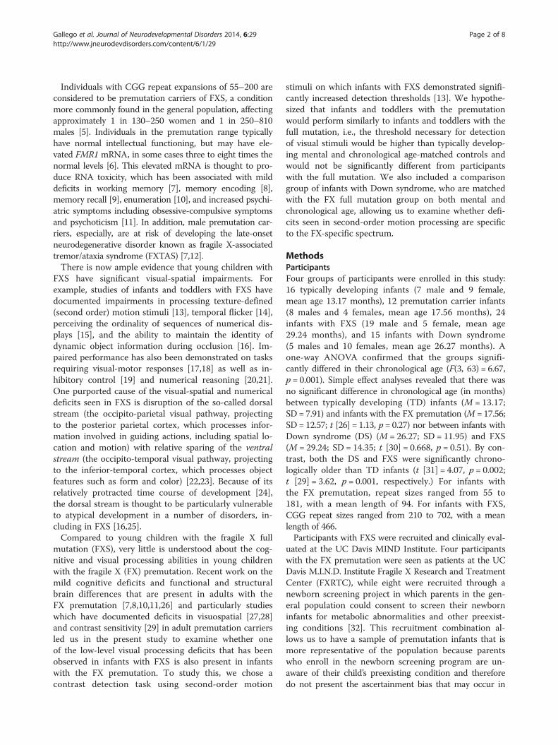
utilized [34] in which stimuli were presented either onthe left or the right side of the screen (see Figure 1) Eachtrial was approximately 3 s in length, with 1 s of thegrating fading in (500 ms) and out (500 ms) of thescreen. An attention getter was presented between trialsto draw the participants' attention to the middle of thescreen. This attention getter was a colorful, centrally lo-cated circle looming in and out accompanied by a single3-s tone. Trials began automatically after the presenta-tion of the attention getter. The presentation includedfour contrast levels (10%, 21%, 31%, or 42%), with thelowest contrast being the most difficult to perceive and
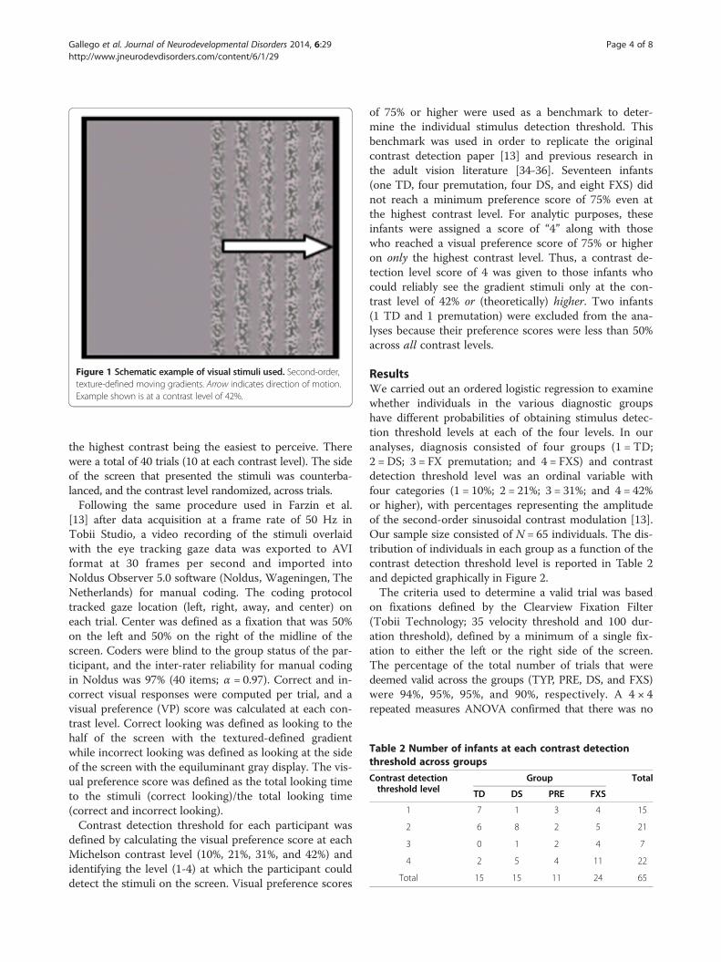
Figure 1 Schematic example of visual stimuli used. Second-order,texture-defined moving gradients. Arrow indicates direction of motion.Example shown is at a contrast level of 42%.
Table 2 Number of infants at each contrast detectionthreshold across groups
Contrast detectionthreshold level
Group Total
TD DS PRE FXS
1 7 1 3 4 15
2 6 8 2 5 21
3 0 1 2 4 7
4 2 5 4 11 22
Total 15 15 11 24 65
Gallego et al. Journal of Neurodevelopmental Disorders 2014, 6:29 Page 4 of 8http://www.jneurodevdisorders.com/content/6/1/29
the highest contrast being the easiest to perceive. Therewere a total of 40 trials (10 at each contrast level). The sideof the screen that presented the stimuli was counterba-lanced, and the contrast level randomized, across trials.Following the same procedure used in Farzin et al.
[13] after data acquisition at a frame rate of 50 Hz inTobii Studio, a video recording of the stimuli overlaidwith the eye tracking gaze data was exported to AVIformat at 30 frames per second and imported intoNoldus Observer 5.0 software (Noldus, Wageningen, TheNetherlands) for manual coding. The coding protocoltracked gaze location (left, right, away, and center) oneach trial. Center was defined as a fixation that was 50%on the left and 50% on the right of the midline of thescreen. Coders were blind to the group status of the par-ticipant, and the inter-rater reliability for manual codingin Noldus was 97% (40 items; α = 0.97). Correct and in-correct visual responses were computed per trial, and avisual preference (VP) score was calculated at each con-trast level. Correct looking was defined as looking to thehalf of the screen with the textured-defined gradientwhile incorrect looking was defined as looking at the sideof the screen with the equiluminant gray display. The vis-ual preference score was defined as the total looking timeto the stimuli (correct looking)/the total looking time(correct and incorrect looking).Contrast detection threshold for each participant was
defined by calculating the visual preference score at eachMichelson contrast level (10%, 21%, 31%, and 42%) andidentifying the level (1-4) at which the participant coulddetect the stimuli on the screen. Visual preference scores
of 75% or higher were used as a benchmark to deter-mine the individual stimulus detection threshold. Thisbenchmark was used in order to replicate the originalcontrast detection paper [13] and previous research inthe adult vision literature [34-36]. Seventeen infants(one TD, four premutation, four DS, and eight FXS) didnot reach a minimum preference score of 75% even atthe highest contrast level. For analytic purposes, theseinfants were assigned a score of “4” along with thosewho reached a visual preference score of 75% or higheron only the highest contrast level. Thus, a contrast de-tection level score of 4 was given to those infants whocould reliably see the gradient stimuli only at the con-trast level of 42% or (theoretically) higher. Two infants(1 TD and 1 premutation) were excluded from the ana-lyses because their preference scores were less than 50%across all contrast levels.
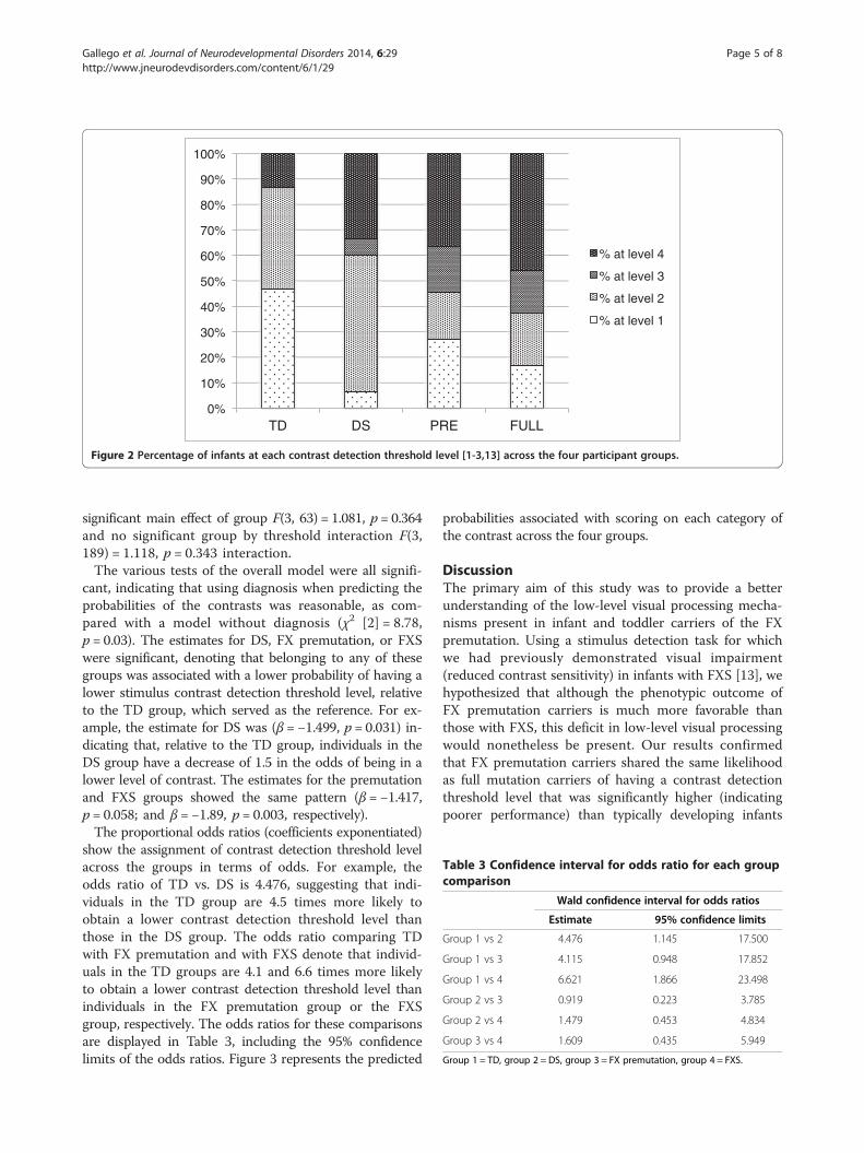
ResultsWe carried out an ordered logistic regression to examinewhether individuals in the various diagnostic groupshave different probabilities of obtaining stimulus detec-tion threshold levels at each of the four levels. In ouranalyses, diagnosis consisted of four groups (1 = TD;2 = DS; 3 = FX premutation; and 4 = FXS) and contrastdetection threshold level was an ordinal variable withfour categories (1 = 10%; 2 = 21%; 3 = 31%; and 4 = 42%or higher), with percentages representing the amplitudeof the second-order sinusoidal contrast modulation [13].Our sample size consisted of N = 65 individuals. The dis-tribution of individuals in each group as a function of thecontrast detection threshold level is reported in Table 2and depicted graphically in Figure 2.The criteria used to determine a valid trial was based
on fixations defined by the Clearview Fixation Filter(Tobii Technology; 35 velocity threshold and 100 dur-ation threshold), defined by a minimum of a single fix-ation to either the left or the right side of the screen.The percentage of the total number of trials that weredeemed valid across the groups (TYP, PRE, DS, and FXS)were 94%, 95%, 95%, and 90%, respectively. A 4 × 4repeated measures ANOVA confirmed that there was no
0%
10%
20%
30%
40%
50%
60%
70%
80%
90%
100%
TD DS PRE FULL
% at level 4
% at level 3
% at level 2
% at level 1
Figure 2 Percentage of infants at each contrast detection threshold level [1-3,13] across the four participant groups.
Table 3 Confidence interval for odds ratio for each groupcomparison
Wald confidence interval for odds ratios
Estimate 95% confidence limits
Group 1 vs 2 4.476 1.145 17.500
Group 1 vs 3 4.115 0.948 17.852
Group 1 vs 4 6.621 1.866 23.498
Group 2 vs 3 0.919 0.223 3.785
Group 2 vs 4 1.479 0.453 4.834
Group 3 vs 4 1.609 0.435 5.949
Group 1 = TD, group 2 = DS, group 3 = FX premutation, group 4 = FXS.
Gallego et al. Journal of Neurodevelopmental Disorders 2014, 6:29 Page 5 of 8http://www.jneurodevdisorders.com/content/6/1/29
significant main effect of group F(3, 63) = 1.081, p = 0.364and no significant group by threshold interaction F(3,189) = 1.118, p = 0.343 interaction.The various tests of the overall model were all signifi-
cant, indicating that using diagnosis when predicting theprobabilities of the contrasts was reasonable, as com-pared with a model without diagnosis (χ2 [2] = 8.78,p = 0.03). The estimates for DS, FX premutation, or FXSwere significant, denoting that belonging to any of thesegroups was associated with a lower probability of having alower stimulus contrast detection threshold level, relativeto the TD group, which served as the reference. For ex-ample, the estimate for DS was (β = −1.499, p = 0.031) in-dicating that, relative to the TD group, individuals in theDS group have a decrease of 1.5 in the odds of being in alower level of contrast. The estimates for the premutationand FXS groups showed the same pattern (β = −1.417,p = 0.058; and β = −1.89, p = 0.003, respectively).The proportional odds ratios (coefficients exponentiated)
show the assignment of contrast detection threshold levelacross the groups in terms of odds. For example, theodds ratio of TD vs. DS is 4.476, suggesting that indi-viduals in the TD group are 4.5 times more likely toobtain a lower contrast detection threshold level thanthose in the DS group. The odds ratio comparing TDwith FX premutation and with FXS denote that individ-uals in the TD groups are 4.1 and 6.6 times more likelyto obtain a lower contrast detection threshold level thanindividuals in the FX premutation group or the FXSgroup, respectively. The odds ratios for these comparisonsare displayed in Table 3, including the 95% confidencelimits of the odds ratios. Figure 3 represents the predicted
probabilities associated with scoring on each category ofthe contrast across the four groups.
DiscussionThe primary aim of this study was to provide a betterunderstanding of the low-level visual processing mecha-nisms present in infant and toddler carriers of the FXpremutation. Using a stimulus detection task for whichwe had previously demonstrated visual impairment(reduced contrast sensitivity) in infants with FXS [13], wehypothesized that although the phenotypic outcome ofFX premutation carriers is much more favorable thanthose with FXS, this deficit in low-level visual processingwould nonetheless be present. Our results confirmedthat FX premutation carriers shared the same likelihoodas full mutation carriers of having a contrast detectionthreshold level that was significantly higher (indicatingpoorer performance) than typically developing infants

TD DS FX PRE FXS
Threshold Level
Figure 3 Predicted probabilities for contrast detection threshold level. Predicted probabilities for contrast detection threshold level [1-3,13]in the four participant groups.
Gallego et al. Journal of Neurodevelopmental Disorders 2014, 6:29 Page 6 of 8http://www.jneurodevdisorders.com/content/6/1/29
matched on mental age. We also tested a comparisongroup of infants with DS who were both chronologicallyand mentally age-matched to the participants with FXSand mentally age-matched to the infants with the FXpremutation, and found that their performance did notdiffer significantly from the two FX groups. This result isconsistent with research showing that individuals withDS exhibit significantly reduced visual acuity and con-trast sensitivity as compared to TD controls [37] andsuggests that this deficit may not be specific to the FXspectrum but a more general deficit found in other de-velopmental disabilities such as Williams syndrome andautism spectrum disorders [30,38].While it was long believed that individuals with the FX
premutation remained cognitively unaffected throughoutadulthood, there have been a number of studies, particu-larly those utilizing brain imaging techniques, which havedocumented measurable differences in brain functioning,across a number of different cognitive domains, in youngadult premutation carriers who are asymptomatic forFXTAS [7,8,26]. Adult male FX premutation carriers havealso been shown to exhibit slower reaction times, evenafter controlling for simple reaction time, in the visuo-spatial tasks of magnitude comparison and enumeration[28]. Perhaps most relevant to the present study, a recentinvestigation has shown that adult female FX premutationcarriers show a ‘dorsal stream deficit’ in that they areselectively impaired on perceptual tests of magnocellular
(so-called M pathway—projecting primarily to dorsal visualstream areas) stimuli, whereas they show intact perform-ance on tests of parvocellular (‘P pathway’—projecting pri-marily to ventral visual stream areas) stimuli [29].The present study is unique in that it is the first to
document dorsal stream processing difficulties in infantsand toddlers with the FX premutation. Our results sug-gest that even in very young premutation carriers, whoin the vast majority of cases are cognitively unaffectedand developing normally, a selective visual motion pro-cessing impairment on second-order motion gradientstimuli is present that is not significantly different fromthat found in those with the FX full mutation. Whilethese results are striking, they beg the important ques-tion of what is the functional importance of such an im-pairment. It may be, as suggested by Keri and Benedek[29], that this represents a psychophysical endophe-notype (a marker of genetic traits that do not resultin observable clinical symptoms) for the FX spectrum ofinvolvement. If so, individual differences in this abilitymay hold clues for differentiating those individuals onthe FX spectrum (which includes premutation carriers,mosaics, and full mutation individuals) who are at riskfor developing more severe phenotypes.As reviewed above, there have been numerous demon-
strations of spatiotemporal deficits in both individualswith FXS and those with the FX premutation. Becauserepresentations of space and time are integral to forming
Gallego et al. Journal of Neurodevelopmental Disorders 2014, 6:29 Page 7 of 8http://www.jneurodevdisorders.com/content/6/1/29
conceptions of number and arithmetic [31], impairedspatiotemporal processing may in fact also underlie theimpairments in numerical processing that have been ob-served in both FXS [21] and in FX premutation carriers[26,39]. In females with FXS, brain activation duringarithmetic processing has also been shown to be relatedto FMRP expression [21], which suggests that impairedprocessing of spatiotemporal information that is medi-ated primarily by the parietal cortex may represent anendophenotype that is modulated by FMR1 gene expres-sion across the FX spectrum. While the present studywas underpowered to study how second-order motionvisual processing may be modulated by variations inFMR1 gene expression, this is an important question forfuture research to address.Despite the spatiotemporal processing impairments in
individuals with the FX premutation demonstrated bothin the present study and elsewhere [28,29], the factremains that individuals with the FX premutation onlyrarely present with overall cognitive functioning thatfalls below the normal range. One might be tempted,then, to dismiss these low-level visual processing impair-ments as unimportant to the individual’s perceptual andcognitive development. However, the presence of suchimpairments, even in an individual whose overall cogni-tive development falls within the normal range, mayforce compensation in the developing neural system,thus changing the landscape of development in waysthat are difficult to measure, though potentially stillimpactful. It is therefore imperative that we continue tostudy and broaden our understanding of the processingcapabilities of individuals with the FX premutation, par-ticularly early in life.
ConclusionsWe tested four groups of infants and toddlers (TD, DS,FX premutation, and FXS) on a second-order motionstimulus detection task for which we had previously dem-onstrated visual impairment (reduced contrast sensitivity)in infants with FXS [13]. As reported previously, infantswith FXS showed motion contrast detection thresholdlevels that were significantly higher than age-matched typ-ically developing control infants. Strikingly, the motion de-tection contrast levels of FX premutation infants were alsosignificantly higher than TD infants, and not significantlydifferent from the group of infants with FXS or with theDS group. The present data, along with other evidence ofimpairments in processing spatiotemporal informationthat comes from the study of adults with the FX premuta-tion, suggest that this type of spatiotemporal processingimpairment may constitute an endophenotype for indi-viduals on the FX spectrum and highlights the need forfurther study of the development of these processes, espe-cially in children with the FX premutation.
AbbreviationsFX: fragile X; FXS: fragile X syndrome; FMRP: fragile X mental retardationprotein; CGG: cytosine-guanine-guanine; DS: Down syndrome; FXTAS: fragileX tremor/ataxia syndrome; VP: visual preference; TD: typically developing.
Competing interestsThe authors declare that they have no competing interests.
Authors’ contributionsPG collected and analyzed the eye tracking data and wrote the first draft ofthe manuscript. JB assisted in the collection and analysis of the eye trackingdata and provided feedback on the drafts of the manuscript. SR conceptualizedand designed the study and wrote the final draft of the manuscript. All authorsread and approved the final manuscript.
Authors’ informationPG has a Masters in Psychology and is a research staff member (assistantspecialist) in SR's Neurocognitive Development lab. JB has a BS in Psychologyand is currently a graduate student in Psychology at UC Davis. SR has a Ph.D.in Psychology and is currently a Professor in the Department of Psychology atUC Davis.
AcknowledgementsWe would like to acknowledge Lena Rothstein for contributing with dataacquisition and coding, Dr. Flora Tassone for providing molecular data,Dr. Emilio Ferrer for providing statistical guidance, research assistantsLina Baranauskaite and Mercy Huang for their assistance in testing participants,and all of the families that volunteered their time for our research study.
Author details1Department of Psychology, University of California Davis, Davis, CA 95618,USA. 2Center for Mind and Brain, University of California Davis, 202 CousteauPlace, Suite 250, Davis, CA 95618, USA. 3M.I.N.D. Institute, University ofCalifornia Medical Center, Sacramento, CA 95817, USA.
Received: 26 November 2013 Accepted: 11 July 2014Published: 30 July 2014
References1. Comery TA, Harris JB, Willems PJ, Oostra BA, Irwin SA, Weiler IJ, Greenough WT:
Abnormal dendritic spines in fragile X knockout mice: maturation andpruning deficits. Proc Natl Acad Sci U S A 1997, 94:5401–5404.
2. Irwin SA, Galvez R, Weiler IJ, Beckel-Mitchener A, Greenough WT: Brainstruction and function of FMR1 protein. In Fragile X syndrome: DiagnosisTreatment and Research. 3rd edition. Edited by Hagerman RJ, Hagerman PJ.Baltimore: The John Hopkins University Press; 2002:191–205.
3. Hagerman R, Au J, Hagerman P: FMR1 premutation and full mutationmolecular mechanisms related to autism. J Neurodev Disord 2011,3:211–224.
4. Hagerman R, Harris S: Autism profiles of males with fragile X syndrome.Am J Ment Retard 2008, 113:427–438.
5. Fernandez-Carvajal I, Walichiewicz P, Xiaosen X, Pan R, Hagerman PJ,Tassone F: Screening for expanded alleles of the FMR1 gene in bloodspots from newborn males in a Spanish population. J Mol Diagn 2009,11:324–329.
6. Tassone F, Hagerman RJ, Taylor AK, Gane LW, Godfrey TE, Hagerman PJ:Elevated levels of FMR1 mRNA in carrier males: a new mechanism ofinvolvement in the fragile-X syndrome. Am J Hum Genet 2000, 66:6–15.
7. Hashimoto R, Backer KC, Tassone F, Hagerman RJ, Rivera SM: An fMRI studyof the prefrontal activity during the performance of a working memorytask in premutation carriers of the fragile X mental retardation 1 genewith and without fragile X-associated tremor/ataxia syndrome (FXTAS).J Psychiatr Res 2012, 45:36–43.
8. Wang JM, Koldewyn K, Hashimoto R-I, Schneider A, Le L, Tassone F, Cheung K,Hagerman P, Hessl D, Rivera SM: Male carriers of the FMR1 premutation showaltered hippocampal-prefrontal function during memory encoding.Front Hum Neurosci 2012, 6:297.
9. Koldewyn K, Hessl D, Adams J, Tassone F, Hagerman PJ, Hagerman RJ,Rivera SM: Reduced hippocampal activation during recall is associatedwith elevated FMR1 mRNA and psychiatric symptoms in men with thefragile X premutation. Brain Imaging Behav 2008, 2:105–116.
Gallego et al. Journal of Neurodevelopmental Disorders 2014, 6:29 Page 8 of 8http://www.jneurodevdisorders.com/content/6/1/29
10. Goodrich-Hunsaker NJ, Wong LM, McLennan Y, Tassone F, Harvey D, Rivera SM,Simon TJ: Adult female fragile X premutation carriers exhibit age- and CGGrepeat length-related impairments on an attentionally based enumerationtask. Front Hum Neurosci 2011, 5:63.
11. Hessl D, Tassone F, Loesch DZ, Berry-Kravis E, Leehey MA, Gane LW, Barbato I,Rice C, Gould E, Hall DA, Grigsby J, Wegelin JA, Harris S, Lewin F, Weinberg D,Hagerman PJ, Hagerman RJ: Abnormal elevation of FMR1 mRNA is associatedwith psychological symptoms in individuals with the fragile X premutation.Am J Med Genet B Neuropsychiatr Genet 2005, 139B:115–121.
12. Hagerman P: Current gaps in understanding the molecular basis ofFXTAS. Tremor Other Hyperkinet Mov (N Y) 2012, 2:1–7.
13. Farzin F, Whitney D, Hagerman RJ, Rivera SM: Contrast detection in infantswith fragile X syndrome. Vision Res 2008, 48:1471–1478.
14. Farzin F, Rivera SM, Whitney D: Resolution of spatial and temporal visualattention in infants with fragile X syndrome. Brain 2011, 134:3355–3368.
15. Owen ER, Baumgartner HA, Rivera SM: Using infrared eye-tracking toexplore ordinal numerical processing in toddlers with fragile X syndrome.J Neurodev Disord 2013, 5:1.
16. Farzin F, Rivera S: Dynamic object representations in infants with andwithout fragile X syndrome. Front Hum Neurosci 2010, 4:12.
17. Cornish KMMF, Cross G: Spatial cognition in males with fragile-Xsyndrome: evidence for a neuropsychological phenotype. Cortex1998, 35:263–271.
18. Scerif G, Cornish K, Wilding J, Driver J, Karmiloff-Smith A: Visual search intypically developing toddlers and toddlers with fragile X or Williamssyndrome. Dev Sci 2004, 7:116–130.
19. Scerif G, Cornish K, Wilding J, Driver J, Karmiloff-Smith A: Delineation ofearly attentional control difficulties in fragile X syndrome: focus onneurocomputational changes. Neuropsychologia 2007, 45:1889–1898.
20. Murphy MM, Mazzocco MMM, Gerner G, Henry AE: Mathematics learningdisability in girls with Turner syndrome or fragile X syndrome. BrainCognit 2006, 61:195–210.
21. Rivera SM, Menon V, White CD, Glaser B, Reiss AL: Functional brainactivation during arithmetic processing in females with fragile Xsyndrome is related to FMR1 protein expression. Hum Brain Mapp 2002,16:206–218.
22. Milner AD, Goodale MA: The Visual Brain in Action. Oxford: Oxford UniversityPress; 1995.
23. Ungerleider LG, Mishkin M: Two cortical visual systems. In Analysis of visualbehavior. Edited by Ingle DJ, Goodale MA, Mansfield RJW. Cambridge: MITPress; 1982:549–586.
24. Atkinson J: The Developing Visual Brain. Oxford: Oxford University Press; 2000.25. Kogan CS, Bertone A, Cornish K, Boutet I, Der Kaloustian VM, Andermann E,
Faubert J, Chaudhuri A: Integrative cortical dysfunction and pervasivemotion perception deficit in fragile X syndrome. Neurology 2004,63:1634–1639.
26. Kim SY, Hashimoto RI, Tassone F, Simon TJ, Rivera SM: Altered neuralactivity of magnitude estimation processing in adults with the fragile Xpremutation. J Psychiatr Res 2013, 47:1909–1916.
27. Hocking DR, Kogan CS, Cornish KM: Selective spatial processing deficitsin an at-risk subgroup of the fragile X premutation. Brain Cogn 2012,79:39–44.
28. Wong LM, Goodrich-Hunsaker NJ, McLennan Y, Tassone F, Harvey D, Rivera SM,Simon TJ: Young adult male carriers of the fragile X premutationexhibit genetically modulated impairments in visuospatial taskscontrolled for psychomotor speed. J Neurodev Disord 2012, 4:26.
29. Keri S, Benedek G: Visual pathway deficit in female fragile X premutationcarriers: a potential endophenotype. Brain Cogn 2009, 69:291–295.
30. Ronconi L, Gori S, Ruffino M, Franceschini S, Urbani B, Molteni M, Facoetti A:Decreased coherent motion discrimination in autism spectrum disorder:the role of attentional zoom-out deficit. PlosOne 2012, 7:1–9.
31. Hubbard EM, Piazza M, Pinel P, Dehaene S: Interactions between numberand space in parietal cortex. Nat Rev 2005, 6:435–448.
32. Sorensen PL, Gane LW, Yarborough M, Hagerman RJ, Tassone F: Newbornscreening and cascade testing for FMR1 mutations. Am J Med Genet A2013, 161A:59–69.
33. Mullen EM: Mullen Scales of Early Learning. Circle Pines, MN: AmericanGuidance Service Inc.; 1995.
34. Teller DY: The forced-choice preferential looking procedure: apsychophysical technique for use with human infants. Infant BehavDev 1979, 2:135–158.
35. Allen HA, Ledgeway T: Attentional modulation of threshold sensitivity tofirst-order motion and second-order motion patterns. Vis Res 2003,43:2927–2936. doi:10.1016/j.visres.2003.07.005.
36. Wichmann FA, Hill J: The psychometric function: II. Bootstrap-basedconfidence intervals and sampling. Percept Psychophys 2001,63:1314–1329.
37. John FM, Bromham NR, Woodhouse JM, Candy TR: Spatial vision deficits ininfants and children with Down syndrome. Invest Ophthalmol Vis Sci 2004,45:1566–1572.
38. Braddick O, Atkinson J, Wattam-Bell J: Normal and anomalous developmentof visual motion processing: motion coherence and ‘dorsal-streamvulnerability’. Neuropsychologia 2003, 41:1769–1784.
39. Lachiewicz AM, Dawson DV, Spiridigliozzi GA, McConkie-Rosell A: Arithmeticdifficulties in females with the fragile X premutation. Am J Med Genet A2006, 140:665–672.
doi:10.1186/1866-1955-6-29Cite this article as: Gallego et al.: Visual motion processing deficits ininfants with the fragile X premutation. Journal of NeurodevelopmentalDisorders 2014 6:29.
Submit your next manuscript to BioMed Centraland take full advantage of:
• Convenient online submission
• Thorough peer review
• No space constraints or color figure charges
• Immediate publication on acceptance
• Inclusion in PubMed, CAS, Scopus and Google Scholar
• Research which is freely available for redistribution
Submit your manuscript at www.biomedcentral.com/submit