Visual field defects in obstructive hydrocephalus · Visualfield defects in obstructive...
7
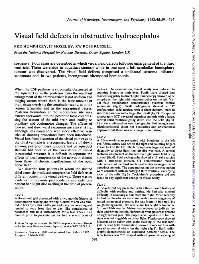
Journal of Neurology, Neurosurgery, and Psychiatry 1982;45:591-597 Visual field defects in obstructive hydrocephalus PRD HUMPHREY, IF MOSELEY, RW ROSS RUSSELL From the National Hospital for Nervous Diseases, Queen Square, London UK SUMMARY Four cases are described in which visual field defects followed enlargement of the third ventricle. Three were due to aqueduct stenosis while in one case a left cerebellar hemisphere tumour was discovered. The visual field defects comprised a unilateral scotoma, bilateral scotomata and, in two patients, incongruous bitemporal hemianopia. When the CSF pathway is chronically obstructed at the aqueduct or in the posterior fossa the resultant enlargement of the third ventricle is not uniform and bulging occurs where there is the least amount of brain tissue overlying the ventricular cavity, as at the lamina terminalis and in the suprapineal recess. Posterior herniation at the suprapineal site may extend backwards into the posterior fossa compres- sing the tectum of the mid brain and leading to pupillary and oculomotor changes. The effects of forward and downward expansion are also striking, although less commonly seen since effective ven- tricular shunting procedures have been introduced. Visual loss from distortion of the visual pathways by the third ventricle is a recognised feature of slowly growing posterior fossa tumours and of aqueduct stenosis but because of the coexistence of raised intracranial pressure it is difficult to separate the effects of local compression of the nerves or chiasm from those of chronic papilloedema of the optic nerve head. We describe four patients in whom the dilated third ventricle produced compressive field defects at different points in the visual pathway. There was no evidence of previous papilloedema and only one patient had slight disc swelling at the time of presen- tation. Case 1 A 9-year-old girl presented with a two months history of deteriorating reading and writing. Central vision was blur- red in both eyes; this had begun suddenly one morning and tended to vary from day to day. She complained of headaches and mild unsteadiness for a few weeks. Ten months prior to presentation she had a severe bout of Address for reprint requests: Dr PRD Humphrey, National Hospi- tal for Nervous Diseases, Queen Square, London WCI 3BG, UK. Received 4 December 1981 and in revised form 5 March 1982. Accepted 10 March 1982. measles. On examination, visual acuity was reduced to counting fingers in both eyes. Pupils were dilated and reacted sluggishly to direct light. Fundoscopy showed optic atrophy on the right with temporal pallor on the left. Vis- ual field examination demonstrated bilateral central scotomata (fig 1). Skull radiographs showed a "J" configuration sella turcica, with a short dorsum, marked suture separation and a large, thin vault (fig 2). Computed tomography (CT) revealed aqueduct stenosis with a large, central third ventricle going down into the sella (fig 3). This was confirmed on ventriculography. Following a ven- triculoperitoneal shunt her headaches and unsteadiness improved but there was no change in her vision. Case 2 A 30-year-old man presented with blindness in the left eye. Visual acuity was 6/9 on the right and counting fingers at two feet on the left. The left pupil was large and reacted sluggishly to direct light; the left disc was pale. A central scotoma was present on the left, the right visual field being normal (fig 4). Skull radiographs showed a "J' sella turcica with a truncated dorsum. CT demonstrated marked enlargement of the third and lateral ventricles suggestive of aqueduct stenosis. The appearances on the ventriculogram were consistent with an enlarged third ventricle, occupying most of the sella (fig 5). Torkildsen's procedure did not result in any significant change in visual acuity. Case 3 A 12-year-old boy presented with a three month history of difficulty with reading and writing. He had also noticed difficulty in receiving a ball from the right. More recently he had had headaches associated with nausea suggestive of raised intracranial pressure. He was found to be small, his weight being on the 1 0th centile and his height between the 3rd and 10th centile. Vision was reduced to 6/60 on the right and 6/9 on the left. Horizontal nystagmus was present on right lateral gaze. The pupils were equal in size but the right reacted sluggishly to direct light. Fundoscopy showed bilateral optic pallor with slight swelling of the disc mar- gins. Visual field examination showed temporal loss with spread to central vision on the right (fig 6). Skull radio- graphs demonstrated an expanded posterior fossa. The sella turcica was "J" shaped without gross shortening of 591 copyright. on 1 July 2019 by guest. Protected by http://jnnp.bmj.com/ J Neurol Neurosurg Psychiatry: first published as 10.1136/jnnp.45.7.591 on 1 July 1982. Downloaded from
Transcript of Visual field defects in obstructive hydrocephalus · Visualfield defects in obstructive...
Journal of Neurology, Neurosurgery, and Psychiatry 1982;45:591-597
Visual field defects in obstructive hydrocephalusPRD HUMPHREY, IF MOSELEY, RW ROSS RUSSELL
From the National Hospital for Nervous Diseases, Queen Square, London UK
SUMMARY Four cases are described in which visual field defects followed enlargement of the thirdventricle. Three were due to aqueduct stenosis while in one case a left cerebellar hemispheretumour was discovered. The visual field defects comprised a unilateral scotoma, bilateralscotomata and, in two patients, incongruous bitemporal hemianopia.
When the CSF pathway is chronically obstructed atthe aqueduct or in the posterior fossa the resultantenlargement of the third ventricle is not uniform andbulging occurs where there is the least amount ofbrain tissue overlying the ventricular cavity, as at thelamina terminalis and in the suprapineal recess.Posterior herniation at the suprapineal site mayextend backwards into the posterior fossa compres-sing the tectum of the mid brain and leading topupillary and oculomotor changes. The effects offorward and downward expansion are also striking,although less commonly seen since effective ven-tricular shunting procedures have been introduced.Visual loss from distortion of the visual pathways bythe third ventricle is a recognised feature of slowlygrowing posterior fossa tumours and of aqueductstenosis but because of the coexistence of raisedintracranial pressure it is difficult to separate theeffects of local compression of the nerves or chiasmfrom those of chronic papilloedema of the opticnerve head.We describe four patients in whom the dilated
third ventricle produced compressive field defects atdifferent points in the visual pathway. There was noevidence of previous papilloedema and only onepatient had slight disc swelling at the time of presen-tation.
Case 1A 9-year-old girl presented with a two months history ofdeteriorating reading and writing. Central vision was blur-red in both eyes; this had begun suddenly one morning andtended to vary from day to day. She complained ofheadaches and mild unsteadiness for a few weeks. Tenmonths prior to presentation she had a severe bout of
Address for reprint requests: Dr PRD Humphrey, National Hospi-tal for Nervous Diseases, Queen Square, London WCI 3BG, UK.
Received 4 December 1981 and in revised form 5 March 1982.Accepted 10 March 1982.
measles. On examination, visual acuity was reduced tocounting fingers in both eyes. Pupils were dilated andreacted sluggishly to direct light. Fundoscopy showed opticatrophy on the right with temporal pallor on the left. Vis-ual field examination demonstrated bilateral centralscotomata (fig 1). Skull radiographs showed a "J"configuration sella turcica, with a short dorsum, markedsuture separation and a large, thin vault (fig 2). Computedtomography (CT) revealed aqueduct stenosis with a large,central third ventricle going down into the sella (fig 3).This was confirmed on ventriculography. Following a ven-triculoperitoneal shunt her headaches and unsteadinessimproved but there was no change in her vision.
Case 2A 30-year-old man presented with blindness in the lefteye. Visual acuity was 6/9 on the right and counting fingersat two feet on the left. The left pupil was large and reactedsluggishly to direct light; the left disc was pale. A centralscotoma was present on the left, the right visual field beingnormal (fig 4). Skull radiographs showed a "J' sella turcicawith a truncated dorsum. CT demonstrated markedenlargement of the third and lateral ventricles suggestive ofaqueduct stenosis. The appearances on the ventriculogramwere consistent with an enlarged third ventricle, occupyingmost of the sella (fig 5). Torkildsen's procedure did notresult in any significant change in visual acuity.
Case 3A 12-year-old boy presented with a three month history ofdifficulty with reading and writing. He had also noticeddifficulty in receiving a ball from the right. More recentlyhe had had headaches associated with nausea suggestive ofraised intracranial pressure. He was found to be small, hisweight being on the 1 0th centile and his height between the3rd and 10th centile. Vision was reduced to 6/60 on theright and 6/9 on the left. Horizontal nystagmus was presenton right lateral gaze. The pupils were equal in size but theright reacted sluggishly to direct light. Fundoscopy showedbilateral optic pallor with slight swelling of the disc mar-gins. Visual field examination showed temporal loss withspread to central vision on the right (fig 6). Skull radio-graphs demonstrated an expanded posterior fossa. Thesella turcica was "J" shaped without gross shortening of
591
copyright. on 1 July 2019 by guest. P
rotected byhttp://jnnp.bm
j.com/
J Neurol N
eurosurg Psychiatry: first published as 10.1136/jnnp.45.7.591 on 1 July 1982. D
ownloaded from
Humphrey, Moseley, Russell
*ii i't,*r .. -pr C
a
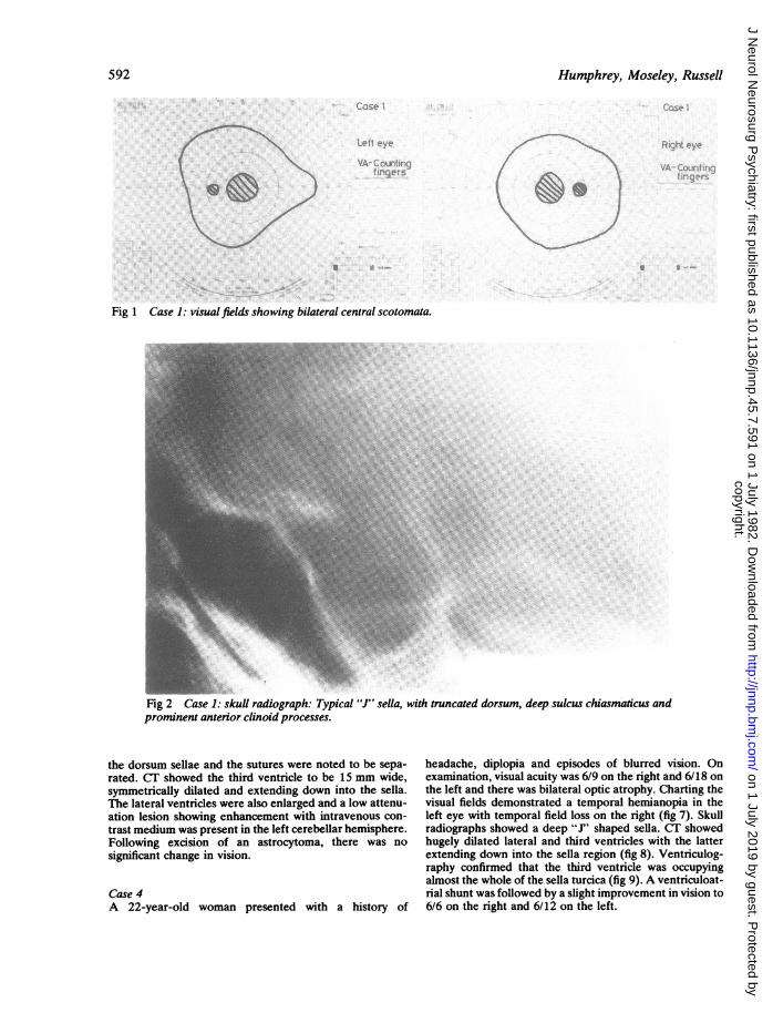
Fig 1 Case 1: visual fields showing bilateral central scotomata.
Fig 2 Case 1: skull radiograph: Typical "J" sella, with truncated dorsum, deep sulcus chiasmaticus andprominent anterior clinoid processes.
the dorsum sellae and the sutures were noted to be sepa-rated. CT showed the third ventricle to be 15 mm wide,symmetrically dilated and extending down into the sella.The lateral ventricles were also enlarged and a low attenu-ation lesion showing enhancement with intravenous con-trast medium was present in the left cerebellar hemisphere.Following excision of an astrocytoma, there was nosignificant change in vision.
Case 4A 22-year-old woman presented with a history of
headache, diplopia and episodes of blurred vision. Onexamination, visual acuity was 6/9 on the right and 6/18 onthe left and there was bilateral optic atrophy. Charting thevisual fields demonstrated a temporal hemianopia in theleft eye with temporal field loss on the right (fig 7). Skullradiographs showed a deep "J" shaped sella. CT showedhugely dilated lateral and third ventricles with the latterextending down into the sella region (fig 8). Ventriculog-raphy confirmed that the third ventricle was occupyingalmost the whole of the sella turcica (fig 9). A ventriculoat-rial shunt was followed by a slight improvement in vision to6/6 on the right and 6/12 on the left.
592
Q. .11lellcNl-
i kk Iw.-v
copyright. on 1 July 2019 by guest. P
rotected byhttp://jnnp.bm
j.com/
J Neurol N
eurosurg Psychiatry: first published as 10.1136/jnnp.45.7.591 on 1 July 1982. D
ownloaded from

Visual field defects in obstructive hydrocephalus
Fig 3 Case 1: CT: there is marked hydrocephalus involving all four ventricles; penrventricular lucencies are present,especially around the frontal horns (black and white arrows). The dilated third ventricle extends right down to thesella region (horizontal arrows).
.Se
Le!t r-yC, U, la
'r0CUi ;
Fig4Case2:visualfieldsshowingleftcentals ict i.
Fig 4 Case 2: visual fields showing left central scotoma.
Discussion
The pathogenesis of visual field defects in patientswith posterior fossa lesions or aqueduct stenosis hasbeen debated since the last century. Cushing andWalker were amongst the first to realise the impor-tance of the dilated third ventricle acting as amechanical compressive and distorting force on thevisual pathways.' They found binasal defects to bethe most frequent result and proposed that thedilated ventricle displaced both optic nerves out-wards against the resistant carotid arteries. The fielddefects shown by their patients indicate a general-ised contraction of the fields more marked on thenasal side, which might well have been the result ofchronic papilloedema.2 They encountered nopatients with bitemporal hemianopia. However,
bitemporal defects were convincingly shown by Sinc-lair and Dott in a patient with postmeningitic obs-tructive hydrocephalus.3 At operation the chiasmwas shown to be broadened, and pushed downwardsand forwards by the bulging anterior wall of thethird ventricle. After ventriculostomy the visionimproved. Hughes, in a careful study of the evolu-tion of the field defect in third ventricular enlarge-ment, stressed the importance of the position of thechiasm.4 He suggested that if the chiasm was post-fixed or in its normal position the enlarging thirdventricle would impinge against the posterior aspectand spread over the superior surface tending to pro-duce indrawing of the outer visual isopters of thetemporal fields (depressional field). If on the otherhand the chiasm was prefixed the ventricle pressedagainst its posterior and inferior aspects sometimes
c 4F ... ,
uiy-,
593
copyright. on 1 July 2019 by guest. P
rotected byhttp://jnnp.bm
j.com/
J Neurol N
eurosurg Psychiatry: first published as 10.1136/jnnp.45.7.591 on 1 July 1982. D
ownloaded from

594 Humphrey, Moseley, Russell
A..~~~~~~~~~~~~.
Fig 5Case 2: ventriculogram, lateral projection: the anterior end ofthe third ventricle,grosslydilated (arrows), occupies the suprasella region, and much of the sella turcica, resting on thedorsum (open arrow).
Case3Ca 3TEST OBJECTS NAME TEST OBJECTS NAME Cs
SIZEOFTESTOBJECTINMM_
WHITE ~~~~~~~~~~DATESIZE OF TEST OBJECTS IN M.M DATE6 WHITE 6I'LUE ~~~~~~~~~~~VISION BLUE VISION
Lefteyes1
10
1000 ..A WhiteWhite~..
19eFig 6 Case 3: bitemporal hemianopia with loss ofcentral vision on the right.
copyright. on 1 July 2019 by guest. P
rotected byhttp://jnnp.bm
j.com/
J Neurol N
eurosurg Psychiatry: first published as 10.1136/jnnp.45.7.591 on 1 July 1982. D
ownloaded from

Visual field defects in obstructive hydrocephalus
TEST OBJECTSSIZE OF TEST OBJECTS IN M MWHITEBLUEREDGREEN
1.
NAME Case 4 TEST OBJECTS9ZE OF TEST OBJECTS IN MM.WHITEBLUEREDGREEN
Right eye
102000
White10
2000
Whate
Fig 7 Case 4: visual fields demonstrating incongruous bitemporal hemianopia.
Fig 8 Case 4: CT: a widened anterior third ventricle(arrow) lies between the anterior clinoid processes, in the
position normally occupied by the pituitary. The dorsumsellae is not seen. The fourth ventricle is normal.
595
NAME
copyright. on 1 July 2019 by guest. P
rotected byhttp://jnnp.bm
j.com/
J Neurol N
eurosurg Psychiatry: first published as 10.1136/jnnp.45.7.591 on 1 July 1982. D
ownloaded from

Humphrey, Moseley, Russell
..
'r
Fig 9 Case 4: ventriculogram. (a) anteroposterior, brow up projection. The lateral ventricles are grosslydilated, as is the third ventricle (open arrows). (b) lateral, brow up projection. An enlarged pituitary fossais almost entirely occupied by the anterior end ofthe third ventricle (arrows), which has eroded the dorsumsellae (open arrow). The dense black central shadow is caused by air in both temporal horns superimposedon the third ventricle.
596
copyright. on 1 July 2019 by guest. P
rotected byhttp://jnnp.bm
j.com/
J Neurol N
eurosurg Psychiatry: first published as 10.1136/jnnp.45.7.591 on 1 July 1982. D
ownloaded from
Visual field defects in obstructive hydrocephalus
bulging beneath it to lie between the optic nerves. Inthis case he proposed that the early field defectswere bitemporal scotomas appearing first in thelower temporal quadrants in the paracentral region.Lassman, Cullen and Howat, describing a patient
with an upper temporal defect in one eye only, indi-cating a lesion near the junction of nerve andchiasm, proposed that the lower aspect of the nerveand chiasm were displaced downwards by thedilated ventricle and were pressed forcibly againstthe planum sphenoidale.5 Junctional defects of thistype are also mentioned by Wagener and Cusick.6
Optic nerve defects are distinctly unusual. Bilat-eral central scotomas are mentioned by Wagenerand Cusick in a catalogue of defects in patients withposterior fossa lesions and a single case wasremarked upon by Pennybacker.7 A patient withcomplete unilateral blindness and a normal con-tralateral field was described by Calogero and Alex-ander.8 There has been some speculation as to how acentral herniation could cause a unilateral opticnerve lesion but the close proximity of the anteriorcerebral arteries, which are frequently unequal insize may result in compression of a distorted opticnerve.Homonymous hemianopias have also been
reported to result from an enlarged third ventricle.In the four patients with cerebellar tumoursdescribed by Weinberger and Webster9 the defectswere congruous and might possibly have been theresult of a posterior cerebral artery occlusion; themechanism suggested by the authors was downwardpressure on the tract by dilated third ventricleagainst the posterior communicating artery.The present report is a further illustration of the
variety of field defects. Two of our patients hadbitemporal hemianopias (both asymmetrical), one aunilateral scotoma and one bilateral centralscotomas. The latter defect is particularly unusualand is a possible source of misdiagnosis becausewhile a progressive unilateral optic nerve lesion orbitemporal hemianopia always suggests a compres-sive lesion, a patient with bilateral central scotomasis more likely to have a demyelinating or toxicneuropathy than a compressive lesion.No firm conclusions can be reached on the
mechanism of visual loss. While enlargement of thethird ventricle into the pituitary fossa is likely to bean important factor in the development of opticnerve, chiasmal or tract lesions, it is doubtful if this
597
is the sole reason for these visual field defects.Schechter and Zingesser showed that in the ven-triculogram in aqueduct stenosis the third ventriclefrequently enlarges to occupy the pituitary fossa.'0Visual defects such as those reported here are, how-ever, rare. The ventriculographic and CT appear-ances in our patients were typical of those found inpatients with obstructive hydrocephalus. All fourpatients showed extension of the hypothalamicrecess into the pituitary fossa rather than extensionof the supraoptic recess over the chiasm. Enlarge-ment of this type would be likely to impinge on theposterior aspect of the chiasm and since the chiasmbears a fixed relationship to the recesses it seemsunlikely that pre or postfixation of the chiasm wouldassert a major influence on the type of field defect.
We are grateful to Mr J Garfield from the WessexNeurological Centre for allowing us to report case 4.
References
'Cushing H, Walker CB. Distortion of the visual fields incases of brain tumour. Arch Ophthalmol1912;41:559-98.
2 Osher RH, Corbett JJ, Schatz NJ, Saisho PJ, Orr LS.Neuro-ophthalmological complications of enlarge-ment of the third ventricle. Br J Ophthalmol1978;62:536-42.
3Sinclair AHH, Dott NM. Hydrocephalus simulatingtumour in the production of chiasmal and other para-hypophysial lesions. Trans Ophthalmol Soc UK1931 ;51:232-46.
Hughes EBC. Some observations on the visual fields inhydrocephalus. J Neuro Neurosurg Psychiatry1946;9:30-39.
5Lassman LP, Cullen JF, Howat JM. Stenosis of theaqueduct of Sylvius. Am J Ophthalmol1960;49:261-6.
6 Wagener HP, Cusick PL. Chiasmal syndromes producedby lesions in the posterior fossa. Arch Ophthalmol1937;18:887-91.
7 Pennybacker J. Stenosis of the aqueduct of Sylvius. ProcSoc Med 1940;33:507-12.
Calogero JA, Alexander E. Unilateral amaurosis in ahydrocephalic child with an obstructed shunt. JNeurosurg 1971;34:236-40.
9 Weinberger LM, Webster JE. Visual field defects associ-ated with cerebellar tumours. Arch Ophthalmol1941;25:128-38.
10 Schechter MM, Zingesser LH. The radiology ofaqueductal stenosis. Radiology, 1967;88:905-16.
copyright. on 1 July 2019 by guest. P
rotected byhttp://jnnp.bm
j.com/
J Neurol N
eurosurg Psychiatry: first published as 10.1136/jnnp.45.7.591 on 1 July 1982. D
ownloaded from