
Viruses Prof. Khaled H. Abu-Elteen Hashemite University.
56
Viruses Prof. Khaled H. Abu- Elteen Hashemite University
-
Upload
martina-hodge -
Category
Documents
-
view
226 -
download
0
Transcript of Viruses Prof. Khaled H. Abu-Elteen Hashemite University.

Viruses
Prof. Khaled H. Abu-Elteen
Hashemite University
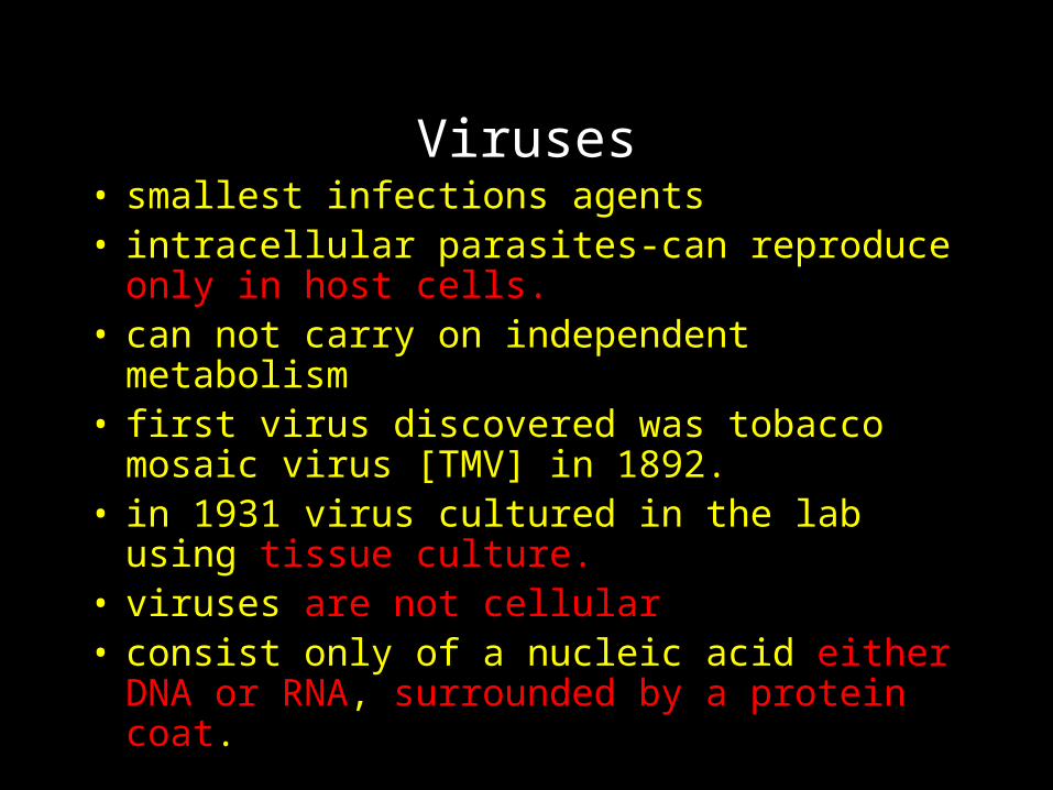
Viruses• smallest infections agents• intracellular parasites-can reproduce only in host
cells. • can not carry on independent metabolism• first virus discovered was tobacco mosaic virus
[TMV] in 1892. • in 1931 virus cultured in the lab using tissue
culture. • viruses are not cellular• consist only of a nucleic acid either DNA or RNA,
surrounded by a protein coat.
Virus facts
• generally more resistant to some disinfectants than most bacteria.
• most are susceptible to heat, except hepatitis virus
• not affected by antibiotics
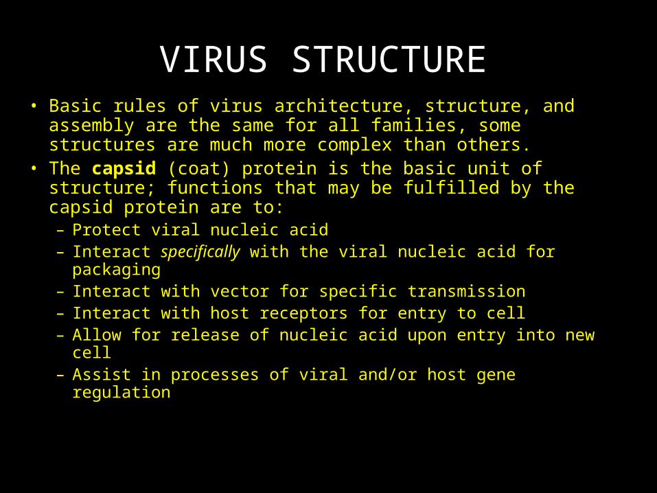
VIRUS STRUCTURE• Basic rules of virus architecture, structure, and assembly
are the same for all families, some structures are much more complex than others.
• The capsid (coat) protein is the basic unit of structure; functions that may be fulfilled by the capsid protein are to:– Protect viral nucleic acid– Interact specifically with the viral nucleic acid for packaging– Interact with vector for specific transmission – Interact with host receptors for entry to cell – Allow for release of nucleic acid upon entry into new cell– Assist in processes of viral and/or host gene regulation
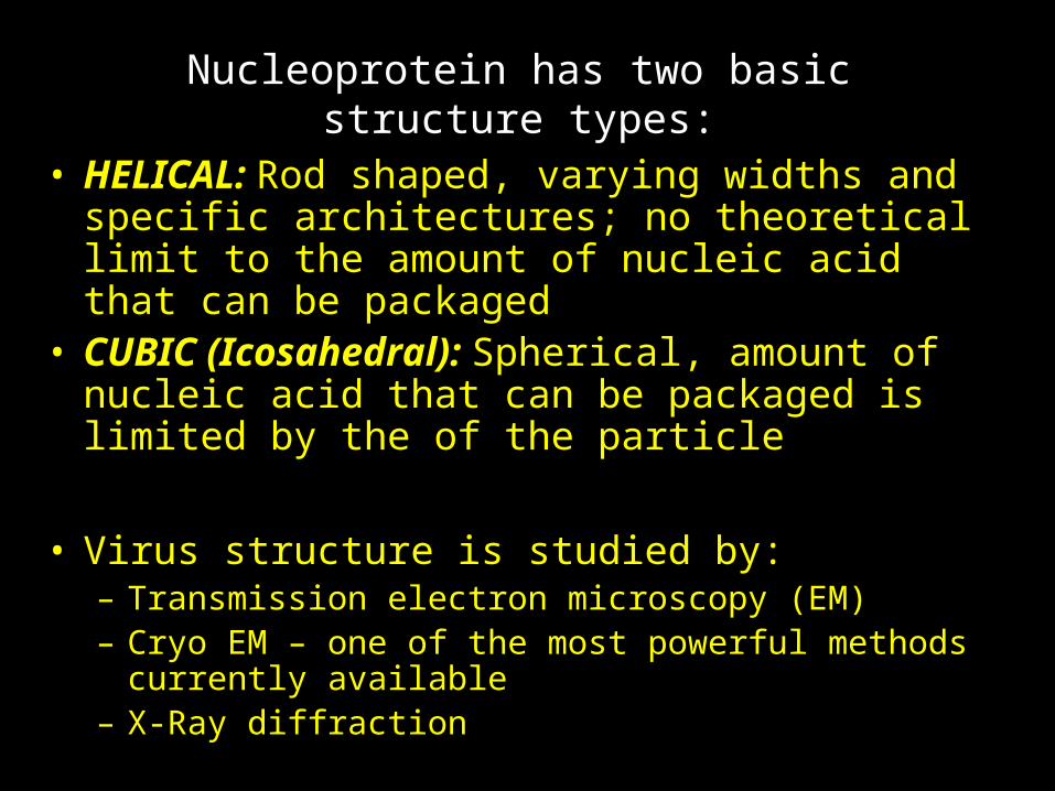
Nucleoprotein has two basic structure types:
• HELICAL: Rod shaped, varying widths and specific architectures; no theoretical limit to the amount of nucleic acid that can be packaged
• CUBIC (Icosahedral): Spherical, amount of nucleic acid that can be packaged is limited by the of the particle
• Virus structure is studied by:– Transmission electron microscopy (EM)– Cryo EM – one of the most powerful methods currently
available– X-Ray diffraction
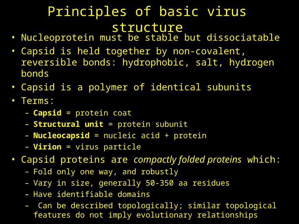
Principles of basic virus structure• Nucleoprotein must be stable but dissociatable• Capsid is held together by non-covalent, reversible bonds:
hydrophobic, salt, hydrogen bonds• Capsid is a polymer of identical subunits• Terms:
– Capsid = protein coat– Structural unit = protein subunit– Nucleocapsid = nucleic acid + protein– Virion = virus particle
• Capsid proteins are compactly folded proteins which:– Fold only one way, and robustly– Vary in size, generally 50-350 aa residues– Have identifiable domains– Can be described topologically; similar topological features do not
imply evolutionary relationships
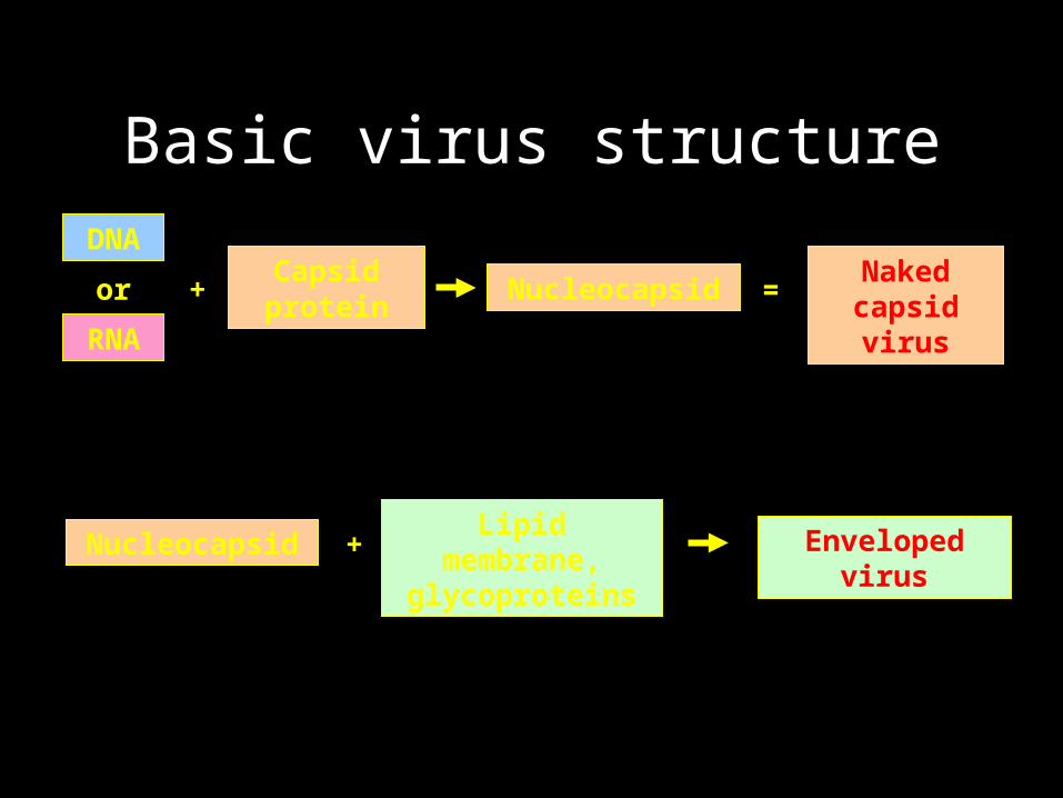
Basic virus structure
Capsid protein
NucleocapsidNaked
capsid virus
DNA
RNA
or =+
NucleocapsidLipid membrane,
glycoproteinsEnveloped virus+
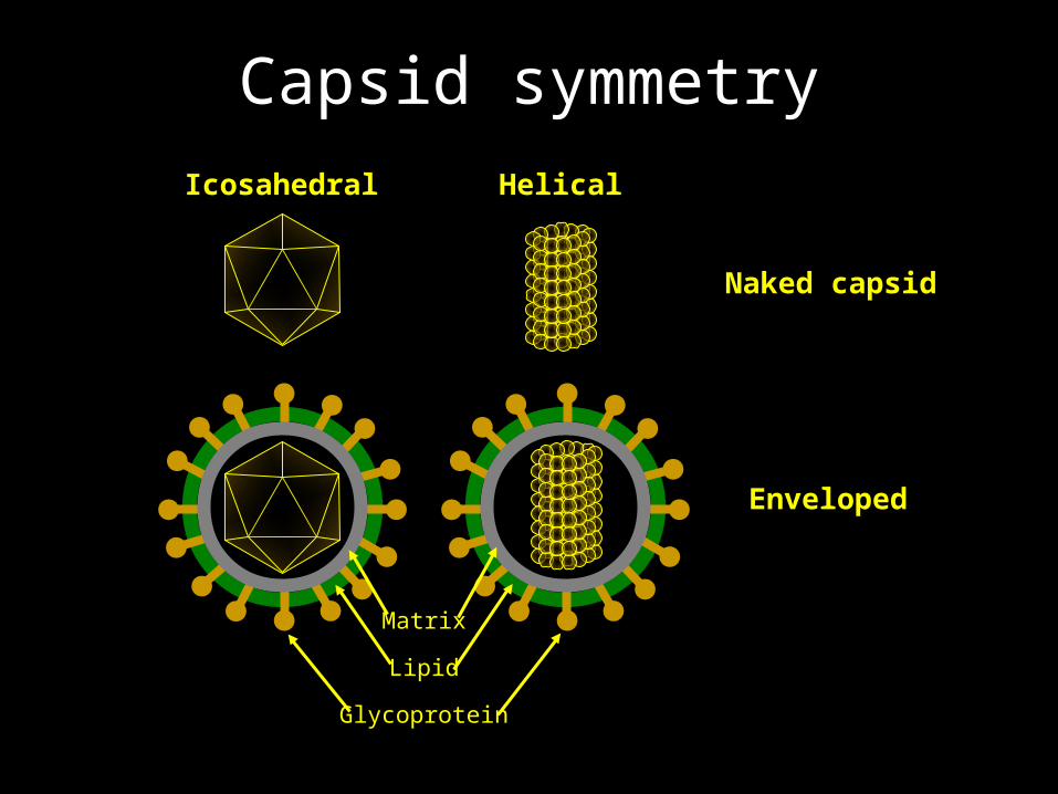
Capsid symmetry
Icosahedral Helical
Naked capsid
Enveloped
Lipid
Glycoprotein
Matrix
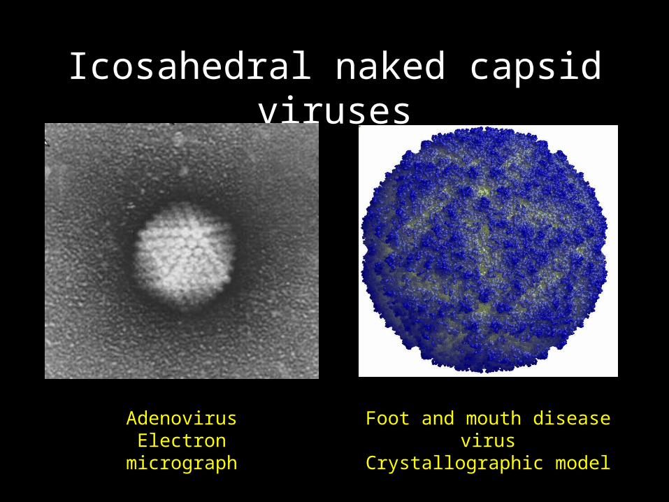
Icosahedral naked capsid viruses
AdenovirusElectron micrograph
Foot and mouth disease virusCrystallographic model
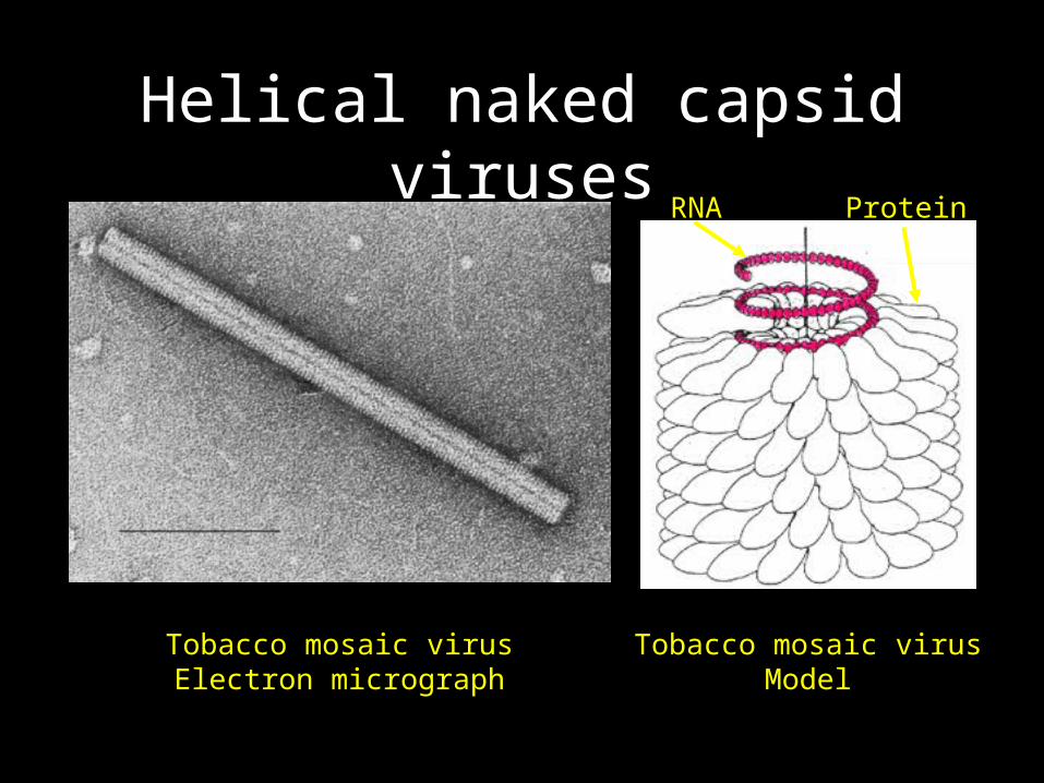
Helical naked capsid viruses
Tobacco mosaic virusElectron micrograph
Tobacco mosaic virusModel
RNA Protein
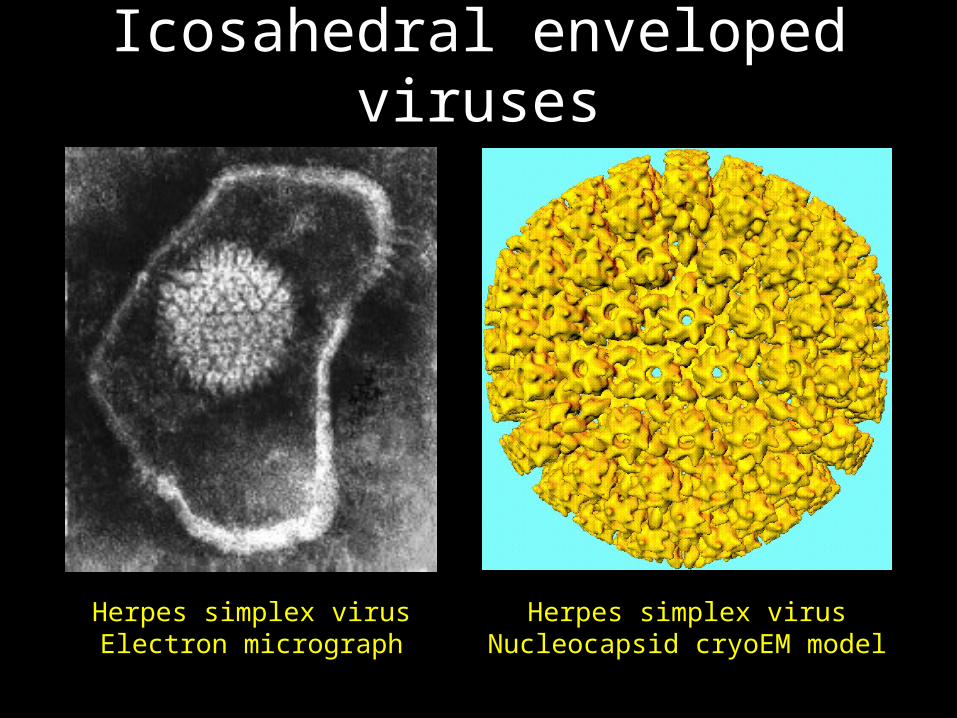
Icosahedral enveloped viruses
Herpes simplex virusElectron micrograph
Herpes simplex virusNucleocapsid cryoEM model
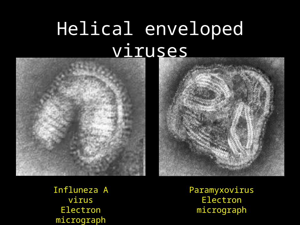
Helical enveloped viruses
Influneza A virusElectron micrograph
ParamyxovirusElectron micrograph

Properties of enveloped viruses
• Envelope is sensitive to– Drying– Heat– Detergents– Acid
• Consequences– Must stay wet during transmission– Transmission in large droplets and secretions– Cannot survive in the gastrointestinal tract– Do not need to kill cells in order to spread– May require both a humoral and a cellular immune
response
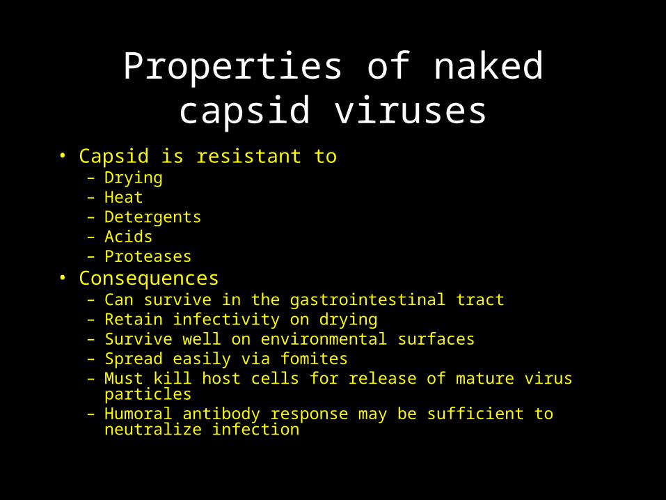
Properties of naked capsid viruses
• Capsid is resistant to– Drying– Heat– Detergents– Acids– Proteases
• Consequences– Can survive in the gastrointestinal tract– Retain infectivity on drying– Survive well on environmental surfaces– Spread easily via fomites– Must kill host cells for release of mature virus particles– Humoral antibody response may be sufficient to neutralize infection
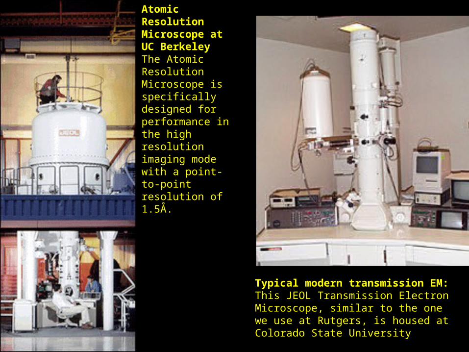
Atomic Resolution Microscope at UC Berkeley The Atomic Resolution Microscope is specifically designed for performance in the high resolution imaging mode with a point-to-point resolution of 1.5Å.
Typical modern transmission EM: This JEOL Transmission Electron Microscope, similar to the one we use at Rutgers, is housed at Colorado State University
Classification of viruses
• on the basis of:• nucleic acid they contain ( DNA or RNA )• the size, shape and structure of the virus • the tissue the infect
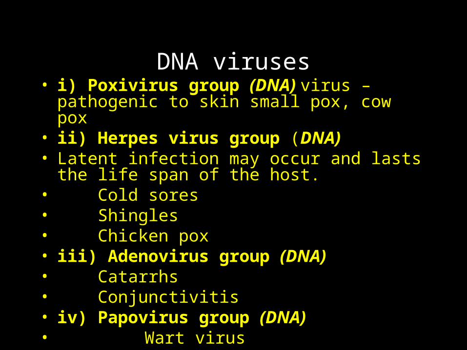
DNA viruses• i) Poxivirus group (DNA) virus – pathogenic to skin
small pox, cow pox• ii) Herpes virus group (DNA) • Latent infection may occur and lasts the life span of
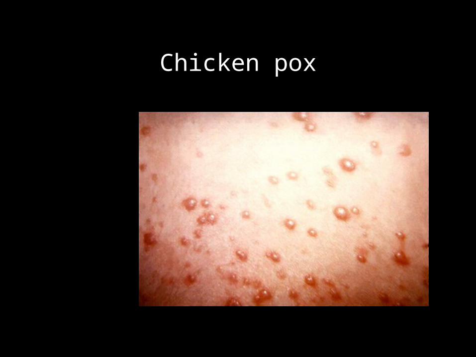
the host. • Cold sores• Shingles• Chicken pox• iii) Adenovirus group (DNA)• Catarrhs• Conjunctivitis• iv) Papovirus group (DNA)• Wart virus
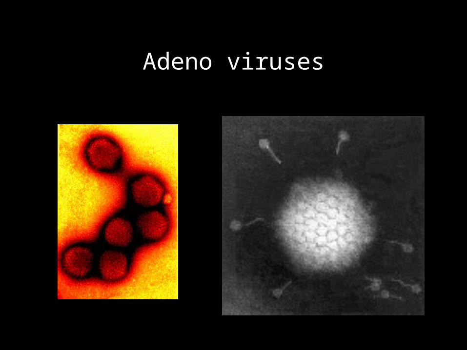
Adeno viruses
Adenovirus-Associated Human Disease
Pharyngitis Acute Respiratory Disease Pneumonia Pharyngoconjunctival Fever Epidemic Keratoconjuntivitis Genitourinary Infections (cervicitis, urethritis ) Gasteroenteritis Some asymptomatic and persistent infection Adenovirus oncogenically tranforms rodent cells but not human cells.
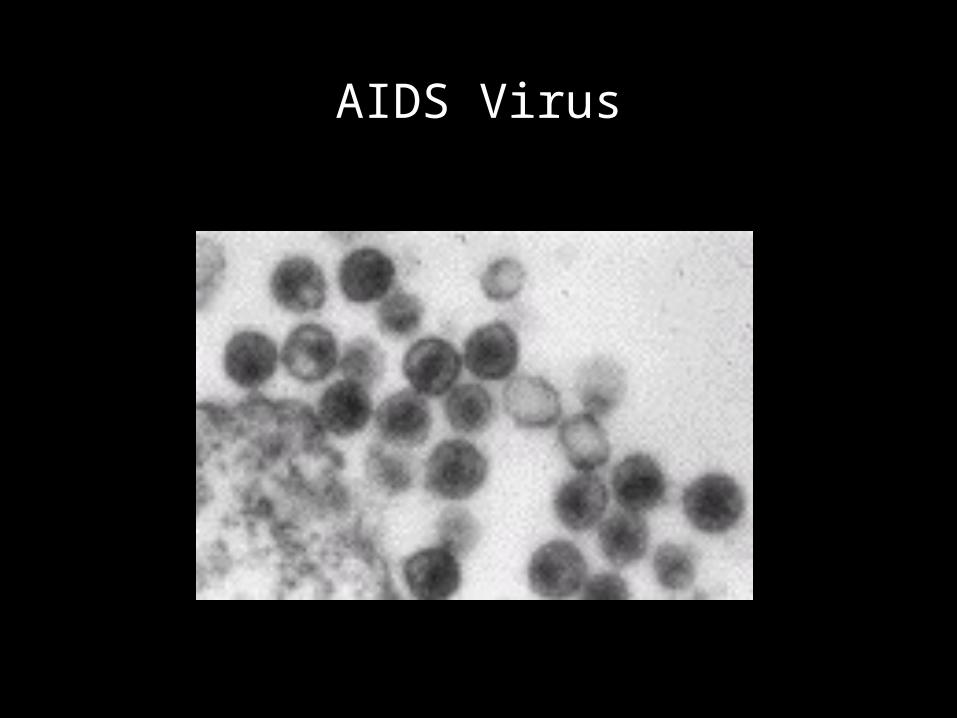
AIDS Virus
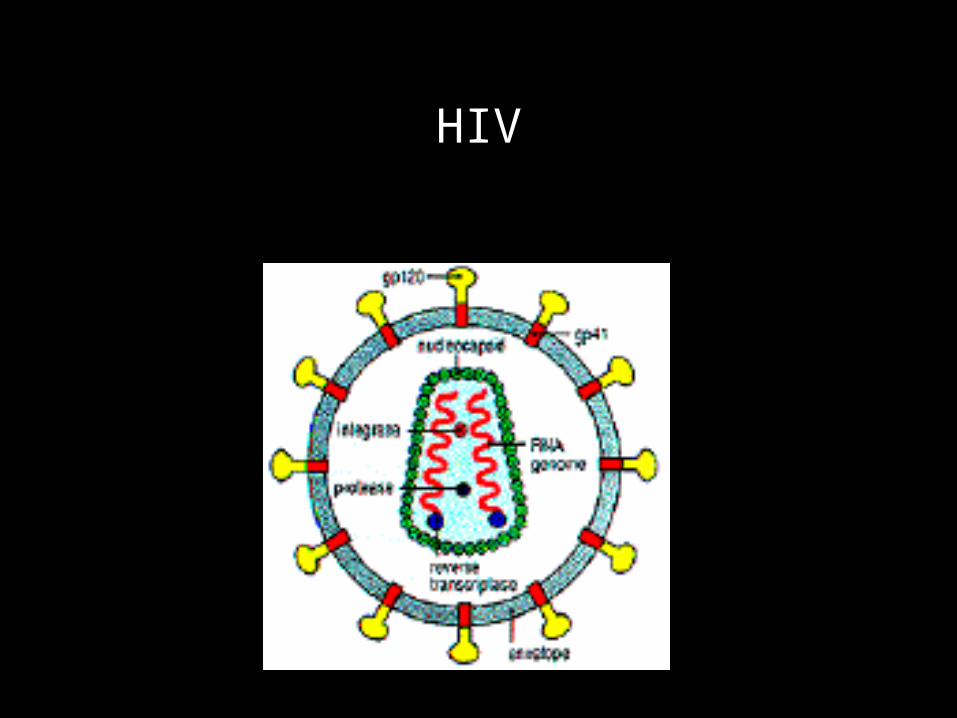
HIV
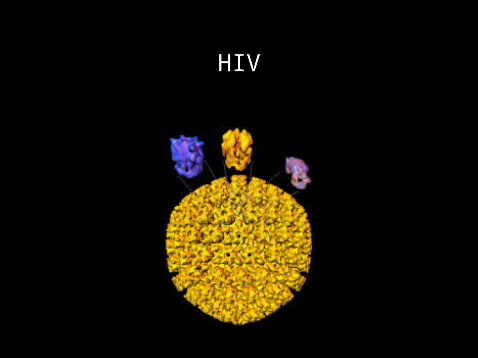
HIV
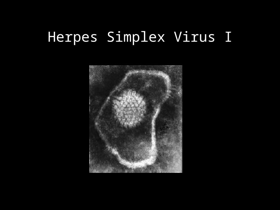
Herpes Simplex Virus I
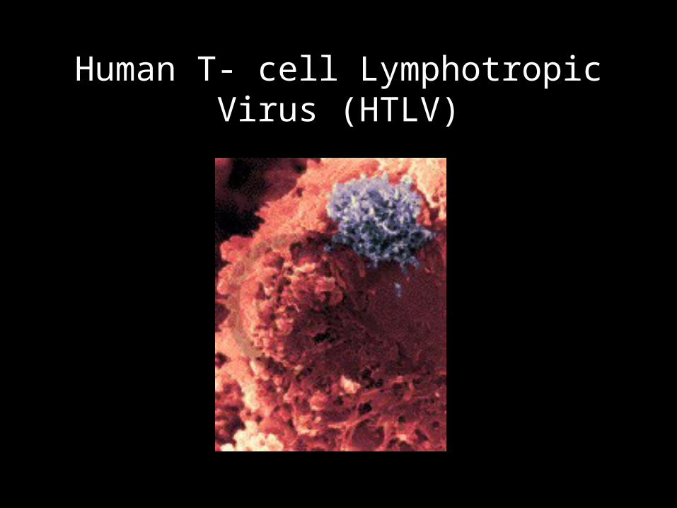
Human T- cell Lymphotropic Virus (HTLV)
Human T- cell Lymphotropic Virus
• HTLV-1 stands for Human T-cell Lymphotropic Virus.
• It is a retrovirus, in the same class of virus as the AIDS virus, HIV-1.
• HTLV-I is associated with a rare form of blood dsycrasia known as Adult T-cell Leukemia/lymphoma (ATLL) and a myelopathy, tropical spastic paresis.
• However, even with infection, fewer than 4% of seropositive persons will experience overt associated disease.
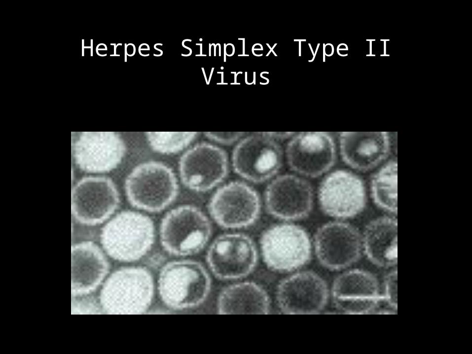
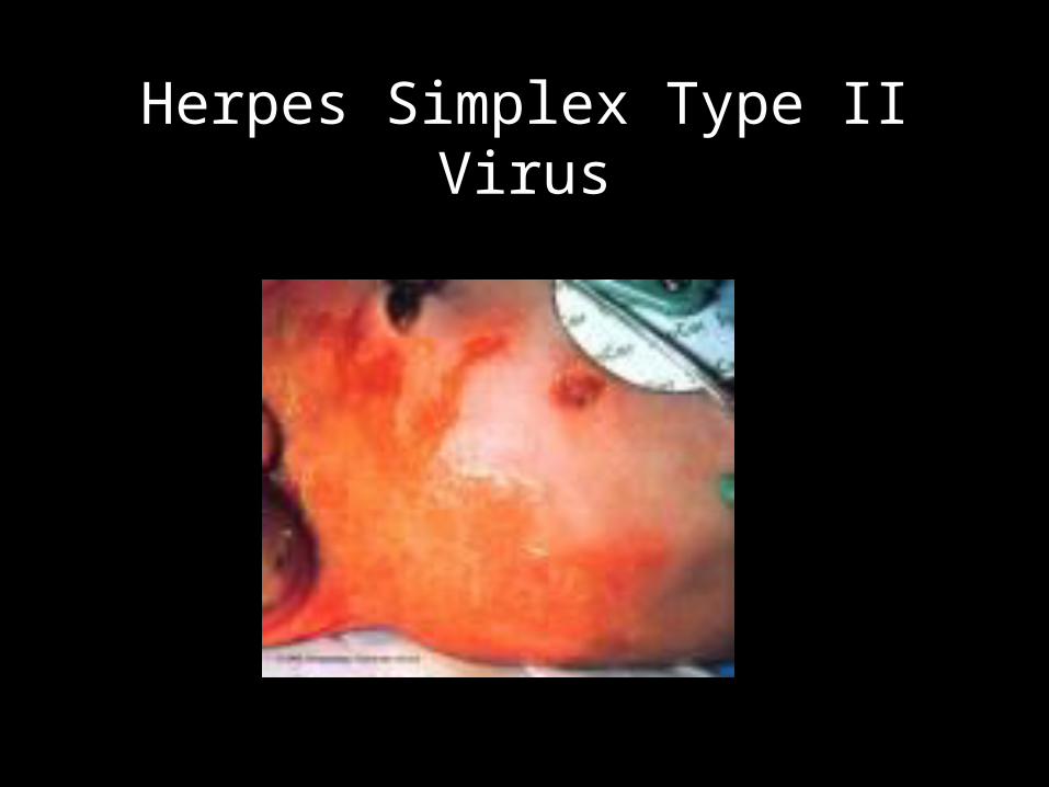
Herpes Simplex Type II Virus
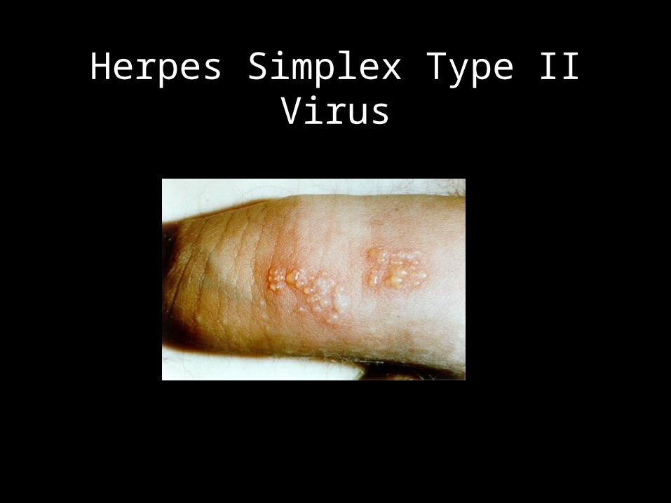
Herpes Simplex Type II Virus
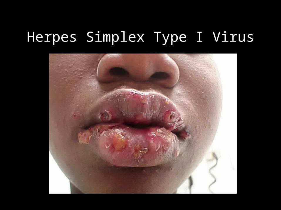
Herpes Simplex Type I Virus
Hepatitis
• Hepatitis• a. chemically induced• b. viral infection A, B, C, D, E, F• Viral hepatitis is the most common liver disease
found worldwide. • Epstein Barr virus• Herpes virus • Cytomegalovirus
Hepatitis B (HBV)
• DNA virus• has an outer surface structure known as hepatitis B
surface antigen (HBs Ag) & an inner core component known as hepatitis B core Antigen (HBc Ag)
• Long incubation period—up to 6 months. • Transmitted through blood contact. • Some modes of transmission as those for HIV.• HBV is very serious illness. • Series of 3 immunizations are given on day 0, 30, 180.
Hepatitis C
• Blood borne pathogen. • Also found in water like HV-A • Many become carriers
Hepatitis D
• Super-infects some patients who are already infected with HBV.
• HBV is required as a helper to initiate infection.
• blood borne.

Hepatitis A Virus
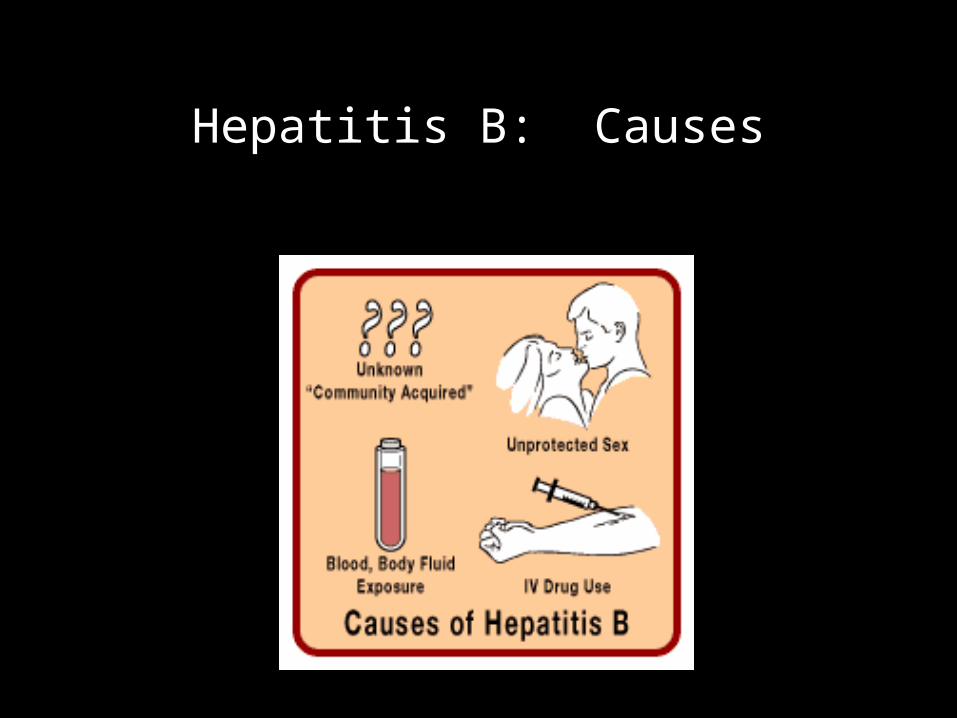
Hepatitis B: Causes
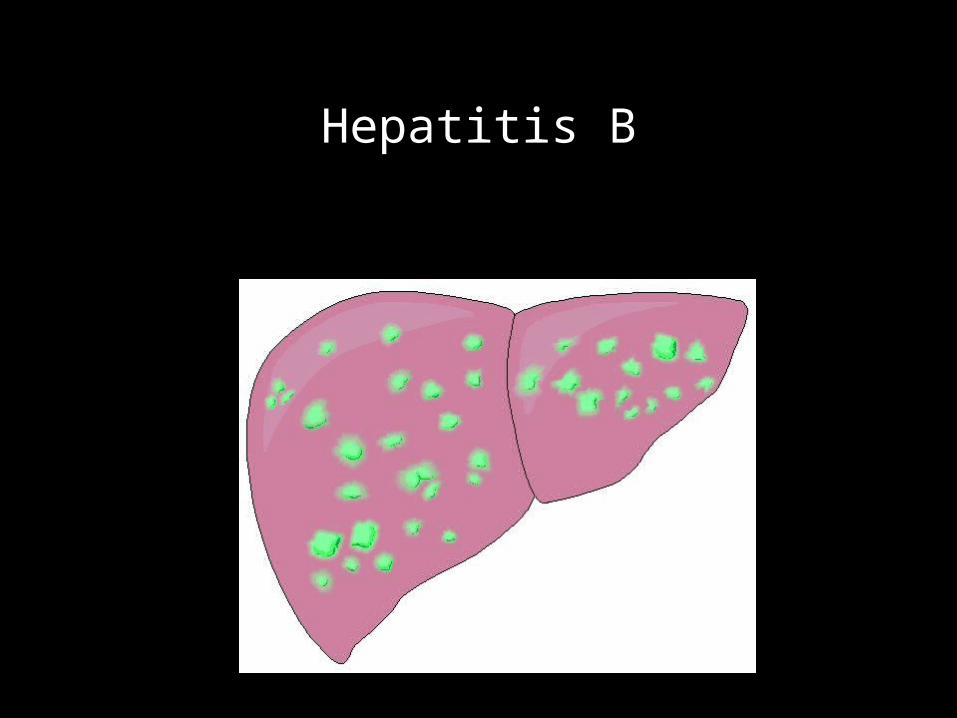
Hepatitis B

Hepatitis C: Getting Tattoos
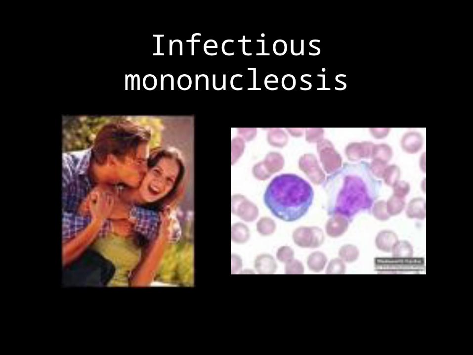
Infectious mononucleosis
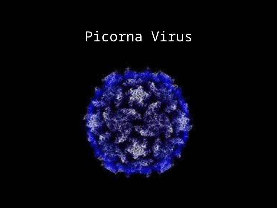
Picorna Virus
Picorna Virus
Primary site of infection is lymphoid tissue associated with the oropharynx and gut (GALT).
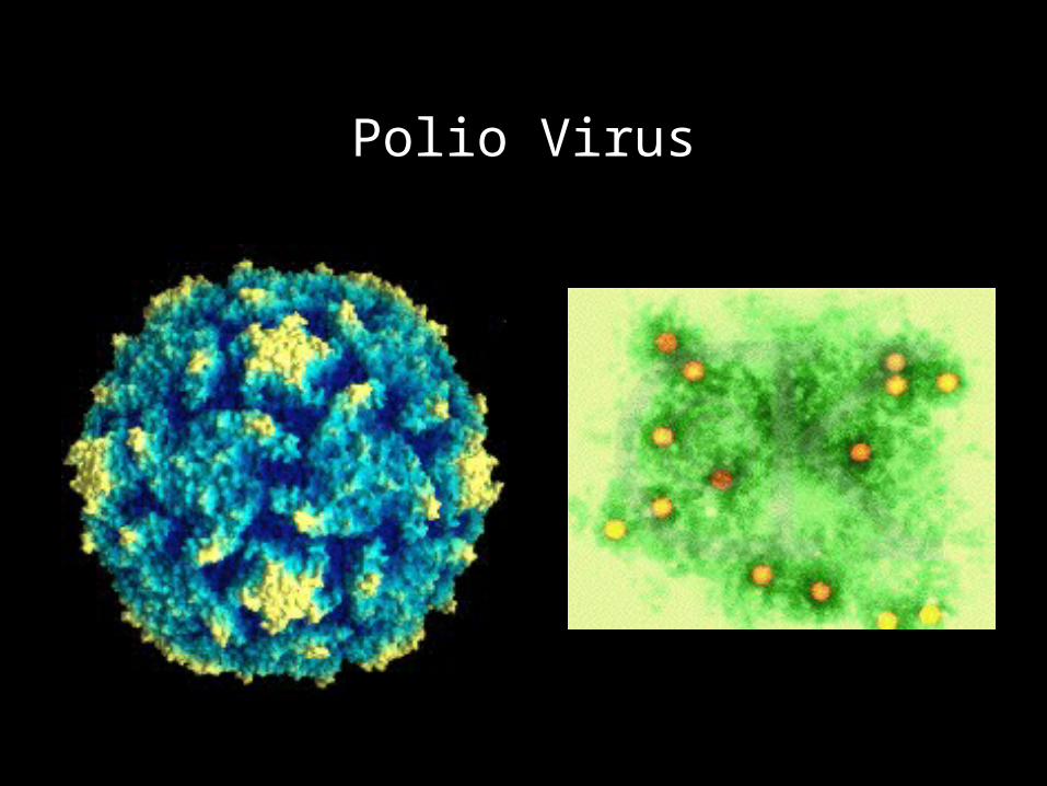
Polio Virus
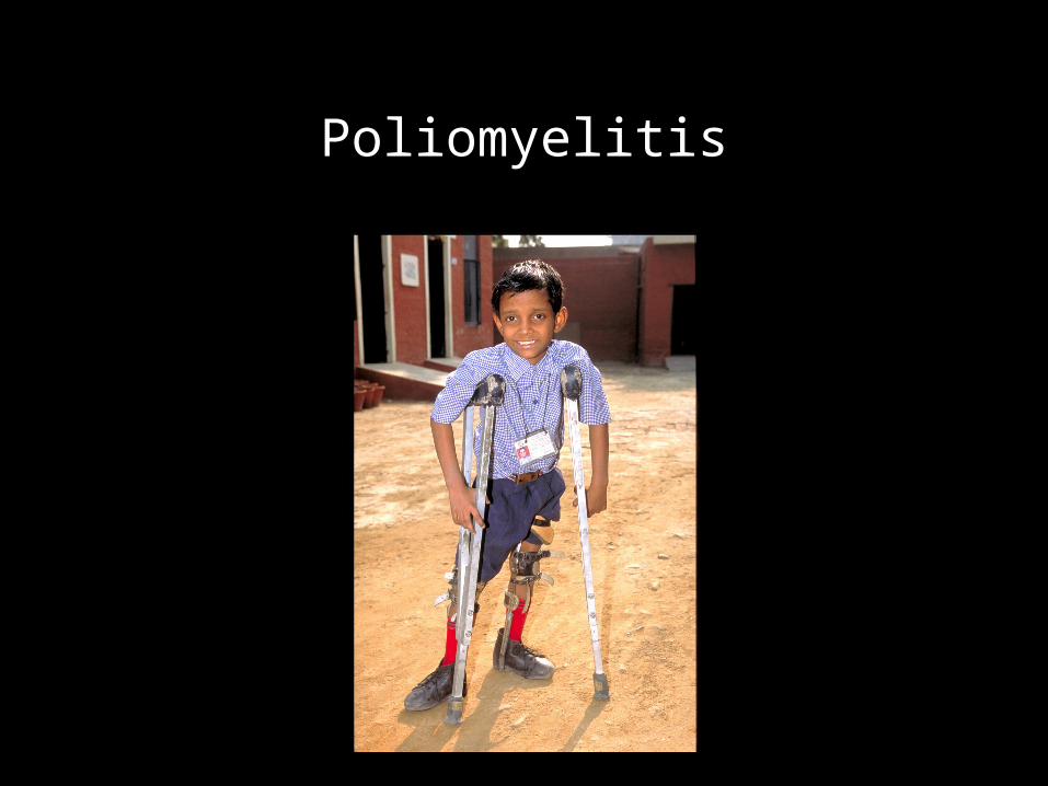
Poliomyelitis
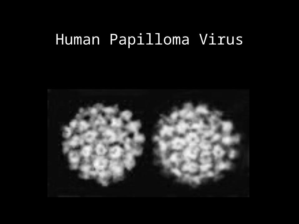
Human Papilloma Virus
Genital Warts - HPV
• Causes genital warts
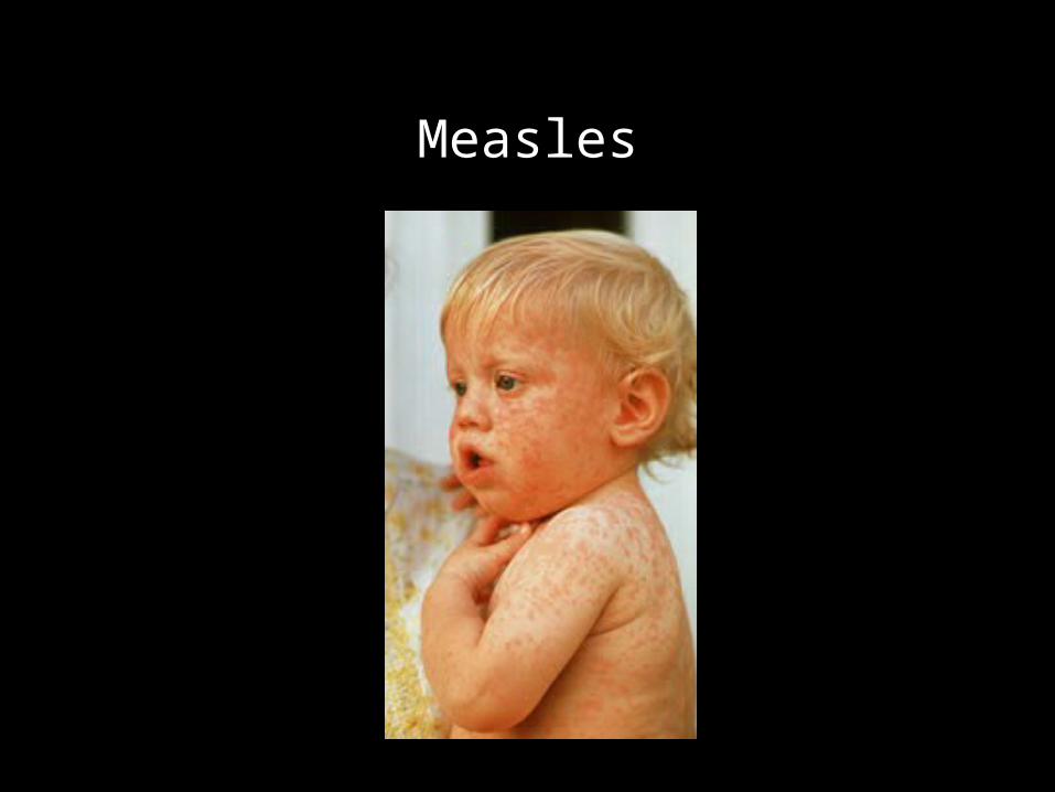
Measles
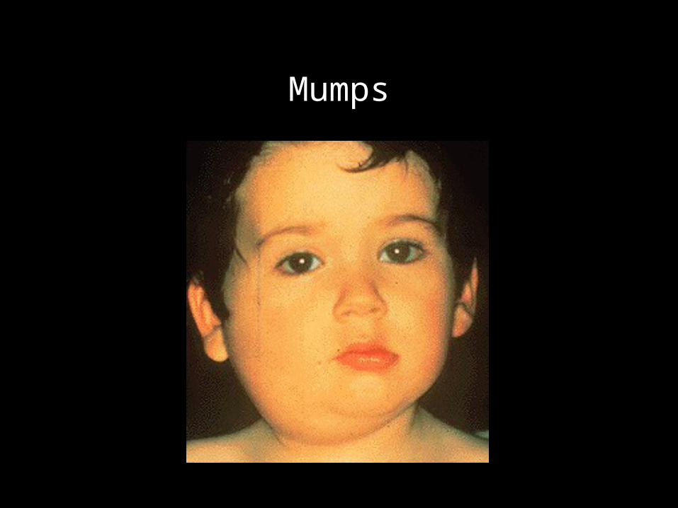
Mumps
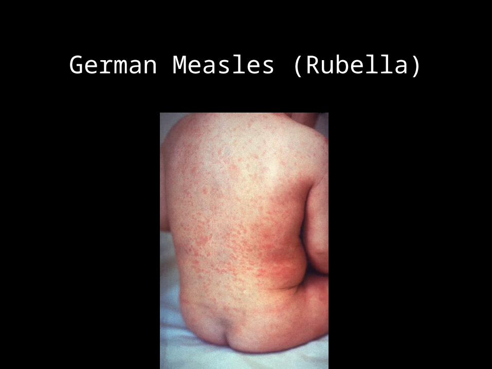
German Measles (Rubella)
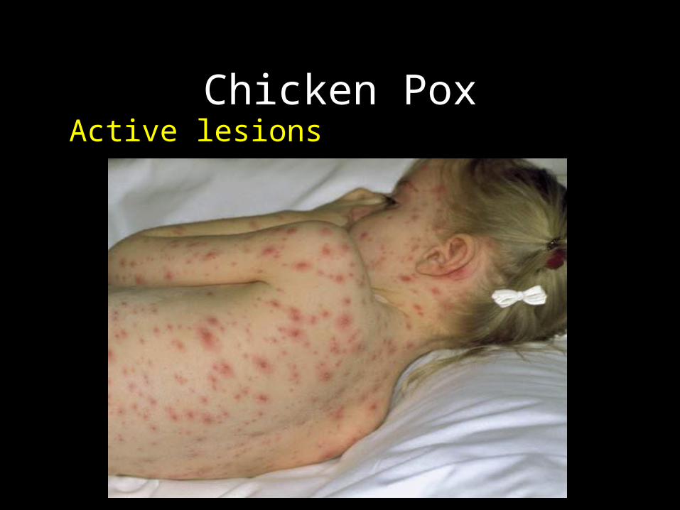
Chicken PoxActive lesions
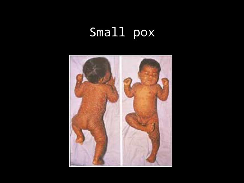
Small pox
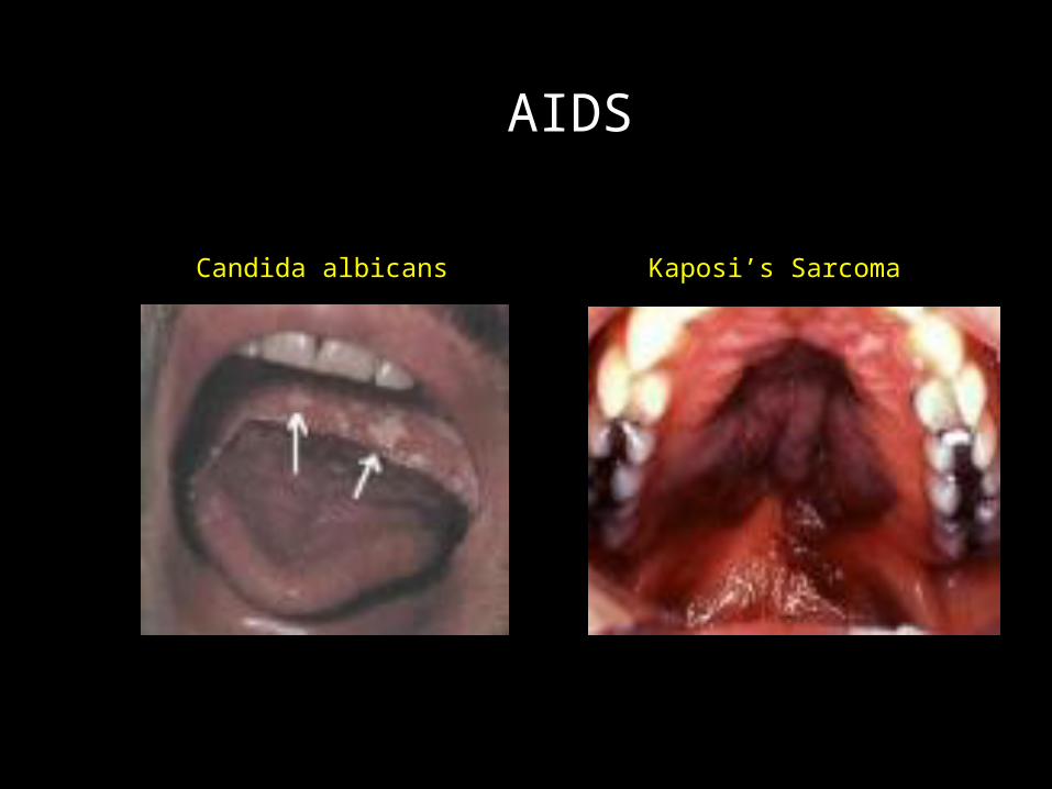
AIDS
Candida albicans Kaposi’s Sarcoma
HIV• Incubation period (the period between becoming
infected and the actual development of the symptoms) • 6 months-5 or more years, up to 10 years. • Sometimes a mild illness--flu like symptoms appears
7-14 days after infection• Sometimes no symptoms appear for years. • It is accepted that once infected with HIV, AIDS will
develop at some time in the future in all cases. • At present there is no cure. • Opportunistic infections associated with AIDS can be
treated.
HIV
• HIV is carried in blood, semen, & body fluids. • usually fatal• known to be dormant for years• certain drug combinations slow the rate of
invasion of the White Blood cells by the virus.• cure is not yet on the horizon • leading cause of death in young adults, aged 25-44
AIDS
• Retrovirus- an RNA virus that carries an enzyme capable of forming DNA from RNA.
• Aids virus infects T. Lymphocytes (Helper T- cells)• patient may be asymptomatic before diagnosis• affects the immune system• patients are prone to develop opportunistic
infections, malignancies, and neurological disorders
• fatal disease• no treatment
AIDS
• More common in I.V. drug users & homosexuals.• Pneumocytic carinii infection and blood vessel
malignancy-Kaposi’s Sarcoma

Atomic Resolution Microscope at UC Berkeley The Atomic Resolution Microscope is specifically designed for performance in the high resolution imaging mode with a point-to-point resolution of 1.5Å.
Typical modern transmission EM: This JEOL Transmission Electron Microscope, similar to the one we use at Rutgers, is housed at Colorado State University