Virus Subtype-Specific Features of Natural Simian ...
11
JOURNAL OF VIROLOGY, Aug. 2007, p. 7913–7923 Vol. 81, No. 15 0022-538X/07/$08.000 doi:10.1128/JVI.00281-07 Copyright © 2007, American Society for Microbiology. All Rights Reserved. Virus Subtype-Specific Features of Natural Simian Immunodeficiency Virus SIV smm Infection in Sooty Mangabeys Cristian Apetrei, 1,2 * Rajeev Gautam, 1 Beth Sumpter, 3 Anders C. Carter, 1 Thaidra Gaufin, 1 Silvija I. Staprans, 3 James Else, 3 Mary Barnes, 1 Robert Cao, Jr., 1 Seema Garg, 3 Jeffrey M. Milush, 4 Donald L. Sodora, 4 Ivona Pandrea, 5,6 and Guido Silvestri 3,7 Divisions of Microbiology 1 and Comparative Pathology, 5 Tulane National Primate Research Center, Covington, Louisiana 70433; Department of Tropical Medicine of the School of Public Health 2 and Department of Pathology, School of Medicine, 6 Tulane University, New Orleans, Louisiana 70112; Emory Vaccine Center and Yerkes National Research Primate Center, Emory University, Atlanta, Georgia 3 ; University of Texas Southwestern Medical Center, Dallas, Texas 4 ; and Department of Pathology and Laboratory Medicine, University of Pennsylvania, Philadelphia, Pennsylvania 19107 7 Received 9 February 2007/Accepted 7 May 2007 Simian immunodeficiency virus (SIV) SIV smm naturally infects sooty mangabeys (SMs) and is the source virus of pathogenic infections with human immunodeficiency virus type 2 (HIV-2) and SIV mac of humans and macaques, respectively. In previous studies we characterized SIV smm diversity in naturally SIV-infected SMs and identified nine different phylogenetic subtypes whose genetic distances are similar to those reported for the different HIV-1 group M subtypes. Here we report that, within the colony of SMs housed at the Yerkes National Primate Research Center, at least four SIV smm subtypes cocirculate, with the vast majority of animals infected with SIV smm subtype 1, 2, or 3, resulting in the emergence of occasional recombinant forms. While SIV smm - infected SMs show a typically nonpathogenic course of infection, we have observed that different SIV smm subtypes are in fact associated with specific immunologic features. Notably, while subtypes 1, 2, and 3 are associated with a very benign course of infection and preservation of normal CD4 T-cell counts, three out of four SMs infected with subtype 5 show a significant depletion of CD4 T cells. The fact that virus replication in SMs infected with subtype 5 is similar to that in SMs infected with other SIV smm subtypes suggests that the subtype 5-associated CD4 T-cell depletion is unlikely to simply reflect higher levels of virus-mediated direct killing of CD4 T-cells. Taken together, this systematic analysis of the subtype-specific features of SIV smm infection in natural SM hosts identifies subtype-specific differences in the pathogenicity of SIV smm infection. The AIDS pandemic originated from zoonotic transmission to humans of simian CD4 T-cell-tropic lentiviruses that infect African monkey species and are collectively defined as simian immunodeficiency viruses (SIV) (12, 24, 26). In marked con- trast to human immunodeficiency virus (HIV)-infected hu- mans, who, if untreated, almost invariably develop progressive CD4 T-cell depletion and AIDS, the African monkey species naturally infected with SIV are generally spared from any signs of disease (reviewed in references 28, 33, 54, and 64). Con- versely, experimental SIV infection of nonnatural host Asian macaque species causes symptoms similar to those described for AIDS patients (simian AIDS) (reviewed in reference 32). At present, it is still unclear why SIV infection is relatively nonpathogenic in natural host monkey species but induces immunodeficiency in nonnatural or recent hosts (including hu- mans). A better understanding of the mechanisms underlying the lack of disease in natural hosts for SIV infection will likely provide important clues as to the pathogenesis of AIDS in HIV-infected individuals (58, 59). Among the natural hosts for SIV infection, the sooty manga- beys (SMs) (Cercocebus atys) are particularly relevant for two reasons. First, SIV smm , the virus infecting SMs, is the root cause of the HIV type 2 (HIV-2) epidemic in humans (12, 25). Second, SIV smm was used to generate the various rhesus ma- caque (Rh)-adapted SIV mac /SIV smm viruses (i.e., SIV mac239 and SIV smm543-3 ) that are commonly used for studies of AIDS pathogenesis and vaccines (reviewed in reference 32). SIV smm - infected SMs typically maintain normal CD4 T-cell counts and do not develop AIDS despite intensive virus replication, with levels of plasma viremia that are as high as, or even higher than, those observed in HIV-infected individuals (11, 51, 56). Therefore, unlike both HIV-infected individuals and experi- mentally SIV-infected Rhs, in which high levels of virus repli- cation predict faster disease progression (31, 35, 39, 40, 47, 66), in SMs the levels of SIV smm viral loads (VLs) do not seem to have a predictive value for the outcome of infection (11, 51, 56). Natural SIV infection of SMs is characterized by near-nor- mal CD4 T-cell counts in both blood and lymph nodes, nor- mal T-cell turnover, preserved bone marrow and thymic func- tion, and absence of the generalized immune activation and bystander T-cell apoptosis that are typical of HIV-infected individuals (11, 56, 60). Similarly, no signs of disease progres- sion and relatively low levels of immune activation were ob- served in SMs experimentally infected with uncloned SIV smm or molecularly cloned SIV mac239 (34, 55). Taken together, these results led to the hypothesis that the attenuated immune activation in SIV-infected SMs is a mechanism that favors the * Corresponding author. Mailing address: Division of Microbiology and Immunology, Tulane National Primate Research Center, 18703 Three Rivers Road, Covington, LA 70433. Phone: (985) 871-6518. Fax: (985) 871-6248. E-mail: [email protected]. Published ahead of print on 16 May 2007. 7913 on March 20, 2018 by guest http://jvi.asm.org/ Downloaded from
Transcript of Virus Subtype-Specific Features of Natural Simian ...
JOURNAL OF VIROLOGY, Aug. 2007, p. 7913–7923 Vol. 81, No. 150022-538X/07/$08.00�0 doi:10.1128/JVI.00281-07Copyright © 2007, American Society for Microbiology. All Rights Reserved.
Virus Subtype-Specific Features of Natural Simian ImmunodeficiencyVirus SIVsmm Infection in Sooty Mangabeys�
Cristian Apetrei,1,2* Rajeev Gautam,1 Beth Sumpter,3 Anders C. Carter,1 Thaidra Gaufin,1Silvija I. Staprans,3 James Else,3 Mary Barnes,1 Robert Cao, Jr.,1 Seema Garg,3
Jeffrey M. Milush,4 Donald L. Sodora,4 Ivona Pandrea,5,6 and Guido Silvestri3,7
Divisions of Microbiology1 and Comparative Pathology,5 Tulane National Primate Research Center, Covington, Louisiana 70433;Department of Tropical Medicine of the School of Public Health2 and Department of Pathology, School of Medicine,6
Tulane University, New Orleans, Louisiana 70112; Emory Vaccine Center and Yerkes National Research Primate Center,Emory University, Atlanta, Georgia3; University of Texas Southwestern Medical Center, Dallas, Texas4; and
Department of Pathology and Laboratory Medicine, University ofPennsylvania, Philadelphia, Pennsylvania 191077
Received 9 February 2007/Accepted 7 May 2007
Simian immunodeficiency virus (SIV) SIVsmm naturally infects sooty mangabeys (SMs) and is the sourcevirus of pathogenic infections with human immunodeficiency virus type 2 (HIV-2) and SIVmac of humans andmacaques, respectively. In previous studies we characterized SIVsmm diversity in naturally SIV-infected SMsand identified nine different phylogenetic subtypes whose genetic distances are similar to those reported for thedifferent HIV-1 group M subtypes. Here we report that, within the colony of SMs housed at the Yerkes NationalPrimate Research Center, at least four SIVsmm subtypes cocirculate, with the vast majority of animals infectedwith SIVsmm subtype 1, 2, or 3, resulting in the emergence of occasional recombinant forms. While SIVsmm-infected SMs show a typically nonpathogenic course of infection, we have observed that different SIVsmmsubtypes are in fact associated with specific immunologic features. Notably, while subtypes 1, 2, and 3 areassociated with a very benign course of infection and preservation of normal CD4� T-cell counts, three out offour SMs infected with subtype 5 show a significant depletion of CD4� T cells. The fact that virus replicationin SMs infected with subtype 5 is similar to that in SMs infected with other SIVsmm subtypes suggests that thesubtype 5-associated CD4� T-cell depletion is unlikely to simply reflect higher levels of virus-mediated directkilling of CD4� T-cells. Taken together, this systematic analysis of the subtype-specific features of SIVsmminfection in natural SM hosts identifies subtype-specific differences in the pathogenicity of SIVsmm infection.
The AIDS pandemic originated from zoonotic transmissionto humans of simian CD4� T-cell-tropic lentiviruses that infectAfrican monkey species and are collectively defined as simianimmunodeficiency viruses (SIV) (12, 24, 26). In marked con-trast to human immunodeficiency virus (HIV)-infected hu-mans, who, if untreated, almost invariably develop progressiveCD4� T-cell depletion and AIDS, the African monkey speciesnaturally infected with SIV are generally spared from any signsof disease (reviewed in references 28, 33, 54, and 64). Con-versely, experimental SIV infection of nonnatural host Asianmacaque species causes symptoms similar to those describedfor AIDS patients (simian AIDS) (reviewed in reference 32).At present, it is still unclear why SIV infection is relativelynonpathogenic in natural host monkey species but inducesimmunodeficiency in nonnatural or recent hosts (including hu-mans). A better understanding of the mechanisms underlyingthe lack of disease in natural hosts for SIV infection will likelyprovide important clues as to the pathogenesis of AIDS inHIV-infected individuals (58, 59).
Among the natural hosts for SIV infection, the sooty manga-beys (SMs) (Cercocebus atys) are particularly relevant for two
reasons. First, SIVsmm, the virus infecting SMs, is the rootcause of the HIV type 2 (HIV-2) epidemic in humans (12, 25).Second, SIVsmm was used to generate the various rhesus ma-caque (Rh)-adapted SIVmac/SIVsmm viruses (i.e., SIVmac239
and SIVsmm543-3) that are commonly used for studies of AIDSpathogenesis and vaccines (reviewed in reference 32). SIVsmm-infected SMs typically maintain normal CD4� T-cell countsand do not develop AIDS despite intensive virus replication,with levels of plasma viremia that are as high as, or even higherthan, those observed in HIV-infected individuals (11, 51, 56).Therefore, unlike both HIV-infected individuals and experi-mentally SIV-infected Rhs, in which high levels of virus repli-cation predict faster disease progression (31, 35, 39, 40, 47, 66),in SMs the levels of SIVsmm viral loads (VLs) do not seemto have a predictive value for the outcome of infection (11,51, 56).
Natural SIV infection of SMs is characterized by near-nor-mal CD4� T-cell counts in both blood and lymph nodes, nor-mal T-cell turnover, preserved bone marrow and thymic func-tion, and absence of the generalized immune activation andbystander T-cell apoptosis that are typical of HIV-infectedindividuals (11, 56, 60). Similarly, no signs of disease progres-sion and relatively low levels of immune activation were ob-served in SMs experimentally infected with uncloned SIVsmm
or molecularly cloned SIVmac239 (34, 55). Taken together,these results led to the hypothesis that the attenuated immuneactivation in SIV-infected SMs is a mechanism that favors the
* Corresponding author. Mailing address: Division of Microbiologyand Immunology, Tulane National Primate Research Center, 18703Three Rivers Road, Covington, LA 70433. Phone: (985) 871-6518. Fax:(985) 871-6248. E-mail: [email protected].
� Published ahead of print on 16 May 2007.
7913
on March 20, 2018 by guest
http://jvi.asm.org/
Dow
nloaded from
preservation of CD4� T-cell homeostasis and, thus, the non-pathogenic state of this chronic infection.
Interestingly, the SIVsmm cross-species transmission to bothhumans and macaques has specific epidemiological and patho-genic features, with some of the strains showing better fitnessin terms of transmission to and replication in the new hosts.Only groups A and B of HIV-2 resulted in epidemics (19, 25),whereas the remaining HIV-2 groups act as epidemiologicaldead ends in humans, with lower pathogenicity and no docu-mented human-to-human transmission (12, 25, 67). Similarly,SIVmac strains clearly have higher pathogenic potential thanother cross-species-transmitted SIVsmm strains both in Rhs andin other macaque species, such as pig-tailed macaques orstump-tailed macaques (32, 42, 46). We and others suggestedthat a key factor beyond these differences in pathogenicity ofcross-species-transmitted SIVsmm in the macaque host may bethe serial passage in vitro and/or in vivo (4, 6, 8, 10, 30).However, the alternative hypothesis that different SIVsmm
strains show intrinsic differences in pathogenicity cannot bedismissed.
The recent discovery of a significant SIVsmm diversity innaturally infected SIVs from primate centers in the UnitedStates (3) offered us the possibility to directly test this laterhypothesis. In this study we have characterized the SIVsmm
diversity within the large colony of naturally SIV-infected SMshoused at the Yerkes National Primate Research Center(YNPRC) and correlated the prognostic markers of diseaseprogression (levels of CD4� T cells, VLs, and T-cell activation)with SIVsmm lineage. We have also performed a longitudinalretrospective study of the dynamics of virus replication in nat-urally SIV-infected SMs from the Tulane National PrimateResearch Center (TNPRC) over a 20-year period. While thesestudies failed to reveal any significant differences between theknown SIVsmm lineages in terms of virus replication or pro-gression to AIDS, a significant association between subtype 5infection and CD4� T-cell depletion was observed.
MATERIALS AND METHODS
Animals. The cross-sectional study included 110 naturally SIV-infected SMsfrom YNPRC and 29 from TNPRC. At least one blood sample was collectedfrom all animals. The retrospective study involved serum samples collected overa 20-year period from 20 naturally infected SMs from the TNPRC, which wereinfected with six different subtypes as follows: eight with subtype 1 (AO24, EO38,EO39, EO41, GO79, M923, M927, and M939), three with subtype 2 (D177,GO80, and M946), three with subtype 3 (AO23, F102, and M942), three withsubtype 4 (G930, G931, and G932), two with subtype 5 (D175 and FO98), andone with subtype 6 (D215). At least five serum samples and up to 18 serumsamples were available from each SM included in this retrospective study. More-over, for the SMs in the TNPRC colony, a prospective study of virus replicationin plasma samples was carried out for 6 years to observe the dynamics of SIVsmm
VLs. All SMs and Rhs housed at the YNPRC and TNPRC were maintained inaccordance with NIH guidelines (44).
Nucleic acid extractions. RNA was extracted from plasma and serum of allSIVsmm-infected SMs from TNPRC and YNPRC using the QIAamp viral RNAkit (QIAGEN, Valencia, CA), as described previously (3).
PCR and sequencing. Nested PCR was performed to obtain amplified frag-ments from the gag, env, and pol regions, as described previously (3). A 793-bpgag fragment was obtained by a nested PCR protocol using GagA/GagB andGagC/GagF primers, as described previously (25). Alternatively, primers GF1/GR1 and GF2A/GR3 (13) were used in a nested PCR to generate a 909-bpfragment in the gag region. These two fragments completely overlapped. Nestedprimers were used for sequencing. A 438-bp fragment in the gp36 env region wasobtained using a nested PCR protocol with the primers EF4/ER1 and EF5A/ER2A (12). A 602-bp pol integrase fragment was obtained using a slight variation
of previously described primers used to amplify divergent SIVs (14–17). Polis4B(5�-CCA GCH CAY AAA GGW ATA GGW GGA AA-3�) and PolORB (5�-ACT GCH CCT TCH CCT TTC CA-3�) were used in the first round of ampli-fication, and Polis4B was again used in a seminested reaction with Unipol2B(5�-CCC CTA TTC CTC CCY TTC TTT TAA-3�). PCR conditions were aspreviously described (3).
PCR products were purified using the QIAquick gel extraction kit or the PCRpurification kit (QIAGEN, Valencia, CA) and sequenced by using direct se-quencing and dye terminator methodologies (ABI PRISM Big Dye TerminatorCycle Sequencing Ready Reaction kit with AmpliTaq FS DNA polymerase[Applied Biosystems, Foster City, CA]) on an automated sequencer (ABI 373,stretch model; Applied Biosystems). Sequencing was performed using the innerprimers of each reaction.
Phylogenetic analysis. The gag, pol, and env nucleotide sequence alignmentswere obtained from the Los Alamos National Laboratory HIV Sequence Data-base (http://hiv-web.lanl.gov). Newly derived SIVsmm sequences were alignedusing the CLUSTALW (63) profile alignment option. The resulting alignmentswere adjusted manually where necessary. Regions of ambiguous alignment andall gap-containing sites were excluded.
Phylogenetic trees were inferred from the nucleotide sequence alignments bythe neighbor-joining method (52) using PAUP* (62) and based upon the HKY85model of nucleotide substitution (29). The reliability of branching order wasassessed by performing 1,000 bootstrap replicates, again utilizing neighbor join-ing and the HKY85 model. Phylogenetic trees were also inferred by maximumlikelihood using PAUP* with models inferred from the alignment using Mod-eltest (50). The neighbor-joining tree topology was used as the starting tree in aheuristic search employing tree-bisection-reconnection branch swapping.
Determination of plasma viral RNA. For the cross-sectional study, SIVsmm RNAquantitation was performed by real-time PCR as follows. RNA was extracted andreverse transcribed as described previously (55). Real-time PCR was performed byamplification of 20 �l cDNA in a 50-�l reaction mixture containing 50 mM KCl, 10mM Tris-HCl (pH 8.3), 4 mM MgCl2, 0.2 �M forward primer, 0.3 �M reverseprimer, 0.1 �M probe, and 5 U of AmpliTaq Gold DNA polymerase (reagents fromApplied Biosystems, Foster City, CA). Primer and probe sequences were targeted tothe 5� untranslated region of the SIVsmm genome: forward primer, 5�-GGCAGGAAAATCCCTAGCAG-3�; reverse primer, 5�-GCCCTTACTGCCTTCACTCA-3�; probe, 5�-(6-carboxyfluorescein)-AGTCCCTGTTCRGGCGCCAA-(6-carboxyl-tetramethylrhodamine). Amplicon accumulation was monitored with an ABIPRISM 7700 sequence detection system (Applied Biosystems) with the followingcycling conditions: 50°C for 2 min, 95°C for 10 min, 40 cycles at 93°C for 30 s, and59.5°C for 1 min. RNA copy number was determined by comparison to an externalstandard curve consisting of in vitro transcripts representing bases 216 to 2106 of theSIVmac239 genome.
For the retrospective study of SIVsmm virus replication dynamics in naturallyinfected SMs, VL was measured on serum stored at �70°C and thawed only oncebefore testing. For the prospective study, VLs were quantified on fresh plasmasamples using the branched DNA (bDNA) assay (Bayer Reference TestingLaboratory, Emeryville, CA) that we have previously reported to properly detectdifferent SIVsmm lineages (2, 3).
Lymphocyte studies and flow cytometry. Whole blood was stained using awhole-blood lysis technique as previously described (48). Monoclonal antibodiesfor flow cytometry which were originally designed to detect human moleculeshave been shown by us and others to be cross-reactive with SMs (55, 56, 65). Theantibodies used were anti-CD4–phycoerythrin (clone SK3), anti-CD4–allophy-cocyanin (clone SK3), and anti-CD8–allophycocyanin (clone SK1) (all fromBecton Dickinson, San Jose, CA) and anti-CD3–phycoerythrin (clone SP34-2)(from BD Pharmingen, San Diego, CA). Flow cytometric acquisition and analysisof samples were performed on at least 100,000 events on a FACSCalibur flowcytometer driven by the CellQuest software package (Becton Dickinson). Anal-ysis of the acquired data was performed using FlowJo software (Tree Star, Inc.,Ashland, OR). Cell blood counts were performed for each animal and each timepoint and were used to determine the absolute numbers of CD4� T cells.
Statistical analyses. VL data and CD4� T-cell levels were displayed using thebox-and-whisker plot method. In all figures displaying box plots, the box extendsfrom the 25th percentile to the 75th percentile, with a horizontal line at themedian, and the whiskers extend down to the 10th percentile and up to the90th percentile. Each outlier is shown as an individual point outside the plots.SigmaPlot 5.0 software (SPSS, Inc., Richmond, CA) was used for box plotconstruction.
The Kruskal-Wallis one-way analysis of variance with the post hoc Tukey testwas used to analyze the statistically significant differences in VLs and CD4�
T-cell counts between SMs infected with different SIVsmm subtypes or uninfectedSMs, with P � 0.05 being considered significant.
7914 APETREI ET AL. J. VIROL.
on March 20, 2018 by guest
http://jvi.asm.org/
Dow
nloaded from
Nucleotide sequence accession numbers. The nucleotide sequences of the gag,pol, and env sequences from SIVsmm-infected SMs were deposited in GenBank(accession numbers EF569684 to EF569963).
RESULTSFour distinct SIVsmm subtypes cocirculate at the YNPRC.
This extensive study of SIVsmm diversity in the colony of SMshoused at the YNPRC was prompted by our recent finding thatSIVsmm diversity at the TNPRC is much higher than was ini-tially believed, with six different phylogenetic lineages resultingfrom independent introductions of SIVsmm and now cocircu-lating at the TNPRC (3). Since part of the TNPRC colony wasestablished with animals from YNPRC, in a previous study weinvestigated the SIVsmm diversity in the YNPRC colony andconfirmed the presence of subtypes 1, 2, 3, and 5 in a smallgroup of samples (3). To better characterize the extent of virusdiversity and to evaluate its biological consequences, we havenow extended this investigation to the entire colony of SIVsmm-infected SMs from YNPRC. To the best of our knowledge, thisassessment of SIV diversity is the largest ever performed in anatural host species. We included in our study plasma samplesfrom 110 SIVsmm-infected SMs from the YNPRC colony. Atleast one fragment was obtained in 105 SIVsmm-infected SMs.For the remaining ones, low VL levels precluded a positivePCR amplification, in spite of repeated attempts. Consistentwith our previous report (3), the sequence analysis of SIVsmm
samples identified subtypes 1, 2, 3, and 5 (Table 1; Fig. 1a to c).Similar to the distribution at the TNPRC, the distribution ofSIVsmm subtypes at the YNPRC is uneven, with subtype 1strains being the major viral form at YNPRC with 51% (54/105). Subtypes 2 and 3 have relatively similar prevalences,accounting for 20% (21/105) and 23% (24/105) of cases, re-spectively. Subtype 5 was detected in only three monkeys (4%).Interestingly, three SMs at the YNPRC showed recombinantprofile patterns, as follows: FDv (2gag/3pol/3pol), FRc (1gag/3env),and FPc (1gag/1pol/2env).
We then compared the relative prevalences of differentSIVsmm subtypes among age groups (Table 1). We did notobserve any significant correlation for the four subtypes cocir-culating at the YNPRC, suggesting that there is no significantdifference in strain transmissibility over time. Interestingly, therecombinant strains are present only in a young animal and intwo very old animals. This result is not entirely unexpected, asthe chances of superinfection and recombination may increasewith the duration of infection.
Similar levels of in vivo viral replication between SIVsmm
strains belonging to different subtypes. We then pooled datarelative to all SIVsmm-infected SMs for which sequence datawere available (i.e., both YNPRC and TNPRC) and comparedthe in vivo viral replication levels for the SIVsmm strains be-longing to different subtypes (Fig. 2). The cross-sectional anal-ysis showed a slight, albeit statistically significant, higher levelof viral replication for subtype 1 strains (average, 203,162SIVsmm RNA copies/ml; limits, 3,730 to 1,909,700 SIVsmm
RNA copies/ml) than for subtype 2 strains (average, 109,858SIVsmm RNA copies/ml; limits, 6,980 to 678,000 SIVsmm RNAcopies/ml; P � 0.025), subtype 3 strains (average, 141,884SIVsmm RNA copies/ml; limits, 460 to 1,010,000 SIVsmm RNAcopies/ml; P � 0.025), and subtype 5 strains (average, 72,901SIVsmm RNA copies/ml; limits, 301 to 187,000 SIVsmm RNA
copies/ml; P � 0.0322). No statistically significant differencewas observed for the recombinant strains (average, 185,613SIVsmm RNA copies/ml; limits, 1,330 to 453,000 SIVsmm RNAcopies/ml; P � 0.5) (Fig. 2). The slightly higher levels of VLobserved in subtype 1-infected SMs might suggest either thatsubtype 1 strains are associated with replicative fitness in vivo(thus also explaining the higher prevalence of this subtype inboth colonies) or that the VL quantification assay used is moresensitive for subtype 1 strains, i.e., the most numerous knownat the time of assay design and optimization.
To further address these issues, we performed a retrospec-tive study of the dynamics of VL in a subset of SIVsmm-infectedSMs housed at TNPRC by using stored serum samples thatwere collected over a 20-year period. Due to the nature ofsamples, the VL levels quantified on sera were lower thanthose obtained by quantification in plasma. Importantly, thisretrospective study failed to reveal any statistically significantdifference in viral replication between subtype 1 (Fig. 3a)strains and non-subtype 1 strains (Fig. 3b). This analysis alsorevealed that the set point levels of virus replication are verystable over long periods of time in chronically SIVsmm-infectedSMs, thus identifying another notable difference between non-pathogenic SIV infection of natural hosts and HIV infection ofhumans, where virus replication tends to increase during thecourse of infection.
As quantification of SIVsmm VL in serum samples may notaccurately reflect the actual level of in vivo viral replication, wealso collected and analyzed a set of longitudinal data inSIVsmm-infected SMs from the TNPRC in which the level ofviral replication was measured in freshly collected plasma sam-ples over a 6-year period. As shown in Fig. 4, VL levels quan-tified on fresh plasma exhibited remarkably well-conservedpatterns and confirmed the lack of any significant differencebetween the different SIVsmm subtypes with respect to trendsof viral replication over time.
SIVsmm subtype 5 infection is associated with significant invivo CD4� T-cell depletion. To then determine whether infec-tion with different SIVsmm subtypes is associated with specificimmunological features, we next compared the levels of CD4�
T cells in the SMs infected with different SIVsmm phylogeneticsubtypes to those measured in non-SIVsmm-infected SMs. Asshown in Fig. 5, no statistically significant difference was ob-served between uninfected SMs (average, 1,061 � 55 CD4� Tcells/�l; limits, 453 to 1,640 CD4� T cells/�l) and those in-fected with subtype 1 (average, 1,016 � 77 CD4� T cells/�l;
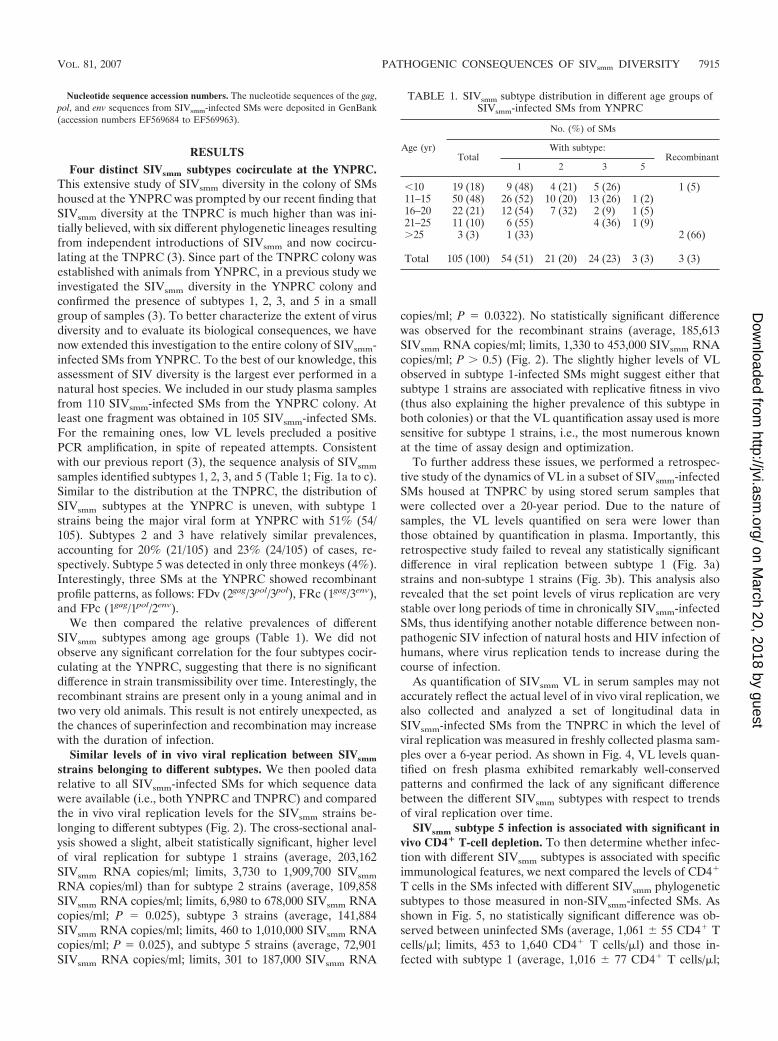
TABLE 1. SIVsmm subtype distribution in different age groups ofSIVsmm-infected SMs from YNPRC
Age (yr)
No. (%) of SMs
TotalWith subtype:
Recombinant1 2 3 5
�10 19 (18) 9 (48) 4 (21) 5 (26) 1 (5)11–15 50 (48) 26 (52) 10 (20) 13 (26) 1 (2)16–20 22 (21) 12 (54) 7 (32) 2 (9) 1 (5)21–25 11 (10) 6 (55) 4 (36) 1 (9)�25 3 (3) 1 (33) 2 (66)
Total 105 (100) 54 (51) 21 (20) 24 (23) 3 (3) 3 (3)
VOL. 81, 2007 PATHOGENIC CONSEQUENCES OF SIVsmm DIVERSITY 7915
on March 20, 2018 by guest
http://jvi.asm.org/
Dow
nloaded from
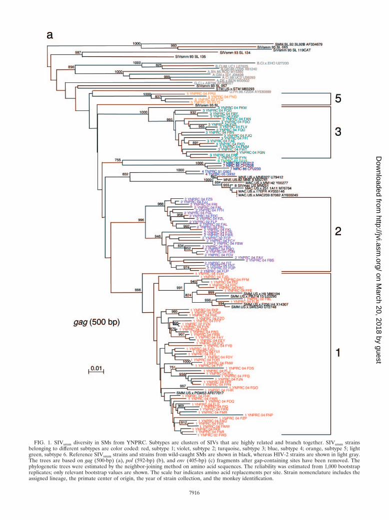
FIG. 1. SIVsmm diversity in SMs from YNPRC. Subtypes are clusters of SIVs that are highly related and branch together. SIVsmm strainsbelonging to different subtypes are color coded: red, subtype 1; violet, subtype 2; turquoise, subtype 3; blue, subtype 4; orange, subtype 5; lightgreen, subtype 6. Reference SIVsmm strains and strains from wild-caught SMs are shown in black, whereas HIV-2 strains are shown in light gray.The trees are based on gag (500-bp) (a), pol (592-bp) (b), and env (405-bp) (c) fragments after gap-containing sites have been removed. Thephylogenetic trees were estimated by the neighbor-joining method on amino acid sequences. The reliability was estimated from 1,000 bootstrapreplicates; only relevant bootstrap values are shown. The scale bar indicates amino acid replacements per site. Strain nomenclature includes theassigned lineage, the primate center of origin, the year of strain collection, and the monkey identification.
7916
on March 20, 2018 by guest
http://jvi.asm.org/
Dow
nloaded from
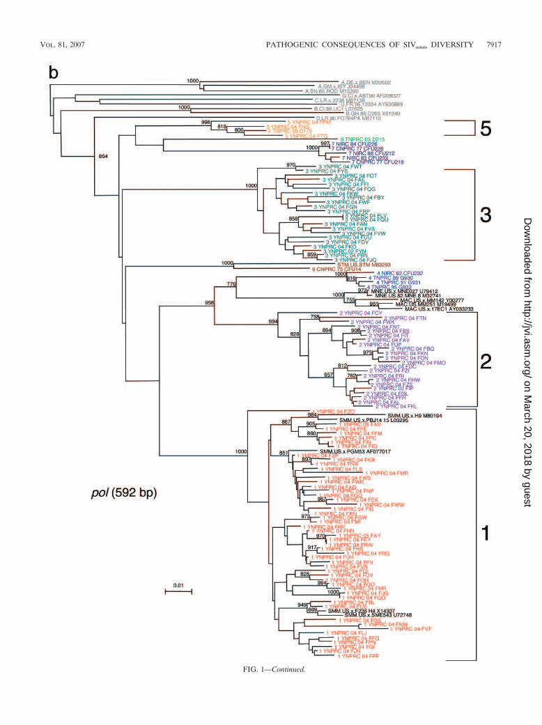
FIG. 1—Continued.
VOL. 81, 2007 PATHOGENIC CONSEQUENCES OF SIVsmm DIVERSITY 7917
on March 20, 2018 by guest
http://jvi.asm.org/
Dow
nloaded from
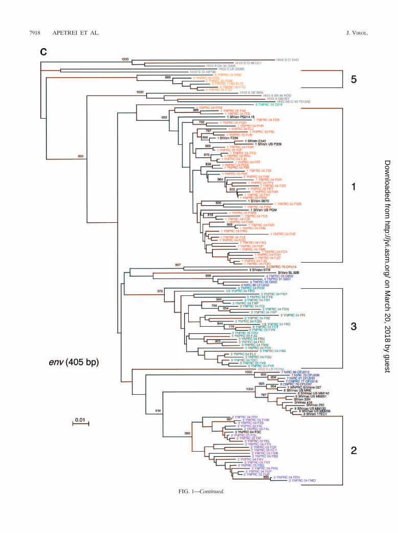
FIG. 1—Continued.
7918 APETREI ET AL. J. VIROL.
on March 20, 2018 by guest
http://jvi.asm.org/
Dow
nloaded from
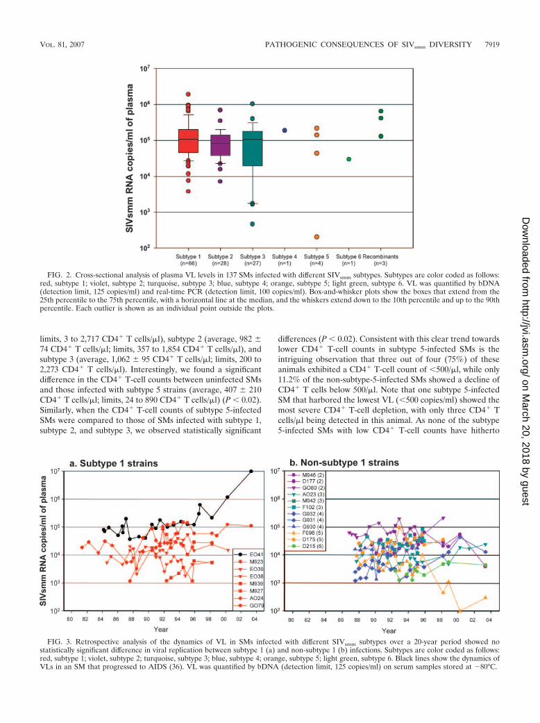
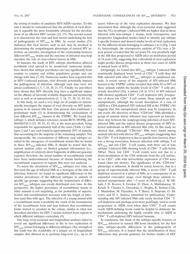
limits, 3 to 2,717 CD4� T cells/�l), subtype 2 (average, 982 �74 CD4� T cells/�l; limits, 357 to 1,854 CD4� T cells/�l), andsubtype 3 (average, 1,062 � 95 CD4� T cells/�l; limits, 200 to2,273 CD4� T cells/�l). Interestingly, we found a significantdifference in the CD4� T-cell counts between uninfected SMsand those infected with subtype 5 strains (average, 407 � 210CD4� T cells/�l; limits, 24 to 890 CD4� T cells/�l) (P � 0.02).Similarly, when the CD4� T-cell counts of subtype 5-infectedSMs were compared to those of SMs infected with subtype 1,subtype 2, and subtype 3, we observed statistically significant
differences (P � 0.02). Consistent with this clear trend towardslower CD4� T-cell counts in subtype 5-infected SMs is theintriguing observation that three out of four (75%) of theseanimals exhibited a CD4� T-cell count of �500/�l, while only11.2% of the non-subtype-5-infected SMs showed a decline ofCD4� T cells below 500/�l. Note that one subtype 5-infectedSM that harbored the lowest VL (�500 copies/ml) showed themost severe CD4� T-cell depletion, with only three CD4� Tcells/�l being detected in this animal. As none of the subtype5-infected SMs with low CD4� T-cell counts have hitherto
FIG. 2. Cross-sectional analysis of plasma VL levels in 137 SMs infected with different SIVsmm subtypes. Subtypes are color coded as follows:red, subtype 1; violet, subtype 2; turquoise, subtype 3; blue, subtype 4; orange, subtype 5; light green, subtype 6. VL was quantified by bDNA(detection limit, 125 copies/ml) and real-time PCR (detection limit, 100 copies/ml). Box-and-whisker plots show the boxes that extend from the25th percentile to the 75th percentile, with a horizontal line at the median, and the whiskers extend down to the 10th percentile and up to the 90thpercentile. Each outlier is shown as an individual point outside the plots.
FIG. 3. Retrospective analysis of the dynamics of VL in SMs infected with different SIVsmm subtypes over a 20-year period showed nostatistically significant difference in viral replication between subtype 1 (a) and non-subtype 1 (b) infections. Subtypes are color coded as follows:red, subtype 1; violet, subtype 2; turquoise, subtype 3; blue, subtype 4; orange, subtype 5; light green, subtype 6. Black lines show the dynamics ofVLs in an SM that progressed to AIDS (36). VL was quantified by bDNA (detection limit, 125 copies/ml) on serum samples stored at �80°C.
VOL. 81, 2007 PATHOGENIC CONSEQUENCES OF SIVsmm DIVERSITY 7919
on March 20, 2018 by guest
http://jvi.asm.org/
Dow
nloaded from
shown any signs of illness, the actual clinical significance of theCD4� T-cell depletion associated with SIVsmm subtype 5 in-fection is still unknown.
DISCUSSION
In this study we have characterized the SIVsmm diversity inthe YNPRC SM colony. Based on a large set of data, we haveinvestigated if viral diversity has any significant impact onSIVsmm pathogenesis in its natural hosts, the SMs. We did notobserve any significant difference in virulence between differ-
ent SIVsmm subtypes in regard to viral replication or diseaseprogression. However, we found a significant correlation be-tween infection with SIVsmm subtype 5 and lower CD4� T-cellcounts, suggesting that at least this virus subtype may influencethe immunological course of infection in the natural SIVsmm
hosts.There are two major reasons to study SIVsmm diversity in
naturally infected SMs. The first reason is that the magnitudeof diversity of different SIVsmm subtypes is similar to that ofHIV-1 group M subtypes (3), thus indicating that SIVsmm sub-types can be used as a model of the effect of virus diversity in
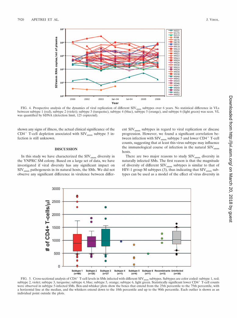
FIG. 4. Prospective analysis of the dynamics of viral replication of different SIVsmm subtypes over 6 years. No statistical difference in VLsbetween subtype 1 (red), subtype 2 (violet); subtype 3 (turquoise), subtype 4 (blue), subtype 5 (orange), and subtype 6 (light green) was seen. VLwas quantified by bDNA (detection limit, 125 copies/ml).
FIG. 5. Cross-sectional analysis of CD4� T-cell levels in SMs infected with different SIVsmm subtypes. Subtypes are color coded: subtype 1, red;subtype 2, violet; subtype 3, turquoise; subtype 4, blue; subtype 5, orange; subtype 6, light green. Statistically significant lower CD4� T-cell countswere observed in subtype 5-infected SMs. Box-and-whisker plots show the boxes that extend from the 25th percentile to the 75th percentile, witha horizontal line at the median, and the whiskers extend down to the 10th percentile and up to the 90th percentile. Each outlier is shown as anindividual point outside the plots.
7920 APETREI ET AL. J. VIROL.
on March 20, 2018 by guest
http://jvi.asm.org/
Dow
nloaded from
the setting of studies of candidate HIV/AIDS vaccines. To thisend it should be remembered that the problem of viral diver-sity is arguably the most formidable obstacle for the develop-ment of an effective HIV vaccine (22, 27). The second reasonis to characterize the virus and host factors underlying the lackof pathogenicity of SIVsmm in SMs. As recent studies (53)indicated that viral factors, such as nef, may be involved indetermining the nonpathogenic phenotype of natural SIV in-fection, an extensive investigation of the relationship betweendifferent SIVsmm strains and the outcome of infection may helpelucidate the role of virus-related factors in SMs.
In humans, the study of HIV subtype distribution allowedworldwide viral spread to be monitored: numerous studieshave shown that subtype prevalence can vary significantly fromcountry to country and within population groups and canchange with time (7, 49). Numerous studies have reported thatin HIV-1-infected patients, viral diversity potentially impactsthe pathogenesis of infection, although such data were notalways confirmed (1, 5, 7, 18, 20, 21, 57). Finally, we and othershave shown that HIV diversity may have a significant impacton the efficacy of currently available diagnostic and monitoringtests as well as of antiretrovirals (5, 7, 18, 37, 38, 41, 49, 61).
In this study, we used a very large set of samples to system-atically investigate the impact of viral diversity on SIV patho-genesis in its natural SM host. Our present data confirmed apreliminary study (3) in demonstrating the cocirculation offour different SIVsmm clusters at the YNPRC. We found thatsubtype 1, which includes reference strains B670, H9/PBj, andH4/660/543-3 (23, 30, 32, 43, 45), is the most prevalent, beingresponsible for approximately half of the cases (Table 1). Sub-types 2 and 3 are each found in approximately 20% of animals,thus accounting for the majority of the remaining samples. Notunexpectedly, the cocirculation of different viral subtypes re-sulted in the emergence of recombinant strains that we foundin three SIVsmm-infected SMs. It should be noted that thecurrent analysis relies on limited genomic information (i.e.,amplification of relatively short fragments of different genomicregions); therefore, the actual number of recombinants couldhave been underestimated because of strains harboring therecombinant sequences in regions that were not analyzed.
To assess the circulation of SIVsmm subtypes over time, wefirst used the age of infected SMs as a surrogate of the time ofinfection; however, we found no significant differences in therelative prevalences of the different subtypes in animals ofspecific age groups, suggesting that the transmission of differ-ent SIVsmm subtypes was evenly distributed over time. In thisperspective, the higher prevalence of recombinant strains inolder animals is not surprising, as the probability of superin-fection and recombination increases with the length of infec-tion (and thus with age). The infection of a young animal witha recombinant strain is probably the result of the transmissionof the recombinant form and may indicate that recombinantforms started to circulate in the colony, similar to what wasdescribed elsewhere for HIV-1 strains isolated from regions inwhich different subtypes cocirculate (9).
Our large cross-sectional and longitudinal analyses failed toreveal any significant difference in the in vivo replication ofSIVsmm strains belonging to different subtypes. One strength ofthis study was the availability of a unique set of longitudinalsamples that allowed us to perform long-term (i.e., up to 20
years) follow-up of the virus replication dynamics. We thusdetermined that, although the cross-sectional study suggestedthat the VLs in subtype 1-infected SMs are higher than in thoseinfected with non-subtype 1 strains, both retrospective andprospective longitudinal studies failed to identify any subtype1-specific features and showed similar levels of viral replicationfor the different strains belonging to subtypes 1 to 6 (Fig. 3 and4). Interestingly, the retrospective analysis of VLs over a 20-year period revealed that the set point VL was peculiarly highin the SM that progressed to AIDS after an incubation periodof 18 years (36), suggesting that a threshold of viral replicationmight predict disease progression in those rare cases of AIDSin natural hosts of SIVs.
Intriguingly, we found that SIVsmm subtype 5-infected SMsconsistently displayed lower levels of CD4� T cells than didSMs infected with other SIVsmm subtypes or uninfected ani-mals. A recent survey of the majority of SIV-infected SMshoused at the YNPRC colony revealed that while 85 to 90% ofthese animals exhibit the healthy levels of CD4� T cells pre-viously described (56), a subset (10 to 15%) of SIV-infectedSMs showed significant CD4� T-cell depletion (60). Irrespec-tive of these low CD4� T-cell counts, these animals are stillasymptomatic, although the recent description of a case ofAIDS in a CD4-depleted, SIV-infected SM at the TNPRC (36)suggests that this asymptomatic phase may be temporary. Inany event, the discovery of these “CD4-low” SMs identified agroup of animals whose infection may represent an interme-diate step between the nonprogressing infection of most SIV-infected SMs and the typical disease progression of HIV-in-fected individuals or SIV-infected Rhs (60). In this study weshowed that, although “CD4-low” SMs were found amonganimals infected with diverse SIVsmm subtypes (suggesting thatthe CD4� T-cell depletion is due to specific host factors), therewas a strong association between infection with subtype 5 ofSIVsmm and low CD4� T-cell counts, with three out of foursubtype 5-infected SMs showing levels of CD4� T cells below500/�l. These low CD4� T-cell counts were not due to adown-modulation of the CD4 molecule from the cell surface,as no CD3� cells with intracellular expression of CD4 werefound (data not shown). The significance of this “CD4-low”phenotype is unknown. It should be noted, however, that in agroup of experimentally infected SMs, a severe CD4� T-celldepletion occurred in a subset of SMs, as a consequence of anexpanded coreceptor usage, even though these animals re-mained asymptomatic after �5 years of follow-up (J. M. Mi-lush, J. D. Reeves, S. Gordon, D. Zhou, A. Muthukumar, D.Kosub, E. Chacko, L. Giavedoni, C. Ibegbu, K. Stefano-Cole,J. Miamidian, M. Paiardini, A. P. Barry, S. Staprans, G. Sil-vestri, and D. L. Sodora, submitted for publication). Under-standing why SIV-infected SMs only rarely develop CD4� T-cell depletion and, perhaps even more puzzlingly, tend to avoidprogression to AIDS even when their CD4� T-cell countsreach AIDS-defining levels could be helpful to delineate themechanisms underlying the highly variable time to AIDS inCD4� T-cell-depleted HIV-infected humans.
In conclusion, this study of the subtype-specific features ofSIVsmm infection in natural SM hosts identified, for the firsttime, subtype-specific differences in the pathogenicity ofSIVsmm infection. It is hoped that the identification of thesedifferences may provide guidance for further studies of HIV
VOL. 81, 2007 PATHOGENIC CONSEQUENCES OF SIVsmm DIVERSITY 7921
on March 20, 2018 by guest
http://jvi.asm.org/
Dow
nloaded from
pathogenesis and vaccines where pathogenic SIVsmm infectionof nonnatural hosts (i.e., Asian macaques) is used as an exper-imental model.
ACKNOWLEDGMENTS
We thank Andrew Lackner and Preston Marx for helpful discussion.We thank Benton Lawson, Meredith Hunter, Nathalia Katz, and theveterinary and animal care staff of TNPRC and YNPRC for theirservice and expertise.
This work was supported by funds from grants RO1 AI065325 andP20 RR020159 (C.A.), RO1 AI52755 and RO1 AI66998 (G.S.), RO1AI064066 and R21 AI069935 (I.P.), and P51 RR000164 (TNPRC) andRR00165 (YNPRC) from the National Institute of Allergy and Infec-tious Diseases and from the National Center for Research Resources.
REFERENCES
1. Apetrei, C., D. Descamps, G. Collin, I. Loussert-Ajaka, F. Damond, M. Duca,F. Simon, and F. Brun-Vezinet. 1998. Human immunodeficiency virus type 1subtype F reverse transcriptase sequence and drug susceptibility. J. Virol.72:3534–3538.
2. Apetrei, C., B. Gormus, I. Pandrea, M. Metzger, P. ten Haaft, L. N. Martin,R. Bohm, X. Alvarez, G. Koopman, M. Murphey-Corb, R. S. Veazey, A. A.Lackner, G. Baskin, J. Heeney, and P. A. Marx. 2004. Direct inoculation ofsimian immunodeficiency virus from sooty mangabeys in black mangabeys(Lophocebus aterrimus): first evidence of AIDS in a heterologous Africanspecies and different pathologic outcomes of experimental infection. J. Virol.78:11506–11518.
3. Apetrei, C., A. Kaur, N. W. Lerche, M. Metzger, I. Pandrea, J. Hardcastle,S. Fakelstein, R. Bohm, J. Kohler, V. Traina-Dorge, T. Williams, S.Staprans, G. Plauche, R. S. Veazey, H. McClure, A. A. Lackner, B. Gormus,D. L. Robertson, and P. A. Marx. 2005. Molecular epidemiology of SIVsm inU.S. primate centers unravels the origin of SIVmac and SIVstm. J. Virol.79:8991–9005.
4. Apetrei, C., N. W. Lerche, I. Pandrea, B. Gormus, M. Metzger, G. Silvestri,A. Kaur, R. Bohm, D. L. Robertson, J. Hardcastle, A. A. Lackner, and P. A.Marx. 2006. Kuru experiments triggered the emergence of pathogenicSIVmac. AIDS 20:317–321.
5. Apetrei, C., I. Loussert-Ajaka, D. Descamps, F. Damond, S. Saragosti, F.Brun-Vezinet, and F. Simon. 1996. Lack of screening test sensitivity duringHIV-1 non-subtype B seroconversions. AIDS 10:F57–F60.
6. Apetrei, C., and P. A. Marx. 2004. Simian retroviral infections in humanbeings. Lancet 364:137–138.
7. Apetrei, C., P. A. Marx, and S. M. Smith. 2004. The evolution of HIV and itsconsequences. Infect. Dis. Clin. N. Am. 18:369–394.
8. Apetrei, C., D. L. Robertson, and P. A. Marx. 2004. The history of SIVs andAIDS: epidemiology, phylogeny and biology of isolates from naturally SIVinfected non-human primates (NHP) in Africa. Front. Biosci. 9:225–254.
9. Butler, I. F., I. Pandrea, P. A. Marx, and C. Apetrei. 2007. HIV genetic diversity:biological and public health consequences. Curr. HIV Res. 5:23–45.
10. Cayabyab, M., G. B. Karlsson, B. A. Etemad-Moghadam, W. Hofmann, T.Steenbeke, M. Halloran, J. W. Fanton, M. K. Axthelm, N. L. Letvin, andJ. G. Sodroski. 1999. Changes in human immunodeficiency virus type 1envelope glycoproteins responsible for the pathogenicity of a multiply pas-saged simian-human immunodeficiency virus (SHIV-HXBc2). J. Virol. 73:976–984.
11. Chakrabarti, L. A., S. R. Lewin, L. Zhang, A. Gettie, A. Luckay, L. N. Martin,E. Skulsky, D. D. Ho, C. Cheng-Mayer, and P. A. Marx. 2000. Normal T-cellturnover in sooty mangabeys harboring active simian immunodeficiency virusinfection. J. Virol. 74:1209–1223.
12. Chen, Z., A. Luckay, D. L. Sodora, P. Telfer, P. Reed, A. Gettie, J. M. Kanu,R. F. Sadek, J. Yee, D. D. Ho, L. Zhang, and P. A. Marx. 1997. Humanimmunodeficiency virus type 2 (HIV-2) seroprevalence and characterizationof a distinct HIV-2 genetic subtype from the natural range of simian immu-nodeficiency virus-infected sooty mangabeys. J. Virol. 71:3953–3960.
13. Chen, Z., P. Telfer, A. Gettie, P. Reed, L. Zhang, D. D. Ho, and P. A. Marx.1996. Genetic characterization of new West African simian immunodefi-ciency virus SIVsm: geographic clustering of household-derived SIV strainswith human immunodeficiency virus type 2 subtypes and genetically diverseviruses from a single feral sooty mangabey troop. J. Virol. 70:3617–3627.
14. Courgnaud, V., B. Abela, X. Pourrut, E. Mpoudi-Ngole, S. Loul, E. Delaporte,and M. Peeters. 2003. Identification of a new simian immunodeficiency viruslineage with a vpu gene present among different Cercopithecus monkeys (C.mona, C. cephus, and C. nictitans) from Cameroon. J. Virol. 77:12523–12534.
15. Courgnaud, V., P. Formenty, C. Akoua-Koffi, R. Noe, C. Boesch, E.Delaporte, and M. Peeters. 2003. Partial molecular characterization of twosimian immunodeficiency viruses (SIV) from African colobids: SIVwrc fromWestern red colobus (Piliocolobus badius) and SIVolc from olive colobus(Procolobus verus). J. Virol. 77:744–748.
16. Courgnaud, V., X. Pourrut, F. Bibollet-Ruche, E. Mpoudi-Ngole, A.Bourgeois, E. Delaporte, and M. Peeters. 2001. Characterization of a novelsimian immunodeficiency virus from guereza colobus monkeys (Colobusguereza) in Cameroon: a new lineage in the nonhuman primate lentivirusfamily. J. Virol. 75:857–866.
17. Courgnaud, V., M. Salemi, X. Pourrut, E. Mpoudi-Ngole, B. Abela, P. Auzel,F. Bibollet-Ruche, B. Hahn, A.-M. Vandamme, E. Delaporte, and M. Peeters.2002. Characterization of a novel simian immunodeficiency virus with a vpugene from greater spot-nosed monkeys (Cercopithecus nictitans) providesnew insights into simian/human immunodeficiency virus phylogeny. J. Virol.76:8298–8309.
18. Damond, F., C. Apetrei, D. Descamps, F. Brun-Vezinet, and F. Simon. 1999.HIV-1 subtypes and plasma RNA quantification. AIDS 13:286–288.
19. Damond, F., C. Apetrei, D. L. Robertson, S. Souquiere, A. Lepretre, S.Matheron, J. C. Plantier, F. Brun-Vezinet, and F. Simon. 2001. Variability ofhuman immunodeficiency virus type 2 (HIV-2) infecting patients living inFrance. Virology 280:19–30.
20. Descamps, D., C. Apetrei, G. Collin, F. Damond, F. Simon, and F. Brun-Vezinet. 1998. Naturally occurring decreased susceptibility of HIV-1 subtypeG to protease inhibitors. AIDS 12:1109–1111.
21. Descamps, D., G. Collin, F. Letourneur, C. Apetrei, F. Damond, I. Loussert-Ajaka, F. Simon, S. Saragosti, and F. Brun-Vezinet. 1997. Susceptibility ofhuman immunodeficiency virus type 1 group O isolates to antiretroviralagents: in vitro phenotypic and genotypic analyses. J. Virol. 71:8893–8898.
22. Desrosiers, R. C. 2004. Prospects for an AIDS vaccine. Nat. Med. 10:221–223.
23. Fultz, P. N. 1994. SIVsmmPBj14: an atypical lentivirus. Curr. Top. Micro-biol. Immunol. 188:65–76.
24. Gao, F., E. Bailes, D. L. Robertson, Y. Chen, C. M. Rodenburg, S. F.Michael, L. B. Cummins, L. O. Arthur, M. Peeters, G. M. Shaw, P. M. Sharp,and B. H. Hahn. 1999. Origin of HIV-1 in the chimpanzee Pan troglodytestroglodytes. Nature 397:436–441.
25. Gao, F., L. Yue, D. L. Robertson, S. C. Hill, H. Hui, R. J. Biggar, A. E.Neequaye, T. M. Whelan, D. D. Ho, G. M. Shaw, P. M. Sharp, and B. H.Hahn. 1994. Genetic diversity of human immunodeficiency virus type 2:evidence for distinct sequence subtypes with differences in virus biology.J. Virol. 68:7433–7447.
26. Gao, F., L. Yue, A. T. White, P. G. Pappas, J. Barchue, A. P. Hanson, B. M.Greene, P. M. Sharp, G. M. Shaw, and B. H. Hahn. 1992. Human infectionby genetically diverse SIVSM-related HIV-2 in West Africa. Nature 358:495–499.
27. Garber, D. A., G. Silvestri, and M. B. Feinberg. 2004. Prospects for an AIDSvaccine: three big questions, no easy answers. Lancet Infect. Dis. 4:397–413.
28. Gordon, S., I. Pandrea, R. Dunham, C. Apetrei, and G. Silvestri. 2005. Thecall of the wild: what can be learned from studies of SIV infection of naturalhosts?, p. 2–29. In T. Leitner, B. Foley, B. Hahn, P. Marx, F. McCutchan, J.Mellors, S. Wolinsky, and B. Korber (ed.), HIV sequence compendium 2004.Theoretical Biology and Biophysics Group, Los Alamos National Labora-tory, Los Alamos, NM.
29. Hasegawa, M., H. Kishino, and T. Yano. 1985. Dating of the human-apesplitting by a molecular clock of mitochondrial DNA. J. Mol. Evol. 22:160–174.
30. Hirsch, V., D. Adger-Johnson, B. Campbell, S. Goldstein, C. Brown, W. R.Elkins, and D. C. Montefiori. 1997. A molecularly cloned, pathogenic, neu-tralization-resistant simian immunodeficiency virus, SIVsmE543-3. J. Virol.71:1608–1620.
31. Hirsch, V. M., T. R. Fuerst, G. Sutter, M. W. Carroll, L. C. Yang, S.Goldstein, M. Piatak, Jr., W. R. Elkins, W. G. Alvord, D. C. Montefiori, B.Moss, and J. D. Lifson. 1996. Patterns of viral replication correlate withoutcome in simian immunodeficiency virus (SIV)-infected macaques: effectof prior immunization with a trivalent SIV vaccine in modified vaccinia virusAnkara. J. Virol. 70:3741–3752.
32. Hirsch, V. M., and P. R. Johnson. 1994. Pathogenic diversity of simianimmunodeficiency viruses. Virus Res. 32:183–203.
33. Johnson, P. R., and V. M. Hirsch. 1991. Pathogenesis of AIDS: the non-human primate model. AIDS 5(Suppl. 2):S43–S48.
34. Kaur, A., R. M. Grant, R. E. Means, H. McClure, M. Feinberg, and R. P.Johnson. 1998. Diverse host responses and outcomes following simian im-munodeficiency virus SIVmac239 infection in sooty mangabeys and rhesusmacaques. J. Virol. 72:9597–9611.
35. Lifson, J. D., M. A. Nowak, S. Goldstein, J. L. Rossio, A. Kinter, G. Vasquez,T. A. Wiltrout, C. Brown, D. Schneider, L. Wahl, A. L. Lloyd, J. Williams,W. R. Elkins, A. S. Fauci, and V. M. Hirsch. 1997. The extent of early viralreplication is a critical determinant of the natural history of simian immu-nodeficiency virus infection. J. Virol. 71:9508–9514.
36. Ling, B., C. Apetrei, I. Pandrea, R. S. Veazey, A. A. Lackner, B. Gormus, andP. A. Marx. 2004. Classic AIDS in a sooty mangabey after an 18-year naturalinfection. J. Virol. 78:8902–8908.
37. Loussert-Ajaka, I., T. D. Ly, M. L. Chaix, D. Ingrand, S. Saragosti, A. M.Courouce, F. Brun-Vezinet, and F. Simon. 1994. HIV-1/HIV-2 seronegativityin HIV-1 subtype O infected patients. Lancet 343:1393–1394.
38. Mauclere, P., F. Damond, C. Apetrei, I. Loussert-Ajaka, S. Souquiere, L.
7922 APETREI ET AL. J. VIROL.
on March 20, 2018 by guest
http://jvi.asm.org/
Dow
nloaded from
Buzelay, P. Dalbon, M. Jolivet, M. Mony Lobe, F. Brun-Vezinet, F. Simon,and F. Barin. 1997. Synthetic peptide ELISAs for detection of and discrim-ination between group M and group O HIV type 1 infection. AIDS Res.Hum. Retrovir. 13:987–993.
39. Mellors, J. W., A. Munoz, J. V. Giorgi, J. B. Margolick, C. J. Tassoni, P.Gupta, L. A. Kingsley, J. A. Todd, A. J. Saah, R. Detels, J. P. Phair, and C. R.Rinaldo, Jr. 1997. Plasma viral load and CD4� lymphocytes as prognosticmarkers of HIV-1 infection. Ann. Intern. Med. 126:946–954.
40. Mellors, J. W., C. R. Rinaldo, Jr., P. Gupta, R. M. White, J. A. Todd, andL. A. Kingsley. 1996. Prognosis in HIV-1 infection predicted by the quantityof virus in plasma. Science 272:1167–1170.
41. Michael, N. L., S. A. Herman, S. Kwok, K. Dreyer, J. Wang, C. Christopher-son, J. P. Spadoro, K. K. Young, V. Polonis, F. E. McCutchan, J. Carr, J. R.Mascola, L. L. Jagodzinski, and M. L. Robb. 1999. Development of cali-brated viral load standards for group M subtypes of human immunodefi-ciency virus type 1 and performance of an improved AMPLICOR HIV-1MONITOR test with isolates of diverse subtypes. J. Clin. Microbiol. 37:2557–2563.
42. Morton, W. R., L. Kuller, R. E. Benveniste, E. A. Clark, C. C. Tsai, M. J.Gale, M. E. Thouless, J. Overbaugh, and M. G. Katze. 1989. Transmission ofthe simian immunodeficiency virus SIVmne in macaques and baboons.J. Med. Primatol. 18:237–245.
43. Murphey-Corb, M., L. N. Martin, S. R. Rangan, G. B. Baskin, B. J. Gormus,R. H. Wolf, W. A. Andes, M. West, and R. C. Montelaro. 1986. Isolation ofan HTLV-III-related retrovirus from macaques with simian AIDS and itspossible origin in asymptomatic mangabeys. Nature 321:435–437.
44. National Research Council. 1996. Guide for the care and use of laboratoryanimals. National Academy Press, Washington, DC.
45. Novembre, F. J., J. De Rosayro, S. P. O’Neil, D. C. Anderson, S. A. Klumpp,and H. M. McClure. 1998. Isolation and characterization of a neuropatho-genic simian immunodeficiency virus derived from a sooty mangabey. J. Vi-rol. 72:8841–8851.
46. Novembre, F. J., V. M. Hirsch, H. M. McClure, P. N. Fultz, and P. R.Johnson. 1992. SIV from stump-tailed macaques: molecular characterizationof a highly transmissible primate lentivirus. Virology 186:783–787.
47. Nowak, M. A., A. L. Lloyd, G. M. Vasquez, T. A. Wiltrout, L. M. Wahl, N.Bischofberger, J. Williams, A. Kinter, A. S. Fauci, V. M. Hirsch, and J. D.Lifson. 1997. Viral dynamics of primary viremia and antiretroviral therapy insimian immunodeficiency virus infection. J. Virol. 71:7518–7525.
48. Pandrea, I., C. Apetrei, J. Dufour, N. Dillon, J. Barbercheck, M. Metzger, B.Jacquelin, R. Bohm, P. A. Marx, F. Barre-Sinoussi, V. M. Hirsch, M. C.Muller-Trutwin, A. A. Lackner, and R. S. Veazey. 2006. Simian immunode-ficiency virus SIVagm.sab infection of Caribbean African green monkeys:new model of the study of SIV pathogenesis in natural hosts. J. Virol.80:4858–4867.
49. Peeters, M., and P. M. Sharp. 2000. Genetic diversity of HIV-1: the movingtarget. AIDS 14(Suppl. 3):S129–S140.
50. Posada, D., and K. A. Crandall. 2001. Selecting the best-fit model of nucle-otide substitution. Syst. Biol. 50:580–601.
51. Rey-Cuille, M. A., J. L. Berthier, M. C. Bomsel-Demontoy, Y. Chaduc, L.Montagnier, A. G. Hovanessian, and L. A. Chakrabarti. 1998. Simian im-munodeficiency virus replicates to high levels in sooty mangabeys withoutinducing disease. J. Virol. 72:3872–3886.
52. Saitou, N., and M. Nei. 1987. The neighbor-joining method: a new methodfor reconstructing phylogenetic trees. Mol. Biol. Evol. 4:406–425.
53. Schindler, M., J. Munch, O. Kutsch, H. Li, M. L. Santiago, F. Bibollet-Ruche, M. C. Muller-Trutwin, F. J. Novembre, M. Peeters, V. Courgnaud, E.Bailes, P. Roques, D. L. Sodora, G. Silvestri, P. M. Sharp, B. H. Hahn, andF. Kirchhoff. 2006. Nef-mediated suppression of T cell activation was lost ina lentiviral lineage that gave rise to HIV-1. Cell 125:1055–1067.
54. Silvestri, G. 2005. Naturally SIV-infected sooty mangabeys: are we closer tounderstanding why they do not develop AIDS? J. Med. Primatol. 34:243–252.
55. Silvestri, G., A. Fedanov, S. Germon, N. Kozyr, W. J. Kaiser, D. A. Garber,H. McClure, M. B. Feinberg, and S. I. Staprans. 2005. Divergent hostresponses during primary simian immunodeficiency virus SIVsm infection ofnatural sooty mangabey and nonnatural rhesus macaque hosts. J. Virol.79:4043–4054.
56. Silvestri, G., D. L. Sodora, R. A. Koup, M. Paiardini, S. P. O’Neil, H. M.McClure, S. I. Staprans, and M. B. Feinberg. 2003. Nonpathogenic SIVinfection of sooty mangabeys is characterized by limited bystander immuno-pathology despite chronic high-level viremia. Immunity 18:441–452.
57. Simon, F., S. Souquiere, F. Damond, A. Kfutwah, M. Makuwa, E. Leroy, P.Rouquet, J. L. Berthier, J. Rigoulet, A. Lecu, P. T. Telfer, I. Pandrea, J. C.Plantier, F. Barre-Sinoussi, P. Roques, M. C. Muller-Trutwin, and C. Ape-trei. 2001. Synthetic peptide strategy for the detection of and discriminationamong highly divergent primate lentiviruses. AIDS Res. Hum. Retrovir.17:937–952.
58. Stebbing, J., B. Gazzard, and D. C. Douek. 2004. Where does HIV live?N. Engl. J. Med. 350:1872–1880.
59. Stevenson, M. 2003. HIV-1 pathogenesis. Nat. Med. 9:853–860.60. Sumpter, B., R. Dunham, S. Gordon, J. Engram, M. Hennessy, A. Kinter, M.
Paiardini, B. Cervasi, N. Klatt, H. McClure, J. M. Milush, S. Staprans, D. L.Sodora, and G. Silvestri. 2007. Correlates of preserved CD4� T cell ho-meostasis during natural, nonpathogenic simian immunodeficiency virus in-fection of sooty mangabeys: implications for AIDS pathogenesis. J. Immu-nol. 178:1680–1691.
61. Swanson, P., V. Soriano, S. G. Devare, and J. Hackett, Jr. 2001. Comparativeperformance of three viral load assays on human immunodeficiency virustype 1 (HIV-1) isolates representing group M (subtypes A to G) and groupO: LCx HIV RNA quantitative, AMPLICOR HIV-1 MONITOR version1.5, and Quantiplex HIV-1 RNA version 3.0. J. Clin. Microbiol. 39:862–870.
62. Swofford, D. 1999. PAUP*. Phylogenetic analysis using parsimony (*andother methods), version 4. Sinauer Associates, Sunderland, MA.
63. Thompson, J. D., D. G. Higgins, and T. J. Gibson. 1994. CLUSTAL W:improving the sensitivity of progressive multiple sequence alignment throughsequence weighting, position-specific gap penalties and weight matrix choice.Nucleic Acids Res. 22:4673–4680.
64. VandeWoude, S., and C. Apetrei. 2006. Going wild: lessons from T-lympho-tropic naturally occurring lentiviruses. Clin. Microbiol. Rev. 19:728–762.
65. Veazey, R., B. Ling, I. Pandrea, H. McClure, A. Lackner, and P. Marx. 2003.Decreased CCR5 expression on CD4� T cells of SIV-infected sooty manga-beys. AIDS Res. Hum. Retrovir. 19:227–233.
66. Watson, A., J. Ranchalis, B. Travis, J. McClure, W. Sutton, P. R. Johnson,S. L. Hu, and N. L. Haigwood. 1997. Plasma viremia in macaques infectedwith simian immunodeficiency virus: plasma viral load early in infectionpredicts survival. J. Virol. 71:284–290.
67. Yamaguchi, J., S. G. Devare, and C. A. Brennan. 2000. Identification of anew HIV-2 subtype based on phylogenetic analysis of full-length genomicsequence. AIDS Res. Hum. Retrovir. 16:925–930.
VOL. 81, 2007 PATHOGENIC CONSEQUENCES OF SIVsmm DIVERSITY 7923
on March 20, 2018 by guest
http://jvi.asm.org/
Dow
nloaded from