
Virus Structure Rotterdam 2018 handouts
11
28-5-2018 VIRUS STRUCTURE Course in Virology Rotterdam 2018 Raoul J. de Groot Division of Virology Faculty of Veterinary Medicine Utrecht University Outline • Historical introduction • Function of the virion • Analysis of virion structure • Analysis of virion proteins Suggested literature • Principles of Virology (2015) Flint, Racaniello, Rall and Skalka eds ASM Press Dmitri Iosifovich Ivanofski (1864-1920) Martinus Willem Beijerinck (1851-1931) Founding Fathers of Virology Lesions in a Tobacco Leaf: Poison or Infectious Agent?
Transcript of Virus Structure Rotterdam 2018 handouts
28-5-2018
VIRUS STRUCTURE
Course in Virology Rotterdam 2018
Raoul J. de GrootDivision of VirologyFaculty of Veterinary MedicineUtrecht University
Outline
• Historical introduction
• Function of the virion
• Analysis of virion structure
• Analysis of virion proteins
Suggested literature
• Principles of Virology (2015) Flint, Racaniello, Rall and Skalka edsASM Press
Dmitri Iosifovich Ivanofski(1864-1920)
Martinus Willem Beijerinck(1851-1931)
Founding Fathers of Virology
Lesions in a Tobacco Leaf: Poison or Infectious Agent?
28-5-2018
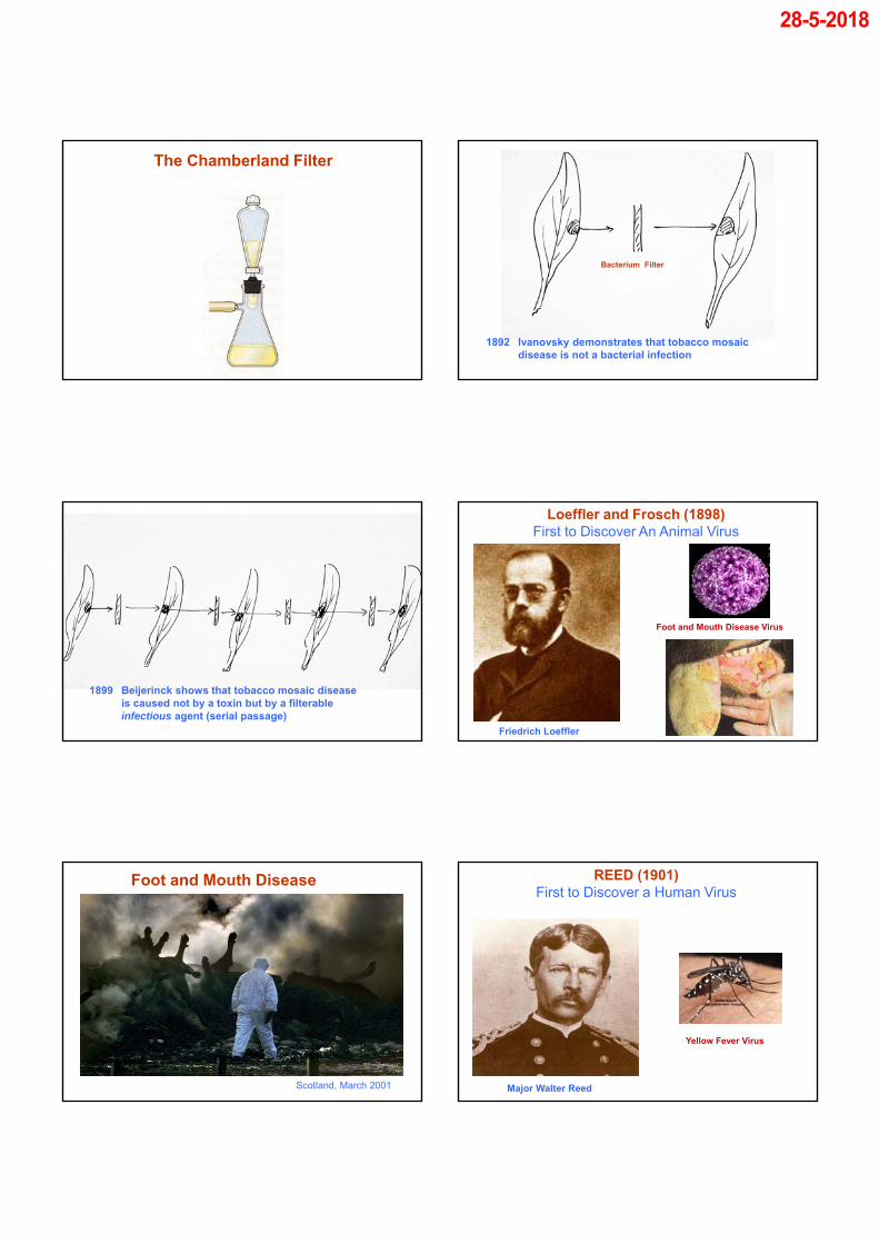
The Chamberland Filter
Bacterium Filter
1892 Ivanovsky demonstrates that tobacco mosaic disease is not a bacterial infection
1899 Beijerinck shows that tobacco mosaic diseaseis caused not by a toxin but by a filterableinfectious agent (serial passage)
Friedrich Loeffler
Loeffler and Frosch (1898)First to Discover An Animal Virus
Foot and Mouth Disease Virus
Scotland, March 2001
Foot and Mouth Disease
Major Walter Reed
REED (1901)First to Discover a Human Virus
Yellow Fever Virus
28-5-2018
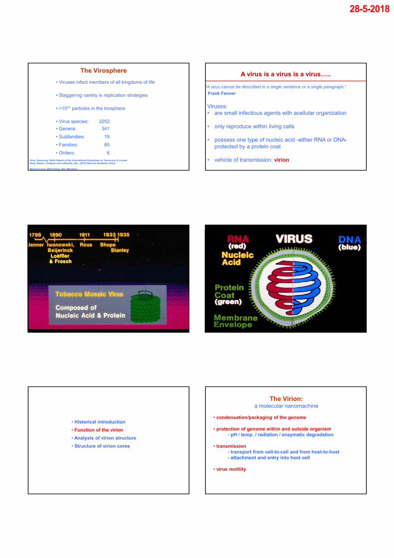
The Virosphere
• Viruses infect members of all kingdoms of life
• Staggering variety in replication strategies
• >1031 particles in the biosphere
• Virus species: 2252
• Genera: 341
• Subfamilies: 19
• Families: 85
• Orders: 6
Virus Taxonomy; Ninth Report of the International Committee on Taxonomy of virusesKing, Adams, Carstens and Lefkowitz, eds. (2011) Elsevier Academic Press
Abrescia et al. (2012) Annu. Rev. Biochem.
A virus is a virus is a virus…..
Viruses:• are small infectious agents with acellular organization
• only reproduce within living cells
• possess one type of nucleic acid -either RNA or DNA-protected by a protein coat
• vehicle of transmission: virion
“A virus cannot be described in a single sentence or a single paragraph.“
Frank Fenner
• Historical introduction
• Function of the virion
• Analysis of virion structure
• Structure of virion cores
The Virion: a molecular nanomachine
• condensation/packaging of the genome
• protection of genome within and outside organism- pH / temp. / radiation / enzymatic degradation
• transmission- transport from cell-to-cell and from host-to-host- attachment and entry into host cell
• virus motility
28-5-2018
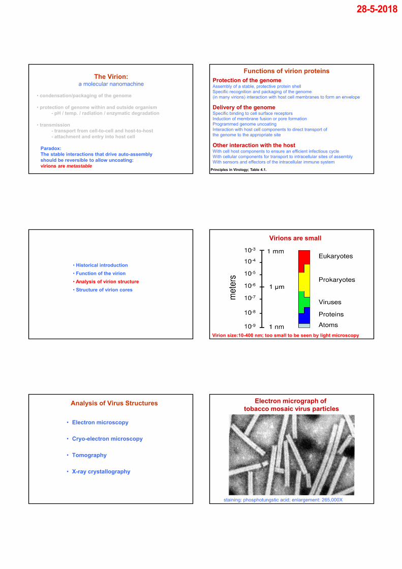
The Virion: a molecular nanomachine
Paradox:The stable interactions that drive auto-assembly should be reversible to allow uncoating: virions are metastable
• condensation/packaging of the genome
• protection of genome within and outside organism - pH / temp. / radiation / enzymatic degradation
• transmission- transport from cell-to-cell and host-to-host- attachment and entry into host cell
Delivery of the genomeSpecific binding to cell surface receptorsInduction of membrane fusion or pore formationProgrammed genome uncoatingInteraction with host cell components to direct transport of the genome to the appropriate site
Functions of virion proteinsProtection of the genomeAssembly of a stable, protective protein shellSpecific recognition and packaging of the genome(in many virions) interaction with host cell membranes to form an envelope
Other interaction with the hostWith cell host components to ensure an efficient infectious cycleWith cellular components for transport to intracellular sites of assemblyWith sensors and effectors of the intracellular immune system
Principles in Virology; Table 4.1.
• Historical introduction
• Function of the virion
• Analysis of virion structure
• Structure of virion cores
Virion size:10-400 nm; too small to be seen by light microscopy
Virions are small
• Electron microscopy
• Cryo-electron microscopy
• Tomography
• X-ray crystallography
Analysis of Virus Structures
staining: phosphotungstic acid; enlargement: 265,000X
Electron micrograph of tobacco mosaic virus particles
28-5-2018
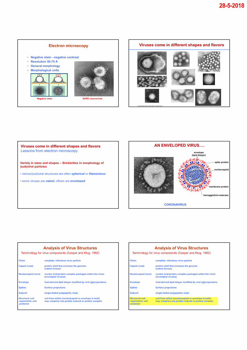
Electron microscopy
SARS coronavirus
• Negative stain→negative contrast
• Resolution 50-75 Å
• General morphology
• Morphological units
Negative stain
Viruses come in different shapes and flavors
Variety in sizes and shapes – Similarities in morphology of (sub)viral particles
• virions/(sub)viral structures are often spherical or filamentous
• some viruses are naked, others are enveloped
Viruses come in different shapes and flavorsLessons from electron microscopy
AN ENVELOPED VIRUS….
CORONAVIRUS
spike protein
hemagglutinin-esterase
membrane protein
nucleocapsid
envelope(lipid bilayer)
Terminology for virus components (Caspar and Klug, 1962)
Analysis of Virus Structures
• Virion complete, infectious virus particle
• Capsid (coat) protein shell that encloses the genome(naked viruses)
• Nucleocapsid (core) nucleic acid-protein complex packaged within the virion(enveloped viruses)
• Envelope host-derived lipid bilayer modified by viral (glyco)proteins
• Spikes Surface projections
• Subunit single folded polypeptide chain
• Structural unit unit from which (nucleo)capsid or envelope is build;asymmetric unit may comprise one protein subunit or protein complexprotomer

28-5-2018
Virus Coatsdifferent coats – common principles
• most viruses appear to be rod-shaped or spherical
• genetic economy
• (nucleo)capsids/cores comprised of multiple copies (≥ 60) of a limited number of viral proteins
• sub-units engage in regular, repetitive non-covalent interactions: symmetry
• two main architectural solutions to build a coat
Helical or Icosahedral Symmetry
Helical Structure
• Repetitive structure, equivalent interactions.Open structure: changes in genome length can be accomodatedby changing the length of the (nucleo)capsid
Helical Structure
• Repetitive structure, equivalent interactions.Open structure: changes in genome length can be accomodatedby changing the length of the (nucleo)capsid
• Example: TMV (a naked virus)Of note: all animal viruses that employ helical symmetry are enveloped
Helical Structure: Tobacco Mosaic Virus
•Single CP•16.3 CP/turn•3nt/CP
ICOSAHEDRAL STRUCTURE
• Most efficient of all possible arrangement to build a symmetrical closed shell with a maximal internal volume from asymmetrical subunits
• Geometric figure with 20 equilateral triangular faces and 12 vortices (corner points)
• Characterized by specific symmetry: contains 2-fold, 3-fold and 5-fold axes of rotational symmetry
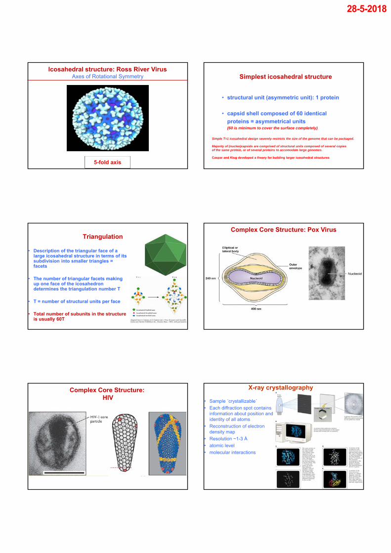
Icosahedral structure: Ross River Virus
28-5-2018
2-fold axis3-fold axis5-fold axis
Axes of Rotational SymmetryIcosahedral structure: Ross River Virus
Simplest icosahedral structure
• structural unit (asymmetric unit): 1 protein
• capsid shell composed of 60 identical
proteins = asymmetrical units(60 is minimum to cover the surface completely)
Simple T=1 icosahedral design severely restricts the size of the genome that can be packaged.
Majority of (nucleo)capsids are comprised of structural units composed of several copies of the same protein, or of several proteins to accomodate large genomes.
Caspar and Klug developed a theory for building larger icosahedral structures
Triangulation
• Description of the triangular face of a large icosahedral structure in terms of its subdivision into smaller triangles = facets
• The number of triangular facets making up one face of the icosahedron determines the triangulation number T
• T = number of structural units per face
• Total number of subunits in the structure is usually 60T
Complex Core Structure: Pox Virus
Complex Core Structure: HIV
X-ray crystallography
• Sample ´crystallizable´
• Each diffraction spot contains information about position and identity of all atoms
• Reconstruction of electron density map
• Resolution ~1-3 Å
• atomic level
• molecular interactions
28-5-2018
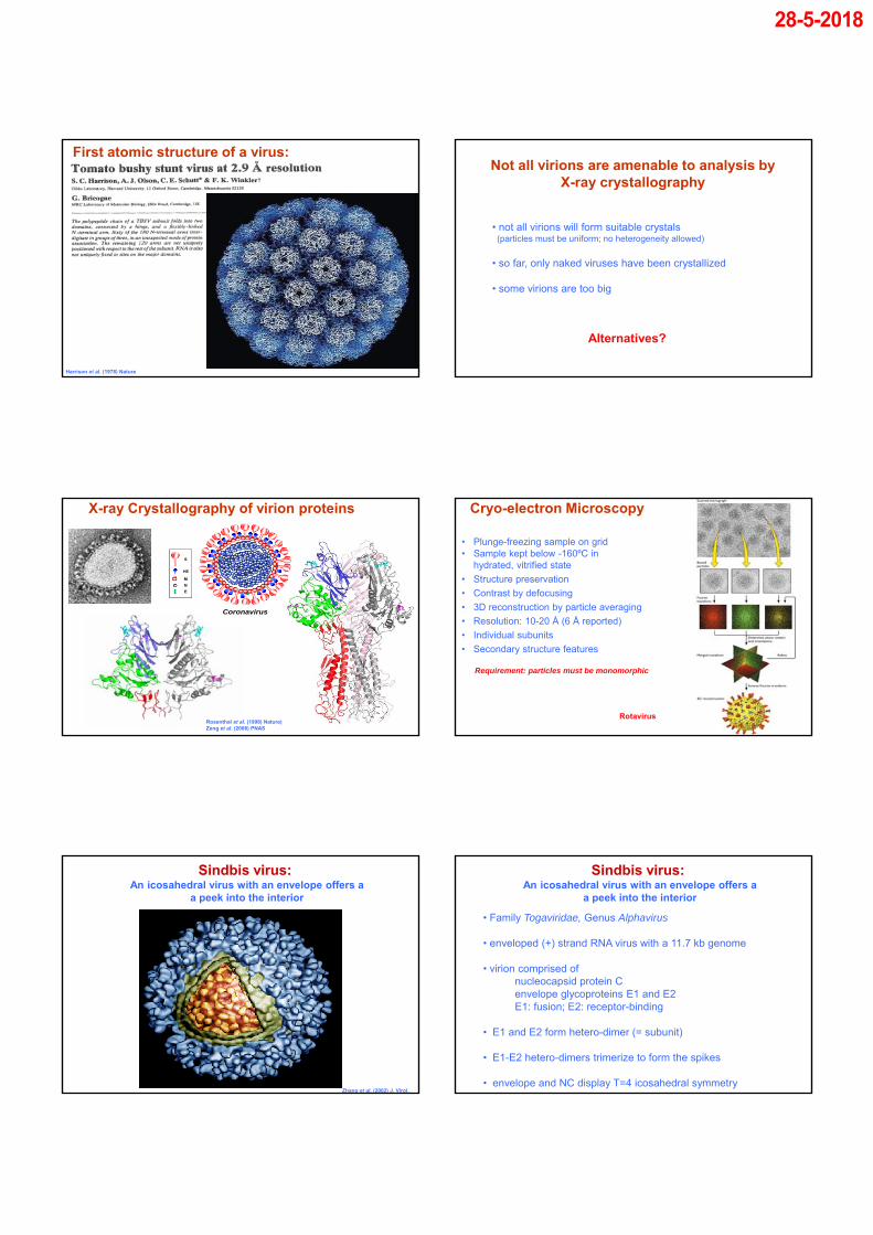
First atomic structure of a virus:
Harrison et al. (1978) Nature
Not all virions are amenable to analysis by X-ray crystallography
• not all virions will form suitable crystals(particles must be uniform; no heterogeneity allowed)
• so far, only naked viruses have been crystallized
• some virions are too big
Alternatives?
X-ray Crystallography of virion proteins
Coronavirus
S
HE
M
N
E
Rosenthal et al. (1998) Nature)Zeng et al. (2008) PNAS
Rotavirus
Cryo-electron Microscopy
• Plunge-freezing sample on grid • Sample kept below -160ºC in
hydrated, vitrified state
• Structure preservation
• Contrast by defocusing
• 3D reconstruction by particle averaging
• Resolution: 10-20 Å (6 Å reported)
• Individual subunits
• Secondary structure features
Requirement: particles must be monomorphic
Sindbis virus: An icosahedral virus with an envelope offers a
a peek into the interior
Zhang et al. (2002) J. Virol.
Sindbis virus: An icosahedral virus with an envelope offers a
a peek into the interior
• Family Togaviridae, Genus Alphavirus
• enveloped (+) strand RNA virus with a 11.7 kb genome
• virion comprised of nucleocapsid protein C envelope glycoproteins E1 and E2 E1: fusion; E2: receptor-binding
• E1 and E2 form hetero-dimer (= subunit)
• E1-E2 hetero-dimers trimerize to form the spikes
• envelope and NC display T=4 icosahedral symmetry

28-5-2018
CryoEM density distribution for Sindbis virus
Zhang W et al. J. Virol. 2002;76:11645-11658
The cryoEM structure of Zika virus at 3.8 Å.
Devika Sirohi et al. Science 2016;352:467-470
Published by AAAS
Not all virions are amenable to analysis by cryo-electron microscopy
How to analyze viruses with a pleiomorphic virion structure?
3D object projection images at different angles
3D reconstruction(tomogram)
• Pleiomorphic viruses
Cryo-electron Tomography
Cryo-electron Tomography
• Pleiomorphic virus: HIV
Briggs et al. (2006) Structure
HIV assembly and virion composition
• Retrovirus
• Enveloped virion with a cone-shaped core encasing the (diploid) RNA genome in association with the NC protein
• Envelope contains ~10 trimeric Envelope (Env) proteins
• Virion assembly is driven by oligomerization of Gag polyprotein, Gag-Pol precurser (~1:10 ratio) and genomic RNA at PM
• Budding generates an immature non-infectious virus particle

28-5-2018
HIV assembly and virion composition
Intra-Virion Processing
of Pol: Protease, RT, Integrase
of Gag: MA (Matrix), CA (capsid), NC (nucleoprotein) and shorter peptides SP1, SP2 and P6
• Proteolytic cleavage of Gag within the virion causes dramatic rearrangements of the interior virion organization and renders the virion infectious
MA CA SP1 NC SP2 P6
Cryo-Electron Tomographical analysis of HIV maturation
Architecture of the mature HIV corePseudo-atomic model based on Cryo-ET and crystallography
~250 hexameric subunits with 9,6-nm hexamer-hexamer spacing and 12 pentamers
Briggs and Kräuschlich (2012) J. Mol. Biol.
Alan Merk, Alberto Bartesaghi, Soojay Banerjee, Veronica Falconieri, Prashant Rao, Mindy I. Davis, Rajan Pragani, Matthew B. Boxer, Lesley A. Earl, Jacqueline L.S. Milne, Sriram Subramaniam (2016) Cell, Volume 165, Issue 7, 2016, 1698–1707
Breaking Cryo-EM Resolution Barriers to Facilitate Drug Discovery
R N Kirchdoerfer et al. Nature 531, 118–121 (2016) doi:10.1038/nature17200
Comparison of structurally related class I viral fusion proteins
A C Walls et al. Nature 1–4 (2016) doi:10.1038/nature16988
3D reconstruction of the coronavirus S trimers determined by single-particle cryoEM Virus Structure
General impression of the design of virus
particles, of the properties and functions of
the structural components, and of how these
structures are being studied.

28-5-2018