Virus-like particles addressed by HBV preS1 sequences
8
1 Virus-like particles addressed by HBV preS1 sequences Gints Kalniņš*, Indulis Cielēns, Regīna Renhofa Latvian Biomedical Research and Study Center, Rātsupītes 1, Riga, Latvia *Corresponding author, E-mail: [email protected] Abstract Targeted delivery of drugs to specific cells is one of the goals of modern nanomedicine. One of the strategies for achieving this is developing small nanocontainers capable of transporting the desired substance to target cells. Virus-like particles (VLPs) have moved forward as perspective delivery tools because of their ability to encapsidate different substances with potential therapeutic effect. VLPs can be easily produced in a large-scale, and their surface can be decorated with different factors mediating the recognition and binding to target cells. We propose the hepatitis B virus preS1 sequence as a potential delivery address. PreS1 is a fragment of HBV L surface protein, and is known for being the main viral specificity determinant. In the current study we focused on virus-like particles derived from bacteriophage AP205 coat protein and hepatitis B virus core proteins. We tested the interaction of preS1 decorated VLP with HepG2, Hek293, Jurkat, Namalwa and BHK-21 eukaryotic cell lines. We found that full-length preS1 can mediate VLP uptake into cells. However, the nature of preS1-mediated VLP uptake is cell-line unspecific and has low efficiency. Key words: hepatitis B virus, preS1 sequence, virus-like particles, targeted delivery. Abbreviations: FBS, fetal bovine serum; HBV, hepatitis B virus; PBS, phosphate buffered saline; VLPs, virus like particles. Environmental and Experimental Biology (2013) 11: 1–8 Original Paper Introduction A number of reports during the last decade have given convincing evidence that hepatitis B virus surface preS1 protein, and even its active part-sequence preS1(21-47), built in nanoparticles or viruses, ensure specific interaction with certain kinds of eukaryotic cells (HepG2, Huh7, CCLP-1, LX-2). Extensive investigations were performed by engineered insertion of preS1 corresponding sequences into viral vectors (Argnani et al. 2004; Markusic et al. 2006; Qi et al. 2007; Murata et al. 2012) with further specific interaction of these vectors with hepatoblastoma HepG2 cells (Argnani et al. 2004; Markusic et al. 2006; Murata et al. 2012) or other liver-derived cell lines (Markusic et al. 2007) as result. Also, a nonviral vector was elaborated and used as an active and specific targeting tool to HepG2 cells providing cell-line specific gene and drug delivery (Miyata et al. 2009). In this last case, synthetic micelle- forming nanoparticles were conjugated with preS1 protein, and the result was nanomaterial capable of inhibiting hepatoma growth in vitro and in vivo. PreS1 conjugation to nanoparticles substantionally enhanced the synergistic inhibitory effect of cancer-specific drug paclitaxel on the human hepatocellular cell line without side effects and it seems that in the future this could be a promising basis of a human hepatocyte-specific drug delivery system in clinical settings (Miyata et al. 2007). HBV L (large) surface protein, along with two other M (middle) and S (small) proteins, are components of the HBV envelope. HBV L consists of three domains – preS1, preS2 and S. All three HBV surface proteins are encoded by a single S gene. It contains three ORFs and therefore can produce three proteins with different lengths. L surface protein was found in infective virinos and also in subviral particles. e amount of subviral particles in serum can reach high levels and even exceed the amount of virus itself. However, the functions of these subviral particles are not yet fully understood. ere are suggestions that these particles suppress HBV infection by activation of cell signaling pathways (Bruns et al. 1998). Biosynthesis of L protein occurs in endoplasmatic reticulum. Aſter translation, transmembranal regions TM2 and TM3/TM4 are integrated in the endoplasmatic reticulum membrane, whereas only a half of all produced TM1 is buried into the membrane (Rapoport et al. 1999; Lambert et al. 2003). As a result, the position of N-terminal part of HBV L surface protein is dual, pointed both inwards and outwards (Schädler, Hildt 2009). e preS1 sequence consists of the first 108 (in some viral genotypes – 119) amino acids of HBV large surface protein. e reason of this difference is various HBV genome lengths among different genotypes. D, E and G genotypes are N-terminally shortened by 11 amino acids (Sominskaya et al. 1992). Amino acids 3 to 77 are crucial for virus ability to infect cells (Le Seyec et al. 1999). Even more, HBV infectivity is determined almost exclusively by preS1 in L-protein, with M and S surface proteins playing only minor roles. Infectivity determinants of the hepatitis B virus domain are confined to the 75 N-terminal amino acids (Blanchet,
Transcript of Virus-like particles addressed by HBV preS1 sequences

11
Virus-like particles addressed by HBV preS1 sequences
Gints Kalniņš*, Indulis Cielēns, Regīna Renhofa
Latvian Biomedical Research and Study Center, Rātsupītes 1, Riga, Latvia
*Corresponding author, E-mail: [email protected]
Abstract
Targeted delivery of drugs to specific cells is one of the goals of modern nanomedicine. One of the strategies for achieving this is developing small nanocontainers capable of transporting the desired substance to target cells. Virus-like particles (VLPs) have moved forward as perspective delivery tools because of their ability to encapsidate different substances with potential therapeutic effect. VLPs can be easily produced in a large-scale, and their surface can be decorated with different factors mediating the recognition and binding to target cells. We propose the hepatitis B virus preS1 sequence as a potential delivery address. PreS1 is a fragment of HBV L surface protein, and is known for being the main viral specificity determinant. In the current study we focused on virus-like particles derived from bacteriophage AP205 coat protein and hepatitis B virus core proteins. We tested the interaction of preS1 decorated VLP with HepG2, Hek293, Jurkat, Namalwa and BHK-21 eukaryotic cell lines. We found that full-length preS1 can mediate VLP uptake into cells. However, the nature of preS1-mediated VLP uptake is cell-line unspecific and has low efficiency.
Key words: hepatitis B virus, preS1 sequence, virus-like particles, targeted delivery.Abbreviations: FBS, fetal bovine serum; HBV, hepatitis B virus; PBS, phosphate buffered saline; VLPs, virus like particles.
Environmental and Experimental Biology (2013) 11: 1–8 Original Paper
Introduction
A number of reports during the last decade have given convincing evidence that hepatitis B virus surface preS1 protein, and even its active part-sequence preS1(21-47), built in nanoparticles or viruses, ensure specific interaction with certain kinds of eukaryotic cells (HepG2, Huh7, CCLP-1, LX-2). Extensive investigations were performed by engineered insertion of preS1 corresponding sequences into viral vectors (Argnani et al. 2004; Markusic et al. 2006; Qi et al. 2007; Murata et al. 2012) with further specific interaction of these vectors with hepatoblastoma HepG2 cells (Argnani et al. 2004; Markusic et al. 2006; Murata et al. 2012) or other liver-derived cell lines (Markusic et al. 2007) as result. Also, a nonviral vector was elaborated and used as an active and specific targeting tool to HepG2 cells providing cell-line specific gene and drug delivery (Miyata et al. 2009). In this last case, synthetic micelle-forming nanoparticles were conjugated with preS1 protein, and the result was nanomaterial capable of inhibiting hepatoma growth in vitro and in vivo. PreS1 conjugation to nanoparticles substantionally enhanced the synergistic inhibitory effect of cancer-specific drug paclitaxel on the human hepatocellular cell line without side effects and it seems that in the future this could be a promising basis of a human hepatocyte-specific drug delivery system in clinical settings (Miyata et al. 2007).
HBV L (large) surface protein, along with two other M (middle) and S (small) proteins, are components of the HBV envelope. HBV L consists of three domains – preS1,
preS2 and S. All three HBV surface proteins are encoded by a single S gene. It contains three ORFs and therefore can produce three proteins with different lengths. L surface protein was found in infective virinos and also in subviral particles. The amount of subviral particles in serum can reach high levels and even exceed the amount of virus itself. However, the functions of these subviral particles are not yet fully understood. There are suggestions that these particles suppress HBV infection by activation of cell signaling pathways (Bruns et al. 1998). Biosynthesis of L protein occurs in endoplasmatic reticulum. After translation, transmembranal regions TM2 and TM3/TM4 are integrated in the endoplasmatic reticulum membrane, whereas only a half of all produced TM1 is buried into the membrane (Rapoport et al. 1999; Lambert et al. 2003). As a result, the position of N-terminal part of HBV L surface protein is dual, pointed both inwards and outwards (Schädler, Hildt 2009).
The preS1 sequence consists of the first 108 (in some viral genotypes – 119) amino acids of HBV large surface protein. The reason of this difference is various HBV genome lengths among different genotypes. D, E and G genotypes are N-terminally shortened by 11 amino acids (Sominskaya et al. 1992).
Amino acids 3 to 77 are crucial for virus ability to infect cells (Le Seyec et al. 1999). Even more, HBV infectivity is determined almost exclusively by preS1 in L-protein, with M and S surface proteins playing only minor roles. Infectivity determinants of the hepatitis B virus domain are confined to the 75 N-terminal amino acids (Blanchet,

G. Kalniņš, I. Cielēns, R. Renhofa
2
Sureau 2007). In other words, activity of the preS domain at viral entry solely depends on the integrity of its first 75 amino acids and thus excludes any function of the matrix domain TMs.
In wild type viruses the second amino acid of preS1 glycine is myristiolated and this myristiolation is essential for viral infectivity in human primary hepatocytes (Pershing et al. 1987; De Falco et al. 2001a). Myristiolation seems to be necessary for fixation of correct preS1 orientation. It was concluded that preS1 natively is unstructured protein with several specific pre-organized motifs into an area of 50 N-terminal amino acids (Chi et al. 2007). There is also evidence that the preS1 non-translocated form is necessary for envelope interaction with the viral capsid (Bruss 1997). The structural and functional properties of HBV preS1 sequence are well reviewed in Glebe and Urban (2007).
The preS1 sequence is important also in formation of the immune responses against the virus infection (Ryu et al. 1997). In early studies, three peptide sequences within the preS1 region were identified, which can induce and elicit T cell proliferation with subsequent T cell-B cell interaction and in vivo antibody production (Milich et al. 1987). These sequences are: a ten amino acid link 1 to10 with amino acids 7 and 8 critical to the process, and region 83 to 106 with at least two T-cell recognition sites. A monoclonal antibody that recognizes an epitope between amino acids 27 and 49 of preS1 was identified (Pontisso et al. 1989). The role of other sequences was shown by use of synthetic peptides (Neurath et al. 1989), where it was found that preS1 (10 to 36) and preS1 (1 to 36) elicited antibodies neutralizing and protectiing against HBV infection.
Other experiments (Petit et al. 1991) strengthened the idea of the functional importance of anti-preS1 (10 to 36) antibodies (i) in blocking the attachment of complete HBV particles to HepG2 cells, and (ii) as an essential factor for effective virus binding to HepG2 cells. Also in the case of woodchuck hepatitis virus envelope protein, a site of host- and cell type specific recognition was identified in the very N-terminal part of the preS1 domain (Jin et al. 1996). It was shown (Glebe et al. 2003) that a monoclonal antibody MA 18/7, directed against preS1 DPAF motif (Chi et al. 2007a), neutralized infectivity of HBV for primary human hepatocytes, and also blocked infection of primary tupaia hepatocytes. Fine mapping of virus-neutralizing epitopes (Maeng et al. 2000) on hepatitis B virus preS1 allowed to identify specific epitopes that induced antibodies that neutralized both adr and ayw subtypes of the HBV. After analyses of a series of deletion mutants of preS1 proteins fused to GST (glutathione S-transferase), two main immunogenic spaces of preS1 were found: sequences 8 to 15 and 26 to 36. One of the newest publications on the searching of neutralizing epitopes in the preS1 attachment site of hepatitis B virus (Bremer et al. 2011) does not seem to be in conflict with findings discussed above, as the authors considered preS1 (2 to 48) peptide as highly immunogenic,
inducing production of protective antibodies, and therefore recommended to include such a sequence into hepatitis B vaccine. A number of immunogenic domains of preS1 were identified in experiments with eight overlapping preS1-GST fusion proteins (Hu et al. 2005). Results in mice gave two main sequences 10 to 48 and 84 to 98. These two fragments had strong immunogenicity also in humans, but in humans there were other fragments with strong immunogenicy. PreS1, especially 23 to 48, stood out among the investigated N- and C- terminal fragments of preS1. Humanized monoclonal antibody HzKR127 binds to preS1 of various clinical HBV isolates, including both adr and ayw subtypes, suggesting broad neutralizing activity (Chi et al. 2007b). This study also revealed that Fab-peptide complex was formed with 36 to 45 amino acids from the preS1 sequence. It was noticed that the role of that preS1 region differ from „accustomed” DPAF (21 to 23), and were well consistent with other studies (Hu et al. 2005). These results clearly points out the complex nature of antibody-preS1 sequence interaction.
A synthetic peptide analog, which corresponds to 21 to 47 amino acids of HBV preS1 sequence, was recognized by both HepG2 cells and anti-HBV antibodies (Neurath et. al. 1986). Another N-terminally acylated 21-residue peptide analog from the very N-terminal part of preS1 also was shown to inhibit HBV infection (Kim et. al. 2008). Sequences of these two functionally active peptides overlap in 21 to 31 amino acids. Systemic mapping of the HBV preS1 domain involved in receptor recognition was performed simultaneously by two researcher groups (Gripon et. al. 2005; Barrera et. al. 2005). In both cases, N-terminal myristiolation of the examined peptides was shown to be critical for the functional activity. Yet, minimal functionally important structural domains found in each of the investigations were not the same, moreover they were fully different without overlapping. In one case (Barrera et al. 2005) it was 16 to 31 amino acids, in the other work it wasa 30 to 59 amino acid sequence (Gripon et. al. 2005). The explanation of such a paradox may lie in the complicacy of the performed experiments, or in the real activity of the sequences. The T7-phage displaying method (Tang et al. 2009) gave another sequence 60 to 108 amino acids of preS1 as most effective in transfecting HepG2 cells in a dose- and time-dependent manner.
The identification of the exact receptor for HBV entry into cells is often narrowed down to the identification of the preS1 receptor. The real picture might not be so simple, several other factors, coreceptors, etc could also be involved in cell-virus interaction. For example, since the preS1 domain of the large envelope protein has a particular sequence homology with Fc of IgA, they could cross-react in binding to the hepatocyte surface (Budkowska et al. 1993). The identification of the exact receptor is hindered by very high HBV infection specificity. HBV naturally infects only human and chimpanzee liver cells. Human primary

Virus-like particles addressed by HBV preS1 sequences
3
hepatocyte cultures can be useful, but they have several disadvantages: limited cultivation time, varying cell quality, low availability and low susceptibility to HBV infection. Few human hepatic cell lines, for example, HepG2, HepaRG, and Huh7, have been useful in studying HBV receptors, and even fewer can ensure permanent HBV infection. It has been reported that receptors for HBV (or HBV receptor-like structures) are present on the surface of human cells of nonhepatic origin, including peripheral blood B-cells, some hematopoietic cell lines, neuroblastoma, and embrional carcinoma (Pontisso et al. 1992). In this study, the presence of such structures was evaluated by inhibitory assays. Nonhepatic cell line lysate effect on interaction between HepG2 cell line and preS1-specific ligands, and HepG2 cell lysate effect on interaction between nonhepatic cell lines and preS1-specific ligands were tested.
Studies have indicated that human hepatocyte membrane can be recognized by anti-preS1 Mabs, giving false positive results (Neurath et al. 1990). For liver of other species (rabbit, rat, chicken, duck, mouse, woodchuck, and also for human HepG2 cells) no staining has been observed, and membranes remained unlabeled.
The main question for us is whether, and if yes, then how to involve other types of nanoparticles in the creation of specific interacting and drug delivery tools. Firstly, we considered self assembling RNA bacteriophage coat protein derived capsids, and, secondly, hepatitis B virus core protein formed particles. Our main aim was to test preS1 as a potential VLP addressing factor. We expected this address to work as a mediator between VLP and cell-specific surface receptor and selectively enhance VLP interaction with the HepG2 cell line.
Materials and methods
Cell culturesHepG2, BHK-21 and Hek293 cells were grown in Dulbecco’s modified Eagle medium supplemented with 10% fetal bovine serum (FBS), 100 U mL–1 penicillin and 100 µg mL–1 streptomycin. Jurkat and Namalwa cells were grown in Roswell Park Memorial Institute medium supplemented with 10% FBS, 100 U mL–1 penicillin and 100 µg mL–1 streptomycin. Cells were maintained at 37 °C, 5% CO2. Cells were harvested at 70 to 80% confluency by trypsinisation. Cell counting was performed using a hemocytometer.
Contact methodAn aliqout of 1 × 10⁶ cells was suspended in 50 μL VLP solution and incubated for 1 h at 37° C. For all experiments, VLP were diluted in phosphate buffered saline (PBS) to ~300 μg mL–1 concentration. After incubation, cells were centrifuged at 1000 × g for 5 min and washed thoroughly with PBS. Finally cells were suspended in 100 μl PBS and poured into black 96-well plates (Nunc). Fluorescence intensity (counts per second) was measured with a Wallac
Victor 3 (PerkinElmer). As positive controls, 50-μL VLP solutions were measured. An aliqout of 1 × 10⁶ cells that were not incubated with VLP were measured as negative controls. Bounded fluorescence from total was calculated using the following formula:
bounded fluorescence = (experiment – negative control) × 100 / positive control.
SDS-PAGE and Western blot analysisSamples were loaded in LAEMLI loading buffer on 15% polyacrylamide gel and electrophoresed at 20 mAh for 2 h in standard Tris-glycine pH 8.3 buffer. Gel was electrotransferred onto a Hybond-C Extra (GE Healthcare) nitrocellulose membrane using a semi-dry system (0.8 mA cm–2). Western blots were done according to general protocols. Membranes were incubated overnight with rabbit polyclonal anti-AP 205 antibodies (dilution 1:1000, provided by Dr. Dace Skarstiņa, Latvian Biomedical Research and Study Center) or with mouse monoclonal anti-preS1 antibodies (dilution 1:100, provided by Ludmila Jackevica, Latvian Biomedical Research and Study Center), then for 1 h with Protein A-peroxidase (Sigma) or Anti-mouse IgG-peroxidase (Sigma) accordingly (both diluted 1:1000). Bands were visualized with 3,3-diaminobenzidine tetrahydrochloride hydrate solution in PBS (1 mg of dye diluted in 20 mL PBS supplemented with 40 µL H2O2).
Confocal microscopyAliqouts of 1 × 10⁵ cells were seeded on 16-well chamber slides (Thermo Scientific). After 24 h, cells were incubated with 50 μL VLP solution for 1 h in 37 °C. For all experiments, VLP were diluted in PBS to 1 μg mL–1 concentration. After incubation, cells were fixed in 2% formaldehyde, stained with Hoechst (Invitrogen) and WGA-Alexa 633 (Invitrogen) and mounted in Pro-Long Antifade medium (Invitrogen). Cells were visualized using a Leica TCS SP5 confocal microscopy system.
ImmunofluorescenceAliqouts of 1 × 10⁶ cells were suspended in 50 μL VLP solution and incubated for 2 h at 37 °C. For all experiments, VLP was diluted in PBS to 1 μg mL–1. After incubation, cells were centrifuged at 1000 × g for 5 min, and washed thoroughly with PBS. Cells were fixed in 2% formaldehyde, washed with PBS and permeabilized with 0.05% Triton X-100. VLP were detected using primary mouse monoclonal anti-AP205 antibodies (provided by Ludmila Jackevica) and secondary fluorescein-labeled rabbit anti-mouse antibodies (Invitrogen). Primary antibodies were diluted 1:4000 in 1% BSA in PBS and incubated for 1 h at 4 °C. Cells were washed with PBS and incubated with secondary antibodies, diluted 1:1000 in 1% BSA in PBS. Cells were washed with PBS and analyzed with a FACS Calibur (BD Biosciences) flow cytometer. BD FACStation software was used for data acquisition.
4
G. Kalniņš, I. Cielēns, R. Renhofa
Results and discussion
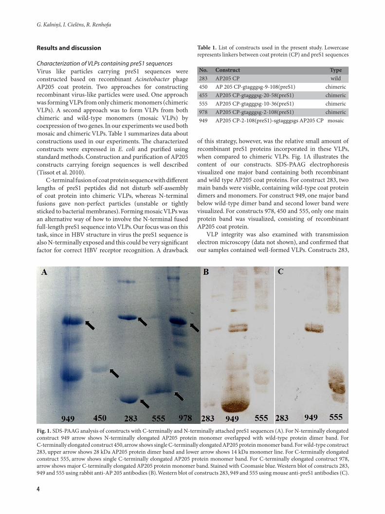
Characterization of VLPs containing preS1 sequencesVirus like particles carrying preS1 sequences were constructed based on recombinant Acinetobacter phage AP205 coat protein. Two approaches for constructing recombinant virus-like particles were used. One approach was forming VLPs from only chimeric monomers (chimeric VLPs). A second approach was to form VLPs from both chimeric and wild-type monomers (mosaic VLPs) by coexpression of two genes. In our experiments we used both mosaic and chimeric VLPs. Table 1 summarizes data about constructions used in our experiments. The characterized constructs were expressed in E. coli and purified using standard methods. Construction and purification of AP205 constructs carrying foreign sequences is well described (Tissot et al. 2010).
C-terminal fusion of coat protein sequence with different lengths of preS1 peptides did not disturb self-assembly of coat protein into chimeric VLPs, whereas N-terminal fusions gave non-perfect particles (unstable or tightly sticked to bacterial membranes). Forming mosaic VLPs was an alternative way of how to involve the N-terminal fused full-length preS1 sequence into VLPs. Our focus was on this task, since in HBV structure in virus the preS1 sequence is also N-terminally exposed and this could be very significant factor for correct HBV receptor recognition. A drawback
of this strategy, however, was the relative small amount of recombinant preS1 proteins incorporated in these VLPs, when compared to chimeric VLPs. Fig. 1A illustrates the content of our constructs. SDS-PAAG electrophoresis visualized one major band containing both recombinant and wild type AP205 coat proteins. For construct 283, two main bands were visible, containing wild-type coat protein dimers and monomers. For construct 949, one major band below wild-type dimer band and second lower band were visualized. For constructs 978, 450 and 555, only one main protein band was visualized, consisting of recombinant AP205 coat protein.
VLP integrity was also examined with transmission electron microscopy (data not shown), and confirmed that our samples contained well-formed VLPs. Constructs 283,
Fig. 1. SDS-PAAG analysis of constructs with C-terminally and N-terminally attached preS1 sequences (A). For N-terminally elongated construct 949 arrow shows N-terminally elongated AP205 protein monomer overlapped with wild-type protein dimer band. For C-terminally elongated construct 450, arrow shows single C-terminally elongated AP205 protein monomer band. For wild-type construct 283, upper arrow shows 28 kDa AP205 protein dimer band and lower arrow shows 14 kDa monomer line. For C-terminally elongated construct 555, arrow shows single C-terminally elongated AP205 protein monomer band. For C-terminally elongated construct 978, arrow shows major C-terminally elongated AP205 protein monomer band. Stained with Coomasie blue. Western blot of constructs 283, 949 and 555 using rabbit anti-AP 205 antibodies (B). Western blot of constructs 283, 949 and 555 using mouse anti-preS1 antibodies (C).
Table 1. List of constructs used in the present study. Lowercase represents linkers between coat protein (CP) and preS1 sequences
No. Construct Type283 AP205 CP wild450 AP 205 CP-gtagggsg-9-108(preS1) chimeric455 AP205 CP-gtagggsg-20-58(preS1) chimeric555 AP205 CP-gtagggsg-10-36(preS1) chimeric978 AP205 CP-gtagggsg-2-108(preS1) chimeric949 AP205 CP-2-108(preS1)-sgtagggsgs AP205 CP mosaic
5
Virus-like particles addressed by HBV preS1 sequences
949 and 555 were analyzed with Western blot, using mouse anti-AP 205 coat (1B) and anti-preS1 (1C) antibodies. These data confirmed that both C-terminally and N-terminally elongated constructs contained AP 205 chimeric coat proteins elongated with preS1 fragments, but constructs without the preS1 attachment contained only wild-type coat proteins.
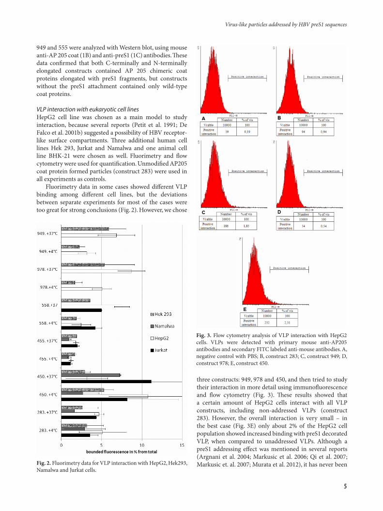
VLP interaction with eukaryotic cell linesHepG2 cell line was chosen as a main model to study interaction, because several reports (Petit et al. 1991; De Falco et al. 2001b) suggested a possibility of HBV receptor-like surface compartments. Three additional human cell lines Hek 293, Jurkat and Namalwa and one animal cell line BHK-21 were chosen as well. Fluorimetry and flow cytometry were used for quantification. Unmodified AP205 coat protein formed particles (construct 283) were used in all experiments as controls.
Fluorimetry data in some cases showed different VLP binding among different cell lines, but the deviations between separate experiments for most of the cases were too great for strong conclusions (Fig. 2). However, we chose
three constructs: 949, 978 and 450, and then tried to study their interaction in more detail using immunofluorescence and flow cytometry (Fig. 3). These results showed that a certain amount of HepG2 cells interact with all VLP constructs, including non-addressed VLPs (construct 283). However, the overall interaction is very small – in the best case (Fig. 3E) only about 2% of the HepG2 cell population showed increased binding with preS1 decorated VLP, when compared to unaddressed VLPs. Although a preS1 addressing effect was mentioned in several reports (Argnani et al. 2004; Markusic et al. 2006; Qi et al. 2007; Markusic et. al. 2007; Murata et al. 2012), it has never been Fig. 2. Fluorimetry data for VLP interaction with HepG2, Hek293,
Namalwa and Jurkat cells.
Fig. 3. Flow cytometry analysis of VLP interaction with HepG2 cells. VLPs were detected with primary mouse anti-AP205 antibodies and secondary FITC labeled anti-mouse antibodies. A, negative control with PBS; B, construct 283; C, construct 949; D, construct 978; E, construct 450.

6
G. Kalniņš, I. Cielēns, R. Renhofa
used for delivery of VLP. In our experiments we found that only a very small fraction of HepG2 cells interact with preS1-decorated VLP in detectable amounts. The reason for this may be because only a very small fraction of HepG2 cells possess HBV receptor-like structures in sufficient amounts. It might also be possible that that receptor affinity, sufficient for HBV binding and infection, is not sufficient for VLP selective binding. In any case, further studies are required to resolve these issues.
Confocal microscopy was performed to characterize VLP localization in cells that interacted efficiently with VLPs (Fig. 4). We performed FITC-labeled VLP incubations with HepG2 and Hek293 cells for 1 hour. Cells were grown on chamber slides, and those with observable FITC labeling were examined closer. We could conclude that construct 555, containing only the immunologically significant region, bound strongly to the HepG2 cell membrane, but was not present in cytosol. However, constructs 978 and 949 (containing additional preS1 regions), were present into cytosol during the same experimental conditions. This effect might be due to the existence of 1 to 9 and/or 37 to 108 prolonged preS1 amino acid regions in constructs 978 and 949, but lacking in construct 555. There are data (Tang et al. 2009) showing that the preS1 amino acid sequence 60-108 can be an addressing factor to HepG2 cells even without the structurally and immunologically important 10-36 amino acid region. Our results confirm that the full-length preS1 sequence is required for more efficient interaction with cells.
In conclusion, our results show that full-length preS1 sequence mediates VLP uptake into HepG2 and Hek293 cells in a cell type unspecific way as uptake of these particles can occur in both HepG2 and Hek293 cells, even despite the fact that Hek293 cells lack any HBV receptors or receptor-like structures. In contrast, the short preS1 21-47 amino acid fragment mediates only adhesion to HepG2 cells, and therefore lacks the ability to mediate VLP uptake into cytosol. However, the overall specific addressing effect of preS1 sequence to HepG2 cells is very low.
Acknowledgements
We thank Arnis Strods for support and assistance with flow cytometry analysis. This work was supported by Latvian National Research Program No. 4 “BIOMEDICINE” Project. 2010-7/7.2.
References
Argnani R., Boccafogli L., Marconi P.C., Manservigi R. 2004. Specific targeted binding of herpes simplex virus type 1 to hepatocytes via the human hepatitis B virus preS1 peptide. Gene Ther. 11: 1087–1098.
Fig. 4. Confocal cross-sections of HepG2 and Hek293 cells, incubated for 1 h with FITC-labeled VLPs. Blue channel, nuclei stained by Hoechst (except E, where cells were stained with LIVE-DEAD); red channel, membranes stained by WGA-Alexa 633; green channel, VLP labeled with FITC. A, construct 949 incubated with HepG2 cells; B, construct 949 incubated with Hek293 cells; C, construct 978 incubated with HepG2 cells; D, construct 978 incubated with Hek293 cells; E, construct 555 incubated with HepG2 cells.

Virus-like particles addressed by HBV preS1 sequences
7
Barrera A., Guerra B., Notvall L., Lanford R.E. 2005. Mapping of the hepatitis B virus pre-S1 domain involved in receptor recognition. J. Virol. 79: 9786–9798.
Blanchet M., Sureau C. 2007. Infectivity determinants of the hepatitis B virus pre-S domain are confined to the N-terminal 75 amino acid residues. J Virol. 81: 5841–5849.
Bremer C.M., Sominskaya I., Skrastina D., Pumpens P., El Wahed A.A., Beutling U., Frank R., Fritz H.J., Hunsmann G., Gerlich W.H., Glebe D. 2011. N-terminal myristoylation-dependent masking of neutralizing epitopes in the preS1 attachment site of hepatitis B virus. J. Hepatol. 55: 29–37.
Bruns M., Miska S., Chassot S., Will H.. 1998. Enhancement of hepatitis B virus infection by noninfectious subviral particles. J. Virol. 72: 1462–1468.
Bruss V. 1997. A short linear sequence in the pre-S domain of the large hepatitis B virus envelope protein required for virion formation. J. Virol. 71: 9350–9357.
Budkowska A., Quan C., Groh F., Bedossa F., Dubreuil P., Bouvet J.P., Pillot J. 1993. Hepatitis B virus (HBV) binding factor in human serum: candidate for a soluble form of hepatocyte HBV receptor. J. Virol. 67: 4316–4322.
Chi S.W., Kim D.H., Lee S.H., Chang I., Han K.H. 2007. Pre-structured motifs in the natively unstructured preS1 surface antigen of hepatitis B virus. Protein Sci. 16: 2108–2017.
Chi S.W., Maeng C.Y., Kim S.J., Oh M.S., Ryu C.J., Kim S.J., Han K.H., Hong H.J., Ryu S.E. 2007. Broadly neutralizing anti-hepatitis B virus antibody reveals a complementarity determining region H3 lid-opening mechanism. Proc. Natl. Acad Sci. USA 104: 9230–9235.
De Falco S., Ruvo M., Verdoliva A., Scarallo A., Raimondo D., Raucci A., Fassina G. 2001. N-terminal myristylation of HBV preS1 domain enhances receptor recognition. J. Pept. Res. 57: 390–400.
De Falco S., Ruvoletto M.G., Verdoliva A., Ruvo M., Raucci A., Marino M., Senatore S., Cassani G., Alberti A., Pontisso P., Fassina G. 2001. Cloning and expression of a novel hepatitis B virus-binding protein from HepG2 cells. J. Biol. Chem. 276: 36613–36623.
Glebe D., Aliakbari M., Krass P., Knoop E.V., Valerius K.P., Gerlich W.H. 2003. Pre-s1 antigen-dependent infection of Tupaia hepatocyte cultures with human hepatitis B virus. J. Virol. 77: 9511–9521.
Glebe D., Urban S. 2007. Viral and cellular determinants involved in hepadnaviral entry. World J. Gastroenterol. 13: 22–38.
Gripon P., Cannie I., Urban S. 2005. Efficient inhibition of hepatitis B virus infection by acylated peptides derived from the large viral surface protein. J. Virol. 79: 1613–1622.
Hu W.G., Wei J., Xia H.C., Yang X.X., Li F., Li G.D., Wang Y., Zhang Z.C. 2005. Identification of the immunogenic domains in HBsAg preS1 region using overlapping preS1 fragment fusion proteins. World J. Gastroenterol. 11: 2088–2094.
Jin Y.M., Churchill N.D., Michalak T.I. 1996. Protease-activated lymphoid cell and hepatocyte recognition site in the preS1 domain of the large woodchuck hepatitis virus envelope protein. J. Gen. Virol. 77: 1837–1846.
Kim D.H., Ni Y., Lee S.H., Urban S., Han K.H. 2008. An anti-viral peptide derived from the preS1 surface protein of hepatitis B virus. BMB Rep. 41: 640–644.
Lambert C., Prange R. 2003. Chaperone action in the posttranslational topological reorientation of the hepatitis B virus large envelope protein: Implications for translocational regulation. Proc. Natl. Acad. Sci. USA 100: 5199–5204.
Le Seyec J., Chouteau P., Cannie I., Guguen-Guillouzo C., Gripon P. 1999. Infection process of the hepatitis B virus depends on the presence of a defined sequence in the pre-S1 domain. J. Virol. 73: 2052–2057.
Maeng C.Y., Ryu C.J., Gripon P., Guguen-Guillouzo C., Hong H.J. 2000. Fine mapping of virus-neutralizing epitopes on hepatitis B virus PreS1. Virology 270: 9–16.
Markusic D.M., van Til N., Kanitz A., Hiralall J., Oude-Elferink R., Seppen J. 2006. Pseudotyping lentiviral vectors with a modified GP64 envelope proteins redirects gene transfer in vitro and in vitro. Mol. Ther. 13: S180.
Markusic D.M., Kanitz A., Oude-Elferink R.P., Seppen J. 2007. Preferential gene transfer of lentiviral vectors to liver-derived cells, using a hepatitis B peptide displayed on GP64. Hum. Gene Ther. 18: 673–679.
Milich D.R., McLachlan A., Moriarty A., Thornton G.B. 1987. A single 10-residue pre-S(1) peptide can prime T cell help for antibody production to multiple epitopes within the pre-S(1), pre-S(2), and S regions of HBsAg. J. Immunol. 138: 4457–4465.
Miyata R., Ueda M., Jinno H., Konno T., Shihara K., Ando N., Kitagawa Y. 2009. Selective targeting by preS1 domain of hepatitis B surface antigen conjugated with phosphorylcholine-based amphiphilic block copolymer micelles as a biocompatible, drug delivery carrier for treatment of human hepatocellular carcinoma with paclitaxel. Int. J. Cancer 124: 2460–2467.
Murata M., Narahara S., Umezaki K., Toita R., Tabata S., Piao J.S., Abe K., Kang J.H., Ohuchida K., Cui L., Hashizume M. 2012. Liver cell specific targeting by the preS1 domain of hepatitis B virus surface antigen displayed on protein nanocages. Int. J. Nanomed. 7: 4353–4362.
Neurath A.R., Kent S.B., Strick N., Parker K. 1986. Identification and chemical synthesis of a host cell receptor binding site on hepatitis B virus. Cell 46: 429–436.
Neurath A.R., Seto B., Strick N. 1989. Antibodies to synthetic peptides from the preS1 region of the hepatitis B virus (HBV) envelope (env) protein are virus-neutralizing and protective. Vaccine 7: 234–236.
Neurath A.R., Strick N., Sproul P., Ralph H.E., Valinsky J. 1990. Detection of receptors for hepatitis B virus on cells of extrahepatic origin. Virology 176: 448–457.
Persing D.H., Varmus H.E., Ganem D. 1987. The preS1 protein of hepatitis B virus is acylated at its amino terminus with myristic acid. J. Virol. 61: 1672–1677.
Petit M.A., Dubanchet S., Capel F., Voet P., Dauguet C., Hauser P. 1991. HepG2 cell binding activities of different hepatitis B virus isolates: inhibitory effect of anti-HBs and anti-preS1(21-47). Virology 180: 483–491.
Pontisso P., Ruvoletto M.G., Gerlich W.H., Heermann K.H., Bardini R., Alberti A. 1989. Identification of an attachment site for human liver plasma membranes on hepatitis B virus particles. Virology 173: 522–530.
Pontisso P., Ruvoletto M.G., Tiribelli C., Gerlich W.H., Ruol A., Alberti A. 1992. The preS1 domain of hepatitis B virus and IgA cross-react in their binding to the hepatocyte surface. J. Gen. Virol. 73: 2041–2045.
Qi P., Han J.X., Lu Y.Q., Wang C.X. 2007. Redirecting retroviral tropism by insertion of hepatitis B virus PreS1 core peptide into the envelope. Arch. Virol. 152: 1721–1730.
Rapoport T.A., Matlack K.E., Plath K., Misselwitz B., Staeck O. 1999. Posttranslational protein translocation across the membrane of the endoplasmic reticulum. Biol. Chem. 380:

G. Kalniņš, I. Cielēns, R. Renhofa
8
1143–1150. Ryu C.J,. Gripon P., Park H.R., Park S.S., Kim Y.K., Guguen-
Guillouzo C., Yoo O.J., Hong H.J. 1997. In vitro neutralization of hepatitis B virus by monoclonal antibodies against the viral surface antigen. J. Med. Virol. 52: 226–233.
Schädler S., Hildt E. 2009. HBV life cycle: entry and morphogenesis. Viruses 1: 185–209.
Sominskaya I., Pushko P., Dreilina D., Kozlovskaya T., Pumpen P. 1992. Determination of the minimal length of preS1 epitope recognized by a monoclonal antibody which inhibits
Received 6 November 2012; received in revised form 16 January 2013; accepted 1 March 2013
attachment of hepatitis B virus to hepatocytes. Med. Microbiol. Immun. 181: 215–226.
Tang K.H., Yusoff K., Tan W.S. 2009. Display of hepatitis B virus PreS1 peptide on bacteriophage T7 and its potential in gene delivery into HepG2 cells. J. Virol. Methods 159: 194–199.
Tissot A.C., Renhofa R., Schmitz N., Cielens I., Meijerink E., Ose V., Jennings G.T., Saudan P., Pumpens P., Bachmann M.F. 2010. Versatile virus-like particle carrier for epitope based vaccines. PLoS One 5: e9809.