Virtual Anatomy of Spinal Disorders by 3-D MRI/CT Fusion ... · - Spondylolisthesis and...
14
5 Virtual Anatomy of Spinal Disorders by 3-D MRI/CT Fusion Imaging Junji Kamogawa and Osamu Kato Shiraishi Hospital/Spine & Sports Center Japan 1. Introduction Both CT and MRI are widely known to be important tools in diagnosing spinal disorders. However, even when three-dimensional data are collected, images are often presented in two dimensions and in monochrome. Furthermore, CT and MRI represent different types of information, and it is very difficult for doctors to mentally fuse these images. We have been working to overcome these challenges, and reported the first 3-D MRI/CT fusion images of the cranio-vertebral junction (Kamogawa, 2009). We have since continued to develop the technique, and have used it to visualize the lumbar nerve root (Misaki, 2009; Yamanaka 2010). However, there are two major problems related to ensuring image quality for accurate diagnosis. The first is the optimal operation of MRI machines in order to obtain the best MRI sequences for each patient. Although it is simple to obtain clear CT images of spinal bone, it is very difficult to obtain such images of neural anatomy (spinal cord, cauda equina and spinal nerve roots) by MRI. Second, we must precisely superimpose the two images (MRI and CT) using computer software to evaluate complex spinal disorders. In this chapter, the methods adopted at our hospital for obtaining, evaluating and displaying 3-D MRI/CT fusion imaging are discussed, focusing on their application to the field of spinal surgery. 2. Aims There are numerous conditions of the spine that cannot readily be diagnosed using regular 2-D MRI. One such condition is spinal deformity (including malformation and tumor) with a focus on bone architecture; another is the secondary changes in the structure or environment of the spinal canal, which is the space inside the spinal column that supplies the nerve tissue. These secondary changes are involved in numerous disorders, such as chondrogenesis, bulging disk, thickened ligament flavum and varix (Table 1). The power of fusion imaging is particularly apparent in the visualization of conditions of the nerve root. Nerve roots are important from a clinical perspective because they are the source of particularly severe pain, and because tethering of a nerve root during procedures to correct the spinal column can cause paralysis. The nerve roots are normally distal to the lateral canal of the spinal column, but this area is difficult to assess by either myelography www.intechopen.com
Transcript of Virtual Anatomy of Spinal Disorders by 3-D MRI/CT Fusion ... · - Spondylolisthesis and...

5
Virtual Anatomy of Spinal Disorders by 3-D MRI/CT Fusion Imaging
Junji Kamogawa and Osamu Kato Shiraishi Hospital/Spine & Sports Center
Japan
1. Introduction
Both CT and MRI are widely known to be important tools in diagnosing spinal disorders. However, even when three-dimensional data are collected, images are often presented in two dimensions and in monochrome. Furthermore, CT and MRI represent different types of information, and it is very difficult for doctors to mentally fuse these images. We have been working to overcome these challenges, and reported the first 3-D MRI/CT fusion images of the cranio-vertebral junction (Kamogawa, 2009). We have since continued to develop the technique, and have used it to visualize the lumbar nerve root (Misaki, 2009; Yamanaka 2010).
However, there are two major problems related to ensuring image quality for accurate diagnosis. The first is the optimal operation of MRI machines in order to obtain the best MRI sequences for each patient. Although it is simple to obtain clear CT images of spinal bone, it is very difficult to obtain such images of neural anatomy (spinal cord, cauda equina and spinal nerve roots) by MRI. Second, we must precisely superimpose the two images (MRI and CT) using computer software to evaluate complex spinal disorders.
In this chapter, the methods adopted at our hospital for obtaining, evaluating and displaying 3-D MRI/CT fusion imaging are discussed, focusing on their application to the field of spinal surgery.
2. Aims
There are numerous conditions of the spine that cannot readily be diagnosed using regular 2-D MRI. One such condition is spinal deformity (including malformation and tumor) with a focus on bone architecture; another is the secondary changes in the structure or environment of the spinal canal, which is the space inside the spinal column that supplies the nerve tissue. These secondary changes are involved in numerous disorders, such as chondrogenesis, bulging disk, thickened ligament flavum and varix (Table 1).
The power of fusion imaging is particularly apparent in the visualization of conditions of the nerve root. Nerve roots are important from a clinical perspective because they are the source of particularly severe pain, and because tethering of a nerve root during procedures to correct the spinal column can cause paralysis. The nerve roots are normally distal to the lateral canal of the spinal column, but this area is difficult to assess by either myelography
www.intechopen.com

Recent Advances in Scoliosis
74
or CT myelography (Fig. 1). Fusion imaging offers a solution to the problem of this hidden imaging zone, in which assessment has not been possible using the techniques available to date.
A. Spinal deformity 1. Congenital scoliosis 2. Adult Deformity
- Degenerative scoliosis - Spondylolisthesis and retrolisthesis - Rotatory subluxation
3. Trauma
B. The secondary changes in structure or environment of the spinal canal 1. Siatica due to
- Single pathological nerve root on degenerative lumbar scoliosis - Lumbar lateral canal stenosis - Far-out syndrome, Double-crash lesion - Spondylolysis - Varix
2. Adhesion due to - Epi-dural membrane formation on dural sac - Failed back surgery syndrome - Re-operation
C. Tumor
Table 1. Indication for Fusion Imaging.
Fig. 1. Hidden imaging zone. Even with myelography or CT myelography (yellow area), the green areas do not show up in detail.
3. Machines
We used an Asteion 4® 4-row CT unit (TOSHIBA, Tochigi, Japan), an Echelon Vega® 1.5-T MRI unit (HITACHI, Tokyo, Japan), and Synapse Vincent® workstation software (FUJIFILM, Tokyo, Japan). Parameters for CT were as follows: tube voltage, 120 kV; tube current, 260 mA; slice thickness, 1 mm; rotational speed, 0.75 s/rotation; and slice thickness for reconstruction, 0.5 mm. The MRI sequences are shown in Table 2.
www.intechopen.com

Virtual Anatomy of Spinal Disorders by 3-D MRI/CT Fusion Imaging
75
BASG RSSG Mode 3D coronal 3D coronal FOV 250 mm 384 mm TR 10.4 ms 18.0 ms TE 5.2 ms 9.2 ms Flip angle 45° 10° Slice thickness 1.3 mm 1.5 mm Recon-pitch 0.65 mm 0.75 mm Recon-slice 100 90 Scan time 6’40’’ 6’45’’
BASG® (Balanced Sarge by HITACHI) RSSG® (RS Sarge by HITACHI)
Table 2. Sequences of MRI
4. Steps for producing 3-D MRI/CT fusion images (procedure for obtaining the image in case 1)
4.1 Data reading
First, the workstation reads both CT and MRI DICOM data.
4.2 Registering
We use the “Superposition” application to automatically place the MRI data onto the CT data. When we use this tool, as it is not exclusively for spinal use, misalignment between the two modalities is possible. We must therefore correct the gap manually on the sagittal, coronal and axial planes (Fig. 2). An automatic superposition tool specifically for use with the spine has not yet been developed.
Fig. 2. Superposition of MRI and CT. The monochrome image is CT, and the green MRI image is superposed onto the CT image. Coronal (A), sagittal (B) and axial (C) planes.
A B C
www.intechopen.com

Recent Advances in Scoliosis
76
4.3 Isolation of vertebral body for 3-D CT
First, we must remove unneeded information (e.g., calcification of aorta) (Fig. 3A, B). We must then separate each L1-5 vertebra in turn from the whole lumbar spine structure in order to use the “Mask editing” application (Fig. 4). For one vertebra, we use the “Slice out”, “Closing and Painting”, and “Surfacing” tools, and it takes about 10 minutes to manually finish one vertebra. The technique for individually isolating the vertebra is extremely useful, as the shape of the apophyseal joint (facet) is clarified at a glance (Fig. 3C, D, E). Colors are then added (Fig. 3F). Using this technique, the vertebrae can be made translucent at any density.
Fig. 3. 3-D CT reconstruction and vertebral isolation. Removal of unnecessary information: before (A) and after (B). Facet surface and neural arch can be evaluated (C, D). Whole lumbar spine without L5 vertebra (E). Finally, color was added (F).
A B C D
E F
www.intechopen.com

Virtual Anatomy of Spinal Disorders by 3-D MRI/CT Fusion Imaging
77
Fig. 4. Extraction of individual vertebrae. Details of the superior and inferior articular process (facet) are carefully colored. Coronal (A), sagittal (B) and axial (C, D) planes.
4.4 3-D representation of spinal cord
We produce a 3-D representation of both the dural sac and the spinal cord using an RSSG® sequence (HITACHI) with a single click on the Workstation (Fig. 5A).
4.5 Mapping spinal nerve roots from 3-D MRI
It is very difficult to obtain and map the spinal nerve roots. We use two MRI sequences: 3-D
myelography of 1.3 mm slice thickness (Balanced SG® by HITACHI) and 3-D myelography
of 1.5 mm slice thickness (RSSG® by HITACHI). There are many methods for obtaining clear
images of spinal nerves on MRI. Careless operation of the MRI will result in failure to obtain
nerve information. First, we place dots on the nerve roots in MPR (multiplanar
reconstruction) using the “Center line editing” application. We then observe a straight nerve
converted by the straight CPR (curved planar reformation) method. Next, we use “Contour
editing” 7-10 times in order to repair the edge of the straight nerve. After obtaining the
finished 3-D nerve root, it is necessary to cut the unnecessary image near the root edge. We
place the finished 3-D nerve on the MPR screen like tracing paper (Fig. 6), filling in the dots
manually. Finally, we obtain a finished 3-D nerve root (Fig. 5B, C, D). It takes about 20
minutes for a radiographer to produce an image of one nerve root, and we typically prepare
images of six nerve roots for both sides of the lumbar spine (the main pathological root, and
the top and bottom roots) for degenerative lumbar scoliosis.
A B C D
www.intechopen.com

Recent Advances in Scoliosis
78
Fig. 5. Nerve tissue extraction. Dural sac and spinal cord (A). Each individual nerve root is extracted (B, C). The nerve root, which is the focus of disease (orange), is swollen and the pressure has led to constriction (C, D).
Fig. 6. Mapping of spinal nerve roots. The green area shows the nerve root being identified.
4.6 Final superposition
We place the 3-D MRI images onto 3-D CT images, and observe the entire lumbar spine in any direction (Fig. 7). A total of 5-8 h is required to produce 3-D fusion imaging.
A B C D
www.intechopen.com

Virtual Anatomy of Spinal Disorders by 3-D MRI/CT Fusion Imaging
79
Fig. 7. Completed 3-D fusion imaging. The bones can be made semi-transparent and vertebrae can be removed at will, and assessment can be performed from any direction. The orange shows the main pathological nerve root. Left anterior oblique view (A) and left posterior oblique view (B).
5. Case presentation
5.1 Case 1
A 72-year-old female, degenerative lumbar scoliosis with L2 compression fracture
The main clinical findings were severe sciatica of the left thigh, a feeling of heaviness in the
lower back, and gait disturbance for 5 meters. Marked radicular pain of the left L3 nerve
root was confirmed, while selective left L3 root block caused reproducible pain, and reduced
her distress for 2 days. No images depicted accurate information of her chief complaint, and
plain roentgenography is shown in Fig. 8. 3-D MRI/CT Fusion Imaging revealed
compression of the left L3 nerve root at the intraforaminal zone. We were able to evaluate
the lesion from any direction at a glance, and were able to obtain clear evidence to suggest
sciatica due to the L3 nerve root (Fig. 9).
A B
www.intechopen.com
Recent Advances in Scoliosis
80
Fig. 8. Plain X-ray image. Previous compression fracture of L2 can be seen. AP view (A) and lateral view (B).
Fig. 9. Left L3 root and relationship with surrounding bony structure. The L3 root can be seen to be under pressure in the foraminal zone (orange). This image is greatly useful in selecting the operative procedure. Anterosuperior view (A) and lateral view (B).
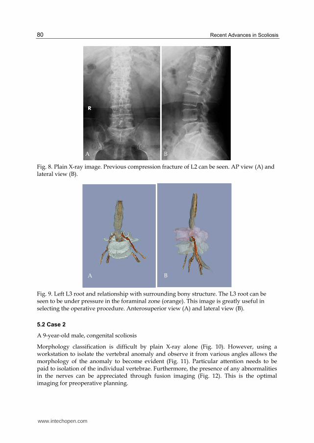
5.2 Case 2
A 9-year-old male, congenital scoliosis
Morphology classification is difficult by plain X-ray alone (Fig. 10). However, using a workstation to isolate the vertebral anomaly and observe it from various angles allows the morphology of the anomaly to become evident (Fig. 11). Particular attention needs to be paid to isolation of the individual vertebrae. Furthermore, the presence of any abnormalities in the nerves can be appreciated through fusion imaging (Fig. 12). This is the optimal imaging for preoperative planning.
A B
A B
www.intechopen.com

Virtual Anatomy of Spinal Disorders by 3-D MRI/CT Fusion Imaging
81
Fig. 10. Plain X-ray image. The details of vertebrae are unclear.
Fig. 11. Isolation of block vertebrae. This is extremely useful for gaining an understanding of the condition and planning osteotomy. Posterior view (A), superior view (B), left lateral view (C) and right lateral view (D).
A B
C D
www.intechopen.com
Recent Advances in Scoliosis
82
Fig. 12. Completed 3-D fusion imaging. If the bone is made semi-transparent, details of the nerve tissue at the surgical site can be seen. Posterior view (A) and anterior view (B). Magnified view (C). The bone are partly cut off to evaluate the nerve roots. The patient was examined for MRI without a sedative.
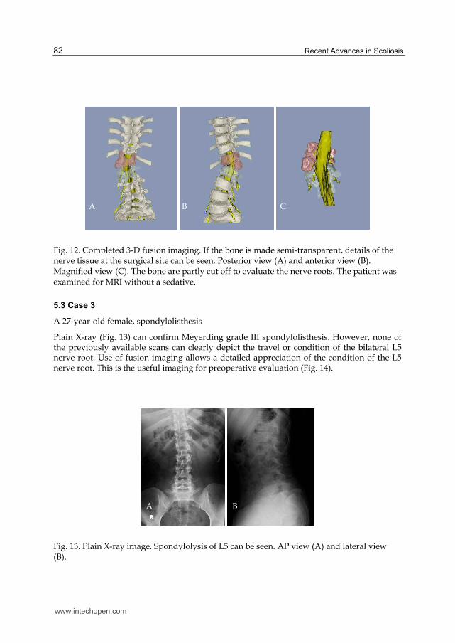
5.3 Case 3
A 27-year-old female, spondylolisthesis
Plain X-ray (Fig. 13) can confirm Meyerding grade III spondylolisthesis. However, none of the previously available scans can clearly depict the travel or condition of the bilateral L5 nerve root. Use of fusion imaging allows a detailed appreciation of the condition of the L5 nerve root. This is the useful imaging for preoperative evaluation (Fig. 14).
Fig. 13. Plain X-ray image. Spondylolysis of L5 can be seen. AP view (A) and lateral view (B).
C B A
A B
www.intechopen.com

Virtual Anatomy of Spinal Disorders by 3-D MRI/CT Fusion Imaging
83
Fig. 14. Completed 3-D fusion imaging. The bone has been made semi-transparent. Bilateral L5 nerve roots are considerably swollen, and the travel of the nerve root is clear at a glance. The relationship between the L5 vertebra and the L5 root is clearly visible. Left lateral view (A), AP view (B) and posterosuperior view (C).
6. Conclusion
The ultimate purpose of our research is to produce images that can easily be interpreted by
anyone at a glance without the use of contrast media. When we examine patients suffering
from sciatica and back pain, or those who have no normal spinal canal structure due to
deformities, it is very difficult to obtain useful images showing the neurological details. MRI
is particularly poor for torsion, curved and rotational deformities of the spine.
In recent years, researchers have attempted to produce clear 3-D images of self-operating
organs such as the heart, large and small intestines, and blood vessels using multi-row, top-
of-the-line CT with the use of contrast media. On the other hand, the fields of spinal surgery
that deal with non-self-operating, complex motions require only 4-16-row CT, but high-
quality MRI units are vital. There are global standards of quality for CT among the
disciplines of internal medicine, brain surgery and radiology; however, no such global
standards yet exist for MRI in spinal surgery. The reason for this is that although taking CT
images is straightforward and the image quality does not vary substantially with different
imaging methods, the necessary conditions and methods for MRI are complex, and even a
good MRI device can be difficult to operate. The nerve roots of cervical vertebrae can be
visualized by utilizing particular conditions (data not shown).
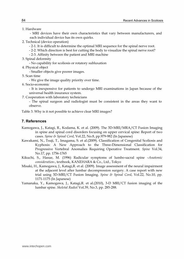
Why is it so difficult to obtain clear MR images of the spine? It is vitally important to obtain
MRI data to produce fusion images, and there are numerous reasons for the poor results of
MRI to date (Table 3).
There may well be spinal pathology that are only detected after the start of spinal surgery.
Nonetheless, if it were possible to demonstrate that the patient’s detailed anatomy could not
be seen satisfactorily in preoperative images, it may also be possible to better interpret
preoperative images. This was the purpose of devising fusion imaging. After the spinal
anatomy is clearly visualized, the pathology can be described, and effective surgical plans
can be formulated. Although our technique is associated with problems such as
reproducibility, convenience and applicability, we intend to continue with further
development in the future.
A B C
www.intechopen.com
Recent Advances in Scoliosis
84
1. Hardware - MRI devices have their own characteristics that vary between manufacturers, and each individual device has its own quirks.
2. Technical (device operation) - 2-1. It is difficult to determine the optimal MRI sequence for the spinal nerve root. - 2-2. Which direction is best for cutting the body to visualize the spinal nerve root? - 2-3. Affinity between the patient and MRI machine
3. Spinal deformity - No capability for scoliosis or rotatory subluxation
4. Physical object - Smaller objects give poorer images.
5. Scan time - We give the image quality priority over time.
6. Socio-economic - It is inexpensive for patients to undergo MRI examinations in Japan because of the universal health insurance system.
7. Cooperation with laboratory technicians - The spinal surgeon and radiologist must be consistent in the areas they want to observe.
Table 3. Why is it not possible to achieve clear MRI images?
7. References
Kamogawa, J., Katagi, R., Kodama, K. et al. (2009). The 3D-MRI/MRA/CT Fusion Imaging in spine and spinal cord disorders focusing on upper cervical spine: Report of two cases. Spine & Spinal Cord, Vol.22, No.8, pp.979-982 (In Japanese)
Kawakami, N., Tsuji, T., Imagama, S. et al.(2009). Classification of Congenital Scoliosis and Kyphosis: A New Approach to the Three-Dimensional Classification for Progressive Vertebral Anomalies Requiring Operative Treatment. Spine Vol.34, No.17, pp. 1756-1765
Kikuchi, S., Hasue, M. (1996) Radicular symptoms of lumbo-sacral spine –Anatomic consideration-, textbook, KANEHARA & Co., Ltd., Tokyo
Misaki, H., Kamogawa, J., Katagi,R. et al. (2009). Image assessment of the neural impairment at the adjacent level after lumbar decompression surgery. A case report with new trial using 3D-MRI/CT Fusion Imaging. Spine & Spinal Cord, Vol.22, No.10, pp. 1171-1175 (In Japanese)
Yamanaka, Y., Kamogawa, J., Katagi,R. et al.(2010), 3-D MRI/CT fusion imaging of the lumbar spine. Skeletal Radiol Vol.39, No.3, pp. 285-288.
www.intechopen.com
Recent Advances in ScoliosisEdited by Dr Theodoros Grivas
ISBN 978-953-51-0595-4Hard cover, 344 pagesPublisher InTechPublished online 09, May, 2012Published in print edition May, 2012
InTech EuropeUniversity Campus STeP Ri Slavka Krautzeka 83/A 51000 Rijeka, Croatia Phone: +385 (51) 770 447 Fax: +385 (51) 686 166www.intechopen.com
InTech ChinaUnit 405, Office Block, Hotel Equatorial Shanghai No.65, Yan An Road (West), Shanghai, 200040, China
Phone: +86-21-62489820 Fax: +86-21-62489821
This book contains information on recent advances in aetiology and pathogenesis of idiopathic scoliosis, forthe assessment of this condition before treatment and during the follow-up, making a note of emergingtechnology and analytical techniques like virtual anatomy by 3-D MRI/CT, quantitative MRI and MoireTopography. Some new trends in conservative treatment and the long term outcome and complications ofsurgical treatment are described. Issues like health related quality of life, psychological aspects of scoliosistreatment and the very important "patient's perspective" are also discussed. Finally two chapters tapping theuntreated early onset scoliosis and the congenital kyphoscoliosis due to hemivertebra are included. It must beemphasized that knowledgeable authors with their contributions share their experience and enthusiasm withpeers interested in scoliosis.
How to referenceIn order to correctly reference this scholarly work, feel free to copy and paste the following:
Junji Kamogawa and Osamu Kato (2012). Virtual Anatomy of Spinal Disorders by 3-D MRI/CT Fusion Imaging,Recent Advances in Scoliosis, Dr Theodoros Grivas (Ed.), ISBN: 978-953-51-0595-4, InTech, Available from:http://www.intechopen.com/books/recent-advances-in-scoliosis/virtual-anatomy-of-spinal-disorders-by-3-d-mri-ct-fusion-imaging

© 2012 The Author(s). Licensee IntechOpen. This is an open access articledistributed under the terms of the Creative Commons Attribution 3.0License, which permits unrestricted use, distribution, and reproduction inany medium, provided the original work is properly cited.