
Frontiers in Zoology BioMed - BioMed Central | The Open Access

BioMed CentralVirology Journal
ss
Open AcceResearchWild type measles virus attenuation independent of type I IFNJohan Druelle*1, Caroline I Sellin1, Diane Waku-Kouomou1,2, Branka Horvat1 and Fabian T Wild1,2Address: 1Inserm, U758, Lyon, F-69365 France ; Ecole Normale Supérieure de Lyon, Lyon, F-69007 France ; IFR128 BioSciences Lyon-Gerland Lyon-Sud, Université de Lyon 1; 21 Avenue Tony Garnier, 69365 Lyon Cedex 07 – France and 2Centre National de Référence pour la Rougeole, Lyon, France
Email: Johan Druelle* - [email protected]; Caroline I Sellin - [email protected]; Diane Waku-Kouomou - [email protected]; Branka Horvat - [email protected]; Fabian T Wild - [email protected]
* Corresponding author
AbstractBackground: Measles virus attenuation has been historically performed by adaptation to cellculture. The current dogma is that attenuated virus strains induce more type I IFN and are moreresistant to IFN-induced protection than wild type (wt).
Results: The adaptation of a measles virus isolate (G954-PBL) by 13 passages in Vero cells induceda strong attenuation of this strain in vivo. The adapted virus (G954-V13) differs from its parentalstrain by only 5 amino acids (4 in P/V/C and 1 in the M gene). While a vaccine strain, EdmonstonZagreb, could replicate equally well in various primate cells, both G954 strains exhibited restrictionto the specific cell type used initially for their propagation. Surprisingly, we observed that bothG954 strains induced type I IFN, the wt strain inducing even more than the attenuated ones,particularly in human plasmacytoid Dendritic Cells. Type I IFN-induced protection from theinfection of both G954 strains depended on the cell type analyzed, being less efficient in the cellsused to grow the viral strain.
Conclusion: Thus, mutations in M and P/V/C proteins can critically affect MV pathogenicity,cellular tropism and lead to virus attenuation without interfering with the α/β IFN system.
BackgroundMass vaccination with live attenuated measles vaccineshas greatly reduced the incidence of this disease and itsassociated pathologies. Most vaccine strains were estab-lished after numerous passages on various cell lines. Dur-ing this period of adaptation, the virus genome mutatedin order to replicate efficiently in cell culture and thus, theoriginal viral phenotype has been modified by mecha-nisms which are still poorly understood. The mutationsobserved in the RNA genome may be responsible for the
replication of the clinical virus in its new host cell at dif-ferent levels: entry, transcription, translation or budding.
Measles virus (MV), one of the leading causes of infantdeath in developing countries, is a member of the Para-myxovirus family. Like other viruses of this family, the MVnegative RNA genome is protected by the N protein. Itsassociation with the replicative complex (P and L pro-teins) constitutes the nucleocapsid. H (haemagglutinin)and F (fusion) proteins are surface glycoproteins, set in alipid envelope, lined by the M (matrix) protein, and are
Published: 3 February 2008
Virology Journal 2008, 5:22 doi:10.1186/1743-422X-5-22
Received: 12 November 2007Accepted: 3 February 2008
This article is available from: http://www.virologyj.com/content/5/1/22
© 2008 Druelle et al; licensee BioMed Central Ltd. This is an Open Access article distributed under the terms of the Creative Commons Attribution License (http://creativecommons.org/licenses/by/2.0), which permits unrestricted use, distribution, and reproduction in any medium, provided the original work is properly cited.
Page 1 of 12(page number not for citation purposes)
Virology Journal 2008, 5:22 http://www.virologyj.com/content/5/1/22
responsible for the attachment and fusion processes. Inaddition to the structural proteins, the MV genomeencodes for two accessory proteins, C and V [1].
For many years, few MV wild-type isolates were availablefor study. This was mainly due to the choice of the cell lineused for virus isolation. Clinical or wild type MVs useCD150 (SLAM) as their main host cell receptor to attachto cells and so are most easily isolated on cell linesexpressing this molecule [2]. The vaccine and vaccine-likestrains, which readily multiply in cells lacking this recep-tor, were shown to be able to use an additional receptor,CD46, a ubiquitously expressed molecule [3,4]. Further, itwas established that a critical amino acid (aa) in the MVH glycoprotein governed the use of the two receptors.Mutation of aa 481 from asparagine to tyrosine permittedthe wild type strains to attach to CD46 [5]. In vivo, wildtype viruses are reported to infect both endothelial andepithelial cells, which do not express CD150. Thus, it isnot clear how the virus gets into the host cell. Two differ-ent hypotheses propose that either the wild type virusenters using CD46 as a low affinity receptor [6,7] or thatthere is another unidentified receptor involved [8-10].
Naniche et al. showed that in contrast to wild type MVstrains, vaccine strains induced high levels of IFN inperipheral blood mononuclear cells (PBMCs) [11]. More-over, the wild type strains were more sensitive to exoge-nous IFN. A number of studies have shown that MV, Cand V accessory proteins may be implicated in both theinhibition of the induction and action of IFN [12-17].However, it is still unknown whether the function of Cand V proteins in the regulation of type I IFN system isaltered after virus attenuation. Schlender et al. showedthat a measles vaccine strain (Schwarz) replicates effi-ciently in plasmacytoid Dendritic Cells (pDCs, the majorproducers of type 1 IFN [18]) and blocks IFN induction byseveral ligands [19]. Nevertheless, the complexity and thediversity of the experimental systems previously usedmade a clear-cut interpretation of these data difficult inthe estimation of the role of type I IFN in the attenuatedMV phenotype.
It was shown that viruses isolated on B95a cells couldinduce in a monkey model all the clinical featuresobserved in humans. However, adaptation of the virus toVero cells attenuated the pathogenicity of the virus[20,21]. Sequence comparison of the 2 viruses showedthat there were 5 aa changes in the polymerase (P/V/C andL) and 3 aa changes in the H [22] affecting the replicationand transcription processes and also the syncytia forma-tion. In a second study, the attenuating mutations wererestricted to the P and M genes [23] with a deletion of theC gene. Recently, Tahara et al. adapted a wt strain to Verocells and observed mutations in the M and L proteins. The
adapted virus could grow in cells that did not expressCD150 but was less efficient in the cell/cell fusion process[24]. The mutation E89K in M was then shown to beimplied in alteration of the interaction between the M andH proteins [25].
In the present study, we compared G954-PBL, a MV wildtype isolate propagated on PBMCs with G954-V13, a virusadapted from G954-PBL by 13 passages on Vero cells.Both strains were shown to have no differences in the Hand F proteins and to use CD150 and not CD46 as areceptor [10]. Sequence analysis of both G954 virusesrevealed that there were 5 mutations located only in the P/V/C and M genes. These mutations render the virus highlyattenuated in vivo. Loss of pathogenicity could be relatedto different aspects of infection. Both G954 strains seemedto be restricted to specific cell types initially used to prop-agate the virus. Interestingly, the vaccine strain, Edmon-ston Zagreb (Ed-Zagreb), used as a control, was morerobust than either of the G954 strains, multiplying in dif-ferent cell types. Surprisingly, despite the differences inthe P/V/C genes and the current belief that viral sensitivityto and induction of type I IFN correlate with an attenuatedphenotype, this study shows the existence of exceptions tothis dogma where virus attenuation is not linked to α/βIFN system.
MethodsVirus strains and cell linesThe wild-type MV strain, G954-PBL (genotype B3.2), wasisolated in Gambia in 1993 and was propagated on acti-vated human PBMCs. The Vero adapted strain, G954-V13was obtained after 13 successive passages of a G954-PBLsample on Vero cells, [10]. MV vaccine Edmonston-Zagreb was kindly provided by D. Forcic and R. Mazuran(Immunology Institute of Zagreb, Croatia). Vesicular sto-matitis virus (VSV) (Indiana strain) was propagated onVero cells.
Measles viruses were titrated on Vero/CD150 cells by thestandard plaque assay method as previously described[10]. For the establishment of viral kinetics, each timepoint was obtained individually. Infections were per-formed at a MOI of 0,1.
B95a, Vero, and Vero/CD150 cells were propagated inDulbecco's modified Eagle's medium (Invitrogen) supple-mented with 2 mM L-glutamine, 100 U of penicillin/ml,0.1 mg of streptomycin/ml,10 mM HEPES, and 10% fetalcalf serum or 2% for infections.
Isolation and infection of human haematopoietic cellsHuman PBMCs were prepared from whole blood ofhealthy donors (Etablissement Français du Sang, Lyon,France) by Ficoll-Hypaque density gradient centrifugation
Page 2 of 12(page number not for citation purposes)
Virology Journal 2008, 5:22 http://www.virologyj.com/content/5/1/22
(Eurobio, France). pDCs were isolated by magnetic acti-vated cell sorting (MACS) using the BDCA-4 dendritic cellisolation kit from Miltenyi Biotec. Prior to positive selec-tion, monocytes, B cells, T cells, NK cells, red cells andmacrophages were depleted by negative selection. Cellswere incubated with an antibody cocktail directed againstCD3, CD8, CD14, CD16, CD19, CD35, CD56 and glyco-phorin-A, then with Biomag Goat anti-mouse IgG mag-netic beads (Quiagen) and finally separated using aBiomag magnet. PDCs were labeled with anti-BDCA-4antibody coupled to colloidal paramagnetic micro beadsand passed through a magnetic separation column (LScolumn; Miltenyi Biotec). The purity of isolated pDCs(BDCA2 positive, CD123-positive) was between 75% and95%. pDCs from individual donors were used separatelyin all experiments and were not pooled. Contaminatingcells were mainly monocytes. After isolation, cells wereinfected for 2 hours, then supernatants were removed andcells were cultivated in RPMI supplemented with 10%FCS at 37°C and 5% CO2 with 10 ng/mL of IL-3 for thepDC (105 cells/mL) and 1 µg/mL PHA, 50 U/mL IL-2 forthe other cells (PBMCs, CD3+CD19+, monocytes ; 105
cells/mL).
Infection of miceHeterozygous one-week-old suckling CD150 transgenicmice [26], backcrossed or not in a type I IFN receptor defi-cient background [27] and their nontransgenic littermateswere infected intranasally (i.n.) by application in bothnares of 10 µl of MV (103 PFU). Clinical signs of diseaseand the weight of the mice were assessed daily for 8 weeksafter infection. Mice were bred at the institute's animalfacility (Plateau de Biologie Experimentale de la Souris,IFR128 BioSciences Lyon-Gerland, France), and in vivoprotocols were certified by the Comité Rhone-Alpesd'Ethique pour l'Expérimentation Animale (CREEA).
Determination of MV-specific antibodies in murine serum by ELISASera were taken from G954-V13 infected mice at 60 daysafter infection from the retro-orbital vein or by intra-car-diac punction and tested for anti-MV antibodies byenzyme-linked immunosorbent assay (ELISA) asdescribed previously [26]. The titer of N-specific antibod-ies in each serum sample was determined using a standardcurve established with sera from mice immunized withMV in complete Freund's adjuvant and expressed in rela-tive units.
Extraction of MV-specific RNAFor quantitative PCR, total RNA was obtained directlyfrom the supernatant of infected cells or control non-infected cells, using the Nucleospin RNA Virus kit (Mach-erey-Nagel, Düren, Germany), according to the manufac-turer's protocol. For in vivo experiments, total RNA was
extracted from murine brains and lungs at 10 days postinfection with RNA-NOW (Biogentex, Ozyme, France)and treated with DNase I (Sigma).
Detection and quantification of MV-specific RNADetection of efficient replication in mice brains (presenceof mRNA coding for N), was performed as previouslydescribed [26]. For determination of viral genome pro-duction, cDNA was obtained using the Superscript II kit(Invitrogen) and further diluted to perform quantitativePCR using a Platinum SYBR Green qPCR super mix uracilDNA glycosylase kit (Invitrogen). The RT reaction wasspecific using the following primer (corresponding to theN region of the genome): 5'-GACATTGACACTGCATC-3'.The Quantitative PCR experiments were performed withthis primer as forward and 5'-GATTCCTGCCATGGCTT-GCAGCC-3' as reverse. QPCR was performed with an ABIPrism 7000 SDS, and results were analyzed using ABIPrism 7000 SDS software available from the Genetic Anal-ysis Platform (IFR128 BioSciences Lyon-Gerland). Inorder to normalize the results, the ubiquitin housekeep-ing gene was quantified [26]. The level of expression ofthe gene of interest in an unknown sample was calculatedfrom the real-time PCR efficiency of primers and the cross-ing point deviation of the unknown sample versus astandard, as described previously [28]. Briefly, thesestandard references were included in each PCR run forevery analyzed gene in order to standardize the PCR runwith respect to RNA integrity, sample loading, and inter-PCR variations. The calculated relative expression repre-sents, therefore, the ratio of the expression level of gene ofinterest versus the expression level of the housekeepinggene. Otherwise, when the level of expression of none ofthe housekeeping genes tested was found to be stable,results were normalized in function of the initial numberof cells.
Nucleic acid sequencingExperiments were performed as described in Kouomou etal [10]. Briefly, PCR products were electrophoresed on a1.2% agarose gel, and then purified using a QIAquick GelExtraction kit (Qiagen, Courtaboeuf, France) followingthe manufacturer's instructions. Purified PCR productswere sequenced with the ABI prism Big Dye TerminatorCycle Sequencing Ready Reaction Kit (PE Biosystems,Langen, Germany). The reaction products were analyzedin an ABI Prism 3100 automatic sequencer (Perkin Elmer,Langen, Germany). The MV G954-PBL and G954-V13sequences were deposited in Genebank under the acces-sion numbers: EF565854 (N gene), EF565855 (P gene,G954-PBL), EF565857 (M gene, G954-PBL), EF565859 (Lgene), EF565856 (P gene, G954-V13) and EF565858 (Mgene, G954-V13).
Page 3 of 12(page number not for citation purposes)
Virology Journal 2008, 5:22 http://www.virologyj.com/content/5/1/22
IFN-α/β detection assayUV-inactivated cell culture supernatants were seriallydiluted (2-fold) and added to confluent Vero monolayercells. After incubation for 24 h at 37°C, the cells wereinfected with VSV at 0.1 PFU/cell. Cytopathic effects weredetermined after fixation with formalin and methyleneblue coloration 24 h later. Titration end-point representeddilutions that gave VSV-induced lysis of 50% of the cells.IFN titers are expressed as International Units per millili-ter with reference to a standard IFN curve obtained usingα-IFN (Sigma).
ResultsAdaptation of wild type MV to Vero cells induced 5 mutations in the P/V/C and M genesIn a previous study [10], we isolated MV (G954-PBL) fromthe lymphocytes of a patient and maintained the isolateeither in PHA-activated human PBMCs or adapted thevirus to Vero cells. During this adaptation to Vero cells, wereported no changes in the amino acid sequences of thetwo viral glycoproteins, H and F [10]. Although the Veroinfected cells expressed large amounts of the two glyco-proteins at the cell surface, no fusion (syncytia) wasobserved. However, infection of Vero cells expressing theMV receptor CD150 readily induced fusion [10]. Threeadditional passages on Vero cells did not modify this viralphenotype. In order to identify the mutations implicatedin the adaptation of the virus (G954-V13) to Vero cells, wesequenced the complete genomes of both viruses. Therewere a total of 5 nucleotide changes which led to codingchanges. These are shown in table 1 (P, E242V ; V, H232D; C, F93S and V130A ; M, E89K).
The Vero-adapted strain, G954-V13, is highly attenuated in vivoIntranasal infection of CD150 transgenic suckling micewith the G954-PBL strain leads to MV spread to differentorgans and to the development of a lethal neurologicalsyndrome [26]. To study the pathogenicity of the G954-V13 Vero-adapted virus, transgenic CD150 suckling micewere inoculated intranasally with either the wild typeG954-PBL or the adapted G954-V13 virus (table 2).Whereas the G954-PBL infected mice died within 15 days
post infection (pi), no deaths were observed for the G954-V13 infected mice during the period of observation (90days). Ten days after infection, when a high level of G954-PBL replication is observed [26], some of the infectedmice were sacrificed and the presence of virus in the differ-ent organs was studied by RT-PCR. In the case of theG954-PBL infected mice, the distribution of the virus wassimilar to that previously described [26]. In the G954-V13infected animals, MV was not detected by the techniqueused. However, at 60 days pi, the mice exhibited anti MV-N antibodies in their sera as detected by ELISA. Infectionwith UV-inactivated virus did not induce antibody pro-duction, strongly suggesting that generation of antibodiesrequires initial replication of G954-V13 after intranasalinfection of CD150 transgenic mice. Therefore, G954-V13replicates in this transgenic model without provoking anypathological effect demonstrating that MV adaptation toVero cells is associated with an important loss of viralpathogenicity in vivo.
Four of the 5 mutations differing G954-PBL and G954-V13, are located in the P/V and C genes. These proteins areknown to interfere with the production and signaling oftype I IFN, suggesting potential importance of type I IFNin G954-V13 attenuation. Therefore, we studied the path-ogenicity of G954 strains in CD150 transgenic micecrossed into a type I IFN receptor KO background. Whileintranasal G954 V13 infection was again not lethal fortransgenic mice, the infection with G954-PBL resulted indeath of all the animals within 11 days (lethal outcomebetween day 9 and day 11). The absence of pathogenicityof G954-V13 in mice lacking type I IFN receptor stronglysuggested that G954 V13 attenuation could be independ-ent of type I IFN.
Adaptation of MV restricts its replication to specific cell typesAlthough the transgenic murine model is a convenientsystem to test different aspects of MV infection, it cannotreflect completely the physiopathology in humans. There-fore, we further analyzed the properties of G954 viruses indifferent primate cell types.
Table 1: Summary of nucleotide and deduced amino acid differences between the G954-PBL and G954-V13 strains.
Nucleotide Amino Acid
Gene Position G954 PBL G954 V13 Protein Position G954 PBL G954 V13
P/V/C 2106 T C C 93 Phe Ser2217 T C C 130 Val Ala2499 C G V 232 His Asp2531 A T P 242 Glu Val
M 3702 G A M 89 Glu Lys
Page 4 of 12(page number not for citation purposes)

Virology Journal 2008, 5:22 http://www.virologyj.com/content/5/1/22
The attenuated phenotype of G954-V13 could reflect itsability to replicate in different tissues. Moreover, the E89Kmutation in the M protein of another wt strain of MV per-mitted an efficient replication in Vero cells while limitingthe cell/cell fusion process [25]. In order to verify if sucha phenomenon was observed with our strains, we com-pared the replication of both G954 viruses in several pri-mate cell types and used the Edmonston Zagreb strain asa vaccine reference.
PBMCs from healthy donors were infected with eitherG954-PBL or G954-V13 or Ed-Zagreb (MOI = 0.1) and theproduction of virus monitored daily (figure 1A). G954-PBL readily infected these cells with a peak of virus pro-duction on day 4. In contrast, G954-V13 virus poorly rep-licated: 100 fold less in these cultures than its parentalstrain. The Ed-Zagreb vaccine strain replicated almost aswell as the wt strain.
The G954-V13 strain grew well in Vero cells (figure 1B),multiplying far more efficiently than G954-PBL. However,we did not observe any difference in syncytia formationbetween G954-PBL and G954-V13. The expression ofCD150 on Vero cells did not modify the kinetics of G954-V13 replication. Similar results were obtained with the Ed-Zagreb vaccine strain. In the case of the wt strain, the avail-ability of CD150 enhanced by almost 100 the yield ofinfectious virus (figure 1C).
All three viruses efficiently replicated in B95a cells, whichexpress CD150 but not CD46. G954 V13 yield was 10times greater than the wt and Ed-Zagreb infections thefirst 2 days of infection (figure 1D). Thus, the restrictionof G954 strains to specific cell types did not seem to relyon known receptor expression.
At day 3 or 4 pi, i.e. when the virus yields were the highest,we performed RT-QPCR analysis on infected cultures. The
number of MV genomes present in those cultures isshown in table 3. G954-V13 and Ed-Zagreb infections ofPBMCs were 20 times less productive than infections ofVero/CD150 cells, while there were 10 fold moregenomes of G954-PBL in infected PBMCs than in theVero/CD150 cells.
Thus, viral adaptation to a specific cell type could belinked to a more efficient production of infectious parti-cles and a greater accumulation/production of genomes incell culture and not necessarily to restrictions at the entrylevel.
Sensitivity of MV G954-PBL and G954-V13 to IFNPrevious studies showed that wild type MV strains aremore sensitive than vaccine strains to type I IFN in PBMCs[11] and that V and C proteins can interfere with type IIFN signaling [13-16]. Therefore, we studied the sensitiv-ity of G954 and Ed-Zagreb strains to type I IFN. As PBMCsare a very heterogeneous cell population, Vero/CD150cells were also included in the study. These cells have theadded advantage of not synthesizing IFN while being sen-sitive to its protective effect which means that any effectcan be correlated to exogenously added IFN. Moreover,we studied the kinetics of infection prior to or followingaddition of type I IFN. This enabled us to study both theinhibition of virus production as well as delay in theestablishment and duration of infection.
Addition of different amounts of type I IFN to Vero/CD150 cells prior to infection revealed that G954 viruseswere inhibited to similar levels. Treatment of cells 48hours prior to infection with 100 IU of type I IFN reducedinfectious virus production by both viruses by approxi-mately 80–90 % and was completely inhibited with 500IU when assayed 3 days after infection (table 4). Pre-treat-ment of the cells with type I IFN for shorter periodsrevealed similar profiles except that higher concentrations
Table 2: Pathogenicity of G954 MV strains in vivo
Mice genotype (no. of mice)
Viral strain Mortality rate (time/days) MV replication (10 days pi) (*)
anti-N response (15 days pi)(†)
brain lung
C57/Bl6 (8) G954 PBL 0 % - - -G954 V13 0 % - - -
CD150 tg (6–8) G954 PBL 100% (9–15 d) +++ + ++G954 V13 0% - - +/++
UV inactivated G954 V13 0% nd nd -CD150/IFNARKO (8–10) G954 PBL 100% (9–11 d) nd nd nd
G954 V13 0% nd nd +/++
(*)determined by RT PCR on N mRNA: +++ > 10× housekeeping gene expression; + > 0,1× housekeeping gene expression ; – beyond limit of detection ; nd not determined(†) determined by ELISA on N specific sera antibodies: ++ between 1 and 10 arbitrary units; + between 0,1 and 1 arbitrary units; – beyond 0,1 arbitrary units
Page 5 of 12(page number not for citation purposes)

Virology Journal 2008, 5:22 http://www.virologyj.com/content/5/1/22
of IFN were required to inhibit viral infection, suggestinga threshold effect. Finally, although the Ed-Zagreb infec-tion was more resistant to a pretreatment of 8 and 24hours than both G954 viruses, it showed similar resist-ance at the 48 h point.
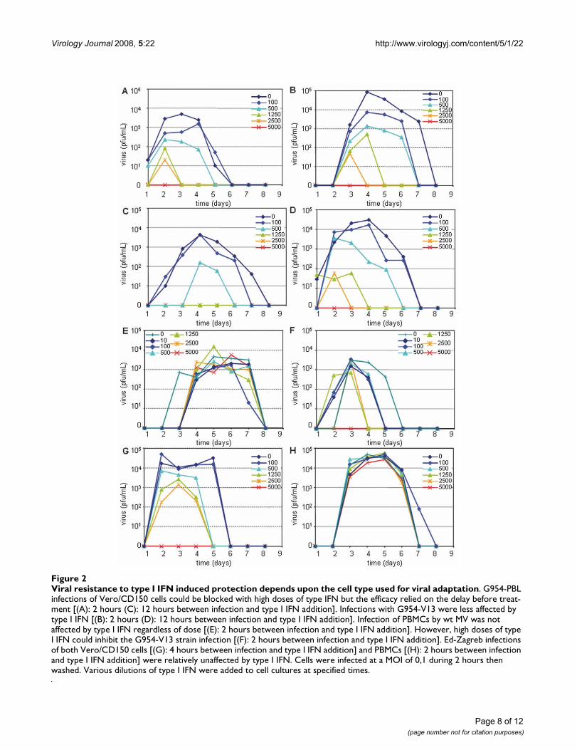
We next examined the effect of type I IFN during the MVinfection. Different quantities of IFN were added to Vero/CD150 cells infected with G954-PBL, G954-V13 or Ed-Zagreb (MOI = 0,1) at 2, 4 or 12 hr post infection (figure2A–D, G). The later the IFN was added the less effect it hadon the inhibition of infectious virus production. Treat-ment with more than 1250 IU/mL of IFN 2 h pi blockedthe infection by the wt strain. The infection was delayedand produced less infectious virus in proportion to thequantity of IFN added. IFN added 12 h after infection didnot slow down the production of infectious MV. In thecase of G954-V13, the effect of type I IFN was much lessimportant. The infection was never delayed, slightly ham-pered and shortened, proportionally to the added dose.Interestingly, the infection by Ed-Zagreb was far morerobust. Independently of the delay between type I IFNtreatment and infection, there was a slight dose effect (fig-ure 2G and unpublished results): the vaccine strain infec-tion of Vero/CD150 cells was less affected by type I IFN.Thus, it appears that type I IFN-induced protection of
Adaptation to a specific cell type limits replication of MVFigure 1Adaptation to a specific cell type limits replication of MV. Replication kinetics of MVs in PBMCs (A), in Vero cells (B), in Vero/CD150 cells (C), in B95a cells (D). For each experiment, 105 cells were infected at a MOI of 0,1. Each time point con-sists of the mean of 2 independent experiments. Vero/CD150 cells were used for the titration.
Table 3: Number of copies of MV genome in different infected cell types
PBMCs Vero/CD150 pDCs
G954 PBL 3,3.105 * 3,8.104 8.103
G954 V13 1,6.105 3,2.106 2.7.103
Ed-Zagreb 1,6.105 3.2.106 8.103
G954 PBL UV < 10 < 10 < 10G954 V13 UV < 10 < 10 < 10
* Data represent the number of copies of MV genomes deduced from RT-QPCR results obtained from infected cultures extractions when maximum PFU were released (8.104 cells infected at 0,1 PFU/cell).
Page 6 of 12(page number not for citation purposes)
Virology Journal 2008, 5:22 http://www.virologyj.com/content/5/1/22
Vero/CD150 cells was less effective in the case of infectionby the Vero adapted strain, G954-V13. This type of resist-ance could be linked with a better adaptation of MV to theVero cell environment.
When the protective effect of type I IFN on MV infectionof PBMCs was studied, the wt strain was not inhibiteddespite the addition of high quantities of IFN (up to 5000IU/mL). On the contrary, infection by G954-V13 strainwas delayed, shortened and of lesser amplitude, propor-tionally to the IFN concentration and totally blocked with5000 IU/mL of type I IFN. Finally, there was no effect oftime between infection and IFN treatment on the virusreplication in PBMCs for both analyzed viruses (figure2E–F and data not shown). Infection of PBMCs by the Ed-Zagreb virus was unaffected by the different conditions oftype I IFN tested (figure 2H). Thus, G954 strains seemedto be rather resistant to type I IFN since the protectiveeffects occurred only when infections were performed oncells less permissive to a specific strain. However, Ed-Zagreb exhibited a strong resistance to type I IFN, inde-pendently of the cell type tested.
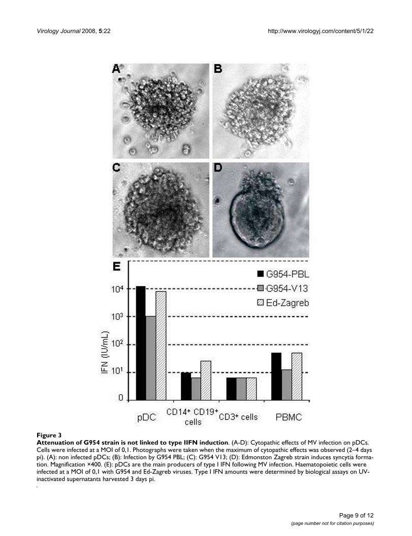
Induction of type I IFN by MV infectionPlasmacytoid Dendritic Cells (pDCs) are the main pro-ducers of type I IFN in blood and lymph nodes. Unstimu-lated pDCs express CD46 at the cell surface, but notCD150 [29]. Schlender et al have shown that pDCs areinfectable by the Edmonston-Schwarz vaccine strain ofMV but do not produce IFN during the first 36 hours afterinfection [19]. To study the permissivity of these cells todifferent MV, pDC cultures were infected for 3 days withG954-PBL, G954-V13 or Ed-Zagreb. Neither G954 virusinduced syncytia formation in the cultures nor were anyinfectious virus particles detectable, whereas infectionwith the Ed-Zagreb strain induced cell fusion withoutinfectious virus production (figure 3A–D ). QuantitativeRT-PCR studies on the infected cells showed that RNA rep-lication/transcription could occur in pDCs (table 3). The
pDCs infected with the wt strain and Ed-Zagreb contained3 fold more genomes than those infected with G954-V13.The presence of MV genomes was not detected after infec-tion with UV-treated virus (table 3) confirming the activereplication of MV in pDCs.
To study the induction of type I IFN by these viruses,PBMCs and fractionated preparations (pDCs, CD14+CD19+, and CD3+ cells) were infected and the produc-tion of type I IFN measured 3 days later (figure 3E). Theinfected pDCs had up to 1,000 fold higher quantities of α/β IFN than the other cells examined. The wild type G954-PBL virus induced 10-fold higher type I IFN amounts thanthe G954-V13 virus in pDCs and equal amounts as Ed-Zagreb. In all tested cell types, each MV strain inducedproduction of type I IFN, although G954-V13 infectioninduced lower level, particularly in PBMCs and pDCs.Altogether, those results demonstrate that the G954-V13attenuation/adaptation was not linked to an enhancedproduction of type I IFN by either of primary humanshaemotopoietic cells analyzed in this study.
DiscussionWe have adapted a wild type MV to Vero cells and shownthat the adapted strain and the parent strain differ fromeach other by only 5 coding mutations. Although the dif-ferences we have observed were located in the P/V/C andM genes, they were different from mutations observed inprevious studies [21-23,30] where viruses were attenuatedby passaging on Vero cells and then tested in a monkeymodel. Our MV adapted to Vero cells (G954-V13) wasstrongly attenuated when inoculated into CD150 trans-genic mice. The E89K mutation in M protein has also beenshown to be present in another Vero adapted strain [24]and was shown to permit the wt strain to replicate in Verocells while provoking limited cell/cell fusion of CD150+
cells. Our results support the proposed importance of theM E89K mutation in replication in Vero cells although weobserved a better cytopathic effect in Vero/CD150 cells
Table 4: Inhibitory efficacy of a type I IFN pre-treatment on Vero/CD150 cells before MV infection
Concentration of IFN added (IU/mL)
Time before infection Strain 5000 1000 500 100 0
8 h G954-PBL 100* 92 95 63 0G954-V13 100 95 89 56 0Ed-Zagreb 100 79 65 58 0
24 h G954-PBL 100 100 100 95 0G954-V13 100 100 100 67 0Ed-Zagreb 100 100 100 43 0
48 h G954-PBL 100 100 100 91 0G954-V13 100 100 100 80 0Ed-Zagreb 100 100 100 81 0
* Inhibition indices were calculated as follow: 100 × [1 – (PFU(type I IFN = x)/PFU(type I IFN = 0)]
Page 7 of 12(page number not for citation purposes)
Virology Journal 2008, 5:22 http://www.virologyj.com/content/5/1/22
Page 8 of 12(page number not for citation purposes)
Viral resistance to type I IFN induced protection depends upon the cell type used for viral adaptationFigure 2Viral resistance to type I IFN induced protection depends upon the cell type used for viral adaptation. G954-PBL infections of Vero/CD150 cells could be blocked with high doses of type IFN but the efficacy relied on the delay before treat-ment [(A): 2 hours (C): 12 hours between infection and type I IFN addition]. Infections with G954-V13 were less affected by type I IFN [(B): 2 hours (D): 12 hours between infection and type I IFN addition]. Infection of PBMCs by wt MV was not affected by type I IFN regardless of dose [(E): 2 hours between infection and type I IFN addition]. However, high doses of type I IFN could inhibit the G954-V13 strain infection [(F): 2 hours between infection and type I IFN addition]. Ed-Zagreb infections of both Vero/CD150 cells [(G): 4 hours between infection and type I IFN addition] and PBMCs [(H): 2 hours between infection and type I IFN addition] were relatively unaffected by type I IFN. Cells were infected at a MOI of 0,1 during 2 hours then washed. Various dilutions of type I IFN were added to cell cultures at specified times.
Virology Journal 2008, 5:22 http://www.virologyj.com/content/5/1/22
Page 9 of 12(page number not for citation purposes)
Attenuation of G954 strain is not linked to type IIFN inductionFigure 3Attenuation of G954 strain is not linked to type IIFN induction. (A-D): Cytopathic effects of MV infection on pDCs. Cells were infected at a MOI of 0,1. Photographs were taken when the maximum of cytopathic effects was observed (2–4 days pi). (A): non infected pDCs; (B): Infection by G954 PBL; (C): G954 V13; (D): Edmonston Zagreb strain induces syncytia forma-tion. Magnification ×400. (E): pDCs are the main producers of type I IFN following MV infection. Haematopoietic cells were infected at a MOI of 0,1 with G954 and Ed-Zagreb viruses. Type I IFN amounts were determined by biological assays on UV-inactivated supernatants harvested 3 days pi.
Virology Journal 2008, 5:22 http://www.virologyj.com/content/5/1/22
rather than defects in syncytia formation [data not shownand [10]]. A recent study shows that this mutation couldaffect MV growth by modifying the interaction between Mand the cytoplasmic tail of the H protein [25]. Since thepredicted domains on H for CD46 and CD150 bindingare close, one could hypothesise that a stronger interac-tion of M with H could change the conformation of H andthus change the affinity of the CD46 binding site (R. Buck-land, personal communication).
Parks et al. sequenced a number of the vaccine strainsderived from the Edmonston isolate and identified aminoacids shared by these attenuated viruses [31]. Eight aminoacid coding changes were common to all vaccine strainsand an additional two were conserved in all except theEdmonston Zagreb strain. They concluded that modula-tion of transcription and replication plays an importantrole in attenuation. Among the mutations found in G954-V13, only M E89K corresponds to an amino acid changeobserved in the transition toward vaccine strains in thisstudy. The observation that the Edmonston Zagreb straincould readily replicate in PBMCs and Vero cells whileresisting to type I IFN induced protection suggests therobustness of vaccine strains. Furthermore, it questionsthe notion of attenuated and vaccine strains since a viruswhich does not induce a pathology in humans could stillexhibit strong deleterious effects in human cell cultures,demonstrating discrepancies of the virus pathogenicity invitro and in vivo. Therefore, these observations beg thequestion of whether a vaccine phenotype can be predictedand engineered at the genetic level by using only in vitroapproach.
Innate immunity is an important early response to viralinfection. The accessory proteins of Paramyxoviruses, Cand V, have been shown to be implicated in the suppres-sion of this response, both in the induction and signallingof type I IFN [12-17,32]. Although in some of those stud-ies, laboratory strains were poor inducers of type I IFN[14], other studies reported that vaccine strains induced10 to 80 times more type I IFN than wt strains after infec-tion of peripheral blood lymphocytes [11,33]. In contrast,in our study, the wild type and attenuated G954-V13viruses as well as vaccine strain Ed-Zagreb induced similarquantities of type I IFN in these cells. We showed that fol-lowing in vitro infection, the major cell population pro-ducing type I IFN was the pDCs for both the wild type andthe attenuated strain. The inhibition of type I IFN produc-tion induced by vaccine MV strains observed in anotherstudy [19] was probably due to the shorter observationperiod (36 hours) than in our study (72 hours). Neverthe-less, we cannot exclude potential interference of MV infec-tion with TLR-induced type I IFN production by pDCs.Furthermore, it may also be possible that G954 forms partof a particular group of wt MV, able of good induction of
type I IFN and then, during its attenuation, this propertyis preserved. Even if better induction and higher sensitiv-ity to type I IFN is an attractive explanation for the mech-anism of viral attenuation, this study strongly suggeststhat it is possible to achieve attenuation without perturb-ing interactions with the innate immune mechanisms.
Our results show that P/V/C mutations are not necessarilylinked to modifications in type I IFN resistance and sug-gest rather that they have a role in the replicative processduring infection. This is in agreement with previous stud-ies on the negative effect of V and C proteins on transcrip-tion and replication [34-36]. The absence of the V proteinwas reported to delay replication [37] and the virus wasless pathogenic in vivo [38,39]. The absence of the C pro-tein reduced the virus yield both in vitro and in vivo [40].Even the M protein has been shown to inhibit the replica-tion process [41]. Recent studies showed that the P pro-tein is involved in STAT1 phosphorylation [42] and thuscan affect type I IFN efficacy. In our case such a role for Pcould not be observed. Moreover, other studies demon-strated that the adaptation of MV to Vero cells couldinduce differences in the amounts of viral proteins pro-duced [43]. More quantitative experiments should be per-formed to assess if such a phenomenon is important inthe adaptation of the G954 viral strain. Therefore, it maybe very likely that differences between G954 strains arelinked to P/V/C and/or M proteins via cell specific restric-tions of viral replication, transcription and translationprocesses.
ConclusionThe present study shows that adaptation of wild type MVto Vero cells induces a strong attenuation in vivo, which isindependent of type I IFN. Identifying the exact role ofeach of the 5 mutations will determine their role in path-ogenicity and could be performed by developing a recom-binant virus strategy. Further analysis of the mechanismsimplicated in the complex process of virus attenuationshould pave the way towards developing new vaccineswith a high capacity to induce specific host immuneresponses.
Competing interestsThe author(s) declare that they have no competing inter-ests.
Authors' contributionsJD participated in the conception of the study and per-formed the majority of the experiments and wrote themanuscript. CIS carried out ELISA assays on mice sera,participated in the in vivo assays and helped to draft themanuscript. DW carried out the nucleic acid sequencingand sequence alignment. BH helped in the design of thestudy, specially the in vivo assays and critically helped to
Page 10 of 12(page number not for citation purposes)
Virology Journal 2008, 5:22 http://www.virologyj.com/content/5/1/22
draft the manuscript. TFW in the conception of the study,its design and coordination and helped to draft the man-uscript. All authors read and approved the final manu-script.
AcknowledgementsWe are grateful to B. Blanquier, Y. Kerdiles, S. Devergnas, B. Dubois (for providing the anti CD14 antibody), T. Duhen, C. Rabourdin-Combe (for providing antibodies and thoughtful discussions) and the personnel of the PBES at ENS-Lyon for their help. We thank D. Gerlier for critical reading the manuscript.
JD was supported by grants from MENRT, CIS was supported by the Fon-dation pour la Recherche Medicale (FRM). This work was supported in part by institutional grants from INSERM, the Institut de Veille Sanitaire and FRM.
References1. Griffin DE: Measles Virus. In Fields Virology 5th edition. D.M.
Knipe, P.M. Howley, D.E. Griffin, R.A. Lamb, M.A. Martin, B. Roizman,and S.E. Straus, Eds; 2006:1551-1585.
2. Tatsuo H, Ono N, Tanaka K, Yanagi Y: SLAM (CDw150) is a cel-lular receptor for measles virus. Nature 2000,406(6798):893-897.
3. Dörig RE, Marcil A, Chopra A, Richardson CD: The human CD46molecule is a receptor for measles virus (Edmonston strain).Cell 1993, 75:295-305.
4. Naniche D, Varior-Krishnan G, Cervoni F, Wild TF, Rossi B, Rabour-din-Combe C, Gerlier D: Human membrane cofactor protein(CD46) acts as a cellular receptor for measles virus. J Virol1993, 67(10):6025-6032.
5. Lecouturier V, Rizzitelli A, Fayolle J, Daviet L, Wild FT, Buckland R:Interaction of measles virus (Halle strain) with CD46: evi-dence that a common binding site on CD46 facilitates bothCD46 downregulation and MV infection. Biochem Biophys ResCommun 1999, 264(1):268-275.
6. Masse N, Barrett T, Muller CP, Wild TF, Buckland R: Identificationof a second major site for CD46 binding in the hemagglutininprotein from a laboratory strain of measles virus (MV):potential consequences for wild-type MV infection. J Virol2002, 76(24):13034-13038.
7. Santiago C, Bjorling E, Stehle T, Casasnovas JM: Distinct kineticsfor binding of the CD46 and SLAM receptors to overlappingsites in the measles virus hemagglutinin protein. J Biol Chem2002, 277(35):32294-32301.
8. Hashimoto K, Ono N, Tatsuo H, Minagawa H, Takeda M, Takeuchi K,Yanagi Y: SLAM (CD150)-independent measles virus entry asrevealed by recombinant virus expressing green fluorescentprotein. J Virol 2002, 76(13):6743-6749.
9. Richardson CD Sarangi, F., Iorio, C.: Studies towards the identifi-cation and characterization of a third receptor for measlesvirus on human and marmoset smooth airway epithelialcells: June 17th - 22nd 2006; Salamanca, Spain. ; 2006.
10. Kouomou DW, Wild TF: Adaptation of wild-type measles virusto tissue culture. J Virol 2002, 76(3):1505-1509.
11. Naniche D, Yeh A, Eto D, Manchester M, Friedman RM, OldstoneMB: Evasion of host defenses by measles virus: wild-type mea-sles virus infection interferes with induction of Alpha/Betainterferon production. J Virol 2000, 74(16):7478-7484.
12. Nakatsu Y, Takeda M, Ohno S, Koga R, Yanagi Y: Translationalinhibition and increased interferon induction in cells infectedwith C protein-deficient measles virus. J Virol 2006,80(23):11861-11867.
13. Palosaari H, Parisien JP, Rodriguez JJ, Ulane CM, Horvath CM: STATprotein interference and suppression of cytokine signaltransduction by measles virus V protein. J Virol 2003,77(13):7635-7644.
14. Shaffer JA, Bellini WJ, Rota PA: The C protein of measles virusinhibits the type I interferon response. Virology 2003,315(2):389-397.
15. Ohno S, Ono N, Takeda M, Takeuchi K, Yanagi Y: Dissection ofmeasles virus V protein in relation to its ability to blockalpha/beta interferon signal transduction. J Gen Virol 2004,85(Pt 10):2991-2999.
16. Takeuchi K, Kadota SI, Takeda M, Miyajima N, Nagata K: Measlesvirus V protein blocks interferon (IFN)-alpha/beta but notIFN-gamma signaling by inhibiting STAT1 and STAT2 phos-phorylation. FEBS Lett 2003, 545(2-3):177-182.
17. Yokota S, Okabayashi T, Yokosawa N, Fujii N: Growth arrest ofepithelial cells during measles virus infection is caused byupregulation of interferon regulatory factor 1. J Virol 2004,78(9):4591-4598.
18. Siegal FP, Kadowaki N, Shodell M, Fitzgerald-Bocarsly PA, Shah K, HoS, Antonenko S, Liu YJ: The nature of the principal type 1 inter-feron-producing cells in human blood. Science 1999,284(5421):1835-1837.
19. Schlender J, Hornung V, Finke S, Gunthner-Biller M, Marozin S,Brzozka K, Moghim S, Endres S, Hartmann G, Conzelmann KK: Inhi-bition of toll-like receptor 7- and 9-mediated alpha/betainterferon production in human plasmacytoid dendritic cellsby respiratory syncytial virus and measles virus. J Virol 2005,79(9):5507-5515.
20. Kobune F, Sakata H, Sugiura A: Marmoset lymphoblastoid cellsas a sensitive host for isolation of measles virus. J Virol 1990,64(2):700-705.
21. Kobune F, Takahashi H, Terao K, Ohkawa T, Ami Y, Suzaki Y, NagataN, Sakata H, Yanamouchi K, Kai C: Nonhuman primate modelsof measles. Laboratory Animal Science 1996, 46(3):315-320.
22. Takeda M, Kato A, Kobune F, Sakata H, Li Y, Shioda T, Sakai Y, Asa-kawa M, Nagai Y: Measles virus attenuation associated withtranscriptional impediment and a few amino acid changes inthe polymerase and accessory proteins. J Virol 1998,72(11):8690-8696.
23. Takeuchi K, Miyajima N, Kobune F, Tashiro M: Comparative nucle-otide sequence analyses of the entire genomes of B95a cell-isolated and vero cell-isolated measles viruses from thesame patient. Virus Genes 2000, 20(3):253-257.
24. Tahara M, Takeda M, Yanagi Y: Contributions of matrix and largeprotein genes of the measles virus edmonston strain togrowth in cultured cells as revealed by recombinant viruses.J Virol 2005, 79(24):15218-15225.
25. Tahara M, Takeda M, Yanagi Y: Altered interaction of the matrixprotein with the cytoplasmic tail of hemagglutinin modu-lates measles virus growth by affecting virus assembly andcell-cell fusion. J Virol 2007, 81(13):6827-6836.
26. Sellin CI, Davoust N, Guillaume V, Baas D, Belin MF, Buckland R, WildTF, Horvat B: High pathogenicity of wild-type measles virusinfection in CD150 (SLAM) transgenic mice. J Virol 2006,80(13):6420-6429.
27. Muller U, Steinhoff U, Reis LF, Hemmi S, Pavlovic J, Zinkernagel RM,Aguet M: Functional role of type I and type II interferons inantiviral defense. Science 1994, 264(5167):1918-1921.
28. Pfaffl MW: A new mathematical model for relative quantifica-tion in real-time RT-PCR. Nucleic Acids Res 2001, 29(9):e45.
29. Facchetti F, Vermi W, Mason D, Colonna M: The plasmacytoidmonocyte/interferon producing cells. Virchows Arch 2003,443(6):703-717.
30. Uejima H, Nakayama T, Komase K: Passage in Vero cells altersthe characteristics of measles AIK-C vaccine strain. Vaccine2006, 24(7):931-936.
31. Parks CL, Lerch RA, Walpita P, Wang HP, Sidhu MS, Udem SA: Com-parison of predicted amino acid sequences of measles virusstrains in the Edmonston vaccine lineage. J Virol 2001,75(2):910-920.
32. Caignard G, Guerbois M, Labernardiere JL, Jacob Y, Jones LM, WildF, Tangy F, Vidalain PO: Measles virus V protein blocks Jak1-mediated phosphorylation of STAT1 to escape IFN-alpha/beta signaling. Virology 2007.
33. Shingai M, Ebihara T, Begum NA, Kato A, Honma T, Matsumoto K,Saito H, Ogura H, Matsumoto M, Seya T: Differential Type I IFN-Inducing Abilities of Wild-Type versus Vaccine Strains ofMeasles Virus. J Immunol 2007, 179(9):6123-6133.
34. Escoffier C, Manie S, Vincent S, Muller CP, Billeter M, Gerlier D:Nonstructural C protein is required for efficient measlesvirus replication in human peripheral blood cells. J Virol 1999,73(2):1695-1698.
Page 11 of 12(page number not for citation purposes)

Virology Journal 2008, 5:22 http://www.virologyj.com/content/5/1/22
Publish with BioMed Central and every scientist can read your work free of charge
"BioMed Central will be the most significant development for disseminating the results of biomedical research in our lifetime."
Sir Paul Nurse, Cancer Research UK
Your research papers will be:
available free of charge to the entire biomedical community
peer reviewed and published immediately upon acceptance
cited in PubMed and archived on PubMed Central
yours — you keep the copyright
Submit your manuscript here:http://www.biomedcentral.com/info/publishing_adv.asp
BioMedcentral
35. Parks CL, Witko SE, Kotash C, Lin SL, Sidhu MS, Udem SA: Role ofV protein RNA binding in inhibition of measles virus minige-nome replication. Virology 2006, 348(1):96-106.
36. Reutter GL, Cortese-Grogan C, Wilson J, Moyer SA: Mutations inthe measles virus C protein that up regulate viral RNA syn-thesis. Virology 2001, 285(1):100-109.
37. Tober C, Seufert M, Schneider H, Billeter MA, Johnston ICD, Niewi-esk S, ter Meulen V, Schneider-Schaulies S: Expression of measlesvirus V protein is associated with pathogenicity and controlof viral RNA synthesis. Journal of Virology 1998, 72(10):8124-8132.
38. Paterson RG, Russell CJ, Lamb RA: Fusion protein of the para-myxovirus SV5: destabilizing and stabilizing mutants offusion activation. Virology 2000, 270(1):17-30.
39. Valsamakis A, Schneider H, Auwaerter PG, Kaneshima H, Billeter MA,Griffin DE: Recombinant meales viruses with mutations in theC, V, or F gene have altered growth phenotypes in vivo. Jour-nal of Virology 1998, 72(10):7754-7761.
40. Takeuchi K, Takeda M, Miyajima N, Ami Y, Nagata N, Suzaki Y, Shah-newaz J, Kadota S, Nagata K: Stringent requirement for the Cprotein of wild-type measles virus for growth both in vitroand in macaques. J Virol 2005, 79(12):7838-7844.
41. Reuter T, Weissbrich B, Schneider-Schaulies S, Schneider-Schaulies J:RNA interference with measles virus N, P, and L mRNAsefficiently prevents and with matrix protein mRNAenhances viral transcription. J Virol 2006, 80(12):5951-5957.
42. Devaux P, von Messling V, Songsungthong W, Springfeld C, CattaneoR: Tyrosine 110 in the measles virus phosphoprotein isrequired to block STAT1 phosphorylation. Virology 2007,360(1):72-83.
43. Sinitsyna OA, Khudaverdyan OE, Steinberg LL, Nagieva FG, Lotte VD,Dorofeeva LV, Rozina EE, Boriskin Yu S: Further-attenuated mea-sles vaccine: virus passages affect viral surface proteinexpression, immunogenicity and histopathology pattern invivo. Res Virol 1990, 141(5):517-531.
Page 12 of 12(page number not for citation purposes)


![Virology Journal BioMed Central - Universiti Malaysia … · Virology Journal Research Open Access ... (LAMR1) interaction with dengue virus serotypes 1, 2 and 3 ... [20]. A schematic](https://static.fdocuments.us/doc/165x107/5adab9c27f8b9a6d7e8d116c/virology-journal-biomed-central-universiti-malaysia-journal-research-open.jpg)


