Viral retinitis - todnet.org
59
Viral Retinitis Carlos Pavesio MD FRCOphth Moofields Eye Hospital
Transcript of Viral retinitis - todnet.org

Viral Retinitis
Carlos Pavesio MD FRCOphth
Moofields Eye Hospital

Varicella Zoster Virus (VZV)
Herpes Simplex Virus 1 and 2 (HSV)
Cytomegalovirus (CMV)
Epstein-Barr Virus (EBV)
HHV-6,7

Holland GN, and the Executive Committee of the American Uveitis Society. Am J Ophthalmol 1994;117:663–7.
M N Muthiah,et al., Br J Ophthalmol 2007;91:1452–1455.

most frequently caused by
VZV and HSV
VZV 2x more common than HSV
reports exist implicating EBV and CMV
Incidence 3 – 4 / million per year ( Sims 2009, Melbourne,
Australia)
1 case per 1.6 to 2.0 million population / year
( Muthiah 2007, BOSU, UK)
J.L. Sims, et al. Clinical and Experimental Ophthalmology 2009; 37: 473–477
M N Muthiah,et al., Br J Ophthalmol 2007;91:1452–1455.

Clinical Findings: Symptoms
rapidly reducing vision (85%),
photophobia (55%),
ocular pain (26%),
flu like symptoms (16%)
NB: Red eye in only 16%

Anterior uveitis with fine or granulomatous KPs
Vitritis -may be severe
Occlusive retinal vasculitis - arteritis with haemorrhages
One or more focus of retinitis located in the retinal periphery with circumferential spread that extend towards the posterior pole
Optic disc swelling
Episcleritis or scleritis


Because dilated fundus examination is not performed / inadequately assesses the periphery
‘Just Acute Anterior Uveitis’
Don’t forget to always examine both eyes


ARN is a clinical diagnosis
So lab tests are not necessary to make the diagnosis
Confirmation by PCR
– vitreous early on: precise identification of virus
– aqueous is positive in 80% of the cases (Tran THC Br J
Ophthalmol 2003)
Also diagnostic vitrectomy may be needed in atypical presentations (usually associated with immunodeficiency) to exclude other agents such as T. gondii / bacteria / masquerade etc (cytology, PCR)


Occlusive arteritis and retinal ischaemia
Periphlebitis and venous thrombosis
Leakage from retinal venules, arterioles, capillaries.
Optic disc swelling
Areas of non-perfusion
Serous retinal detachment (uncommon)


24 to 80% (when no Rx)
Mostly within 3 months/1 year, but much longer intervals reported
As compared to no treatment, treatment with systemic acyclovir decreases the risk of fellow eye involvement from 70% to 13%.
Bilateral at presentation
Time 0
5 days 20 days 26 years 3 years
Bilateral after many years
Interval between eyes Palay et al. Am J Ophthalmol 1991;112:250 –255.

Second eye once treatment started
Lau et al. 22 patients
5/22 (22.7%) had BARN
2/22 presented with BARN
3/22 developed ARN in the fellow eye on day 14 (day 9 on acyclovir treatment), day 53 (day 53 on acyclovir treatment), and day 166 (day 110 after 56 days on acyclovir treatment), after first-eye presentation.
Lau C, Missotten T, Salzmann, J, Lightman S. Ophthalmology 2007;114:756–762

AIDS patients with only mild immune dysfunction and elevated CD4 counts
ARN is more extensive, bilateral 90%, less responsive to antiviral agents, tendency for recurrence same eye, retinal detachment 80%, poor visual acuity, dismal prognosis
Vrabec TR. Surv Ophthalmol. 2004 Mar-Apr;49(2):131-57.

HSV 1 and HSV 2
Younger individuals
Exudative retinal detachment
Association with HSV encephalitis or
meningitis

Brain-to-eye transmission of the herpesvirus
The overall interval between the encephalitis and ARN was 20.6 months average (range 14 days to 5 years).
T. Vandercam; et al.,Neurology® 2008;71:1268–1274

Immunocompetent 50 year old lady
Vitreous CMV PCR positive (negative for HSV/VZV)
Initially treated with acyclovir, progressed intravitreal foscarnet then Intravitreal and iv gancyclovir and oral pred
2 months later RD phaco-vity-oil
VA 6/36 6/12 at 12 months
Peripheral 360° necrosis and severe panuveitis

Complications
Retinal detachment in 75%
Ischemic neuropathy, optic atrophy
Arterial occlusions
Macular damage
Cataract
Glaucoma
Phthysis bulbi

All current treatment options for ARN are based on anecdotal evidence.
Antiviral agents
Corticosteroids
Management and prevention of RD

Immediate start
Intravitreal antiviral
Systemic therapy – acute phase (10-14 days)
Intravenous acyclovir: 10mg/kg tds
Oral valacyclovir: 2gr tds
Systemic therapy following acute phase
Oral acyclovir: 800mg, 5x for 12 weeks
Oral valacyclovir: 1gr, 3x for 12 weeks

The vitreous concentrations achieved in non-inflamed eyes are within the reported inhibitory ranges for most strains of HSV (1 and 2) and VZV
If oral Rx: Use 1-2g tds-valacyclovir
Rapid good absorption
Always watch out for non response
Consider increasing dose and adding intravitreal Foscarnet (2.4mg)
VALTREX GLAXOSMITHKLINE

Indefinite therapy:
Immunocompromised (depending on reason)
Previous history of Herpetic encephalitis?

Famciclovir (500 mg tds) Oral administration of famciclovir (Famvir,
Novartis Pharmaceuticals) vitreous concentrations of penciclovir (Denavir; GlaxoSmithKline) within the inhibitory ranges for HSV-1, HSV-2 and VZV
What is the advantage? famciclovir has a better oral bioavailability and
pharmackinetic profile then acyclovir. Less toxicity at high dose of valacyclovir and
valganciclovir May be useful in acyclovir-resistant VZV-ARN
but penciclovir has a similar dependence on thymidine kinase for activation
AJO 2009;148:38-42

Any role?
Optic disc involvement: pale swelling
Intense inflammation?
When to start
Delay 24-48 hours

result of combination of retinal necrosis and vitreoretinal traction,
breaks can occur posteriorly as well as at the vitreous base….usually at junction of diseased and healthy retina
Presentation: 0 days - 4 years following diagnosis of ARN (median 4-5 weeks).
RD treatment: vity / silicone oil Optic nerve or Photoreceptor damage limits
final visual results despite anatomical success

Unclear
because the conclusions are based on
small case series of 5 to 13 eyes
weak level of evidence
active retinitis will progress through laser scars
worse disease greater vitritis lose the view can’t laser
? Laser group has milder ARN Han DP, et al., Arch Ophthalmol 1987;105:1051– 4. Sternberg P Jr, et al., Ophthalmology 1988;95:1389 –93. Crapotta JA etal.,. Retina 1993;13:208 –13. Hudde etal., Ophthalmologe 1998;95:473–7. McDonald HR, et.al, Br J Ophthalmol 1991;75:455– 8. Park and Pavesio, Br J Ophthalmol 2008 92;9:1161-2

ARN studies compared laser treatment against no laser treatment in two heterogenic study populations.
Milder cases of ARN with limited vitreous opacification and/or retinitis did better after laser treatment compared with more severe cases of retinitis, which did worse without laser, resulting in higher rates of retinal detachment.
Role of pre-existing posterior vitreous separation unknown
May do some harm? hole formation and, possibly, promote RD

81 eyes, 74 patients
Visual outcome in HSV-ARN (33 eyes) better than in VZV-ARN (48 eyes).
2.5-fold greater chance of RRD in VZV-ARN > HSV-ARN
Intravitreal foscarnet useful adjunctive treatment reduced rate of RRD The results for VZV-ARN showed a 40% reduction in
RRD frequency between the foscarnet group and the standard treatment group (54% vs 75%; P 0.23)
Ophthalmology 2009

Milder disease at initiation of therapy in 77% of the eyes, less than 25% of retina
involved
Final VA was 20/40 or better in 6 eyes (46%), and 20/400 or better in 12 eyes (92%).
Better initial VA = Better final VA
Crapotta JA, et al., Retina. 1993;13(3):208-13.
N= 13 eyes, Follow-up period = 3 to 21 months

Progressive Outer Retinal Necrosis VZV retinitis

Defined in AIDS patients
Minimal inflammation
Multifocal retinitis, grey-white opacified lesions in the outer retina that rapidly progress and involve the periphery
Rapid progression to involve second eye
Bilateral in 70%


1st line iv Ganciclovir & Foscarnet
Plus intravitreal ganciclovir +/- foscarnet
2nd line iv Cidofovir
Plus intravitreal ganciclovir +/- foscarnet
Some eyes may not respond to multi drug therapy even if patient responds to HAART

Visual loss due to:
early macular involvement
progressive infection,
retinal detachment (70–85% of patients)
optic nerve sheath effusion,
Blindness is bilateral in 59%.
Rapid: within days or weeks
2/3 patients NPL. (Engstrom et al 1994)
Engstrom RE Jr. et al. Ophthalmology. 1994;101:1488–1502.

PORN (VZV-PORN) with preservation of 20/20 vision after combination
Antiviral treatment ganciclovir implant
intravenous acyclovir (10 mg/kg tds)
intravitreal foscarnet (2.4 mg)
HAART
20/20 visual acuity at 1 year

Immunocompromised individuals CD4 < 50 Rare above 100
Clinical disease: reactivation of latent infection or newly acquired
Most common ocular infection in AIDS patients
More common in homosexual men
Reduction of number of cases since HAART


CMV is a slowly progressive infection, moving an average 250-350 μm/week. Response to treatment is indicated by transition from white active retinitis to inactive disease. There may be persistent whitening within the border that does not represent active disease. Presence of retinal oedema and small satellite lesions are best for differentiating white scar from reactivation.

Movement of disease can occur with minimal retinal whitening - importance of photographs.

Nucleoside analog
Depends on phospholylation by viral kinase
Virustatic
Effect is reversible
Poor oral absorption
Complications: neutropenia, thrombocytopenia, central line infection

Dose:
Induction: 5mg/kg BD for 2-3 weeks
Maintenance: 5mg/kg OD, 7 days/week
6mg/kg OD, 5/7 days/week

A pyrophosphate analog
Inhibits DNA polymerase & reverse transcriptase
Does not require phosphorilation
Effect also virustatic and reversible
Intrinsic activity against HIV
Complications: nephrotoxicity, electrolyte shifts, anaemia and central line infection.

Dose:
Induction: 90mg/kg BD for 2-3 weeks
Maintenance:120 mg/kg OD, 7 days/week
Infusion must be slow. Hydration with 1 L saline.

Nucleotide analog
Dose: Induction - 5mg/kg/week for 2 weeks
Maintenance - 5mg/kg every 2 weeks
Toxicity is primarily renal
Probenecid
No effect on viremia
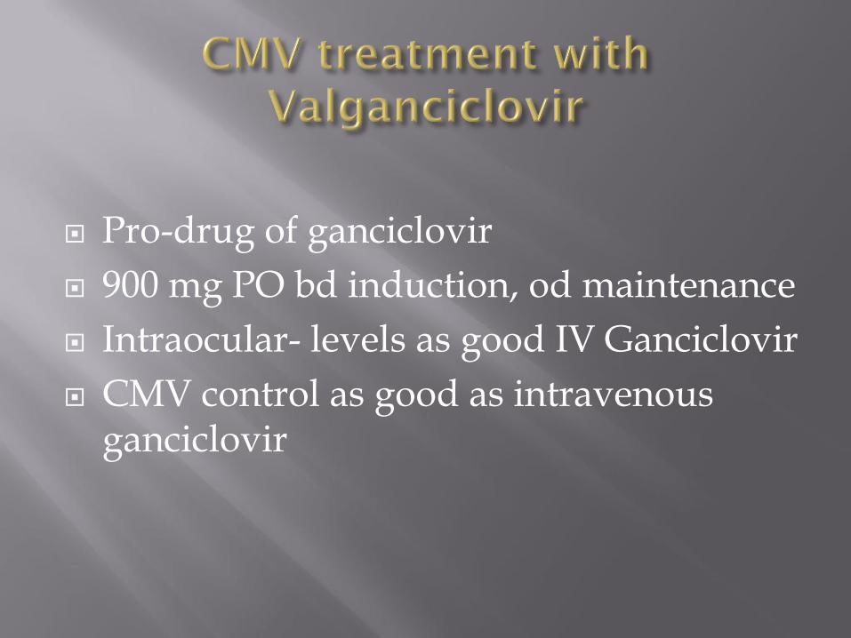
Pro-drug of ganciclovir
900 mg PO bd induction, od maintenance
Intraocular- levels as good IV Ganciclovir
CMV control as good as intravenous ganciclovir

Ensure correct diagnosis!
Ensure patient compliance
Consider admission for intravenous ganciclovir
Exclude bioavailability problem
Malabsorption / GI causes
Try Oral VGV + intravitreal ganciclovir (2mg in 0.1) Induction: 2 per 1st week, then weekly for 3 weeks.
Maintenance: every 2 -3 weeks

The only definitive treatment for CMV is immune restoration
Individualize Cease VGV once CD4 > 100 for at least 3 months (
some say > 200)
CMV reactivation reported with CD4 >100 6% per year CD4 100 to 200 (Jabs et al, Ophthalmology 2000)
Lifelong vigilance CMV likely to reactivate if CD4 drops below 100

Rationale
Site of clinical CMV disease is primarily the eye
Systemic drug penetration into the eye is poor

Ganciclovir: 2mg in 0.05-0.1ml
Foscarnet: 2.4mg in 0.1ml
Cidofovir: 20μg in 0.1ml + probenecid
Fomivirsen (ISIS-2922)

Ganciclovir implant (Vitrasert):
4.5mg of the drug
releases drug at 1μg/hour
lasts for 6-8 months
when to replace
technique


2020
19191818

• More common in HAART era
• CD4 >50,usually in 100’s
• Signs:
– A.C. activity,
– Vitritis
– Cystoid macular oedema
– Epiretinal membrane
– Optic disc neovascularisation
• Spectrum: mild to severe
Nguyen et al. Am J Ophthalmol 2000 May; 129(5):634-9

Immune Recovery Uveitis

Early recognition
Appropriate and aggressive therapy
Watch out for complications