Viral Pneumonia - Med Study Group
10
Respiratory System Virology Lecture – 3 – Viral Pneumonia Brief Introduction: - In general Pneumonia is an inflammatory condition of the lung affecting primarily the microscopic air sacs known as alveoli. It is usually caused by infection with viruses or bacteria and less commonly other microorganisms, certain drugs and other conditions such as autoimmune diseases. - In the past pneumonia has been divided into typical and atypical pneumonia. -Typical pneumonia has been characterized by: sudden onset, coughing, shortness of breath, and fever. Gram stain test and culture will usually provide positive results for streptococcus pneumonia (pneumococcus). -Atypical pneumonia characteristics: gradual symptoms, myalgia, runny nose, and chest X-ray shows bilateral infiltrates. - Later on, they found that atypical pneumonia is caused by viruses then they found out that there are some types of bacteria that can also cause bilateral infiltrates. They also found out that some viruses cause lobar pneumonia (unilateral affecting one lung). - Conclusion: Bacterial pneumonia is usually lobar, but can also be bilateral . Viral pneumonia is usually bilateral, but can also be lobar . - Atypical/Typical pneumonia is an old classification, but it is still used as it has been a common classification in the past. -As we said in the past: They used to link atypical pneumonia to viral infections while gram stain tests which were positive to pneumococcus bacteria and responsive to antibiotic were named typical, while those non-responsive to antibiotic were viral. - Viral Pneumonias are caused by different viruses that overlap in their clinical presentations (they even overlap with bacterial pneumonia).
Transcript of Viral Pneumonia - Med Study Group
Respiratory System Virology
Lecture – 3 –
Viral Pneumonia
Brief Introduction:
- In general Pneumonia is an inflammatory condition of the lung affecting primarily the microscopic air sacs known as alveoli. It is usually caused by infection with viruses or bacteria and less commonly other microorganisms, certain drugs and other conditions such as autoimmune diseases. - In the past pneumonia has been divided into typical and atypical pneumonia. -Typical pneumonia has been characterized by: sudden onset, coughing, shortness of breath, and fever. Gram stain test and culture will usually provide positive results for streptococcus pneumonia (pneumococcus). -Atypical pneumonia characteristics: gradual symptoms, myalgia, runny nose, and chest X-ray shows bilateral infiltrates. - Later on, they found that atypical pneumonia is caused by viruses then they found out that there are some types of bacteria that can also cause bilateral infiltrates. They also found out that some viruses cause lobar pneumonia (unilateral affecting one lung). - Conclusion:
Bacterial pneumonia is usually lobar, but can also be bilateral. Viral pneumonia is usually bilateral, but can also be lobar.
- Atypical/Typical pneumonia is an old classification, but it is still used as it has been a common classification in the past. -As we said in the past: They used to link atypical pneumonia to viral infections while gram stain tests which were positive to pneumococcus bacteria and responsive to antibiotic were named typical, while those non-responsive to antibiotic were viral. - Viral Pneumonias are caused by different viruses that overlap in their clinical presentations (they even overlap with bacterial pneumonia).
-Sometimes it’s hard or even impossible to differentiate between viral and bacterial pneumonia clinically. -Range of severity: Mild and self-limited illness life-threatening illness
Signs and Symptoms of Viral Pneumonia: They are the same as in any other pneumonia (non-specific)
Fever Chills Nonproductive cough (it could be productive) Rhinitis Myalgia Headaches Fatigue
Physical Examination:
Tachypnea or dyspnea Tachycardia or bradycardia Wheezing (sometime only heard by stethoscope) Crepitation (a certain sound in lungs that indicates the presence of fluid in lungs) Dullness to percussion (diminished resonance on percussion; also a peculiar percussion sound which lacks the normal resonance) Decreased breath sounds Cyanosis (due to a decrease in the oxygen level) Rash Respiratory distress
The 3 Most Important Viruses in Pneumonia: (very important to know)
1) Influenza:
- Most common cause of viral pneumonia. - Characterized by what’s known as primary influenza pneumonia: Persistent cough, sore throat, headache, myalgia and malaise for 3-5 days. Symptoms may worsen with time, and new signs and symptoms appear (dyspnea and cyanosis) Conclusion: cough followed by pneumonia, then the pneumonia symptoms we’ve mentioned previously (coughing, headache, etc.) increase, and the most important about it is that the respiratory symptoms are severe. - X-ray can be helpful in diagnosis.

2) Respiratory Syncytial Virus (RSV): - Appears in winter and spring. - The most common cause of pneumonia and lower respiratory tract infection in infants and children. - Present with fever, non-productive cough, otaligia (earache or ear pain), anorexia, and dyspnea. - 2nd most common cause of viral pneumonia in adults. -Wheezes and crepitation are common -Causes bronchiolitis in infants (which causes wheezes) 3) Parainfluenza Virus (PIV): - Appears in spring. - 2nd in importance only to RSV as a cause of lower respiratory tract disease in children and pneumonia and bronchiolitis in infants <6 months. -Conclusion: In Adults: The most common cause of viral pneumonia is influenza followed by RSV. In Children: The most common cause of viral pneumonia is RSV followed by PIV.
Diagnosis:
The patient could present to you while he/she is in a stage of URT infection or LRT infection:
- URT infection: Percentage of having pneumonia is low. Usually they present with minimal cough, runny nose, fever, myalgia, and they are able to perform their daily routine activities. (If runny nose is the dominant symptom common cold). No wheezing.
- LRT infection: Patients present with severe cough, dyspnea, chest pain, productive cough, difficulty in performing their daily routines. (May follow an URT infection)
How to determine if it’s Bacterial or Viral? Usually it’s hard to differentiate clinically. A bilateral pneumonia is usually viral, but as we said this is not a specific way
of differentiation as some bacterial infections could also cause bilateral pneumonia.
Streptococcus pneumonia (bacteria): usually sudden in onset (1-2 days are enough for the symptoms to appear).
Viral pneumonias: usually insidious in onset. Why do we care about determining the etiology (bacterial or viral pneumonia)?

For treatment causes: - Admitting the patient to the hospital. - If there is a specific treatment. (Such as in the case of influenza and
bacterial infections)
Lab Diagnosis:
1) Cytological changes:
- Changes in the epithelial cells of lungs or those in secretions; inclusion bodies (intranuclear or cytoplasmic according to the involved virus).
- Intranuclear inclusions are usually present in cells infected with DNA viruses. - Cytoplasmic inclusions are usually present in cells infected with RNA viruses.
- The presence of inclusion bodies indicates a high probability of a viral infection. - Some inclusion bodies are specific for certain types of viruses.
2) Viral culture:
- Cell culture: Tubes containing 1 layer of cells (eg: monkey cells, rabbit cells, etc., depending on the viruses being isolated). Cultures are designed in specific ways which are specific for certain viruses.
- Secretions put on a cell culture wait for 1-2 days check the tube cell destruction or change in shape (bulgy) virus is present.
- Viral cultures aren’t present in all labs; they are usually found in research labs, expensive, are not frequently needed.
3) Rapid Antigen Detection:
- We get secretions containing viral antigens a substance that reacts with the antigen is added we check for change
- A poorly sensitive and poorly specific test.
- A common test for influenza viruses but we don’t use it anymore although it’s still present!
4) Polymerase Chain Reaction (PCR): - The best in the field. - We get secretions from the patient PCR - The problem with PCR is that it's expensive and not available in all labs.
- PCR's spectrum for diagnosing different viruses depends on its type/kit; some PCRs have a special kit for 12 respiratory tract viruses (Inflenza, Parainfluenza, RSV, Adenovirus, Human Metapneumovirus ...etc)
- PCR can be used to diagnose both viral and bacterial pneumonia, but the bacterial diagnosis by PCR is not that well developed yet.
5) Serology: - Example: ELISA - looking for antibodies. - The problem with antibodies is that they need time to appear and might not be sensitive. - Usually used for confirming the diagnosis and for epidemiological studies. - There are specific Abs for influenza, RSV, etc. which we detect by serology. 6) Radiography:
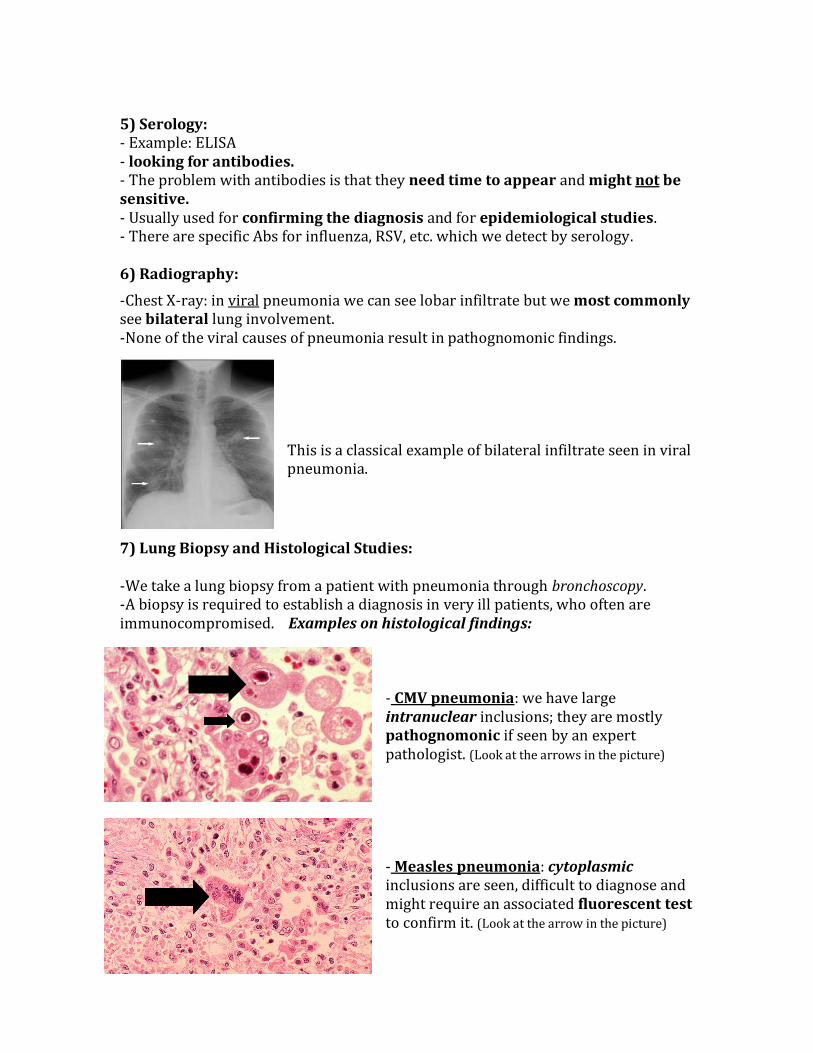
-Chest X-ray: in viral pneumonia we can see lobar infiltrate but we most commonly see bilateral lung involvement. -None of the viral causes of pneumonia result in pathognomonic findings.
This is a classical example of bilateral infiltrate seen in viral pneumonia.
7) Lung Biopsy and Histological Studies: -We take a lung biopsy from a patient with pneumonia through bronchoscopy. -A biopsy is required to establish a diagnosis in very ill patients, who often are immunocompromised. Examples on histological findings:
- CMV pneumonia: we have large intranuclear inclusions; they are mostly pathognomonic if seen by an expert pathologist. (Look at the arrows in the picture) - Measles pneumonia: cytoplasmic inclusions are seen, difficult to diagnose and might require an associated fluorescent test to confirm it. (Look at the arrow in the picture)
But in general inclusion bodies are usually seen in CMV pneumonia.
Management of Viral Pneumonia: 1- Supportive treatment:
Oxygen. Rest. Antipyretics. Analgesics. Nutrition. Close observation: ICU maybe, depending on the severity. IV fluids. Mechanical ventilation: in case of hypoxia.
2- Specific treatment: - Influenza Virus pneumonia: It's the only virus that has an approved treatment; which is: Oseltamavir and Zanamavir. (Were discussed thoroughly in the previous lecture) - RSV pneumonia: Ribavarin (an antiviral) is the only effective agent for RSV. But there are conflicting data regarding its efficacy. - Parainfleunza Virus pneumonia: Mainly treated by supportive treatment. Ribavarin might be used; as they have noticed a decrease in the shedding of the virus when it's applied. But the benefit is not 100% proven.
Clinical Case: [A 52 year old woman presented with fever, cough, shortness of breath, a vesicular rash of multiple stages, the rash started on the trunk then it spread to the extremities, and chest X-ray is bilateral.] Diagnosis: age and the multiple staged rash are indicators of atypical chicken pox and the chest X-ray indicates pneumonia, therefore it is Varicella Zoster Pneumonia. We confirm the diagnosis by a Tzanck smear –which is a sample of the fluid in the lesions put under the microscope– and we will observe multinuclear giant cells (epithelial cells with large inclusions bodies). These cells are highly suggestive of Varicella Zoster Virus.

Epidemiology: - Viral pneumonia's most affected age groups are infants/children and the elderly. - The healthy young and middle-aged adults are least affected. (Therefore if it's set on a curve, it will be high in childhood, low in adulthood and high again in old age.) - Viruses account for the largest proportion of childhood pneumonias. - But in adults, viruses are the second most common cause of pneumonia (after Streptococcus Pneumoniae) ranging from 13-50% of diagnosed cases. (Remember that the most common cause of viral pneumonia is Influenza Virus then RSV. But in children RSV is the most common cause of pneumonia followed by PIV.)
Incidence: - There has been an increased incidence of viral pneumonia recently, due to: Improved diagnostic techniques (especially PCR). Increase in immunocompromised patients (patients of BM transplant,
AIDS, patients on chemotherapy...) - The severity of the disease depends on the virulence of the viral mechanism (e.g.: influenza virus is more virulent than rhinovirus), it also depends on the age of the patient and his immunity. - It's very important to differentiate immunocompetent patients from immunocompromised; as they are more prone to all kinds of infectious diseases. Even the mechanism of the viral infection is more virulent and severe in them. - The most common causes of pneumonia in immunocompetent patients are: Influenza Virus, RSV, Adenovirus, and PIV.
- The most important types of Influenza Virus that cause pneumonia are A & B (esp. in pandemics and outbreaks caused by type A). Type C is rarely involved. - There is a recent increase in importance of other viruses that might cause pneumonia like: Rhinovirus: it's usually community acquired Coronavirus: its types are MERS and SARS. Human Metapneumovirus.
The importance of these other viruses will increase as diagnostic tests such as PCR become more widely available in the future.

Pathophysiology How do viruses cause pneumonia?
Contamination , then
Viruses reach the respiratory tract
Viruses multiply in the epithelium of the upper airway
The virus might infect the lung directly or by viremia (secondarily infect the lung by means of airway secretions or hematogenous spread) - Severe pneumonias may result in extensive consolidation of the lungs with varying degrees of hemorrhage. The hemorrhage might be so severe that it develops into bloody pleural effusions. - The infection also causes damage to the lungs. The mechanism of damage depends on the virus involved: Some viruses are mainly cytopathic, DIRECTLY affecting the lungs. Others will cause damage to the lungs due to their immune response (e.g. RSV)
- Damage to the respiratory tract might stimulate the host to release humoral factors some viruses for example increase the amount of histamine and IgE secreted causing wheezing, allergic reaction, which in turn exacerbates COPD and asthma (e.g. RSV)
-cell mediated immunity is important for the recovery from certain respiratory viral infections. Now let’s talk briefly about influenza and adenoviruses
Influenza virus: - Infection by influenza virus leads to cell death, especially in the upper airway. - Infection of lung parenchyma - Hemorrhage of lungs - Mucociliary clearance is impaired, so bacterial adherence to respiratory epithelium occurs more easily. - Infection impairs T lymphocytes, neutrophils, and macrophages function causing cell death, infection of the lungs’ parenchyma, hemorrhage in the lungs, impair mucociliaray clearance and this might lead to bacterial superinfection. So how can the influenza virus lead to bacterial superinfection? By impairing mucociliary clearance, T lymphocytes, machrophages, neutrophils... so the patient becomes immunocompromised and might easily develop a bacterial infection on top of the previous viral infection

Adenovirus: - Little is known regarding pathogenicity of adenovirus. - Outbreaks of adenovirus are frequent in crowded places and in military.
Viral pneumonia in the elderly: Elderly are at higher risk of viral pneumonia, because of Co-morbidities. decreased immunity (may impair viral clearance allowing the spread of the
virus to the lower respiratory tract). They have weak muscles
Viral Transmission: Depends on the type of the virus. As we said in the common cold: -hand to hand contact, fomites , small particle aerosol (e.g. Influenza and Adenovirus) -Environmental factors: (e.g. Enterovirus, Adenovirus, Rhinovirus) -Direct contact with contaminated objects (e.g. chicken pox virus VZV) … you need direct contact with the skin lesions, or the breath of the patient. -Transplantation of contaminated organs or blood products (e.g. CMV) -Through blood (e.g. CMV) - Hematogenous - - Reactivation of a latent infection (e.g. CMV, HSV) … if the patient is immunocompromised and has herpes simplex virus, the virus might reach the lung and get reactivated ; sometimes the HSV reach the lung through aspiration. - By health care (e.g. 20% of SARS cases are health care workers, Measles, Adenovirus, Parainfluenza, RSV) -Nosocomial pneumonia can be due to SARS, Measles, Adenovirus (As mentioned literally from the slides: Adenovirus, Influenza virus, measles virus, PIV, RSV, Rhinovirus, and VZV are easily transmitted during hospital stays and cause nosocomial pneumonia). Did measles disappear in Jordan? Last year we found some cases due to an outbreak in the north of the country
Pulmonary Host Defense: 1-Mechanical barriers: -Hairs in the nostrils (vibrissae)

- Mucociliary clearance - Sharp-angle branching of the central airways. All these mechanical barriers might impair viruses and foreign bodies from reaching to the lungs and causing infection there. 2- Humoral immunity: Especially IgA (in the secretions and mucosa), IgM (alveolar immunoglobin) 3- Phagocytic cells: Neutrophils and Macrophages: phagocytose the foreign body and present the antigenic part on its surface for the sake of lymphocytes. 4-Alveolar macrophages: provide the first defense in internalizing and degrading the viral pathogens. They act as antigen-presenting cells and opsonin-producing cells. 5-Cell mediated immunity: the most important defense mechanism against the intracellular viral pathogens. -CD8 cells attack infected cells (Please notice the difference between the cilia before and after infection with influenza A virus.)
Special Thanks to Sally Al-Khateeb and May Al-Mukhtar. Done By: Zaina Obeidat, Mai Bsoul and Rana Talj.
Good Luck
Cilia are seen before the infection
Cilia are absent after the infection