Violet Red Bile Agar with MUG - Becton Dickinson€¦ · Difco™ & BBL™ Manual, 2nd Edition...
2
Difco™ & BBL™ Manual, 2nd Edition Violet Red Bile Agar with MUG Intended Use Violet Red Bile Agar with MUG is used for enumerating Escherichia coli and total coliform bacteria in food and dairy products. Summary and Explanation Violet Red Bile Agar is specified in standard methods proce- dures to enumerate coliforms in food and dairy products. 1-3 In 1982, Feng and Hartman developed a rapid fluorogenic assay for Escherichia coli by incorporating 4-methylumbelliferyl-β-D- glucuronide (MUG) into Lauryl Tryptose Broth. 4 Incorporating MUG into Violet Red Bile Agar permits the detection of E. coli among the coliform colonies. 2, 3 Standard methods procedures include Violet Red Bile Agar with MUG for detecting E. coli in food and dairy products by fluorescence. 1-3 Principles of the Procedure Violet Red Bile Agar contains peptone as a source of carbon, nitrogen, vitamins and minerals. Yeast extract supplies B-complex vitamins which stimulate bacterial growth. Bile salts and crystal violet inhibit gram-positive bacteria. Lactose is a carbohydrate source. Neutral red is a pH indicator. MUG (4-methylumbelliferyl-β-D-glucuronide) is a substrate used for detecting glucuronidase activity. Agar is the solidifying agent. E. coli produces the enzyme glucuronidase which hydrolyzes MUG to yield a fluorogenic compound detectable with long-wave UV light (366 nm). Typical strains of E. coli (red colonies surrounded by a bile precipitate) exhibit blue fluorescence. Non-E. coli coliforms may produce red colonies with zones of precipitated bile but they are MUG negative. User Quality Control Escherichia coli ATCC ™ 25922 INOCULUM COLONY FLUOR- ORGANISM ATCC ™ CFU RECOVERY COLOR ESCENCE Enterobacter 13048 30-300 Good Pink, may – aerogenes have bile ppt. Escherichia coli 25922 30-300 Good Deep red, + with bile ppt. Staphylococcus 25923 3×10 2 -10 3 Marked to – – aureus complete inhibition Identity Specifications Difco ™ Violet Red Bile Agar with MUG Dehydrated Appearance: Reddish beige, free-flowing, homogeneous. Solution: 4.16% solution, soluble in purified water upon boiling. Solution is reddish-purple, clear to slightly opalescent, without significant precipitate. Prepared Appearance: Reddish-purple, clear to slightly opalescent, no significant precipitate. Reaction of 4.16% Solution at 25°C: pH 7.4 ± 0.2 Cultural Response Difco ™ Violet Red Bile Agar with MUG Prepare the medium per label directions. Inoculate and incubate at 32 ± 2°C for 22-26 hours.
-
Upload
truongkhanh -
Category
Documents
-
view
233 -
download
3
Transcript of Violet Red Bile Agar with MUG - Becton Dickinson€¦ · Difco™ & BBL™ Manual, 2nd Edition...
Difco™ & BBL™ Manual, 2nd Edition
Violet Red Bile Agar with MUG
Intended UseViolet Red Bile Agar with MUG is used for enumerating Escherichia coli and total coliform bacteria in food and dairy products.
Summary and ExplanationViolet Red Bile Agar is specified in standard methods proce-dures to enumerate coliforms in food and dairy products.1-3 In 1982, Feng and Hartman developed a rapid fluorogenic assay for Escherichia coli by incorporating 4-methylumbelliferyl-β-D-glucuronide (MUG) into Lauryl Tryptose Broth.4 Incorporating MUG into Violet Red Bile Agar permits the detection of E. coli among the coliform colonies.2, 3
Standard methods procedures include Violet Red Bile Agar with MUG for detecting E. coli in food and dairy products by fluorescence.1-3
Principles of the ProcedureViolet Red Bile Agar contains peptone as a source of carbon, nitrogen, vitamins and minerals. Yeast extract supplies B-complex vitamins which stimulate bacterial growth. Bile salts and crystal violet inhibit gram-positive bacteria. Lactose is a carbohydrate source. Neutral red is a pH indicator. MUG (4-methylumbelliferyl-β-D-glucuronide) is a substrate used for detecting glucuronidase activity. Agar is the solidifying agent.
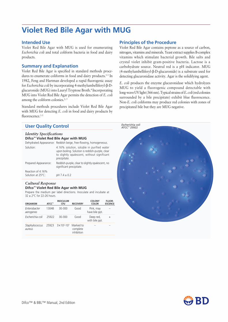
E. coli produces the enzyme glucuronidase which hydrolyzes MUG to yield a fluorogenic compound detectable with long-wave UV light (366 nm). Typical strains of E. coli (red colonies surrounded by a bile precipitate) exhibit blue fluorescence. Non-E. coli coliforms may produce red colonies with zones of precipitated bile but they are MUG negative.
User Quality Control Escherichia coliATCC™ 25922
InoCUlUM Colony FlUoR- oRGAnISM ATCC™ CFU RECoVERy ColoR ESCEnCE
Enterobacter 13048 30-300 Good Pink, may –aerogenes have bile ppt.
Escherichia coli 25922 30-300 Good Deep red, + with bile ppt.
Staphylococcus 25923 3×10²-103 Marked to – –aureus complete inhibition
Identity SpecificationsDifco™ Violet Red Bile Agar with MUGDehydrated Appearance: Reddish beige, free-flowing, homogeneous.
Solution: 4.16% solution, soluble in purified water upon boiling. Solution is reddish-purple, clear to slightly opalescent, without significant precipitate.
Prepared Appearance: Reddish-purple, clear to slightly opalescent, no significant precipitate.
Reaction of 4.16% Solution at 25°C: pH 7.4 ± 0.2
Cultural ResponseDifco™ Violet Red Bile Agar with MUGPrepare the medium per label directions. Inoculate and incubate at 32 ± 2°C for 22-26 hours.

Difco™ & BBL™ Manual, 2nd Edition
FormulaDifco™ Violet Red Bile Agar with MUG
Approximate Formula* Per LiterYeast Extract ............................................................... 3.0 gPeptone ...................................................................... 7.0 gBile Salts No. 3 ............................................................ 1.5 gLactose ..................................................................... 10.0 gSodium Chloride ......................................................... 5.0 gAgar ......................................................................... 15.0 gNeutral Red ................................................................. 0.03 gCrystal Violet............................................................... 2.0 mgMUG (4-methylumbelliferyl-β-D-glucuronide) .............. 0.1 g
*Adjusted and/or supplemented as required to meet performance criteria.
Directions for Preparation from Dehydrated Product1. Suspend 41.6 g of the powder in 1 L of purified water. Mix
thoroughly. 2. Heat with frequent agitation and boil for 1 minute to
completely dissolve the powder. DO NOT AUTOCLAVE.3. Cool to 45-50°C and use immediately.4. Test samples of the finished product for performance using
stable, typical control cultures.
Procedure1. Process each specimen as appropriate for that specimen.1-3
2. Incubate plates at 35°C for 22-26 hours.3. Examine plates for growth and fluorescence.
Expected ResultsColiform organisms form purplish-red colonies that are generally surrounded by a reddish zone of precipitated bile. When examined under long-wave fluorescent light, MUG-positive colonies are surrounded by a bluish fluorescent halo. MUG-negative colonies lack the fluorescent halo.
E. coli colonies are red surrounded by a zone of precipitated bile and fluoresce blue under long-wave UV light.
Salmonella and Shigella strains that produce glucuronidase may be encountered infrequently but these are generally lactose negative and appear as colorless colonies which may fluoresce.
limitations of the Procedure1. Glucuronidase-negative strains of E. coli have been encountered.5-7
Similarly, glucuronidase-positive strains of E. coli that do not fluoresce have been reported.8
2. Strains of Salmonella and Shigella that produce glucuronidase may infrequently be encountered.9 These strains must be distinguished from E. coli on the basis of other parameters; e. g., gas production, lactose fermentation or growth at 44.5°C.
References1. Davidson, Roth, and Gambrel-Lenarz. 2004. In Wehr and Frank (ed.). Standard methods for the
microbiological examination of dairy products, 17th ed. American Public Health Association, Wash-ington, D.C.
2. Kornacki and Johnson. 2001. In Downes and Ito (ed.). Compendium of methods for the microbiological examination of foods, 4th ed. American Public Health Association, Washington, D.C.
3. U.S. Food and Drug Administration. 2001. Bacteriological analytical manual, online. AOAC Interna-tional, Gaithersburg, Md.
4. Feng and Hartman. 1982. Appl. Environ. Microbiol. 43:1320.5. Chang, Brill and Lum. 1989. Appl. Environ. Microbiol. 55:335.6. Hansen and Yourassowsky. 1984. J. Clinical Microbiol. 20:1177.7. Kilian and Bulow. 1976. Acta Pathol. Microbiol. Scand. Sect. B 84:245. 8. Mates and Shaffer. 1989. J. Appl. Bacteriology 67:343.9. Damare, Campbell and Johnston. 1985. J. Food Sci. 50:1736.
AvailabilityDifco™ Violet Red Bile Agar with MUGBAM CoMPF
Cat. No. 229100 Dehydrated –500 g
BBl™ Violet Red Bile Agar with MUGBAM CoMPF
Cat. No. 299128 Prepared Bottle (200 mL) – Pkg. of 10