Vimentin-dependent utilization of LDL-cholesterol in human adrenal
13
1440 Journal of Lipid Research Volume 40, 1999 Vimentin-dependent utilization of LDL-cholesterol in human adrenal tumor cells is not associated with the level of expression of apoE, sterol carrier protein-2, or caveolin Tricia A. Holwell,* Susan C. Schweitzer,* Mary E. Reyland, * ,† and Robert M. Evans 1, * Department of Pathology,* School of Medicine, and Department of Basic Sciences and Oral Research, † School of Dentistry, University of Colorado Health Sciences Center, 4200 East 9th Avenue, Denver, CO 80262 Abstract SW-13 adrenal tumor cells that lack detectable in- termediate filaments (IF-free) exhibit an impaired capacity to esterify lipoprotein-derived cholesterol compared with cells that contain vimentin filaments. IF-free cells were found to synthesize and secrete significant amounts of apoE, while apoE secretion was nearly undetectable in cell lines that spontaneously express vimentin. However, stable transfectants that express a mouse vimentin cDNA exhib- ited elevated levels of cholesterol esterification and apoE secretion compared with untransfected IF-free cells, indi- cating that apoE secretion is not directly related to the ca- pacity of these cells to esterify cholesterol. Some of the cell lines that differed in the level of apoE synthesis and secre- tion had similar levels of apoE mRNA, suggesting that the differences in expression involve a post-transcriptional mechanism. Treatment of these cells with forskolin and IBMX, 8br-cAMP, or TPA had no effect on apoE secretion. The level of sterol carrier protein-2 (SCP 2 ) synthesis and the distribution of SCP 2 between membrane and soluble cellular fractions was not observably different in cells that contained or lacked vimentin. SW-13 cell lines contained little or no detectable caveolin-1 or caveolin-2. These studies demonstrate that the difference in the capacity of these ad- renal tumor cells that contain or lack vimentin filaments to esterify low density lipoprotein-cholesterol is not obviously associated with the level of expression or distribution of apoE, SCP 2 , or caveolins.—Holwell, T. A., S. C. Schweitzer, M. E. Reyland, and R. M. Evans. Vimentin-dependent utiliza- tion of LDL-cholesterol in human adrenal tumor cells is not associated with the level of expression of apoE, sterol car- rier protein-2, or caveolin. J. Lipid Res. 1999. 40: 1440– 1452. Supplementary key words apolipoprotein E • sterol carrier protein-2 • caveolins • cholesterol esterification • vimentin • intermediate fila- ments • adrenal cortical cells Lipoprotein-derived cholesterol is an important sub- strate for steroidogenesis (1). Although little is known about the mechanisms responsible for the post-lysosomal transport of cholesterol (2), studies of adrenal tumor cells have indicated that the intermediate filament (IF) compo- nent of the cell cytoskeleton may be involved (3). Fluores- cence microscopy of lipid droplets and IFs in Y-1 adrenal cells has shown that the cholesteryl ester-rich lipid drop- lets often co-localize with filaments of the vimentin type (4). When viewed by whole mount electron microscopy, the vimentin IFs appear to form contacts with the surface of the lipid droplets in primary adrenal cells (5). Exami- nation of the lipid metabolism of SW-13 adrenal tumor cells that either contain or lack cytoplasmic IFs has dem- onstrated that expression of a vimentin-type IF-network had a specific effect on cholesterol metabolism. IF-free SW-13 cell lines were found to have a lower capacity to esterify lipoprotein-derived cholesterol, compared with cells that contained vimentin filaments (6). In these stud- ies the impaired capacity of IF-free adrenal tumor cells to utilize lipoprotein cholesterol was restored in cell lines that expressed a mouse vimentin cDNA. The difference in the capacity of SW-13 cells to utilize lipoprotein choles- terol was not associated with a difference in receptor- mediated endocytosis of lipoprotein or the inherent capacity of the cells to esterify cholesterol, but rather ap- peared to be associated with post-lysosomal transport (6). The post-lysosomal events that mediate the intracellular movement of lipoprotein-derived cholesterol have not been well characterized (reviewed in 2, 7, 8). Studies of the human disease Niemann-Pick type C (see ref. 9), mutant cells selected for defective cholesterol transport (see 10, 11), and the effect a variety of inhibitors (12) have indicated that the transport of cholesterol derived from lipoprotein may occur independently from the movement of endogenously produced cholesterol, but very little is known about the mechanism or mechanisms. Although there is strong evidence that lipoprotein- Abbreviations: SCP 2 , sterol carrier protein-2; apoE, apolipoprotein E; TPA, phorbol-12-myristate-13-acetate; IBMX, 3-isobutyl-1-methyl- xanthine; IF, intermediate filament; PKA, protein kinase A; PKC, pro- tein kinase C. 1 To whom correspondence should be addressed. by guest, on April 10, 2019 www.jlr.org Downloaded from
Transcript of Vimentin-dependent utilization of LDL-cholesterol in human adrenal
1440 Journal of Lipid Research
Volume 40, 1999
Vimentin-dependent utilization of LDL-cholesterol in human adrenal tumor cells is not associated with the level of expression of apoE, sterol carrier protein-2, or caveolin
Tricia A. Holwell,* Susan C. Schweitzer,* Mary E. Reyland,*
,†
and Robert M. Evans
1,
*
Department of Pathology,* School of Medicine, and Department of Basic Sciences and Oral Research,
†
School of Dentistry, University of Colorado Health Sciences Center, 4200 East 9th Avenue, Denver, CO 80262
Abstract SW-13 adrenal tumor cells that lack detectable in-termediate filaments (IF-free) exhibit an impaired capacityto esterify lipoprotein-derived cholesterol compared withcells that contain vimentin filaments. IF-free cells werefound to synthesize and secrete significant amounts ofapoE, while apoE secretion was nearly undetectable in celllines that spontaneously express vimentin. However, stabletransfectants that express a mouse vimentin cDNA exhib-ited elevated levels of cholesterol esterification and apoEsecretion compared with untransfected IF-free cells, indi-cating that apoE secretion is not directly related to the ca-pacity of these cells to esterify cholesterol. Some of the celllines that differed in the level of apoE synthesis and secre-tion had similar levels of apoE mRNA, suggesting that thedifferences in expression involve a post-transcriptionalmechanism. Treatment of these cells with forskolin andIBMX, 8br-cAMP, or TPA had no effect on apoE secretion.The level of sterol carrier protein-2 (SCP
2
) synthesis andthe distribution of SCP
2
between membrane and solublecellular fractions was not observably different in cells thatcontained or lacked vimentin. SW-13 cell lines contained littleor no detectable caveolin-1 or caveolin-2. These studiesdemonstrate that the difference in the capacity of these ad-renal tumor cells that contain or lack vimentin filaments toesterify low density lipoprotein-cholesterol is not obviouslyassociated with the level of expression or distribution of
apoE, SCP
2
, or caveolins.
—Holwell, T. A., S. C. Schweitzer,M. E. Reyland, and R. M. Evans.
Vimentin-dependent utiliza-tion of LDL-cholesterol in human adrenal tumor cells is notassociated with the level of expression of apoE, sterol car-rier protein-2, or caveolin.
J. Lipid Res.
1999.
40:
1440–1452.
Supplementary key words
apolipoprotein E
•
sterol carrier protein-2
•
caveolins
•
cholesterol esterification
•
vimentin
•
intermediate fila-ments • adrenal cortical cells
Lipoprotein-derived cholesterol is an important sub-strate for steroidogenesis (1). Although little is knownabout the mechanisms responsible for the post-lysosomaltransport of cholesterol (2), studies of adrenal tumor cellshave indicated that the intermediate filament (IF) compo-
nent of the cell cytoskeleton may be involved (3). Fluores-cence microscopy of lipid droplets and IFs in Y-1 adrenalcells has shown that the cholesteryl ester-rich lipid drop-lets often co-localize with filaments of the vimentin type(4). When viewed by whole mount electron microscopy,the vimentin IFs appear to form contacts with the surfaceof the lipid droplets in primary adrenal cells (5). Exami-nation of the lipid metabolism of SW-13 adrenal tumorcells that either contain or lack cytoplasmic IFs has dem-onstrated that expression of a vimentin-type IF-networkhad a specific effect on cholesterol metabolism. IF-freeSW-13 cell lines were found to have a lower capacity toesterify lipoprotein-derived cholesterol, compared withcells that contained vimentin filaments (6). In these stud-ies the impaired capacity of IF-free adrenal tumor cells toutilize lipoprotein cholesterol was restored in cell linesthat expressed a mouse vimentin cDNA. The difference inthe capacity of SW-13 cells to utilize lipoprotein choles-terol was not associated with a difference in receptor-mediated endocytosis of lipoprotein or the inherentcapacity of the cells to esterify cholesterol, but rather ap-peared to be associated with post-lysosomal transport (6).
The post-lysosomal events that mediate the intracellularmovement of lipoprotein-derived cholesterol have notbeen well characterized (reviewed in 2, 7, 8). Studies ofthe human disease Niemann-Pick type C (see ref. 9),mutant cells selected for defective cholesterol transport(see 10, 11), and the effect a variety of inhibitors (12)have indicated that the transport of cholesterol derivedfrom lipoprotein may occur independently from themovement of endogenously produced cholesterol, butvery little is known about the mechanism or mechanisms.Although there is strong evidence that lipoprotein-
Abbreviations: SCP
2
, sterol carrier protein-2; apoE, apolipoproteinE; TPA, phorbol-12-myristate-13-acetate; IBMX, 3-isobutyl-1-methyl-xanthine; IF, intermediate filament; PKA, protein kinase A; PKC, pro-tein kinase C.
1
To whom correspondence should be addressed.
by guest, on April 10, 2019
ww
w.jlr.org
Dow
nloaded from
Holwell et al.
ApoE, SCP
2
, caveolin, and cholesterol esterification in adrenal tumor cells 1441
derived cholesterol transport involves specific cytoplasmiccomponents, it is not clear whether this involves vesicle-mediated transport or another carrier molecule. There isevidence that proteins such as apolipoprotein E (apoE)(13, 14), sterol carrier protein 2 (SCP
2
) (15, 16), and cave-olin-1 (8) could be involved, but the precise role of theseproteins in intracellular cholesterol transport has notbeen determined.
The importance of secreted apoE in cholesterol trans-port between peripheral tissues and the liver is well estab-lished (17) and there is now evidence that apoE expres-sion also affects the intracellular utilization of cholesterolthat is required for the synthesis of steroid hormones (18,19). Steroidogenic tissues are a site of significant apoEsynthesis (18). In the adrenal gland, apoE mRNA levelsare directly related to the level of cholesteryl ester storesand inversely related to the level of steroidogenesis (20).Purified apoE added to primary rat ovarian cell cultureshas been reported to inhibit LH-dependent androgen syn-thesis (21), and expression of a human apoE gene inmouse Y-1 adrenal cells has been shown to produce a sig-nificant suppression of steroidogenesis (13). In Y-1 cells,apoE expression was found to be associated with an in-creased cellular cholesterol content and decreased effluxof free cholesterol, indicating that apoE expression can af-fect intracellular cholesterol homeostasis (14). Recently,Swarnakar et al. (22) reported that apoE expression in Y-1cells was associated with an increase in the selective up-take of cholesteryl ester derived from LDL. While thesestudies have suggested that apoE can affect some aspectsof intracellular cholesterol metabolism, the mechanism bywhich a secreted protein could mediate these effectsremains unknown.
Numerous studies have suggested a role for SCP
2
in in-tracellular sterol transport (7). The SCP
2
gene encodesboth a 58 kD and a 15 kD protein (23). The 58 kD proteinis localized within peroxisomes, while the 15 kD SCP
2
pro-tein is proteolytically cleaved to a 13.2 kD form that isfound in mitochondria, cytosol, the cytosolic surface ofperoxisomes (24, 25) and is the form that has been impli-cated in intracellular cholesterol trafficking. While it maynot be required for the intracellular movement of choles-terol (26), the 13.2 SCP
2
protein has been shown to en-hance sterol transfer between various membranes in vitro(27, 28), and to stimulate steroidogenesis and esterifica-tion of plasma membrane cholesterol when expressed incells (15, 29). Although there is no known connection be-tween SCP
2
and the cytoskeleton, there is at least a super-ficial similarity in the phenotype of SCP
2
-deficient and IF-deficient cells. CHO cell mutants that lack peroxisomesand are deficient in the 13.2 kD SCP
2
protein have beenreported to exhibit an increase in cholesterol synthesisand increased cholesterol efflux, while uptake and hydrol-ysis of LDL-cholesterol was not affected (30). SW-13 adre-nal tumor cells that lack IFs also exhibit an increase incholesterol synthesis and efflux without apparent effectson the uptake and hydrolysis of LDL-cholesterol (6).
Caveolae are specialized clatherin-free areas of plasmamembrane that are rich in cholesterol and glycolipids,
and appear to be involved a variety of transport processes(31, 32) including transcytosis of LDL in brain capillaryendothelial cells (33). Caveolin-1 is a major caveolae coatprotein that has been shown to bind cholesterol (34), andappears to cycle between the cell surface and the Golgicomplex (35, 36). This has led to speculation that caveo-lin may be involved in trafficking and cholesterol homeo-stasis (36). The subcellular distribution of caveolin hasbeen shown to be affected by agents that disrupt microtu-bules and IFs (32), however, the possibility of an interac-tion between IFs and caveolin or caveolae has not beendirectly examined.
To try to gain some insight as to how vimentin IFs affectthe metabolism of cholesterol, studies were carried out todetermine whether the differences in the capacity of SW-13 adrenal tumor cells that contain or lack vimentin fila-ments to utilize lipoprotein-derived cholesterol could berelated to the expression or distribution of apoE or SCP
2
.We have found that SW-13 cells that contain or lack vi-mentin IFs have similar levels of SCP
2
and little or no cave-olin-1 or caveolin-2. While these cell lines were found todiffer substantially in apoE synthesis and secretion, therewas no direct relationship between the level of apoE secre-tion and the capacity of these cells to esterify lipoprotein-derived cholesterol. These observations indicate that theeffect of vimentin-type IFs on the capacity of SW-13 celllines to utilize lipoprotein-derived cholesterol is not obvi-ously associated with the level of expression or subcellulardistribution of apoE, SCP
2
, or caveolins 1 or 2.
MATERIALS AND METHODS
Cells and cell culture
SW-13 cell lines that spontaneously contain (vim
1
) or lack (IF-free) vimentin filaments (37, 38) were grown in monolayer cul-ture in a lipid-free medium consisting of a 1:1 mixture of Ham’sF12:Dulbecco’s MEM containing either 5% CPSR-1 (Sigma) or20 ml/L TCM (Celox), 0.5 mg /ml fetuin, and 10
m
g/ml gen-tamicin. Additional cell lines stably transfected with a mouse vi-mentin cDNA (6) were grown in medium containing 5% CPSR-1or 20 ml/L TCM, 0.5 mg/ml fetuin, and 200
m
g/ml G418. Pre-liminary experiments indicated that the relative rates of choles-terol esterification in SW-13 cell lines were similar in mediumsupplemented with either CPSR-1 or TCM (data not shown).Low density lipoprotein (LDL) was purified from human plasmaby KBr density gradient ultracentrifugation by the method ofGoldstein, Basu, and Brown (39). Forskolin (Sigma), 3-isobutyl-1-methyl-xanthine (IBMX) (Sigma), 8-bromo-cAMP (BoehringerMannheim), chelerythrine (LC Laboratories), and phorbol-12-myristate-13-acetate (TPA)(Sigma) were added to cultures at theindicated concentrations.
Immunoprecipitation
Approximately 2
3
10
6
SW-13 cells that spontaneously con-tain or lack vimentin were plated per 10 cm dish, 48 h prior toradiolabeling. The stable transfectant cell lines grow more slowlyand were plated at an initial density of 3
3
10
6
cells per 10 cmdish. The cells were radiolabeled with 50–100
m
Ci/ml [
35
S]me-thionine in 4 ml of complete medium for the indicated times. Inexperiments that involved secreted protein, the culture mediumwas recovered and centrifuged at approximately 500
g
for 5 min.
by guest, on April 10, 2019
ww
w.jlr.org
Dow
nloaded from
1442 Journal of Lipid Research
Volume 40, 1999
The resulting supertnatant was recovered and PMSF and leupep-tin was added to final concentrations of 0.2 m
m
and 5.0
m
g/ml,respectively. The medium was centrifuged again at 10,000
g
for 5min. Triton X-100 was added to the resulting supernatant to a fi-nal concentration of 1% and this solution was centrifuged againat 5000
g
for 10 min. In experiments involving immunoprecipi-tation from monolayer cells, the monolayer was rinsed 3 timeswith PBS. The cells were then lysed in 1 ml of 145 m
m
NaCl, 20m
m
Tris-HCl, pH 7.4, 1 m
m
EDTA (TBS) containing 0.5% TritonX-100, 0.5 m
m
PMSF, 0.5
m
g/ml leupeptin. The cell lysate wascentrifuged at 14,000
g
for 2–5 min, and the supernatant was re-covered for immunoprecipitation. For each experiment, thesamples were normalized to contain the same amount of
35
S-labeled protein. Fifty
m
l of goat anti-apoE (Calbiochem), or 4
m
gmonoclonal anti-caveolin-1 (Transduction Laboratories), or anti-caveolin-2 (Transduction Laboratories), 10
m
l of rabbit anti-SCP
2
(gift from J. Billheimer) diluted 1:10 in TBS, was added to 0.5–1.5 ml of sample (either culture medium or monolayer Triton-lysate). For anti-caveolin-1 and caveolin-2 immunoprecipitates,the antibodies were diluted in the lysis buffer. Similarly dilutednonimmune goat or rabbit serum (NGS or NRS) was added toduplicate samples as a negative control. Preliminary experi-ments determined that increasing the antibody concentrationdid not result in increased recovery of apoE or SCP
2
. The sam-ples were incubated for 1.5 h at 4
8
C. For anti-caveolin-1 andcaveolin-2 immunoprecipitates, 12.5
m
l of rabbit anti-mouse se-rum was then added as a bridge antibody and incubated an addi-tional 1 h. Two hundred
m
l of blocked rProteinA-agarose (Repli-gen) slurry (100
m
l packed volume) was then added to eachsample and mixed for at least 1 h at 4
8
C. The samples were thencentrifuged at 11,000
g
for 1–2 min. The supernatant was re-moved and the agarose immunoprecipitate was resuspended in1 ml TBS. This washing step was repeated 3 or 4 times. The finalprecipitate was resuspended in 15
m
l of 0.25
m
Tris, 40% glyc-erol, 6% SDS, 2 m
m
DTT, pH 6.8, and stored at
2
20
8
C. Precipi-tated proteins were analyzed by SDS-PAGE (40) in 12.5% (forSCP
2
) or 10% (for apoE and caveolin-1) polyacrylamide gels.After electrophoresis, gels were stained with Coomassie blue,destained, and infiltrated with Amplify (Amersham) or 1
m
sali-cylic acid. The gels were dried and
35
S-labeled proteins weredirectly quantitated using an imaging scanner (Bioscan System200). The gels were scanned for 30 min per lane using the two-dimensional analysis program. The
35
S radioactivity for each in-dividual protein band was determined using the manual mode.The gels were then fluorographed on Hyperfilm (Amersham) at
2
70
8
C. In experiments where the radiolabeled monolayer wasnot used for immunoprecipitation, the monolayer was rinsed 2times with PBS, 1.0 ml of 2% SDS, 0.5% deoxycholate, 1 m
m
EDTA, 1 m
m
dithiothreitol was added, and the resulting cell ly-sate was recovered. These cell lysates were then used to deter-mine
35
S incorporated into TCA-precipitable protein.
RNA isolation and Northern analysis
Cells were solubilized in 4
m
guanidinium thiocyanate, 5 m
m
Na citrate, 0.5% sarcosyl, 0.1
m
2-mercaptoethanol, and totalRNA was prepared by pelleting the lysate through a 5.7
m
CsClcushion in an SW41 rotor at 150,000
g
for 16 h. The RNA (20
m
g/lane) was separated on agarose formaldehyde gels, trans-ferred to Nytran (Scheicher and Schuell), and cross-linkedwith ultraviolet radiation. To determine apoE mRNA levels, theblots were hybridized with a
32
P-labeled human apoE cDNAprobe. The blots were then stripped and re-probed with a ratglyceraldehyde phosphate dehydrogenase cDNA probe as acontrol for RNA loading (13). Probes were radiolabeled with[
a
32
P]-dCTP using a random primer kit (Stratagene). Afterhybridization,
32
P-labeled bands on the dried Nytran were di-
rectly quantitated using a Molecular Dynamics PhosphorIm-ager. The blots were autoradiographed on Hyperfilm (Amer-sham) at
2
70
8
C.
Cell cholesterol content and esterification
In some experiments, the secretion of apoE, cell cholesterolcontent and esterification were measured. Cells were labeledwith [
35
S]methionine as described above. After 20 h, 0.5
m
Ci/ml[1-
14
C]oleate–BSA (39) was added to the medium and the cellswere incubated in the presence of both radiolabels for 4 h. Themedium was then removed for immunoprecipitation of[
35
S]apoE. The cell monolayer was rinsed 2 times with TBS, 50
m
g of cholesteryl heptadecanoate was added as an internal stan-dard, and then extracted twice with hexane–isopropyl alcohol3:2. The combined extracts were dried under nitrogen, resus-pended in chloroform, and an aliquot was taken for analysis byeither TLC or HPLC. Neutral lipids were separated by TLC onactivated Silica Gel G plates (Analtech) using a solvent system ofhexane–ethyl ether–acetic acid 60:40:1 (v/v). The lipids were vi-sualized by spraying the chromatograms with an aqueous solu-tion of 0.5% 8-anilino-1-naphthalenesulfonic acid and detectedwith UV light. Lipids were identified by co-chromatography withappropriate purified standards added to each sample (Avanti).Areas of the chromatograms representing resolved individual lip-ids were scraped into vials and radioactivity was determined in 4ml of Ecolume (ICN Radiochemicals) by scintillation counting.For HPLC analysis, samples were dried under nitrogen, resus-pended in 20
m
l of chloroform, and diluted with 190
m
l of isopro-pyl alcohol–acetonitrile 1:1 (v/v). The labeled cholesterol andcholesteryl esters were separated by HPLC as described byVercaemst, Union, and Rosseneu (41) on a 4.6
3
125 mm Asa-hipak C18 column using an isopropyl alcohol–acetonitrilemobile phase at 0.6 ml/min. Eluted cholesterol and cholesterylesters were detected by absorption at 214 nm and identified byco-elution with cholesterol, cholesteryl oleate, and cholesterylheptadecanoate standards (Sigma). Peak areas were deter-mined using a Gilson 714 analytical program and corrected forrecovery using the cholesteryl heptadecanoate internal stan-dard. [
14
C]oleate incorporated into triglycerides and choles-teryl oleate was detected using an in-line radiodetector (
b
-Ram,IN/US systems) and peak areas were determined using the Gil-son 714 analytical program. The extracted cell monolayer wasdissolved in 0.1 N NaOH, neutralized with 1
m
HCl, 0.2
m
phos-phate, and aliquots were taken for protein determination by theBradford assay (42).
Immunofluorescence microscopy
Cells were plated on sterile glass cover slips. The cells wererinsed with warm PBS and then fixed in freshly prepared 4%paraformaldehyde in PHEM buffer (60 mm PIPES, 25 m
m
HEPES, 10 m
m
EGTA, 3 m
m
MgCl
2
, pH 6.1) (43) for 5 min atroom temperature. The monolayers were then extracted withPHEM buffer containing 0.15% Triton X-110 for 2 min, and thenrinsed with PBS. For indirect immunofluorescence, the cellswere preincubated in PBS containing 1% ovalbumin and 1%normal goat serum for 10 min, and then incubated with mono-clonal anti-apoE for 40 min. The cells on cover slips were thenrinsed in PBS and incubated with secondary antibody for 30 min.Monoclonal anti-human apoE (clone 2034, Bioreclamation) wasused at 10
m
g/ml. Lissamine–rhodamine-conjugated goat anti-mouse IgG (Boehringer Mannheim), diluted 1:100, was used asthe second antibody. All antibodies were diluted in PBS contain-ing 1% ovalbumin and 1% normal goat serum. The cover slipswere mounted in Immunomount (Lerner Labs) and viewed onan Olympus microscope equipped with epifluorescence optics.Photographic exposures were made for 40 sec using Kodak TMax
by guest, on April 10, 2019
ww
w.jlr.org
Dow
nloaded from
Holwell et al.
ApoE, SCP
2
, caveolin, and cholesterol esterification in adrenal tumor cells 1443
400 film and the film processed with an exposure index of 1200using Kodak HC-110 developer.
RESULTS
Vimentin expressing and IF-free SW-13 cell lines secrete different amounts of apoE
In previous studies we have found that SW-13/cl.2 cellsthat lack detectable cytoplasmic intermediate filaments(IF-free) had a substantially lower capacity to esterify LDL-cholesterol than SW-13/cl.1 cells that contain vimentinIFs (vim
1
). Moreover, expression of a mouse vimentin trans-gene in T3M cells, derived from the IF-free cl. 2 cell line, in-creased the capacity of these cells to esterify LDL-cholesterol.To determine whether the difference in intracellular cho-lesterol metabolism in vim
1
and IF-free SW-13 cells couldbe associated with differences in apoE expression, anti-apoE immunoprecipitation experiments were carried outto determine whether these cells express detectable levelsof apoE. Initial experiments were performed with cl.1vim
1
, and IF-free cl.2 cells because these cell lines exhibitsimilar growth characteristics. As a positive control, thehepatoma HepG2 cell line that has been reported to se-crete high levels of apoE (44) was also examined. Culturemedium from [
35
S]methionine-labeled cl.2 IF-free cellscontained significant levels of [
35
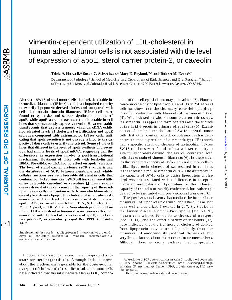
S]apoE (
Fig. 1
), al-though the amount of apoE that was produced was clearlyless than that obtained from the hepatoma cell line. Littleif any detectable [
35
S]apoE was immunoprecipitated fromcl.1 vim
1
cell medium under identical conditions. Treat-ment of cells with LDL stimulated apoE expression in allthree cell lines. Quantitation of [
35
S]apoE immunoprecip-itated in triplicate samples indicated that LDL treatmentapproximately doubled the level of apoE secreted by theIF-free cl.2 cells (data not shown). Because [
35
S]apoE wasbarely detectable in the medium from cl.1 vim
1
cells inthe absence of LDL, it was not possible to accurately deter-mine the actual level of induction of apoE secretion inthese cells after LDL treatment. When the level of apoEsecretion was compared in cultures treated with LDL, theIF-free cl.2 cell medium contained more [
35
S]apoE thanthe medium from the cl.1 vim
1
cells. Anti-apoE immuno-precipitation from cell lysates indicated that [
35
S]apoEwas easily detected in HepG2 cell lysates, and could alsobe detected as a much less abundant band in the IF- freecl.2 cells. However, [
35
S]apoE was undetectable in cl.1vim1 cell lysates, indicating that the low level of apoE se-cretion by these cells was not associated with a detectableaccumulation of intracellular apoE. Comparison of the[35S]apoE immunoprecipitated from the culture mediumand cell lysates of the cl.2 and HepG2 cells indicated thatmost of the radiolabeled apoE was present in the culturemedium. In addition, the [35S]apoE immunoprecipitatedfrom the monolayers was 2–3 kD smaller than the apoEband detected in the culture medium (see also Fig. 5).This is consistent with the increase in apparent molecularweight associated with glycosylation of the secreted formof apoE (44).
As the observed difference in the capacity of cl.1 vim1
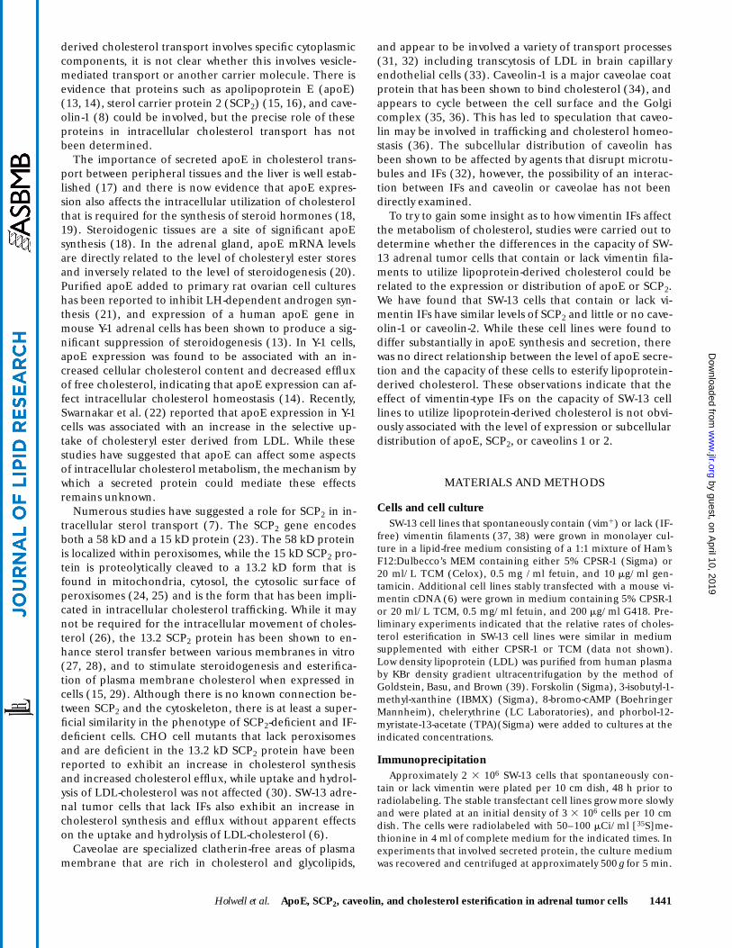
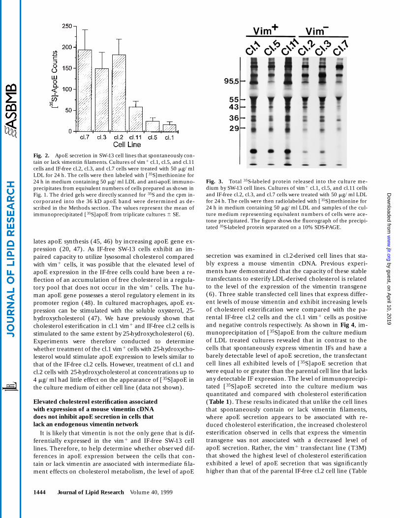
and IF-free cl.2 cells to secrete apoE could simply be a re-flection of clonal variation between individual cell lines,apoE secretion was compared in four additional, indepen-dently isolated SW-13 cell lines that spontaneously containor lack vimentin IFs, where the absence of filaments is as-sociated with a lower level of cholesterol esterification (6).As shown in Fig. 2, quantitation of the [35S]apoE immuno-precipitated from the culture medium of LDL treated cul-tures indicated that all three IF-free cell lines exhibitedsignificant levels of apoE secretion, while apoE was barelydetectable in the culture medium from the three vim1
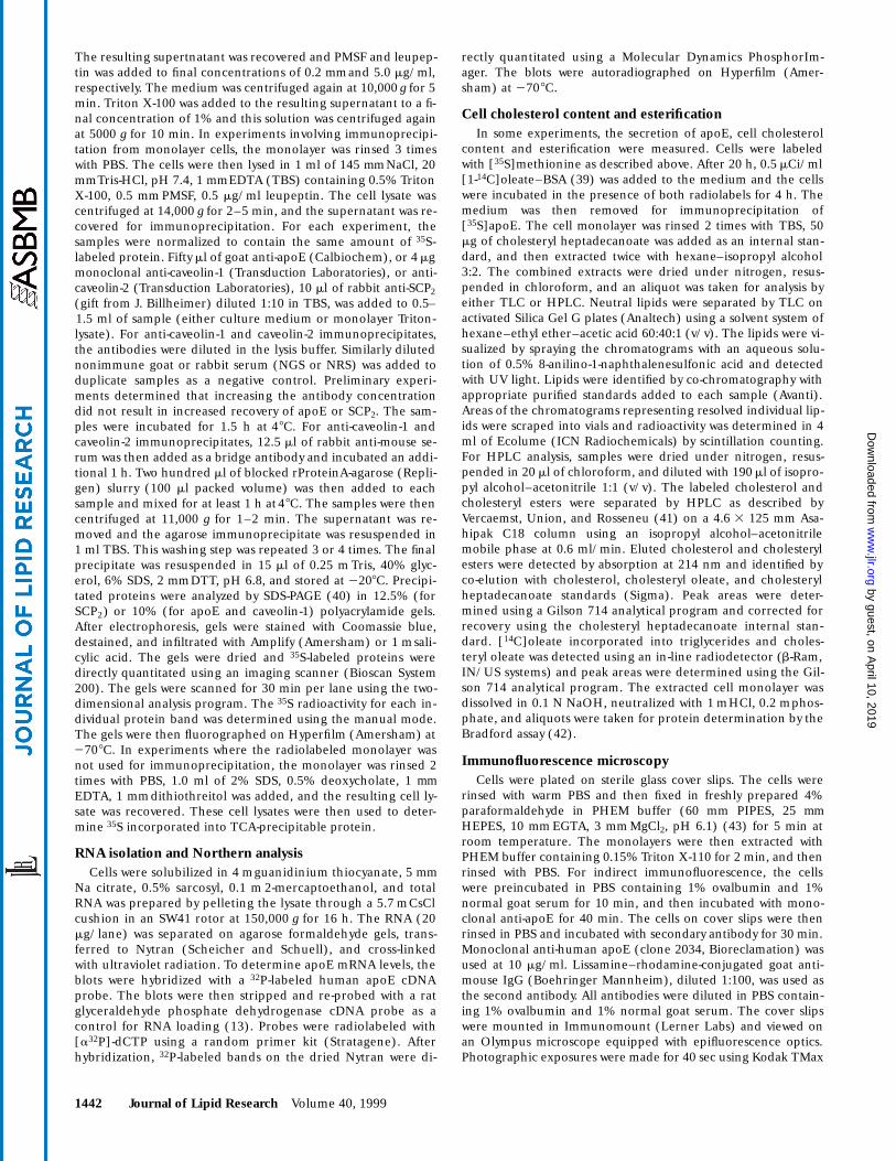
cell lines. Examination of total 35S-labeled protein in theculture medium (Fig. 3) showed no prominent quantita-tive or qualitative differences in total protein secretionbetween the cell lines. This indicated that the observeddifference in secretion of apoE in these cells was not asso-ciated with an overall difference in secretion of radiola-beled proteins.
The stimulation of apoE secretion observed in the cl.2cells treated with LDL is consistent with previous studiesindicating that cholesterol loading of macrophages stimu-
Fig. 1. Immunoprecipitation of apoE from the culture mediumand cell lysates of SW-13/cl.1 (vim1), SW-13/cl.2 (IF-free), andHepG2 cells. Anti-apoE immunoprecipitation of culture medium(Medium) and cell lysates (Cells) was performed from cells labeledfor 24 h with 100 mCi/ml [35S]methionine. The figure shows thefluorograph of a 10% SDS-polyacrylamide gel of anti-apoE immu-noprecipitates from untreated cultures (lane 1), anti-apoE (lane 2),and non-immune serum (lane 3) precipitates from cultures treatedwith 50 mg/ml LDL. The immunoprecipitations of the cl.1 and cl.2cell preparations represent material from equivalent numbers ofcells. The position of the immunoprecipitated apoE is indicated( ). The mobilities of MW standards are indicated in kD.➤
by guest, on April 10, 2019
ww
w.jlr.org
Dow
nloaded from
1444 Journal of Lipid Research Volume 40, 1999
lates apoE synthesis (45, 46) by increasing apoE gene ex-pression (20, 47). As IF-free SW-13 cells exhibit an im-paired capacity to utilize lysosomal cholesterol comparedwith vim1 cells, it was possible that the elevated level ofapoE expression in the IF-free cells could have been a re-flection of an accumulation of free cholesterol in a regula-tory pool that does not occur in the vim1 cells. The hu-man apoE gene possesses a sterol regulatory element in itspromoter region (48). In cultured macrophages, apoE ex-pression can be stimulated with the soluble oxysterol, 25-hydroxycholesterol (47). We have previously shown thatcholesterol esterification in cl.1 vim1 and IF-free cl.2 cells isstimulated to the same extent by 25-hydroxycholesterol (6).Experiments were therefore conducted to determinewhether treatment of the cl.1 vim1 cells with 25-hydroxycho-lesterol would stimulate apoE expression to levels similar tothat of the IF-free cl.2 cells. However, treatment of cl.1 andcl.2 cells with 25-hydroxycholesterol at concentrations up to4 mg/ml had little effect on the appearance of [35S]apoE inthe culture medium of either cell line (data not shown).
Elevated cholesterol esterification associated with expression of a mouse vimentin cDNAdoes not inhibit apoE secretion in cells thatlack an endogenous vimentin network
It is likely that vimentin is not the only gene that is dif-ferentially expressed in the vim1 and IF-free SW-13 celllines. Therefore, to help determine whether observed dif-ferences in apoE expression between the cells that con-tain or lack vimentin are associated with intermediate fila-ment effects on cholesterol metabolism, the level of apoE
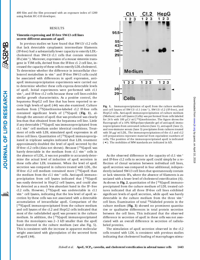
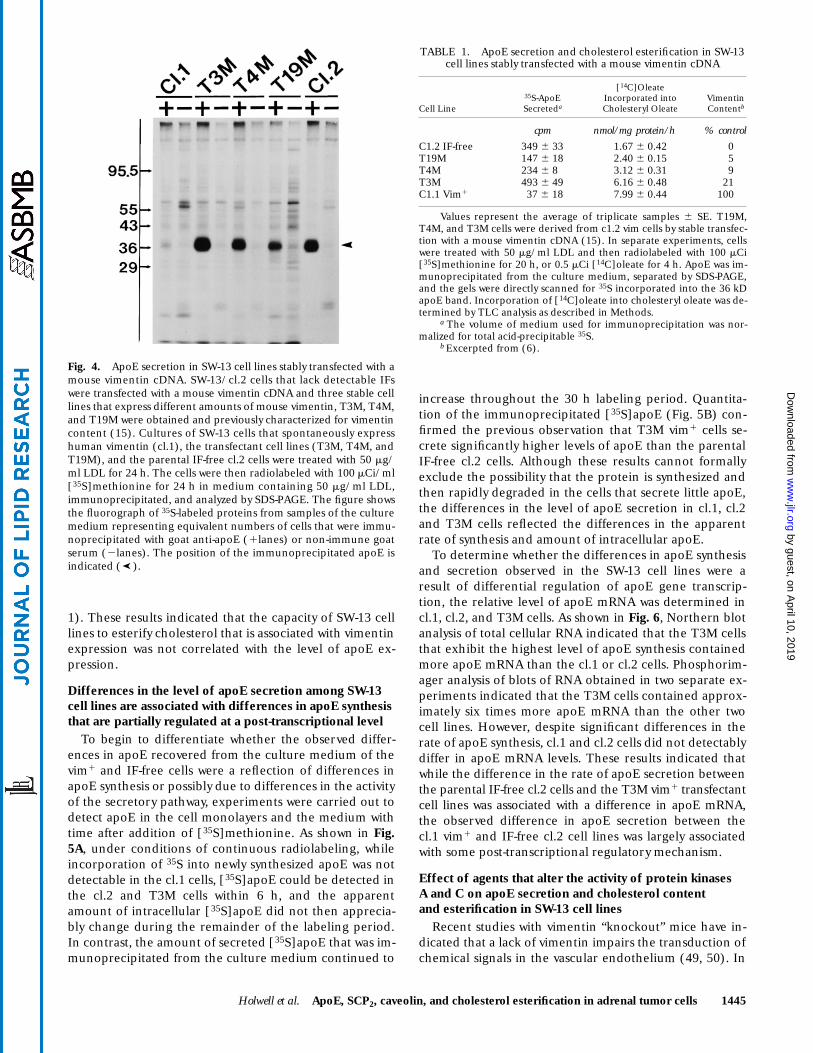
secretion was examined in cl.2-derived cell lines that sta-bly express a mouse vimentin cDNA. Previous experi-ments have demonstrated that the capacity of these stabletransfectants to esterify LDL-derived cholesterol is relatedto the level of the expression of the vimentin transgene(6). Three stable transfected cell lines that express differ-ent levels of mouse vimentin and exhibit increasing levelsof cholesterol esterification were compared with the pa-rental IF-free cl.2 cells and the cl.1 vim1 cells as positiveand negative controls respectively. As shown in Fig 4, im-munoprecipitation of [35S]apoE from the culture mediumof LDL treated cultures revealed that in contrast to thecells that spontaneously express vimentin IFs and have abarely detectable level of apoE secretion, the transfectantcell lines all exhibited levels of [35S]apoE secretion thatwere equal to or greater than the parental cell line that lacksany detectable IF expression. The level of immunoprecipi-tated [35S]apoE secreted into the culture medium wasquantitated and compared with cholesterol esterification(Table 1). These results indicated that unlike the cell linesthat spontaneously contain or lack vimentin filaments,where apoE secretion appears to be associated with re-duced cholesterol esterification, the increased cholesterolesterification observed in cells that express the vimentintransgene was not associated with a decreased level ofapoE secretion. Rather, the vim1 transfectant line (T3M)that showed the highest level of cholesterol esterificationexhibited a level of apoE secretion that was significantlyhigher than that of the parental IF-free cl.2 cell line (Table
Fig. 2. ApoE secretion in SW-13 cell lines that spontaneously con-tain or lack vimentin filaments. Cultures of vim1 cl.1, cl.5, and cl.11cells and IF-free cl.2, cl.3, and cl.7 cells were treated with 50 mg/mlLDL for 24 h. The cells were then labeled with [35S]methionine for24 h in medium containing 50 mg/ml LDL and anti-apoE immuno-precipitates from equivalent numbers of cells prepared as shown inFig. 1. The dried gels were directly scanned for 35S and the cpm in-corporated into the 36 kD apoE band were determined as de-scribed in the Methods section. The values represent the mean ofimmunoprecipitated [35S]apoE from triplicate cultures 6 SE.
Fig. 3. Total 35S-labeled protein released into the culture me-dium by SW-13 cell lines. Cultures of vim1 cl.1, cl.5, and cl.11 cellsand IF-free cl.2, cl.3, and cl.7 cells were treated with 50 mg/ml LDLfor 24 h. The cells were then radiolabeled with [35S]methionine for24 h in medium containing 50 mg/ml LDL and samples of the cul-ture medium representing equivalent numbers of cells were ace-tone precipitated. The figure shows the fluorograph of the precipi-tated 35S-labeled protein separated on a 10% SDS-PAGE.
by guest, on April 10, 2019
ww
w.jlr.org
Dow
nloaded from
Holwell et al. ApoE, SCP2, caveolin, and cholesterol esterification in adrenal tumor cells 1445
1). These results indicated that the capacity of SW-13 celllines to esterify cholesterol that is associated with vimentinexpression was not correlated with the level of apoE ex-pression.
Differences in the level of apoE secretion among SW-13 cell lines are associated with differences in apoE synthesis that are partially regulated at a post-transcriptional level
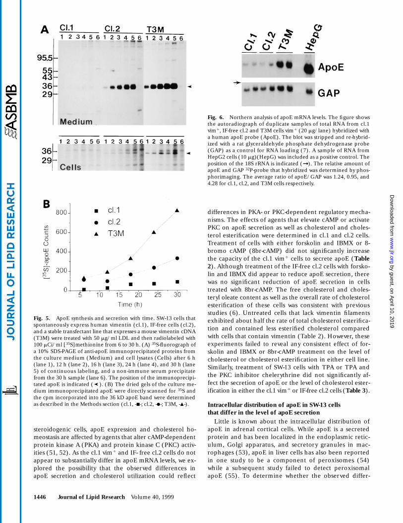
To begin to differentiate whether the observed differ-ences in apoE recovered from the culture medium of thevim1 and IF-free cells were a reflection of differences inapoE synthesis or possibly due to differences in the activityof the secretory pathway, experiments were carried out todetect apoE in the cell monolayers and the medium withtime after addition of [35S]methionine. As shown in Fig.5A, under conditions of continuous radiolabeling, whileincorporation of 35S into newly synthesized apoE was notdetectable in the cl.1 cells, [35S]apoE could be detected inthe cl.2 and T3M cells within 6 h, and the apparentamount of intracellular [35S]apoE did not then apprecia-bly change during the remainder of the labeling period.In contrast, the amount of secreted [35S]apoE that was im-munoprecipitated from the culture medium continued to
increase throughout the 30 h labeling period. Quantita-tion of the immunoprecipitated [35S]apoE (Fig. 5B) con-firmed the previous observation that T3M vim1 cells se-crete significantly higher levels of apoE than the parentalIF-free cl.2 cells. Although these results cannot formallyexclude the possibility that the protein is synthesized andthen rapidly degraded in the cells that secrete little apoE,the differences in the level of apoE secretion in cl.1, cl.2and T3M cells reflected the differences in the apparentrate of synthesis and amount of intracellular apoE.
To determine whether the differences in apoE synthesisand secretion observed in the SW-13 cell lines were aresult of differential regulation of apoE gene transcrip-tion, the relative level of apoE mRNA was determined incl.1, cl.2, and T3M cells. As shown in Fig. 6, Northern blotanalysis of total cellular RNA indicated that the T3M cellsthat exhibit the highest level of apoE synthesis containedmore apoE mRNA than the cl.1 or cl.2 cells. Phosphorim-ager analysis of blots of RNA obtained in two separate ex-periments indicated that the T3M cells contained approx-imately six times more apoE mRNA than the other twocell lines. However, despite significant differences in therate of apoE synthesis, cl.1 and cl.2 cells did not detectablydiffer in apoE mRNA levels. These results indicated thatwhile the difference in the rate of apoE secretion betweenthe parental IF-free cl.2 cells and the T3M vim1 transfectantcell lines was associated with a difference in apoE mRNA,the observed difference in apoE secretion between thecl.1 vim1 and IF-free cl.2 cell lines was largely associatedwith some post-transcriptional regulatory mechanism.
Effect of agents that alter the activity of protein kinasesA and C on apoE secretion and cholesterol content and esterification in SW-13 cell lines
Recent studies with vimentin “knockout” mice have in-dicated that a lack of vimentin impairs the transduction ofchemical signals in the vascular endothelium (49, 50). In
Fig. 4. ApoE secretion in SW-13 cell lines stably transfected with amouse vimentin cDNA. SW-13/cl.2 cells that lack detectable IFswere transfected with a mouse vimentin cDNA and three stable celllines that express different amounts of mouse vimentin, T3M, T4M,and T19M were obtained and previously characterized for vimentincontent (15). Cultures of SW-13 cells that spontaneously expresshuman vimentin (cl.1), the transfectant cell lines (T3M, T4M, andT19M), and the parental IF-free cl.2 cells were treated with 50 mg/ml LDL for 24 h. The cells were then radiolabeled with 100 mCi/ml[35S]methionine for 24 h in medium containing 50 mg/ml LDL,immunoprecipitated, and analyzed by SDS-PAGE. The figure showsthe fluorograph of 35S-labeled proteins from samples of the culturemedium representing equivalent numbers of cells that were immu-noprecipitated with goat anti-apoE (1lanes) or non-immune goatserum (2lanes). The position of the immunoprecipitated apoE isindicated ( ).➤
TABLE 1. ApoE secretion and cholesterol esterification in SW-13 cell lines stably transfected with a mouse vimentin cDNA
Cell Line35S-ApoESecreteda
[14C]OleateIncorporated intoCholesteryl Oleate
Vimentin Contentb
cpm nmol/mg protein/h % control
C1.2 IF-free 349 6 33 1.67 6 0.42 0T19M 147 6 18 2.40 6 0.15 5T4M 234 6 8 3.12 6 0.31 9T3M 493 6 49 6.16 6 0.48 21C1.1 Vim1 37 6 18 7.99 6 0.44 100
Values represent the average of triplicate samples 6 SE. T19M,T4M, and T3M cells were derived from c1.2 vim cells by stable transfec-tion with a mouse vimentin cDNA (15). In separate experiments, cellswere treated with 50 mg/ml LDL and then radiolabeled with 100 mCi[35S]methionine for 20 h, or 0.5 mCi [14C]oleate for 4 h. ApoE was im-munoprecipitated from the culture medium, separated by SDS-PAGE,and the gels were directly scanned for 35S incorporated into the 36 kDapoE band. Incorporation of [14C]oleate into cholesteryl oleate was de-termined by TLC analysis as described in Methods.
a The volume of medium used for immunoprecipitation was nor-malized for total acid-precipitable 35S.
b Excerpted from (6).
by guest, on April 10, 2019
ww
w.jlr.org
Dow
nloaded from
1446 Journal of Lipid Research Volume 40, 1999
steroidogenic cells, apoE expression and cholesterol ho-meostasis are affected by agents that alter cAMP-dependentprotein kinase A (PKA) and protein kinase C (PKC) activ-ities (51, 52). As the cl.1 vim1 and IF- free cl.2 cells do notappear to substantially differ in apoE mRNA levels, we ex-plored the possibility that the observed differences inapoE secretion and cholesterol utilization could reflect
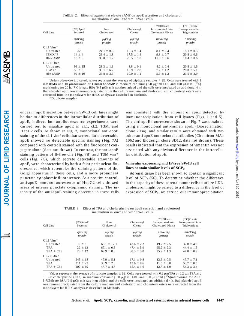
differences in PKA- or PKC-dependent regulatory mecha-nisms. The effects of agents that elevate cAMP or activatePKC on apoE secretion as well as cholesterol and choles-terol esterification were determined in cl.1 and cl.2 cells.Treatment of cells with either forskolin and IBMX or 8-bromo cAMP (8br-cAMP) did not significantly increasethe capacity of the cl.1 vim1 cells to secrete apoE (Table2). Although treatment of the IF-free cl.2 cells with forsko-lin and IBMX did appear to reduce apoE secretion, therewas no significant reduction of apoE secretion in cellstreated with 8br-cAMP. The free cholesterol and choles-teryl oleate content as well as the overall rate of cholesterolesterification of these cells was consistent with previousstudies (6). Untreated cells that lack vimentin filamentsexhibited about half the rate of total cholesterol esterifica-tion and contained less esterified cholesterol comparedwith cells that contain vimentin (Table 2). However, theseexperiments failed to reveal any consistent effect of for-skolin and IBMX or 8br-cAMP treatment on the level ofcholesterol or cholesterol esterification in either cell line.Similarly, treatment of SW-13 cells with TPA or TPA andthe PKC inhibitor chelerythrine did not significantly af-fect the secretion of apoE or the level of cholesterol ester-ification in either the cl.1 vim1 or IF-free cl.2 cells (Table 3).
Intracellular distribution of apoE in SW-13 cells that differ in the level of apoE secretion
Little is known about the intracellular distribution ofapoE in adrenal cortical cells. While apoE is a secretedprotein and has been localized in the endoplasmic retic-ulum, Golgi apparatus, and secretory granules in mac-rophages (53), apoE in liver cells has also been reportedin one study to be a component of peroxisomes (54)while a subsequent study failed to detect peroxisomalapoE (55). To determine whether the observed differ-
Fig. 5. ApoE synthesis and secretion with time. SW-13 cells thatspontaneously express human vimentin (cl.1), IF-free cells (cl.2),and a stable transfectant line that expresses a mouse vimentin cDNA(T3M) were treated with 50 mg/ml LDL and then radiolabeled with100 mCi/ml [35S]methionine from 6 to 30 h. (A) 35S-fluorograph ofa 10% SDS-PAGE of anti-apoE immunoprecipitated proteins fromthe culture medium (Medium) and cell lysates (Cells) after 6 h(lane 1), 12 h (lane 2), 16 h (lane 3), 24 h (lane 4), and 30 h (lane5) of continuous labeling, and a non-immune serum precipitatefrom the 30 h sample (lane 6). The position of the immunoprecipi-tated apoE is indicated ( ). (B) The dried gels of the culture me-dium immunoprecipitated apoE were directly scanned for 35S andthe cpm incorporated into the 36 kD apoE band were determinedas described in the Methods section (cl.1, -j-; cl.2, -d-; T3M, -m-).
➤
Fig. 6. Northern analysis of apoE mRNA levels. The figure showsthe autoradiograph of duplicate samples of total RNA from cl.1vim1, IF-free cl.2 and T3M cells vim1 (20 mg/lane) hybridized witha human apoE probe (ApoE). The blot was stripped and re-hybrid-ized with a rat glyceraldehyde phosphate dehydrogenase probe(GAP) as a control for RNA loading (7). A sample of RNA fromHepG2 cells (10 mg)(HepG) was included as a positive control. Theposition of the 18S rRNA is indicated (➞). The relative amount ofapoE and GAP 32P-probe that hybridized was determined by phos-phorimaging. The average ratio of apoE/GAP was 1.24, 0.95, and4.28 for cl.1, cl.2, and T3M cells respectively.
by guest, on April 10, 2019
ww
w.jlr.org
Dow
nloaded from
Holwell et al. ApoE, SCP2, caveolin, and cholesterol esterification in adrenal tumor cells 1447
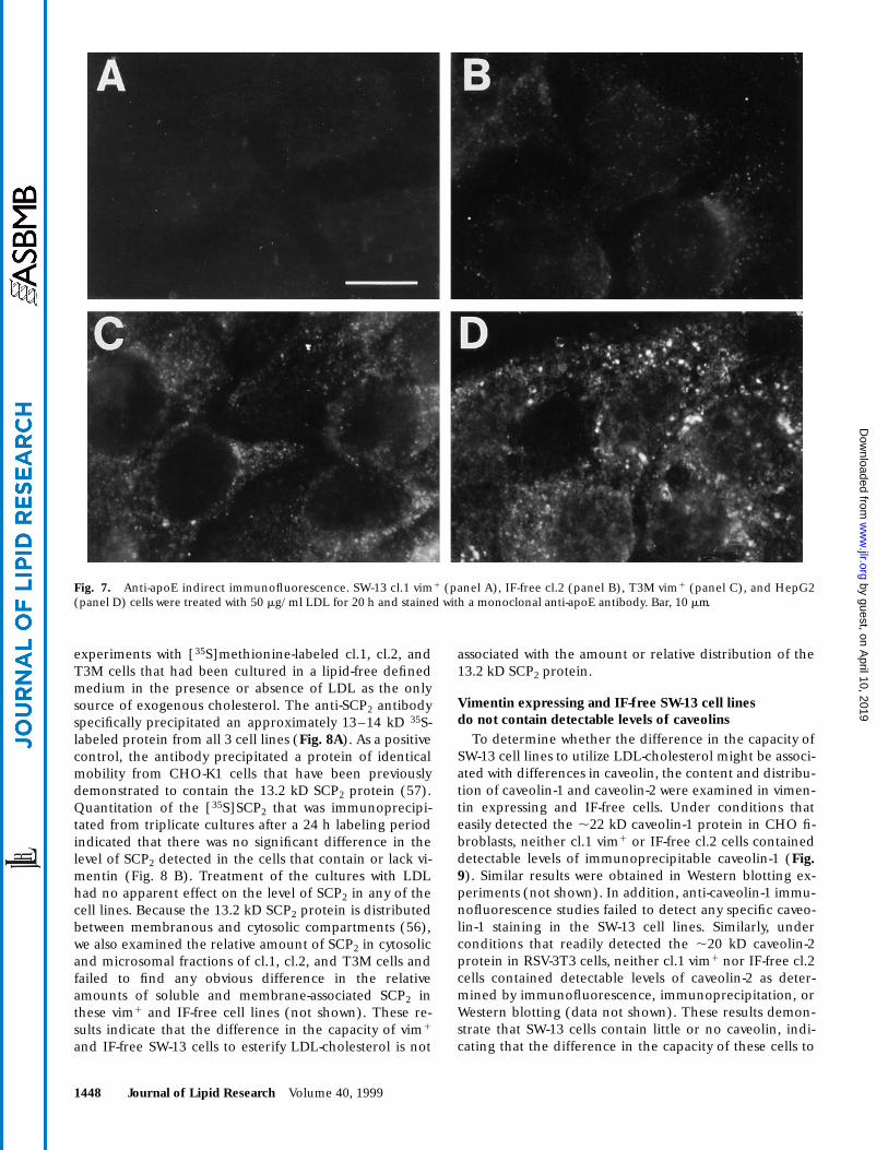
ences in apoE secretion between SW-13 cell lines mightbe due to differences in the intracellular distribution ofapoE, indirect immunofluorescence experiments werecarried out to visualize apoE in cl.1, cl.2, T3M, andHepG2 cells. As shown in Fig. 7, monoclonal anti-apoEstaining of the cl.1 vim+ cells that secrete little detectableapoE showed no detectable specific staining (Fig. 7A)compared with controls stained with the fluorescent con-jugate alone (data not shown). In contrast, the anti-apoEstaining pattern of IF-free cl.2 (Fig. 7B) and T3M vim1
cells (Fig. 7C), which secrete detectable amounts ofapoE, were characterized by both a faint perinuclear flu-orescence, which resembles the staining pattern of theGolgi apparatus in these cells, and a more prominentpunctate cytoplasmic fluorescence. As a positive control,anti-apoE immunofluorescence of HepG2 cells showedareas of intense punctate cytoplasmic staining. The in-tensity of the anti-apoE staining observed in these cells
was consistent with the amount of apoE detected byimmunoprecipitation from cell lysates (Figs. 1 and 5).The anti-apoE fluorescence shown in Fig. 7 was obtainedusing a monoclonal anti-human apoE (Bioreclamationclone 2034), and similar results were obtained with twoother anti-apoE monoclonal antibodies (Chemicon MAb1062 and Biodesign clone 3D12, data not shown). Theseresults indicated that the expression of vimentin was notassociated with any obvious difference in the intracellu-lar distribution of apoE.
Vimentin expressing and IF-free SW-13 cell lines contain similar levels of SCP2
Adrenal tissue has been shown to contain a significantlevel of SCP2 (56). To determine whether the differencein the capacity of these adrenal tumor cells to utilize LDL-cholesterol might be related to a difference in the level ofexpression of SCP2, we carried out immunoprecipitation
TABLE 2. Effect of agents that elevate cAMP on apoE secretion and cholesterolmetabolism in vim1 and vim2 SW-13 cells
Cell Line[35S]ApoESecreted
Free Cholesterol
CholesterylOleate
[14C]Oleate Incorporated into Cholesteryl Oleate
[14C]Oleate Incorporated into
Triglycerides
cpm/mgprotein
mg/mgprotein
mg/mgprotein
nmol/mgprotein
nmol/mgprotein
C1.1 Vim1
Untreated 26a 24.1 6 0.5 16.3 6 1.4 8.1 6 0.4 15.1 6 0.5IBMX/F 14 6 4 26.4 6 1.8 17.5 6 1.4 9.8 6 0.7 20.8 6 0.68br-cAMP 18 6 5 33.0 6 1.7 20.5 6 1.0 11.0 6 0.6 18.4 6 0.6
C1.2 IF-freeUntreated 96 6 15 28.3 6 1.1 8.8 6 0.1 4.2 6 0.4 20.8 6 1.6IBMX/F 56 6 8 33.1 6 6.2 15.9 6 2.8 7.2 6 1.2 29.8 6 5.18br-cAMP 99 6 10 33.8 6 3.5 10.0 6 1.1 5.9 6 1.2 23.5 6 3.9
Unless otherwise indicated, values represent the average of triplicate samples 6 SE. Cells were treated with 1mm IBMX and 10 mm forskolin or 1 mm 8 br-cAMP in medium containing 50 mg/ml LDL and 100 mCi/ml [35S]methionine for 20 h. [14C]oleate BSA (0.5 mCi/ml) was then added and the cells were incubated an additional 4 h.Radiolabeled apoE was immunoprecipitated from the culture medium and cholesterol and cholesteryl esters wereextracted from the monolayers for HPLC analysis as described in Methods.
a Duplicate samples.
TABLE 3. Effect of TPA and chelerythrine on apoE secretion and cholesterolmetabolism in vim1 and vim2 SW-13 cells
Cell Line[35S]ApoE Secreted
Free Cholesterol
Cholesteryl Oleate
[14C]Oleate Incorporated into Cholesteryl Oleate
[14C]OleateIncorporated into
Triglycerides
cpm/mg protein
mg/mg protein
mg/mgprotein
nmol/mgprotein
nmol/mgprotein
C1.1 Vim1
Untreated 9 6 3 63.1 6 12.1 42.6 6 2.2 19.2 6 2.5 32.0 6 4.0TPA 22 6 13 67.1 6 0.8 47.4 6 5.9 25.2 6 2.3 44.4 6 1.5TPA 1 Che 23 6 12 69.9 6 8.3 38.3 6 3.0 25.2 6 1.3 47.8 6 0.9
C1.2 IF-freeUntreated 245 6 18 47.9 6 5.1 17.1 6 0.8 12.6 6 0.5 47.7 6 7.1TPA 211 6 22 38.9 6 2.3 13.6 6 0.6 11.5 6 0.8 50.7 6 0.5TPA 1 Che 207 6 19 43.7 6 4.3 15.4 6 1.6 12.5 6 1.0 54.3 6 3.4
Values represent the average of triplicate samples 6 SE. Cells were treated with 0.2 mm TPA or 0.2 mm TPA and10 mm chelerythrine (Che) in medium containing 50 mg/ml LDL and 100 mCi/ml [35S]methionine for 20 h.[14C]oleate BSA (0.5 mCi/ml) was then added and the cells were incubated an additional 4 h. Radiolabeled apoEwas immunoprecipitated from the culture medium and cholesterol and cholesteryl esters were extracted from themonolayers for HPLC analysis as described in Methods.
by guest, on April 10, 2019
ww
w.jlr.org
Dow
nloaded from
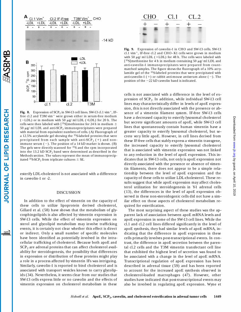
1448 Journal of Lipid Research Volume 40, 1999
experiments with [35S]methionine-labeled cl.1, cl.2, andT3M cells that had been cultured in a lipid-free definedmedium in the presence or absence of LDL as the onlysource of exogenous cholesterol. The anti-SCP2 antibodyspecifically precipitated an approximately 13–14 kD 35S-labeled protein from all 3 cell lines (Fig. 8A). As a positivecontrol, the antibody precipitated a protein of identicalmobility from CHO-K1 cells that have been previouslydemonstrated to contain the 13.2 kD SCP2 protein (57).Quantitation of the [35S]SCP2 that was immunoprecipi-tated from triplicate cultures after a 24 h labeling periodindicated that there was no significant difference in thelevel of SCP2 detected in the cells that contain or lack vi-mentin (Fig. 8 B). Treatment of the cultures with LDLhad no apparent effect on the level of SCP2 in any of thecell lines. Because the 13.2 kD SCP2 protein is distributedbetween membranous and cytosolic compartments (56),we also examined the relative amount of SCP2 in cytosolicand microsomal fractions of cl.1, cl.2, and T3M cells andfailed to find any obvious difference in the relativeamounts of soluble and membrane-associated SCP2 inthese vim1 and IF-free cell lines (not shown). These re-sults indicate that the difference in the capacity of vim1
and IF-free SW-13 cells to esterify LDL-cholesterol is not
associated with the amount or relative distribution of the13.2 kD SCP2 protein.
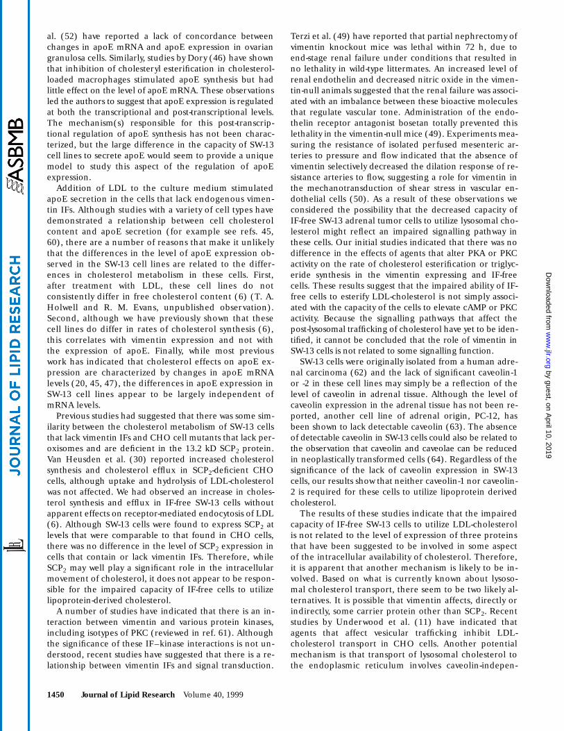
Vimentin expressing and IF-free SW-13 cell lines do not contain detectable levels of caveolins
To determine whether the difference in the capacity ofSW-13 cell lines to utilize LDL-cholesterol might be associ-ated with differences in caveolin, the content and distribu-tion of caveolin-1 and caveolin-2 were examined in vimen-tin expressing and IF-free cells. Under conditions thateasily detected the ,22 kD caveolin-1 protein in CHO fi-broblasts, neither cl.1 vim1 or IF-free cl.2 cells containeddetectable levels of immunoprecipitable caveolin-1 (Fig.9). Similar results were obtained in Western blotting ex-periments (not shown). In addition, anti-caveolin-1 immu-nofluorescence studies failed to detect any specific caveo-lin-1 staining in the SW-13 cell lines. Similarly, underconditions that readily detected the ,20 kD caveolin-2protein in RSV-3T3 cells, neither cl.1 vim1 nor IF-free cl.2cells contained detectable levels of caveolin-2 as deter-mined by immunofluorescence, immunoprecipitation, orWestern blotting (data not shown). These results demon-strate that SW-13 cells contain little or no caveolin, indi-cating that the difference in the capacity of these cells to
Fig. 7. Anti-apoE indirect immunofluorescence. SW-13 cl.1 vim1 (panel A), IF-free cl.2 (panel B), T3M vim1 (panel C), and HepG2(panel D) cells were treated with 50 mg/ml LDL for 20 h and stained with a monoclonal anti-apoE antibody. Bar, 10 mm.
by guest, on April 10, 2019
ww
w.jlr.org
Dow
nloaded from
Holwell et al. ApoE, SCP2, caveolin, and cholesterol esterification in adrenal tumor cells 1449
esterify LDL-cholesterol is not associated with a differencein caveolin-1 or -2.
DISCUSSION
In addition to the effect of vimentin on the capacity ofthese cells to utilize lipoprotein derived cholesterol,Gillard et al. (58) have shown that the metabolism of gly-cosphingolipids is also affected by vimentin expression inSW-13 cells. While the effect of vimentin expression onsterol and glycolipid metabolism may involve traffickingevents, it is certainly not clear whether this effect is director indirect. Only a small number of specific moleculeshave been identified as potentially involved in the intra-cellular trafficking of cholesterol. Because both apoE andSCP2 are adrenal proteins that can affect cholesterol avail-ability for steroidogenesis, the possibility that differencesin expression or distribution of these proteins might playa role in a process affected by vimentin IFs was intriguing.Similarly, caveolin-1 is reported to bind cholesterol and isassociated with transport vesicles known to carry glycolip-ids (34). Nevertheless, it seems clear from our studies thatSW-13 cells express little or no caveolin and the effects ofvimentin expression on cholesterol metabolism in these
cells is not associated with a difference in the level of ex-pression of SCP2. In addition, while individual SW-13 celllines may characteristically differ in levels of apoE expres-sion, this is not directly associated with the presence or ab-sence of a vimentin filament system. IF-free SW-13 cellshave a decreased capacity to esterify lysosomal cholesterolbut secrete significant amounts of apoE, while SW-13 celllines that spontaneously contain human vimentin have agreater capacity to esterify lysosomal cholesterol, but se-crete very little apoE. However, in cell lines derived fromthese IF-free cells that stably express a vimentin transgene,the increased capacity to esterify lysosomal cholesterolthat is associated with vimentin expression was not linkedto any reduction in the level of apoE expression. This in-dicates that in SW-13 cells, not only is apoE expression notdirectly associated with the presence or absence of vimen-tin filaments, there does not appear to be a simple rela-tionship between the level of apoE expression and thecapacity of these cells to utilize LDL-cholesterol. These re-sults suggest that while apoE expression may affect choles-terol utilization for steroidogenesis in Y-1 adrenal cells(13), the differences in the level of apoE expression ob-served in these non-steroidogenic cells did not have a sim-ilar effect on those aspects of cholesterol metabolism re-quired for esterification.
The most surprising aspect of these studies was the ap-parent lack of association between apoE mRNA levels andapoE expression in some of the SW-13 cell lines. While thecl.1 and cl.2 cell lines differed significantly in the level ofapoE synthesis, they had similar levels of apoE mRNA, in-dicating that the difference in apoE expression in thesecells primarily involves post-transcriptional events. In con-trast, the difference in apoE secretion between the paren-tal cl.2 cells and the T3M vimentin transfectant cell linethat exhibited the highest level of secretion was found tobe associated with a change in the level of apoE mRNA.Transcriptional regulation of apoE expression has beendescribed in adrenal tissue (59) and has been reportedto account for the increased apoE synthesis observed incholesterol-loaded macrophages (47). However, otherstudies have indicated that post-transcriptional events mayalso be involved in regulating apoE expression. Wyne et
Fig. 8. Expression of SCP2 in SW-13 cell lines. SW-13 cl.1 vim1, IF-free cl.2 and T3M vim1 were grown either in serum-free medium(2LDL) or in medium with 50 mg/ml LDL (+LDL) for 20 h. Thecells were then labeled with [35S]methionine for 24 h in medium 650 mg/ml LDL and anti-SCP2 immunoprecipitates were preparedwith material from equivalent numbers of cells. (A) Fluorograph ofa 12.5% acrylamide gel showing the 35S-labeled proteins that wereprecipitated from each sample with anti-SCP2 (1) and non-immune serum (2). The position of a 14 kD marker is shown. (B)The gels were directly scanned for 35S and the cpm incorporatedinto the 13.2 kD SCP2 band were determined as described in theMethods section. The values represent the mean of immunoprecip-itated 35S-SCP2 from triplicate cultures 6 SE.
Fig. 9. Expression of caveolin-1 in CHO and SW-13 cells. SW-13cl.1 vim1, IF-free cl.2 and CHO- K1 cells were grown in mediumwith 50 mg/ml LDL (1LDL) for 48 h. The cells were labeled with[35S]methionine for 4 h in medium containing 50 mg/ml LDL andanti-caveolin-1 immunoprecipitates were prepared from count-matched samples. The figure shows the fluorograph of a 10% acry-lamide gel of the 35S-labeled proteins that were precipitated withanti-caveolin-1 (1) or rabbit anti-mouse antiserum alone (2). Theposition of the ,22 kD caveolin band is indicated.
by guest, on April 10, 2019
ww
w.jlr.org
Dow
nloaded from
1450 Journal of Lipid Research Volume 40, 1999
al. (52) have reported a lack of concordance betweenchanges in apoE mRNA and apoE expression in ovariangranulosa cells. Similarly, studies by Dory (46) have shownthat inhibition of cholesteryl esterification in cholesterol-loaded macrophages stimulated apoE synthesis but hadlittle effect on the level of apoE mRNA. These observationsled the authors to suggest that apoE expression is regulatedat both the transcriptional and post-transcriptional levels.The mechanism(s) responsible for this post-transcrip-tional regulation of apoE synthesis has not been charac-terized, but the large difference in the capacity of SW-13cell lines to secrete apoE would seem to provide a uniquemodel to study this aspect of the regulation of apoEexpression.
Addition of LDL to the culture medium stimulatedapoE secretion in the cells that lack endogenous vimen-tin IFs. Although studies with a variety of cell types havedemonstrated a relationship between cell cholesterolcontent and apoE secretion (for example see refs. 45,60), there are a number of reasons that make it unlikelythat the differences in the level of apoE expression ob-served in the SW-13 cell lines are related to the differ-ences in cholesterol metabolism in these cells. First,after treatment with LDL, these cell lines do notconsistently differ in free cholesterol content (6) (T. A.Holwell and R. M. Evans, unpublished observation).Second, although we have previously shown that thesecell lines do differ in rates of cholesterol synthesis (6),this correlates with vimentin expression and not withthe expression of apoE. Finally, while most previouswork has indicated that cholesterol effects on apoE ex-pression are characterized by changes in apoE mRNAlevels (20, 45, 47), the differences in apoE expression inSW-13 cell lines appear to be largely independent ofmRNA levels.
Previous studies had suggested that there was some sim-ilarity between the cholesterol metabolism of SW-13 cellsthat lack vimentin IFs and CHO cell mutants that lack per-oxisomes and are deficient in the 13.2 kD SCP2 protein.Van Heusden et al. (30) reported increased cholesterolsynthesis and cholesterol efflux in SCP2-deficient CHOcells, although uptake and hydrolysis of LDL-cholesterolwas not affected. We had observed an increase in choles-terol synthesis and efflux in IF-free SW-13 cells withoutapparent effects on receptor-mediated endocytosis of LDL(6). Although SW-13 cells were found to express SCP2 atlevels that were comparable to that found in CHO cells,there was no difference in the level of SCP2 expression incells that contain or lack vimentin IFs. Therefore, whileSCP2 may well play a significant role in the intracellularmovement of cholesterol, it does not appear to be respon-sible for the impaired capacity of IF-free cells to utilizelipoprotein-derived cholesterol.
A number of studies have indicated that there is an in-teraction between vimentin and various protein kinases,including isotypes of PKC (reviewed in ref. 61). Althoughthe significance of these IF–kinase interactions is not un-derstood, recent studies have suggested that there is a re-lationship between vimentin IFs and signal transduction.
Terzi et al. (49) have reported that partial nephrectomy ofvimentin knockout mice was lethal within 72 h, due toend-stage renal failure under conditions that resulted inno lethality in wild-type littermates. An increased level ofrenal endothelin and decreased nitric oxide in the vimen-tin-null animals suggested that the renal failure was associ-ated with an imbalance between these bioactive moleculesthat regulate vascular tone. Administration of the endo-thelin receptor antagonist bosetan totally prevented thislethality in the vimentin-null mice (49). Experiments mea-suring the resistance of isolated perfused mesenteric ar-teries to pressure and flow indicated that the absence ofvimentin selectively decreased the dilation response of re-sistance arteries to flow, suggesting a role for vimentin inthe mechanotransduction of shear stress in vascular en-dothelial cells (50). As a result of these observations weconsidered the possibility that the decreased capacity ofIF-free SW-13 adrenal tumor cells to utilize lysosomal cho-lesterol might reflect an impaired signalling pathway inthese cells. Our initial studies indicated that there was nodifference in the effects of agents that alter PKA or PKCactivity on the rate of cholesterol esterification or triglyc-eride synthesis in the vimentin expressing and IF-freecells. These results suggest that the impaired ability of IF-free cells to esterify LDL-cholesterol is not simply associ-ated with the capacity of the cells to elevate cAMP or PKCactivity. Because the signalling pathways that affect thepost-lysosomal trafficking of cholesterol have yet to be iden-tified, it cannot be concluded that the role of vimentin inSW-13 cells is not related to some signalling function.
SW-13 cells were originally isolated from a human adre-nal carcinoma (62) and the lack of significant caveolin-1or -2 in these cell lines may simply be a reflection of thelevel of caveolin in adrenal tissue. Although the level ofcaveolin expression in the adrenal tissue has not been re-ported, another cell line of adrenal origin, PC-12, hasbeen shown to lack detectable caveolin (63). The absenceof detectable caveolin in SW-13 cells could also be related tothe observation that caveolin and caveolae can be reducedin neoplastically transformed cells (64). Regardless of thesignificance of the lack of caveolin expression in SW-13cells, our results show that neither caveolin-1 nor caveolin-2 is required for these cells to utilize lipoprotein derivedcholesterol.
The results of these studies indicate that the impairedcapacity of IF-free SW-13 cells to utilize LDL-cholesterolis not related to the level of expression of three proteinsthat have been suggested to be involved in some aspectof the intracellular availability of cholesterol. Therefore,it is apparent that another mechanism is likely to be in-volved. Based on what is currently known about lysoso-mal cholesterol transport, there seem to be two likely al-ternatives. It is possible that vimentin affects, directly orindirectly, some carrier protein other than SCP2. Recentstudies by Underwood et al. (11) have indicated thatagents that affect vesicular trafficking inhibit LDL-cholesterol transport in CHO cells. Another potentialmechanism is that transport of lysosomal cholesterol tothe endoplasmic reticulum involves caveolin-indepen-
by guest, on April 10, 2019
ww
w.jlr.org
Dow
nloaded from
Holwell et al. ApoE, SCP2, caveolin, and cholesterol esterification in adrenal tumor cells 1451
dent vesicular transport which is influenced by vimentinexpression.
We would like to thank Jeffrey Billheimer (DuPont) for theanti-SCP 2 antibody. This work was supported by NIH grantHL51850 (RME) and an American Heart Grant in Aid (MER).
Manuscript received 9 December 1998 and in revised form 26 March 1999.
REFERENCES
1. Faust, J. R., J. L. Goldstein, and M. S. Brown. 1977. Receptor-medi-ated uptake of low density lipoprotein and utilization of its choles-terol for steroid synthesis in cultured mouse adrenal cells. J. Biol.Chem. 252: 4861–4871.
2. Liscum, L., and K. W. Underwood. 1995. Intracellular cholesteroltransport and compartmentation. J. Biol. Chem. 270: 15443–15446.
3. Evans, R. M. 1994. Intermediate filaments and lipoprotein choles-terol. Trends Cell Biol. 4: 149–151.
4. Almahbobi, G., L. J. Williams, and P. F. Hall. 1992. Attachment ofsteroidogenic lipid droplets to intermediate filaments in adrenalcells. J. Cell Sci. 101: 383–393.
5. Almahbobi, G. 1995. Adhesion of intermediate filaments and lipiddroplets in adrenal cells studied by field emission scanning elec-tron microscopy. Cell Tissue Res. 281: 387–390.
6. Sarria, A. J., S. R. Panini, and R. M. Evans. 1992. A functional rolefor vimentin filaments in the metabolism of lipoprotein-derivedcholesterol in human SW-13 cells. J. Biol. Chem. 267: 19455–19463.
7. Schroeder, F., A. A. Frolov, E. J. Murphy, B. P. Atshaves, J. R. Jeffer-son, L. Pu, W. G. Wood, W. B. Foxworth, and A. B. Kier. 1996. Re-cent advances in membrane cholesterol domain dynamics andintracellular cholesterol trafficking. Proc. Soc. Exp. Biol. Med. 213:150–177.
8. Fielding, C. J., and P. E. Fielding. 1997. Intracellular cholesteroltransport. J. Lipid Res. 38: 1503–1521.
9. Pentchev, P. G., R. O. Brady, E. J. Blanchette-Mackie, M. T. Vanier,E. D. Carstea, C. C. Parker, E. Goldin, and C. F. Roff. 1994. TheNiemann-Pick C lesion and its relationship to the intracellular dis-tribution and utilization of LDL cholesterol. Biochim. Biophys. Acta.1225: 235–243.
10. Spillane, D. M., J. W. Reagan, N. J. Kennedy, D. L. Schneider, andT. Y. Chang. 1995. Translocation of both lysosomal LDL-derivedcholesterol and plasma membrane cholesterol to the endoplasmicreticulum for esterification may require common cellular factorsinvolved in cholesterol egress from the acidic compartments (lyso-somes endosomes). Biochim. Biophys. Acta. 1254: 283–294.
11. Underwood, K. W., N. L. Jacobs, A. Howley, and L. Liscum. 1998.Evidence for a cholesterol transport pathway from lysosomes toendoplasmic reticulum that is independent of the plasma mem-brane. J. Biol. Chem. 273: 4266–4274.
12. Liscum, L. 1990. Pharmacological inhibition of the intracellulartransport of low-density lipoprotein-derived cholesterol in Chinesehamster ovary cells. Biochim. Biophys. Acta. 1045: 40–48.
13. Reyland, M. E., J. T. Gwynne, P. Forgez, M. M. Prack, and D. L. Wil-liams. 1991. Expression of the human apolipoprotein E gene sup-presses steroidogenesis in mouse Y1 adrenal cells. Proc. Natl. Acad.Sci. USA. 88: 2375–2379.
14. Prack, M. M., G. H. Rothblat, S. K. Erickson, M. E. Reyland, andD. L. Williams. 1994. Apolipoprotein E expression in Y1 adrenalcells is associated with increased intracellular cholesterol contentand reduced free cholesterol efflux. Biochemistry. 33: 5049–5055.
15. Yamamoto, R., C. B. Kallen, G. O. Babalola, H. Rennert, J. T. Bill-heimer, and J. F. Strauss III. 1991. Cloning and expression of acDNA encoding human sterol carrier protein 2. Proc. Natl. Acad.Sci. USA. 88: 463–467.
16. Ossendorp, B. C., and K. W. A. Wirtz. 1993. The non-specific lipid-transfer protein (sterol carrier protein 2) and its relationship toperoxisomes. Biochimie. 75: 191–200.
17. Mahley, R. W. 1988. Apolipoprotein E: cholesterol transport pro-tein with expanding role in cell biology. Science. 240: 622–630.
18. Blue, M-L., D. L. Williams, S. Zucker, S. A. Khan, and C. B. Blum.1983. Apolipoprotein E synthesis in human kidney, adrenal gland,and liver. Proc. Natl. Acad. Sci. USA. 80: 283–287.
19. Newman, T. C., P. A. Dawson, L. L. Rudel, and D. L. Williams.1985. Quantitation of apolipoprotein E mRNA in the liver and pe-ripheral tissues of nonhuman primates. J. Biol. Chem. 260: 2452–2457.
20. Prack, M. M., M. Nicosia, D. L. Williams, and J. T. Gwynne. 1991.Relationship between apolipoprotein E mRNA expression and tis-sue cholesterol content in rat adrenal gland. J. Lipid Res. 32: 1611–1618.
21. Dyer, C. A., and L. K. Curtiss. 1988. Apoprotein E-rich high densitylipoproteins inhibit ovarian androgen synthesis. J. Biol. Chem. 263:10965–10973.
22. Swarnakar, S., M. E. Reyland, J. Deng, S. Azhar, and D. L. Williams.1998. Selective uptake of low density lipoprotein-cholesteryl esteris enhanced by inducible apolipoprotein E expression in culturedmouse adrenocortical cells. J. Biol. Chem. 273: 12140–12147.
23. Ohba, T., H. Rennert, S. M. Pfeifer, Z. He, R. Yammamoto, J. A.Holt, J. T. Billheimer, and J. F. Strauss. 1994. The structure of thehuman sterol carrier protein X/sterol carrier protein 2 gene(SCP2). Genomics. 24: 370–374.
24. van Amerongen, A., M. van Noort, J. R. C. M. van Beckhoven, F. F. G.Rommerts, J. Orly, and K. W. A. Wirtz. 1989. The subcellular distri-bution of the nonspecific lipid transfer protein (sterol carrier pro-tein 2) in rat liver and adrenal gland. Biochim. Biophys. Acta. 1001:243–248.
25. Keller, G. A., T. J. Scallen, D. Clarke, P. A. Maher, S. K. Krisans, andS. J. Singer. 1989. Subcellular localization of sterol carrier protein-2 in rat hepatocytes: its primary localization to peroxisomes. J. CellBiol. 108: 1353–1361.
26. Johnson, W. J., and M. P. Reinhart. 1994. Lack of requirement forsterol carrier protein-2 in the intracellular trafficking of lysosomalcholesterol. J. Lipid Res. 35: 563–573.
27. van Amerongen, A., R. A. Demel, J. Westerman, and K. W. A.Wirtz. 1989. Transfer of cholesterol and oxysterol derivatives bythe nonspecific lipid transfer protein (sterol carrier protein 2): astudy on its mode of action. Biochim. Biophys. Acta. 1004: 36–46.
28. Butko, P., T. J. Scallen, and F. Schroeder. 1990. Acidic phospholi-pids strikingly potentiate sterol carrier protein 2 mediated inter-membrane sterol transfer. Biochemistry. 29: 4070–4077.
29. Murphy, E. J., and F. Schroeder. 1997. Sterol carrier protein-2 me-diated cholesterol esterification in transfected L-cell fibroblasts.Biochim. Biophys. Acta. 1345: 283–292.
30. van Heusden, G. P. H., J. R. C. M. van Beckhoven, R. Thieringer,C. R. H. Raetz, and K. W. A. Wirtz. 1992. Increased cholesterol syn-thesis in Chinese hamster ovary cells deficient in peroxisomes. Bio-chim. Biophys. Acta. 1126: 81–87.
31. Anderson, R. G. W., B. A. Kamen, K. G. Rothberg, and S. W. Lacey.1992. Potocytosis: sequestration and transport of small moleculesby caveolae. Science. 255: 410–411.
32. Parton, R. G., B. Joggerst, and K. Simons. 1994. Regulated inter-nalization of caveolae. J. Cell Biol. 127: 1199–1215.
33. Dehouck, B., L. Fenart, M-P. Dehouck, A. Pierce, G. Torpier, andR. Cecchelli. 1997. A new function for the LDL receptor:transcyto-sis of LDL across the blood-brain barrier. J. Cell Biol. 138: 877–889.
34. Murata, M., J. Peranen, R. Schreiner, F. Wieland, T. V. Kurzchalia,and K. Simons. 1995. VIP21/caveolin is a cholesterol-binding pro-tein. Proc. Natl. Acad. Sci. USA. 92: 10339–10343.
35. Kurzchalia, T. V., P. Dupree, R. G. Parton, R. Kellner, H. Virta, M.Lehnert, and K. Simons. 1992. VIP21, a 21-kD membrane proteinis an integral component of trans-Golgi-network-derived transportvesicles. J. Cell Biol. 118: 1003–1014.
36. Smart, E. J., Y. S. Ying, P. A. Conrad, and R. G. W. Anderson. 1994.Caveolin moves from caveolae to the Golgi apparatus in responseto cholesterol oxidation. J. Cell Biol. 127: 1185–1197.
37. Sarria, A. J., S. K. Nordeen, and R. M. Evans. 1990. Regulated ex-pression of vimentin cDNA in cells in the presence and absenceof a pre-existing vimentin filament network. J. Cell Biol. 111: 553–566.
38. Sarria, A. J., J. G. Lieber, S. K. Nordeen, and R. M. Evans. 1994.The presence or absence of a vimentin-type intermediate filamentnetwork affects the shape of the nucleus in human SW-13 cells.J. Cell Sci. 107: 1593–1607.
39. Goldstein, J. L., S. K. Basu, and M. S. Brown. 1983. Receptor-medi-ated endocytosis of low-density lipoprotein in cultured cells. MethodsEnzymol. 98: 241–260.
40. Laemmli, U. K. 1970. Cleavage of structural proteins during the as-sembly of the head of bacteriophage T4. Nature. 227: 680–685.
41. Vercaemst, R., A. Union, and M. Rosseneu. 1991. Separation and
by guest, on April 10, 2019
ww
w.jlr.org
Dow
nloaded from
1452 Journal of Lipid Research Volume 40, 1999
quantitation of free cholesterol and cholesterol esters in a macro-phage cell line by high performance liquid chromatography.J. Chromatogr. 494: 43–52.
42. Bradford, M. 1976. A rapid and sensitive method for the quantita-tion of microgram quantities of protein utilizing the principle ofprotein dye binding. Anal. Biochem. 72: 248–254.
43. Schliwa, M., and J. van Blerkom. 1981. Structural interaction ofcytoskeletal components. J. Cell Biol. 90: 222–235.
44. Zannis, V. I., J. L. Breslow, T. R. SanGiacomo, D. P. Aden, and B. B.Knowles. 1981. Characterization of the major apolipoproteins se-creted by two human hepatoma cell lines. Biochemistry. 20: 7089–7096.
45. Mazzone, T., and C. Reardon. 1994. Expression of heterologoushuman apolipoprotein E by J774 macrophages enhances choles-terol efflux to HDL3. J. Lipid Res. 35: 1345–1353.
46. Dory, L. 1989. Synthesis and secretion of apoE in thioglycolate-elicited mouse peritoneal macrophages: effect of cholesterolefflux. J. Lipid Res. 30: 809–816.
47. Mazzone, T., K. Basheeruddin, and C. Poulos. 1989. Regulation ofmacrophage apolipoprotein E gene expression by cholesterol. J.Lipid Res. 30: 1055–1064.
48. Smith, J. D., A. Melian, T. Leff, and J. Breslow. 1988. Expression ofthe human apolipoprotein E gene is regulated by multiple positiveand negative elements. J. Biol. Chem. 263: 8300–8308.
49. Terzi, F., D. Henrion, E. Colucci-Guyon, P. Federici, C. Babinet,B. I. Levy, P. Briand, and G. Friedlander. 1997. Reduction of renalmass is lethal in mice lacking vimentin. Role of endothelin-nitricoxide imbalance. J. Clin. Invest. 100: 1520–1528.
50. Henrion, D., F. Terzi, K. Matrougui, M. Duriez, C. M. Boulanger, E.Colucci-Guyon, C. Babinet, P. Briand, G. Friedlander, P. Poitevin,and B. I. Levy. 1997. Impaired flow-induced dilation in mesentericresistance arteries from mice lacking vimentin. J. Clin. Invest. 100:2909–2914.
51. Freeman, D. A. 1987. Cyclic AMP mediated modification of choles-terol traffic in Leydig tumor cells. J. Biol. Chem. 262: 13061–13068.
52. Wyne, K. L., J. R. Schreiber, A. L. Larsen, and G. S. Getz. 1989.Regulation of apolipoprotein E biosynthesis by cAMP and phorbolester in rat ovarian granulosa cells. J. Biol. Chem. 264: 981–989.
53. Werb, Z., R. Takemura, P. E. Stenberg, and D. F. Bainton. 1989. Di-rected exocytosis of secretory granules containing apolipoprotein E
to the adherent surface and basal vacuoles of macrophages spread-ing on immobile immune complexes. Am. J. Pathol. 134: 661–670.
54. Hamilton, R. L., J. S. Wong, L. S. S. Guo, S. Krisans, and R. J. Havel.1990. Apolipoprotein E localization in rat hepatocytes by immu-nogold labeling of cryothin sections. J. Lipid Res. 31: 1589–1603.
55. Nicosia, M., M. M. Prack, and D. L. Williams. 1992. Differentialregulation of apolipoprotein-E messenger RNA in Zona Fascicu-lata of rat adrenal gland determined by in situ hybridization. Mol.Endocrinol. 6: 288–298.
56. van Heusden, G. P. H., K. Bos, and K. W. A. Wirtz. 1990. The occur-rence of soluble and membrane-bound non-specific lipid transferprotein (sterol carrier protein 2) in rat tissues. Biochim. Biophys.Acta. 1046: 315–321.
57. van Heusden, G. P. H., K. Bos, C. R. H. Raetz, and K. W. A. Wirtz.1990. Chinese hamster ovary cells deficient in peroxisomes lackthe nonspecific lipid transfer protein (sterol carrier protein 2). J.Biol. Chem. 265: 4105–4110.
58. Gillard, B. K., R. Clement, E. Colucci-Guyon, C. Babinet, G.Schwarzmann, T. Taki, T. Kasama, and D. M. Marcus. 1998. De-creased synthesis of glycosphingolipids in cells lacking vimentinintermediate filaments. Exp. Cell Res. 242: 561–572.
59. Dahan, S., J. P. Ahluwalia, L. Wong, B. I. Posner, and J. J. M. Ber-geron. 1994. Concentration of intracellular hepatic apolipopro-tein E in Golgi apparatus saccular distensions and endosomes. J.Cell Biol. 127: 1859–1869.
60. Basu, S. K., J. L. Goldstein, and M. S. Brown. 1983. Independentpathways for secretion of cholesterol and apolipoprotein E bymacrophages. Science. 219: 871–873.
61. Evans, R. M. 1998. Vimentin: the conundrum of the intermediatefilament gene family. Bioessays. 20: 79–86.
62. Leibovitz, A., W. B. McCombs, D. Johnston, C. E. McCoy, and J. C.Stinson. 1973. New human cancer cell culture lines. I. SW-13, small-cell carcinoma of the adrenal cortex. J. Natl. Cancer Inst. 51: 691–697.
63. Scherer, P. E., R. Y. Lewis, D. Volontes, J. A. Engelman, F. Galbiati,J. Couet, D. S. Kohtz, E. van Donselaar, P. Peters, and M. P. Lisanti.1997. Cell-type and tissue specific expression of caveolin-2. J. Biol.Chem. 272: 29337–29346.
64. Koleske, A. J., D. Baltimore, and M. P. Lisanti. 1995. Reduction ofcaveolin and caveolae in oncogenically transformed cells. Proc.Natl. Acad. Sci. USA. 92: 1381–1385.
by guest, on April 10, 2019
ww
w.jlr.org
Dow
nloaded from