Viktor's Notes – Spondylosisneurosurgeryresident.net/Spin. Spinal Disorders/Spin13... · Web...
15
SPONDYLOSIS Spin13 (1) Spondylosis Last updated: September 5, 2017 ETIOPATHOPHYSIOLOGY....................................................1 Mechanisms of damage / irritation to neural structures..........2 EPIDEMIOLOGY..........................................................2 Cervical Spondylosis............................................ 3 CLINICAL FEATURES..................................................... 3 Cervical Spondylosis............................................ 3 Lumbar Spondylosis.............................................. 4 DIAGNOSIS............................................................5 PLAIN X-RAY........................................................ 5 Cervical Spondylosis............................................ 5 Lumbar Spondylosis.............................................. 5 MRI............................................................... 5 Cervical Spondylosis............................................ 5 Lumbar Spondylosis.............................................. 7 CT MYELOGRAPHY...................................................... 7 Cervical Spondylosis............................................ 7 Lumbar Spondylosis.............................................. 8 DIFFERENTIAL DIAGNOSIS................................................. 8 CONSERVATIVE TREATMENT................................................. 8 SURGICAL TREATMENT – CERVICAL SPONDYLOSIS.................................9 SURGICAL TREATMENT – LUMBAR SPONDYLOSIS..................................9 PROGNOSIS............................................................9 Cervical Spondylosis............................................ 9 SPECIAL ENTITIES......................................................9 DIFFUSE IDIOPATHIC SKELETAL HYPEROSTOSIS (S. DIFFUSE IDIOPATHIC SKELETAL HYPEROSTOSIS, FORESTIER DISEASE).......................................9 OSSIFICATION OF POSTERIOR LONGITUDINAL LIGAMENT (OPLL).....................9 SPONDYLOSIS : A) ankylosis of vertebra B) any degenerative spinal lesion. C) progressive degeneration of intervertebral discs, leading to proliferative changes of surrounding structures CSM – cervical spondylotic myelopathy. ETIOPATHOPHYSIOLOGY Degenerative changes of spine universally accompany aging ! see p. Spin11 >> Most are sequelae of intervertebral disc degeneration - LOSS OF DISC HEIGHT causes: 1) narrowed intervertebral foramina. 2) increased load on vertebral bodies → reactive vertebral changes → osteophytes. most osteophytes are anterior or lateral in projection. osteophytes reduce range of movement and may result in spontaneous fusion.
Transcript of Viktor's Notes – Spondylosisneurosurgeryresident.net/Spin. Spinal Disorders/Spin13... · Web...

SPONDYLOSIS Spin13 (1)
SpondylosisLast updated: September 5, 2017
ETIOPATHOPHYSIOLOGY.........................................................................................................................1
Mechanisms of damage / irritation to neural structures....................................................................2
EPIDEMIOLOGY........................................................................................................................................2
Cervical Spondylosis.........................................................................................................................3
CLINICAL FEATURES................................................................................................................................3
Cervical Spondylosis.........................................................................................................................3
Lumbar Spondylosis.........................................................................................................................4
DIAGNOSIS................................................................................................................................................5
PLAIN X-RAY..........................................................................................................................................5
Cervical Spondylosis.........................................................................................................................5
Lumbar Spondylosis.........................................................................................................................5
MRI........................................................................................................................................................5
Cervical Spondylosis.........................................................................................................................5
Lumbar Spondylosis.........................................................................................................................7
CT MYELOGRAPHY.................................................................................................................................7
Cervical Spondylosis.........................................................................................................................7
Lumbar Spondylosis.........................................................................................................................8
DIFFERENTIAL DIAGNOSIS......................................................................................................................8
CONSERVATIVE TREATMENT..................................................................................................................8
SURGICAL TREATMENT – CERVICAL SPONDYLOSIS..............................................................................9
SURGICAL TREATMENT – LUMBAR SPONDYLOSIS................................................................................9
PROGNOSIS...............................................................................................................................................9
Cervical Spondylosis.........................................................................................................................9
SPECIAL ENTITIES....................................................................................................................................9
DIFFUSE IDIOPATHIC SKELETAL HYPEROSTOSIS (S. DIFFUSE IDIOPATHIC SKELETAL HYPEROSTOSIS, FORESTIER DISEASE)..............................................................................................................................9
OSSIFICATION OF POSTERIOR LONGITUDINAL LIGAMENT (OPLL).........................................................9
SPONDYLOSIS :
A) ankylosis of vertebra
B) any degenerative spinal lesion.
C) progressive degeneration of intervertebral discs, leading to proliferative changes of surrounding structures
CSM – cervical spondylotic myelopathy.
ETIOPATHOPHYSIOLOGY
Degenerative changes of spine universally accompany aging!
see p. Spin11 >>
Most are sequelae of intervertebral disc degeneration - LOSS OF DISC HEIGHT causes:
1) narrowed intervertebral foramina.
2) increased load on vertebral bodies → reactive vertebral changes → osteophytes.
most osteophytes are anterior or lateral in projection.
osteophytes reduce range of movement and may result in spontaneous fusion.
3) increased load on facet & uncovertebral (Luschka) joints → hypertrophic osteoarthritic changes.
remodelling of articular surfaces → instability → forward slippage of upper on lower vertebra.
synovial cysts are frequently solid (cartilaginous or myxomatous) - can be confused with migratory disc fragments or intraspinal tumor; attachment to joint space is characteristic.
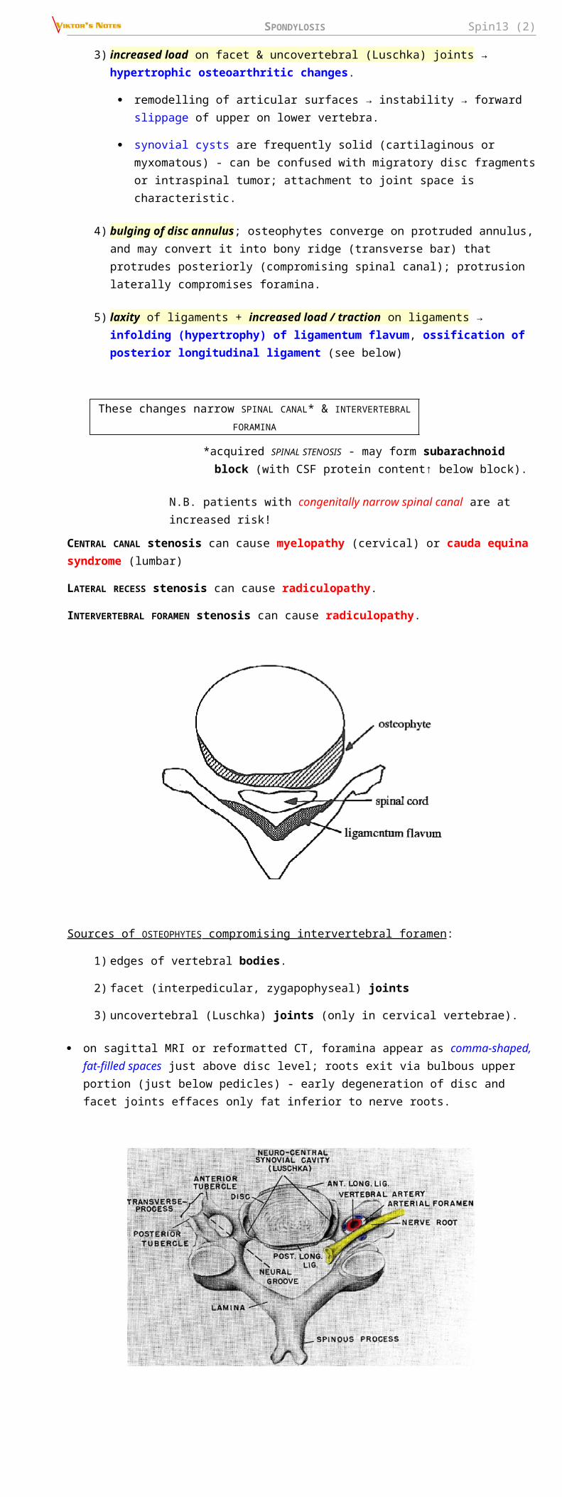
4) bulging of disc annulus; osteophytes converge on protruded annulus, and may convert it into bony ridge (transverse bar) that protrudes posteriorly (compromising spinal canal); protrusion laterally compromises foramina.
SPONDYLOSIS Spin13 (2)
5) laxity of ligaments + increased load / traction on ligaments → infolding (hypertrophy) of ligamentum flavum, ossification of posterior longitudinal ligament (see below)
These changes narrow SPINAL CANAL* & INTERVERTEBRAL FORAMINA
*acquired SPINAL STENOSIS - may form subarachnoid block (with CSF protein content↑ below block).
N.B. patients with congenitally narrow spinal canal are at increased risk!
CENTRAL CANAL stenosis can cause myelopathy (cervical) or cauda equina syndrome (lumbar)
LATERAL RECESS stenosis can cause radiculopathy.
INTERVERTEBRAL FORAMEN stenosis can cause radiculopathy.
Sources of OSTEOPHYTES compromising intervertebral foramen:
1) edges of vertebral bodies.
2) facet (interpedicular, zygapophyseal) joints
3) uncovertebral (Luschka) joints (only in cervical vertebrae).
on sagittal MRI or reformatted CT, foramina appear as comma-shaped, fat-filled spaces just above disc level; roots exit via bulbous upper portion (just below pedicles) - early degeneration of disc and facet joints effaces only fat inferior to nerve roots.

SPONDYLOSIS Spin13 (3)
MECHANISMS of damage / irritation to neural structures
A) STATIC mechanical factor - direct compression (by stenosis of spinal canal & foramina) → distorted / flattened spinal cord (spondylotic bars may leave deep indentations on ventral surface of spinal cord).
compression is usually intermittent (or intermittently accentuated by neck movement).
cord substance is relatively inelastic - retains impression of impinging agent even when contact is removed.
cord damage is sustained only when sagittal diameter of cord is reduced by > 50%.
in thoracic region, far greater compression is tolerated (because of reduced mobility of this part of spine) - cord becomes focally molded around calcified masses (which can occupy 60% of spinal canal) with no clinical abnormality.
H: decompressive surgery
B) DYNAMIC mechanical factor - rubbing* (repeated trauma) on protruding structures (that may not themselves be severely compressive) → demyelination of spinal columns.
*cephalad / caudal cord movement in course of normal flexion and extension, traction by dentate ligaments
posterior columns demyelinate above compression; corticospinal tracts - below compression.
H: surgical fusion
C) ISCHEMIA secondary to compression - arterial deprivation and/or venous stasis → ischemic neuronal loss in central gray matter (sometimes syringomyelia can be found); root sleeves may be thickened and rootlets adherent.
subluxation of zygapophyseal joints may compress vertebral arteries.
EPIDEMIOLOGYRISK FACTORS:
1) aging - major risk factor!!!
2) prior trauma (usually no history of significant trauma)
3) prior disc herniation
4) cervical dystonia
5) congenital spinal anomalies
6) systemic arthritic disorders
7) obesity
8) occupations that expose to vibration

SPONDYLOSIS Spin13 (4)
9) heavy labor
Spondylotic changes increase with advancing age:
age 20-30 yrs – 5-10% have changes on radiographs
N.B. spondylosis can begin in persons as young as 20 years!
age 45 yrs – 50%
age 59 yrs – 85% men (70% women)
age 70 yrs – 97% men (93% women).
vs. disc herniations – highest incidence in 30-50 yrs.
CERVICAL SPONDYLOSIS
PREVALENCE is rising.
most common cause of spinal cord dysfunction in patients > 55 yrs.
most common cause of nontraumatic spastic paraparesis / quadriparesis.
– in one series, 23.6 % of patients with nontraumatic paraparesis / quadriparesis had CSM.
CLINICAL FEATURESSpondylotic changes become clinically important when they cause local pain and / or neurological dysfunction (MYELOPATHY, RADICULOPATHIES). see p. Spin11 >>
patients can have either myelopathy or radiculopathy, or combination of both.
lumbar spondylosis cannot cause myelopathy; instead, cauda equina can be damaged!
ONSET insidious, COURSE slowly progressive
Spondylosis clinically ≈ disc herniation with protracted course. further see PROGNOSIS >>
CERVICAL SPONDYLOSIS
N.B. occasionally patient presents with catastrophic onset of quadriparesis or paraparesis after neck trauma (esp. fall).
Axial neck pain ± myelopathy and / or radiculopathy
1. NECK PAIN (present in 90% cases)
neck pain is axial; root pain is uncommon.
may be prominent (exacerbated by any movements*).
*vs. disc herniation – pain during extension and lateral flexion toward painful side (side of herniation)
some limitation of neck mobility.
± Lhermitte’s sign.
anterior osteophytes may produce dysphagia.
2. ARMS (depending on level of myelopathy and degree of root involvement):
1) sensory loss may follow simple radicular pattern or, more commonly, patchy distribution (multiple root and cord involvement!) often in “glove” distribution!
2) weakness:
a) LMN with fasciculations and atrophy (esp. in hands)
b) UMN with brisk reflexes* - less severe than in legs.
*absence of jaw jerk ↑ helps to differentiate from general hyperreflexia
clumsiness with fine motor skills (buttoning, writing)
slow, stiff opening and closing of fist.
inverted radial reflex (pathognomonic): flexion of fingers in response to brachioradialis reflex.
“finger escape” sign: with eyes closed and fingers kept adducted, 5th finger begins to abduct.
sensory level can be detected in ≈ 40% patients.
3. LEGS :
1) spastic weakness (proximal) with clonus, positive Babinski & Hoffmann (“dynamic Hoffmann’s sign” more sensitive)
2) sensory loss (esp. vibratory and position sense; occasionally pinprick sensation) & paresthesias (almost always below ankle)
coughing or straining exacerbates leg weakness.
elderly patient may present for gait problems or falls (rather than as direct complaint).
bowel / bladder dysfunction are uncommon?

SPONDYLOSIS Spin13 (5)
Slowly progressive spastic gait disorder + hand numbness and loss of fine motor control in patient > 50 yrs = CERVICAL SPONDYLOTIC MYELOPATHY until proven otherwise.
SYNDROMES
1. Motor syndrome: corticospinal tract and anterior horns with minimal or no sensory deficit.
2. Central cord syndrome: motor and sensory deficit (upper extremities > lower extremities).
3. Brown-Sequard syndrome (in asymmetric narrowing of spinal canal).
4. Brachialgia and cord syndrome: radicular upper extremity pain with LMN weakness, some associated long tract involvement (motor and/or sensory).
5. Transverse syndrome (most frequent “end-stage” syndrome): corticospinal and spinothalamic tracts, posterior columns, ± segmental anterior horns.
QUANTIFICATION
1. Hand dynamometry
2. Nine hole peg test
3. 30-meter walk test - measuring time and number of steps (objective, reproducible)
4. NURICK disability score
5. Modified JAPANESE ORTHOPAEDIC ASSOCIATION functional score (mJOA)
≥ 15 – mild

SPONDYLOSIS Spin13 (6)
12-14 – moderate
< 12 – severe
6. Neck Disability Index (NDI)
7. Berg Balance Scale (BBS)
8. Quality of life (nonspecific for CSM) - Medical Outcomes Study Short Form-36 (SF36v2)
LUMBAR SPONDYLOSIS
spinal canal stenosis is usually confined to one or two lumbar levels:
a) most common syndrome - isolated L4-5 disorder with L5 radiculopathy (unilateral or bilateral);
b) L3-4 segment is affected less often (either alone or in combination with L4-5 stenosis);
c) other levels are rarely affected.
symptoms may be episodic.
Lumbar spondylosis usually produces no symptoms - when back or sciatic pains are complaints, lumbar spondylosis usually is unrelated finding!
1. BACK PAIN (present in > 50% cases) is not dominant symptom.
2. LUMBAR RADICULOPATHY
leg pain (bilateral or unilateral).
straight leg-raising is limited in few cases.
leg weakness is rare (many show weakness of isolated muscles)
urinary incontinence is rare.
characteristic symptom (almost all patients!) – PSEUDOCLAUDICATION ( s . NEUROGENIC INTERMITTENT CLAUDICATION) - unilateral or bilateral discomfort in buttock / thigh / leg on walking or prolonged standing (postural claudication).
patients use words “pain”, “numbness”, “weakness”', but there is often no objective sensory loss or focal muscle weakness.
discomfort is relieved within minutes by lying down, sitting*, or flexing at waist* (N.B. pain may persist in recumbency until spine is flexed).
discomfort persists if patient stops walking but does not flex spine**.
no loss of pulses**, no trophic skin changes in feet**.
PATHOGENESIS:
1) spine hyperextension (when walking) increases disc protrusion, causes infolding of ligamentum flavum, narrows spinal canal and foramina.
2) leg muscle exercise → ↑blood flow to lumbar cord → root vessels dilate but are confined by bony changes → compress roots.
3) root microvascular deficiency - activity-related increases in metabolic rate of nerve roots cannot be met.
*vs. disc herniation pain
**vs. vascular claudication
DIAGNOSISIt is very important to establish best possible correlations between clinical findings and imaging abnormalities - high rate of radiological spondylosis in asymptomatic populations!
Intervertebral foramen must be reduced < 30% of normal to cause root compression
other criteria: posterior disk height < 4 mm, foraminal height < 15 mm.
PLAIN X-RAY
(include oblique views for neural foramina!)
- show degenerative changes of bony elements, but do not reveal relationship of these to neural structures!
radiological features of osteoarthritis (if present) are identical to other synovial joints - joint space narrowing, subchondral sclerosis and cyst formation, osteophyte formation.
"vacuum phenomenon" - gas within apophyseal joint / intervertebral disc - pathognomonic for advanced degenerative process!

SPONDYLOSIS Spin13 (7)
CERVICAL SPONDYLOSIS
simple flexion - extension films (performed with care!) can demonstrate spinal instabilities (that are not apparent on MRI or CT myelography!).
Osteophytes at C5-6 interspace:
LUMBAR SPONDYLOSIS
A. Lateral osteophytes at each level but most marked at L2-3 and L3-4 with narrowing of disk space (esp. L2-3).
B. Narrowing and irregularity of disk spaces, large osteophytes anteriorly at L2-5.
C. Gas shadows (arrow).
MRI
- easiest noninvasive means of diagnosis! - can demonstrate dimensions of spinal canal and foramina + distortion of spinal cord and roots.
T1 & T2 – what gives compression – osteophytes vs. soft herniated disk (will desiccate in time → spontaneous improvement)
gadolinium enhancement – only to exclude alternative lesions.
CERVICAL SPONDYLOSIS
N.B. imaging must be high enough (to demonstrate craniocervical junction)!
Most important features:
1. CSF effacement (obliteration of subarachnoid space) & spinal cord deformation (compression)
2. Focal cord atrophy:
1) reduction in transverse CORD AREA (esp. ≤ 45 mm2)
2) reduction in sagittal CORD DIAMETER
Sagittal diameter* of cervical canal < 9-10 mm - cord compression is probably present.
*most severely compromised between posterior-inferior edge of vertebral body and anterior-superior edge of subjacent lamina.
combination of focal reduction in sagittal cord diameter by 50% + obliteration of posterior subarachnoid space ≈ clinical myelopathy.
widening of transverse cord diameter usually implies at least 50% reduction in sagittal diameter!
3. T2 signal↑ within cord substance - reflects cord damage (myelomalacia).
bright focal T2 signal mainly in central areas (on axial images - appearance of ”snake eyes”).

SPONDYLOSIS Spin13 (8)
frequently disappears after decompressive surgery with good outcome (but T2 signal↑ per se is not indication for surgery).
Cervical spondylosis, left C6 radiculopathy:
A. Sagittal T2-MRI - hypointense osteophyte which protrudes from C5-6 level into thecal sac, displacing spinal cord posteriorly (white arrow).
B. Axial MRI - high signal of right C5-6 intervertebral foramen contrasts with narrow high signal of left C5-6 intervertebral foramen produced by osteophytic spurring (arrows):
Focal spinal cord compression from single osteophyte at C3-4 level - dense calcification typical of segmental ossification of posterior longitudinal ligament (B. CT; A. T1-MRI):
Ossification of posterior longitudinal ligament (T2-MRI) - mild spinal cord compression by thickened posterior longitudinal ligament (white arrowheads) within spinal
Cervical spondylotic myelopathy with myelomalacia (T2-MRI): moderate compression of spinal cord at C3–4 level; focal increased signal in cord substance; on axial image -

SPONDYLOSIS Spin13 (9)
canal (black arrowhead): appearance of ‘snake eyes’ (black arrowheads):
LUMBAR SPONDYLOSIS
74-year-old man with neurogenic claudication - severe lumbar stenosis (T2-MRI): degenerative changes at multiple levels with severe spinal stenosis and crowding of cauda equina:
CT myelography
- used to answer any questions that remain after MRI.
Myelography in spinal cord compression has slight risk that existing myelopathy may worsen and become permanent!
MYELOMALACIA - intramedullary contrast penetration and retention (best shown on delayed postmyelography CT).
CERVICAL SPONDYLOSIS
Cervical foraminal stenosis (CT myelogram): with cutoff of right C6 root.
Cervical spondylotic myelopathy (CT myelography): spinal cord (arrowhead) is deformed and contrast medium has accumulated within it. Extensive cervical laminectomy 6 years earlier had produced no appreciable improvement:

SPONDYLOSIS Spin13 (10)
LUMBAR SPONDYLOSIS
High-grade lumbar L4-5 stenosis: A. Myelogram. B. Postmyelographic CT - circumferential stenosis (disc bulging, enlarged facets, ligamentum flavum hypertrophy).
DIFFERENTIAL DIAGNOSIS- particularly important when dealing with condition that is commonly present as asymptomatic radiological finding!
1. Multiple Sclerosis – younger age, fluctuating course, early bladder symptoms, visual complaints, mental status changes.
2. Amyotrophic Lateral Sclerosis – LMN signs are evident from beginning, but spasticity predominates in few; muscle atrophy and increased reflexes in same myotome strongly suggest ALS; bulbar symptoms or signs!!!; absent sensory loss!!!
5% ALS patients undergo cervical laminectomy!
3. Primary Lateral Sclerosis.
4. Subacute Combined Degeneration of Spinal Cord – deficits are often primarily sensory; hypersegmented PMN, macrocytic anemia.
5. Spinal AVM, spinal dural AV fistula (can cause myelopathy) – seen on MRI.
6. AIDS Myelopathy – most patients are young; ascending sensory disorder.
7. Tabes Dorsalis
8. HTLV-I Myelopathy (Tropical Spastic Paraparesis) – slowly progressive spastic paraparesis with early bladder involvement in patient from endemic region.
9. Familial (Hereditary) Spastic Paraplegia – autosomal dominant disorder.

SPONDYLOSIS Spin13 (11)
10. Syringomyelia - segmental loss of spinothalamic modalities.
11. Compressive Lesions (e.g. meningiomas, schwannomas, epidural abscess)
12. Compressive Lesions at Craniocervical Junction:
1) Chiari malformation
2) atlanto-occipital or atlanto-axial instability (e.g. in RA)
13. Normal pressure hydrocephalus
N.B. in young patients (< 40 yrs) tumors, spinal A-V malformations, and congenital anomalies are more common causes of neck pain than is cervical spondylosis!!!
CONSERVATIVE TREATMENT1. Immobilization:
a) cervical – firm cervical collar.
b) lumbar – absolute bed rest.
2. Heat, massage, cervical traction – see p. S20 >>
3. NSAIDs for pain.
4. Epidural steroid injections - for major radicular pain; questionable value for lumbar and cervical radiculopathies (in multiple studies).
Patients with cervical spondylosis are at increased risk of tetraplegia after minor trauma!
SURGICAL TREATMENT – CERVICAL SPONDYLOSISINDICATIONS
1) intractable radiculopathy (esp. motor)
2) if myelopathy progresses / remains severe* despite conservative measures.
N.B. surgery is for myelopathy (not for neck pain!)
*surgery is most effective when performed early (< 6 months symptom duration) for all degrees of CSM!
ACDF vs. PT in cervical radiculopathy
Engquist M “A 5- to 8-year randomized study on the treatment of cervical radiculopathy: anterior cervical decompression and fusion plus physiotherapy versus physiotherapy alone” J Neurosurg Spine. 2016 Aug 26:1-9
the aim of this study was to evaluate the 5-8-year outcome of ACDF combined with a structured PT program vs. the same PT program alone in patients with cervical radiculopathy.
patients were randomized to ACDF + PT (30 patients) or to PT alone (29 patients).
both treatment groups experienced significant improvement over baseline for all outcome measures but in some measures ACDF did better:
Improvement at 5-8 years ACDF + PT PT p value
Neck Disability Index [NDI] 21% (95% CI 14-28) 11% (95% CI 4-18) 0.03
neck pain VAS 39 mm (95% CI 26-53) 19 mm (95% CI 7-30) 0.01
arm pain VAS 33 mm (95% CI 18-49) 19 mm (95% CI 7-32) 0.1
health state EQ-5D questionnaire 0.29 (95% CI 0.13-0.45)
0.14 (95% CI 0.01-0.27)
0.12
patient global assessment - self-rating by patients - patients rated their symptoms as "better" or "much better"
93% 62% 0.005
VAS = visual analog scale
SURGICAL TREATMENT – LUMBAR SPONDYLOSISINDICATIONS
- pain / claudication / radiculopathy severe enough to impede quality of life despite conservative measures

SPONDYLOSIS Spin13 (12)
PROGNOSIS
CERVICAL SPONDYLOSIS
Natural course of CSM for any given individual is variable - precise prognostication is not possible
in 75% patients course is progressive (gradual or stepwise), although many (even severe cases) achieve static period and remain stable for many years (or even improve spontaneously*).
*60–70% fibrocartilaginous masses of discogenic origin can diminish in size or disappear completely over few weeks or months.
N.B. if osteophytes disappear, look for aortic aneurysm - can cause pressure erosions of adjacent vertebrae!
patients with spinal hypermobility are more likely to deteriorate without surgery.
surgery results :
25-75% patients improve;
5-50% patients worsen! (even adequately decompressed spinal cord may demonstrate progression of myelopathy although probably slower than natural history!)
SPECIAL ENTITIES
DIFFUSE IDIOPATHIC SKELETAL HYPEROSTOSIS (s. DIFFUSE IDIOPATHIC SKELETAL HYPEROSTOSIS, FORESTIER disease)
- generalized spinal and extraspinal articular disorder characterized by calcification and ossification of ligaments, particularly of anterior longitudinal ligament.
OSSIFICATION OF POSTERIOR LONGITUDINAL LIGAMENT (OPLL)
- variant of cervical spondylosis (may be focal or diffuse)
most common in Asians.
surgical removal is often difficult (adherent to dura mater – warn patient about CSF leak!) – use cautiously high speed drill.
if OPLL extends at C2 and above, impossible to remove calcified ligament – use laminectomy up to occipital bone decompression.
Ossification of the Posterior Longitudinal Ligament:
http://www.medscape.com/viewarticle/739284?src=mp&spon=26
Conservative Management of Ossification of the Posterior Longitudinal Ligament: A Review:
http://www.medscape.com/viewarticle/739285?src=mp&spon=26
Surgical Management of Cervical Ossification of the Posterior Longitudinal Ligament: Natural History and the Role of Surgical Decompression and Stabilization:
http://www.medscape.com/viewarticle/739286?src=mp&spon=26
Ossification of the Posterior Longitudinal Ligament
Pathogenesis, Management, and Current Surgical Approaches: A Review
http://www.medscape.com/viewarticle/739292?src=mp&spon=26
BIBLIOGRAPHY for ch. “Spinal Disorders” → follow this LINK >>
Viktor’s Notes℠ for the Neurosurgery Resident
Please visit website at www.NeurosurgeryResident.net


![[PPT]PowerPoint Presentation - WordPress.com · Web viewLast modified by faiyaz.ahmed ...](https://static.fdocuments.us/doc/165x107/5ae12dc87f8b9a6e5c8e6515/pptpowerpoint-presentation-viewlast-modified-by-faiyazahmed-.jpg)