VIII LUNG CANCERedmedia.emory.edu/GStaton/Lung CA.pdfEpidemiology and Etiology In the United States,...
16
Jeffrey Crawford, m.d. Definition and Classifications Bronchogenic carcinoma of the lung—lung cancer—compris- es a group of malignant neoplasms that arise from bronchial ep- ithelium. The four major pathologic cell types of lung cancer are small cell carcinoma, adenocarcinoma, squamous cell carcino- ma, and large cell carcinoma. Because they have overlapping clinical behavior and response to treatment, adenocarcinoma, squamous cell carcinoma, and large cell carcinoma are general- ly grouped together in the category of non–small cell lung can- cer (NSCLC). NSCLC represents 75% to 80% of all cases of lung cancer. Classification systems for the four major types of lung cancer have been formulated by the World Health Organiza- tion, the Armed Forces Institute of Pathology, and the Working Party for Lung Cancer [see Table 1]. Epidemiology and Etiology In the United States, lung cancer is the second most common cancer in both men and women, surpassed only by prostate cancer in men and breast cancer in women. For 2003, a total of 169,400 new cases were predicted [see Table 2]. Lung cancer was expected to constitute 14% of all cancer diagnoses in men and 12% of those in women. However, lung cancer is the leading cause of cancer deaths, accounting for 31% and 25% of all can- cer-related deaths in men and women, respectively. For 2003, expected deaths from lung cancer were 154,900 [see Table 2]. 1 The epidemiology of lung cancer in the United States direct- ly reflects patterns in cigarette smoking, albeit with a 10- to 15- year lag time. 2 Over recent decades, the prevalence of cigarette smoking in men has decreased from nearly 50% to approxi- mately 25%, and the incidence of lung cancer in men has de- clined somewhat. During that same period, the prevalence of cigarette smoking in women has declined only 11%, to approx- imately 22%, and the incidence of lung cancer in women is only now leveling off. In men, the incidence of lung cancer peaked in 1984, at 86.5 per 100,000 population, and by 1996 had declined to 70 per 100,000 population. For women, the incidence in 1996 was 42.3 per 100,000 population. Since 1987, more women have died each year from lung cancer than from breast cancer, and the margin between the two diseases continues to widen. Esti- mates suggest that in 2003, over 50% more women died of lung cancer than of breast cancer. 1 Unfortunately, cigarette smoking became increasingly popu- lar in teenagers in the 1990s. In the United States, the prevalence of cigarette smoking in high-school students increased during the 1990s, peaking during 1996 to 1997, then began a gradual decline. 3 The popularity of smoking varied by ethnicity and race. In a 1999 survey of high-school students, smoking rates were 15.8% in blacks, 25.8% in Hispanics, and 32.8% in whites. 4 smoking cessation and lung cancer Cigarette smoking continues to contribute to the risk of lung cancer long after a person has stopped smoking. The American Cancer Society evaluated this relationship in a 6-year prospec- tive study involving more than 900,000 persons. 5 This study in- cluded persons who had never smoked, current smokers, and former smokers. As expected, the risk of dying of lung cancer was lower in patients who had quit smoking early in life than in those who quit later on, and the risk was significantly lower in those who quit than in those who did not. In a person who smoked 26 cigarettes a day starting at 17 years of age and stopped smoking between the ages of 30 and 49, the risk of death from lung cancer is slightly greater than that for persons who never smoked. For a person quitting smoking between the ages of 50 and 64, the risk of death from lung cancer plateaus at the risk level at the time of quitting and remains level until about the age of 75, when the risk appears to increase further. In this mod- el, the annual lung cancer mortality for current smokers at age 75 is 1% for men and 0.5% for women, which is approximately 20 times higher than that of nonsmokers. Nonsmokers (i.e., persons with a lifetime exposure of less than 100 cigarettes) have a rela- © 2004 WebMD, Inc. All rights reserved. February 2004 Update ACP Medicine ONCOLOGY:VIII Lung Cancer–1 VIII LUNG CANCER Squamous Cell* Carcinoma Spindle cell variant Well differentiated Moderately differentiated Poorly differentiated Well differentiated Moderately differentiated Poorly differentiated Table 1—Major Classifications of Lung Cancer Small Cell † Cancer Oat cell Intermediate cell Combined Lymphocyte-like (oat cell) Polygonal (intermediate) Combined (usually squamous) Lymphocyte-like (oat cell) Intermediate cells (fusiform, polygonal, others) System World Health Organization, No. 2 (WHO–No. 2) Armed Forces Institute of Pathology (AFIP) Working Party for Lung Cancer (WPLC) Adenocarcinoma Acinar Papillary Bronchioloalveolar Solid carcinoma with mucin Well differentiated Moderately differentiated Poorly differentiated Bronchioloalveolar Well differentiated Moderately differentiated Poorly differentiated Bronchioloalveolar/papillary Large Cell Carcinoma Giant cell Clear cell Undifferentiated Giant cell Clear cell With mucin production With stratification Giant cell Clear cell Note: both the WHO–No. 2 and AFIP systems have a fifth category, adenosquamous cell carcinoma; benign lesions, dysplasia, carcinoma in situ, carcinoid tumors, soft tissue sarcomas, and other respiratory tract lesions, which account for only a few percent of all lung cancers, are not included in this table. *For the WPLC system, the classification is epidermoid. † For the WPLC system, the classification is small cell anaplastic. Non–Small Cell Lung Cancer
Transcript of VIII LUNG CANCERedmedia.emory.edu/GStaton/Lung CA.pdfEpidemiology and Etiology In the United States,...

Jeffrey Crawford, m.d.
Definition and Classifications
Bronchogenic carcinoma of the lung—lung cancer—compris-
es a group of malignant neoplasms that arise from bronchial ep-
ithelium. The four major pathologic cell types of lung cancer are
small cell carcinoma, adenocarcinoma, squamous cell carcino-
ma, and large cell carcinoma. Because they have overlapping
clinical behavior and response to treatment, adenocarcinoma,
squamous cell carcinoma, and large cell carcinoma are general-
ly grouped together in the category of non–small cell lung can-
cer (NSCLC). NSCLC represents 75% to 80% of all cases of lung
cancer. Classification systems for the four major types of lung
cancer have been formulated by the World Health Organiza-
tion, the Armed Forces Institute of Pathology, and the Working
Party for Lung Cancer [see Table 1].
Epidemiology and Etiology
In the United States, lung cancer is the second most common
cancer in both men and women, surpassed only by prostate
cancer in men and breast cancer in women. For 2003, a total of
169,400 new cases were predicted [see Table 2]. Lung cancer was
expected to constitute 14% of all cancer diagnoses in men and
12% of those in women. However, lung cancer is the leading
cause of cancer deaths, accounting for 31% and 25% of all can-
cer-related deaths in men and women, respectively. For 2003,
expected deaths from lung cancer were 154,900 [see Table 2].1
The epidemiology of lung cancer in the United States direct-
ly reflects patterns in cigarette smoking, albeit with a 10- to 15-
year lag time.2 Over recent decades, the prevalence of cigarette
smoking in men has decreased from nearly 50% to approxi-
mately 25%, and the incidence of lung cancer in men has de-
clined somewhat. During that same period, the prevalence of
cigarette smoking in women has declined only 11%, to approx-
imately 22%, and the incidence of lung cancer in women is only
now leveling off.
In men, the incidence of lung cancer peaked in 1984, at 86.5
per 100,000 population, and by 1996 had declined to 70 per
100,000 population. For women, the incidence in 1996 was 42.3
per 100,000 population. Since 1987, more women have died
each year from lung cancer than from breast cancer, and the
margin between the two diseases continues to widen. Esti-
mates suggest that in 2003, over 50% more women died of lung
cancer than of breast cancer.1
Unfortunately, cigarette smoking became increasingly popu-
lar in teenagers in the 1990s. In the United States, the prevalence
of cigarette smoking in high-school students increased during
the 1990s, peaking during 1996 to 1997, then began a gradual
decline.3 The popularity of smoking varied by ethnicity and
race. In a 1999 survey of high-school students, smoking rates
were 15.8% in blacks, 25.8% in Hispanics, and 32.8% in whites.4
smoking cessation and lung cancer
Cigarette smoking continues to contribute to the risk of lung
cancer long after a person has stopped smoking. The American
Cancer Society evaluated this relationship in a 6-year prospec-
tive study involving more than 900,000 persons.5 This study in-
cluded persons who had never smoked, current smokers, and
former smokers. As expected, the risk of dying of lung cancer
was lower in patients who had quit smoking early in life than in
those who quit later on, and the risk was significantly lower in
those who quit than in those who did not. In a person who
smoked 26 cigarettes a day starting at 17 years of age and
stopped smoking between the ages of 30 and 49, the risk of death
from lung cancer is slightly greater than that for persons who
never smoked. For a person quitting smoking between the ages
of 50 and 64, the risk of death from lung cancer plateaus at the
risk level at the time of quitting and remains level until about the
age of 75, when the risk appears to increase further. In this mod-
el, the annual lung cancer mortality for current smokers at age 75
is 1% for men and 0.5% for women, which is approximately 20
times higher than that of nonsmokers. Nonsmokers (i.e., persons
with a lifetime exposure of less than 100 cigarettes) have a rela-
© 2004 WebMD, Inc. All rights reserved.
February 2004 Update
ACP MedicineONCOLOGY:VIII Lung Cancer–1
V I I I L U N G C A N C E R
Squamous Cell* Carcinoma
Spindle cell variant
Well differentiated
Moderately differentiated
Poorly differentiated
Well differentiated
Moderately differentiated
Poorly differentiated
Table 1—Major Classifications of Lung Cancer
Small Cell† Cancer
Oat cell
Intermediate cell
Combined
Lymphocyte-like (oat cell)
Polygonal (intermediate)
Combined (usually squamous)
Lymphocyte-like (oat cell)
Intermediate cells (fusiform,polygonal, others)
System
World Health Organization,No. 2 (WHO–No. 2)
Armed Forces Institute ofPathology (AFIP)
Working Party for LungCancer (WPLC)
Adenocarcinoma
Acinar
Papillary
Bronchioloalveolar
Solid carcinoma with mucin
Well differentiated
Moderately differentiated
Poorly differentiated
Bronchioloalveolar
Well differentiated
Moderately differentiated
Poorly differentiated
Bronchioloalveolar/papillary
Large Cell Carcinoma
Giant cell
Clear cell
Undifferentiated
Giant cell
Clear cell
With mucin production
With stratification
Giant cell
Clear cell
Note: both the WHO–No. 2 and AFIP systems have a fifth category, adenosquamous cell carcinoma; benign lesions, dysplasia, carcinoma in situ, carcinoid tumors, softtissue sarcomas, and other respiratory tract lesions, which account for only a few percent of all lung cancers, are not included in this table.
*For the WPLC system, the classification is epidermoid.†For the WPLC system, the classification is small cell anaplastic.
Non–Small Cell Lung Cancer

tive risk of lung cancer of 0.05 or less as compared with current
smokers. For former smokers, the relative risk of lung cancer
death depends on the age of smoking cessation. The risk was
0.45 for smokers who quit in their early 60s, 0.2 for those who
stopped smoking in their early 50s, and 0.1 for those who
stopped smoking in their 30s. All available data indicate that the
lung cancer risk for former smokers is still consistently greater
than for those who never smoked. Stopping smoking at any age
can reduce lung cancer mortality, but the risk reduction is much
greater for smokers who quit at a younger age.
In addition to age effects, there is a dose-response relation-
ship for smoking and lung cancer. The risk for lung cancer in-
creases with the duration of smoking and the number of ciga-
rettes smoked. Earlier age of starting to smoke, deeper inhala-
tion, and use of cigarettes that are unfiltered or have a high tar
and nicotine content also increase the risk of lung cancer. In the
current United States population over the age of 50, 23% are
current smokers and 35% are former smokers. Because both
groups remain at elevated risk for lung cancer for their life-
times, clinicians should take an accurate quantitative smoking
history in all patients.
genetic susceptibility and molecular mechanisms
The risk of lung cancer is affected by genetic susceptibility.
Women smokers may be at higher risk for the development of
lung cancer than men with a similar smoking history. Further-
more, lung cancer mortality appears to be higher in African
Americans.
Mechanisms for genetic susceptibility to lung cancer include
genes that govern smoking behavior, which affect dopamine re-
ward mechanisms related to nicotine and nicotine metabolism, as
well as gender6; individual capacity for carcinogen metabolism;
germline mutations coding for dysfunctional genes; and capacity
to repair DNA damage from carcinogens. Several genetic abnor-
malities have been associated with lung cancers [see Table 3].
lung cancer in nonsmokers
Given the dominant role of cigarette smoking in the etiology
of lung cancer, determining the risk posed by other substances
is difficult.7 As many as 25% of cases of lung cancer in non-
smokers may result from second-hand tobacco smoke. A small
percentage of lung cancers result from occupational exposure
to carcinogens, including asbestos, arsenic, cadmium, chromi-
um, radiation, radon, and chemicals such as chromoethyl
ether. Heavy residential exposure to radon may be synergistic
with cigarette smoking in promoting lung cancer, but the risk
from residential radon for nonsmokers remains unclear.8
Pathophysiology and Pathogenesis
The prevalences of histologic subtypes of lung cancer in men
and women have changed in ways that mirror the changes in
smoking habits. In the early studies that established the associa-
tion between smoking and lung cancer, cigarettes were unfiltered,
most of the participants were men, and squamous cell carcinoma
was the most common cell type. Now, with filtered cigarettes
widely popular and larger numbers of women smoking, adeno-
carcinoma is the most common type of lung cancer in both young
men and women. This changing pattern of histology correlates
temporally with the change from unfiltered to filtered cigarettes
and with reductions in the tar and nicotine content of cigarettes.
Those changes in cigarette manufacturing have led to deeper in-
halation of smoke into the lungs, which exposes the distal airways
more heavily to the carcinogenic influences of tobacco smoke.
Other factors likely play a part as well. In nonsmokers, adenocar-
cinomas are the most common histologic type of lung cancer.
The initiation of carcinogenesis from cigarette smoke is relat-
ed to a complex mixture of carcinogens and tumor promoters
combined with the delivery vehicle of inhalation. Serial studies
of bronchial epithelium in smokers demonstrate an evolution
from dysplasia to metaplasia to neoplastic changes.9-12 Each stage
has been associated with a number of genetic alterations, and the
pivotal mechanisms are a topic of intense investigation. Factors
associated with genetic susceptibility have yet to be identified
and may emerge from studies of lung cancer in nonsmokers.
Thus, the main clinical criterion for susceptibility remains a his-
tory of current or former smoking.
© 2004 WebMD, Inc. All rights reserved.
February 2004 Update
ACP MedicineONCOLOGY:VIII Lung Cancer–2
Men
Women
Total
Table 2 Epidemiology of Lung Cancer in the
United States, 20031
New Cases
90,200
79,200
169,400
Deaths
89,200
65,700
154,900
Abnormal Genes
Oncogenes
K-rasmyc family
HER-2/neu
Tumor suppressor genes
p53
Rb3p
Mutation
Point mutation (codon 12)
DNA amplification/overexpression
Increased expression of p185neu
Deletion
Point mutation
Overexpression
Deletion
Deletion
Table 3—Selected Molecular Genetic Abnormalities Associated with Lung Cancer
NSCLC
30
10
25
50
15
50
SCLC
Not reported
10–40
Not reported
80
> 90
90
Frequency of Abnormal Expression (%)
NSCLC—non–small cell lung cancer—SCLC—small cell lung cancer

Prevention
primary prevention
Given that 87% of cases of lung cancer occur in smokers and
that the risk of lung cancer is lower by at least 20-fold in persons
who have never smoked, the obvious strategy for primary pre-
vention is to keep young persons from starting to smoke and to
promote smoking cessation in smokers of all ages [see CE:III Re-ducing Risk of Injury and Disease]. Although public health mea-
sures that discourage smoking in public places and in the work-
place, as well as the development of negative societal attitudes
toward smoking, are helpful in reducing the prevalence of
smoking in adults, progress against smoking has been slow and
teenage smoking rates remain unacceptably high.
secondary prevention
The use of nutritional supplements by smokers as a strategy
to reduce lung cancer was suggested by an epidemiologic asso-
ciation of lower serum levels of β-carotene, vitamin E, and
retinoids with a higher risk of lung cancer.13 Unfortunately, in
clinical trials, these agents did not reduce lung cancer risk.
One of the best known trials, the CARET (β-Carotene and
Retinol Efficacy Trial), comprised over 18,000 smokers of both
sexes randomized to receive a retinoid drug, retinol palmitate,
in combination with β-carotene or placebo.14 In this trial, pa-
tients who received β-carotene and retinol palmitate had a high-
er rate of development of lung cancer (relative risk = 1.36) and
higher lung cancer mortality (relative risk = 1.59). In another
placebo-controlled trial, from Finland, that studied the effects of
vitamin E and β-carotene, smokers who received β-carotene
were more likely to develop lung cancer (relative risk = 1.16).
Vitamin E produced no effect. The risk of harm from β-carotene
in this trial was more pronounced in heavy smokers. In a place-
bo-controlled trial in patients with resected stage I NSCLC, the
use of 13-cis-retinoic acid increased the rate of lung cancer recur-
rence and mortality in patients who continued to smoke. Thus,
at present, no evidence supports recommending vitamins to
prevent lung cancer, and there is some evidence that β-carotene
and retinoids may have harmful effects in smokers, as well as in
persons with occupational exposure to asbestos.15
Diagnosis
screening
Most patients with lung cancer present with advanced inop-
erable disease. Screening for detection of lung cancer at an ear-
lier stage is therefore an attractive idea, especially because per-
sons at high risk for lung cancer can be readily identified by a
smoking history.
Early studies of screening produced disappointing results.
Randomized trials of screening, conducted in the United States
and in the former Czechoslovakia, suggested that chest x-ray
alone was not a satisfactory screening tool to detect early lung
cancer tumors. 16 Curable tumors are often too small or indis-
tinct to be detected on a standard chest x-ray.
Spiral CT scanning may be a more sensitive technique for
lung cancer screening. With this technique, radiologists obtain
a low-resolution image of the entire thorax in a single breath-
hold, with low radiation exposure and relatively rapid through-
put compared with standard CT scans. A number of studies
have demonstrated the feasibility of spiral CT scanning in
screening for lung cancer. In the Early Lung Cancer Action Pro-
ject (ELCAP), 1,000 asymptomatic persons older than 60 years
with a smoking history of 10 or more pack-years underwent
both spiral CT and chest x-ray.17 CT detected malignant nod-
ules in 2.7% of the patients, compared with 0.6% by chest x-ray.
Benign nodules were detected at a rate of 20.6% by CT versus
6.1% by chest x-ray, so careful follow-up is critical for avoiding
unnecessary biopsy. A Mayo Clinic study of spiral CT18 also
demonstrated enhanced detection of malignant nodules, most
of which were early-stage lung cancer, but an even higher yield
of benign nodules (60%), which emphasizes the potential
drawback of this technique.
At present, no data from randomized trials exist to allow an
evidence-based recommendation either for or against lung can-
cer screening. Despite encouraging results from nonrandom-
ized trials, several issues remain to be addressed, including
lead-time bias, generalization to a broader population, applica-
tion to younger patients at lower risk of lung cancer, and long-
term benefit in terms of lower lung cancer mortality. Further-
more, a decision and cost-effectiveness analysis has suggested
that the cost of implementing such a strategy would be substan-
tial.19 Currently, spiral CT screening cannot be recommended
except in the context of a clinical trial. Other new technologies
that deserve consideration as potential screening methods in-
clude analysis of sputum cytology by molecular markers and
localization of tumors by fluorescence bronchoscopy.20
The National Cancer Institute is currently enrolling patients
in the National Lung Screening Trial (NLST), a randomized,
controlled trial that will compare standard chest x-rays with
spiral CT as a screening method for lung cancer.21 The NLST
will enroll 50,000 current or former smokers between the ages
of 55 and 74 years at clinical trial sites throughout the United
States. Study participants will receive either a chest x-ray or a
spiral CT once a year for 3 years and will then undergo moni-
toring until 2009. The researchers will be looking for a reduc-
tion in mortality of 20% or more with either modality. In addi-
tion to the screenings, some NLST centers will test for biologic
markers that may have potential for screening.
clinical manifestations and laboratory studies
The signs and symptoms of lung cancer vary with the
anatomic location of the tumor, its extension into surrounding
structures, metastatic spread, and the systemic effects of para-
neoplastic syndromes. Unfortunately, only 6% of patients with
lung cancer are asymptomatic at the time of diagnosis. The re-
mainder of the patients present with symptoms resulting from
regional spread of the tumor, mediastinal lymph node involve-
ment, or distant metastases.
Pulmonary Manifestations
The most common manifestation of the primary tumor is
cough, which results from endobronchial erosion and irrita-
tion. Others are, in decreasing order of frequency, dyspnea,
chest pain, hemoptysis, and postobstructive pneumonia or
pneumonitis [see Table 4]. Centrally located tumors also typical-
ly cause stridor, wheezing, hemoptysis, dyspnea, or chest pain,
often central in location. Occlusion of the airway by a tumor
can lead to a postobstructive infiltrate or pneumonia. Large tu-
mors may cavitate and present as a lung abscess.
Manifestations of Intrathoracic Disease
Intrathoracic extension of the tumor or spread to mediastinal
© 2004 WebMD, Inc. All rights reserved.
February 2004 Update
ACP MedicineONCOLOGY:VIII Lung Cancer–3

lymph nodes may produce a variety of symptoms [see Table 4].
Although individually these symptoms occur in fewer than
10% of patients with lung cancer, collectively they represent
significant complications of locally advanced NSCLC, either at
diagnosis or during the subsequent disease course. Hoarseness
may result from invasion of the recurrent laryngeal nerve and
resultant vocal cord paralysis. Dysphagia may be a sign of
compression of the esophagus. Extensive tumor involvement
of the right mediastinal lymph nodes often results in the supe-
rior vena cava syndrome, which is characterized by plethoric
appearance; distention of the venous drainage of the arm and
neck; and edema of the face, neck, and arms. Vena caval ob-
struction usually progresses gradually, allowing the develop-
ment of collateral venous drainage that may be detected on
physical examination.
Shoulder and arm pain from superior sulcus (Pancoast) tu-
mor syndrome is a commonly misdiagnosed sign of lung can-
cer. The pain results from local extension of a tumor in the apex
of the lung, with involvement of the eighth cervical and first
thoracic nerves. Unfortunately, this condition is often mistaken
for arthritis. In many cases, careful physical examination will
identify ipsilateral Horner syndrome, which is characterized
by ptosis, meiosis, and anhydrosis. The Horner syndrome is re-
lated to paravertebral extension and sympathetic nerve in-
volvement of the tumors.
Pleuritic pain and chest wall pain occur most commonly in
patients with primary tumors in the lung periphery that spread
to the pleura and, in some cases, extend directly to the chest
wall. Associated pleural effusion may occur in such cases; large
effusions may cause dyspnea. Malignant pericardial effusions
may also develop and can cause cardiac tamponade.
Paraneoplastic syndromes A minority of lung cancer pa-
tients present with paraneoplastic manifestations. The biology
of these syndromes remains poorly characterized, but the syn-
dromes appear to be cytokine-mediated responses to antigens
from the intrathoracic lung tumor, rather than the result of dis-
tant spread of cancer.
The most common paraneoplastic feature associated with
lung cancer is clubbing of the fingers from periosteal swelling
of the distal phalanges, which may occur in 5% to 15% of pa-
tients. In a small percentage of patients, clubbing may be part
of a symptomatic hypertrophic osteoarthropathy. These pa-
tients often complain of a distal symmetrical arthritis that most
commonly involves the ankles or knees but can also involve
the wrists, elbows, and other joints. Misdiagnosis of this condi-
tion as a strictly rheumatologic phenomenon often results in
delayed recognition of the underlying neoplasm.
Although weight loss and fatigue are commonly an indication
of distant metastasis, they can also represent a paraneoplastic
phenomenon that occasionally occurs even with early-stage tu-
mors. Especially in patients with small cell lung cancer (SCLC),
paraneoplastic manifestations can also take the form of specific
neurologic syndromes, such as the Lambert-Eaton syndrome.
These patients present with muscle weakness, a variety of pe-
ripheral neuropathies, and central nervous system involvement
such as subacute cerebellar degeneration or limbic encephalitis.
Another category of neoplastic syndromes relates to aber-
rant hormone or peptide production by lung cancer tumor
cells. The most common of these is hyponatremia secondary to
production of antidiuretic hormone (SIADH). Hypercalcemia
can result from tumors that secrete parathyroid hormone; and
Cushing syndrome, from tumors that secrete adrenocorti-
cotropic hormone. In general, these hormonal syndromes are
more common in SCLC than in NSCLC, because of the neu-
roendocrine nature of SCLC. However, hypercalcemia can
have a range of causes—including both remote effects and di-
rect interactions between tumor and bone—and is much more
common in NSCLC than in SCLC.
Manifestations of Extrathoracic Disease
Extrathoracic manifestations of lung cancer relate to the ex-
tent and site of distant spread [see Table 4]. The most common
of these are anorexia, weight loss, and fatigue. Bone pain com-
monly accompanies metastasis to bone, but with the increased
use of imaging, asymptomatic bony metastases are commonly
found. Liver abnormalities may be detected on clinical exami-
nation or on laboratory or imaging studies, but they are gener-
ally asymptomatic. The frequency of CNS involvement varies
with the extent of other known disease, with a low incidence in
patients who have no nodal spread of cancer. However, in pa-
tients with other signs of mediastinal or distant involvement,
the incidence of occult brain metastases is in the range of 5% to
15%, even in NSCLC.
Occasionally, flank pain will be a presenting feature of
adrenal metastases. Although flank pain occurs in fewer than
10% of patients, the adrenal gland is the most frequent site of
distant metastatic spread of lung cancer, as detected by CT
imaging. Adrenal insufficiency is an unusual but potentially fa-
tal complication of adrenal metastasis from lung cancer, and it
is often overlooked because the weight loss and fatigue it caus-
es are common features in lung cancer patients. In selected cas-
es, an adrenal stimulation test may identify patients with limit-
ed reserve who may benefit from steroid-replacement therapy.
Patients who have bronchial carcinoid tumors metastatic to
liver or other sites may experience the carcinoid syndrome.
This dramatic but rare syndrome is characterized by episodic
© 2004 WebMD, Inc. All rights reserved.
February 2004 Update
ACP MedicineONCOLOGY:VIII Lung Cancer–4
Site of Tumor Involvement
Pulmonary
Intrathoracic
Extrathoracic
Table 4 Common Signs and Symptoms of Lung
Cancer at Diagnosis
Signs or Symptoms
Cough
Dyspnea
Chest pain
Hemoptysis
Pneumonia/pneumonitis
Hoarseness
Dysphagia
Facial/arm swelling
Shoulder/arm pain
Pleural/chest wall pain
Pleural/pericardial effusion
Paraneoplastic syndromes
Anorexia/weight loss
Generalized weakness
Bone pain
Liver abnormalities
Headache/CNS abnormalities
Flank pain
Other (e.g., subcutaneous nod-ule, distant lymph nodes)
Percentage ofPatients Affected
50–75
30–40
25–40
15–30
10–25
< 10
< 10
< 10
< 10
< 10
< 10
< 10
30–50
20–40
20–30
10–20
5–15
< 10
< 10

flushing that may be associated with abdominal pain, diarrhea,
and wheezing.
clinical staging
When the results of the clinical examination and chest x-ray
indicate early-stage lung cancer, imaging studies may be limit-
ed to a chest CT. However, in patients who have clinical, labo-
ratory, or radiologic signs of regional tumor spread, a search
for occult bone and CNS metastases is warranted. For patients
with suspected metastatic disease, the standard imaging evalu-
ation should include a chest CT with images through the
adrenal glands, a bone scan, and a CT or MRI scan of the brain.
The role of PET scanning in the evaluation of lung cancer pa-
tients is currently under study. F-18 fluorodeoxyglucose (FDG)
uptake is greater in malignant cells than in normal, benign
cells. Several series have suggested that FDG-PET imaging can
be very useful in determining whether abnormalities seen on
CT—particularly in the adrenal gland and bone—likely repre-
sent metastatic disease.22 The sensitivity and specificity for me-
diastinal lymph node metastases is still being clarified. PET
scans are also useful for evaluation of solitary pulmonary nod-
ules, with a sensitivity of 90% to 95% and specificity of 80% to
100% for the detection of cancer. Because PET imaging can de-
tect unsuspected metastatic disease in 11% to 14% of patients
and thus help avoid futile surgery in these cases, Medicare in
the United States provides coverage for FDG-PET for the stag-
ing of NSCLC.23 Meanwhile, PET technology is evolving rapid-
ly, improving its sensitivity for the detection of smaller lesions.
Although PET scanning can detect lesions between 0.5 and 1.0
cm, most series have limited the analysis to lesions greater than
1.0 cm. In addition, techniques that incorporate simultaneous
CT and PET image analysis are currently being developed.
The diagnostic approach used to confirm the presence of lung
cancer and determine the subtype depends on the clinical stage
at presentation. In patients with advanced disease, a needle
biopsy of a metastatic site (e.g., liver, bone, or a subcutaneous
nodule) is often the best choice, providing both confirmation of
the diagnosis and identification of the disease stage. In patients
with no extrathoracic signs of cancer, the choice of initial diag-
nostic procedure often depends on whether the patient is likely
to be a candidate for surgery. For the surgical population, the
primary diagnostic procedure in most patients should be a bron-
choscopy and mediastinoscopy by the thoracic surgeon, to deter-
mine the type and stage of cancer with respect to mediastinal
lymph node involvement, as well as to determine resectability.
For patients with more peripheral lung masses or solitary pul-
monary nodules, the procedure of choice for confirming the
presence of cancer and the prospects for definitive surgery is an
initial needle biopsy performed under radiologic guidance or re-
section by video-assisted thoracoscopic surgery (VATS). In pa-
tients with solitary pulmonary nodules, biopsy may show that
the cause is not cancer but rather a benign tumor or an inflam-
matory, infectious, or congenital disorder [see Table 5].
For patients who have evidence of bulky intrathoracic dis-
ease but who are not likely to be surgical candidates, the pre-
ferred method of evaluation is bronchoscopy. During the bron-
choscopy, the surgeon may perform brushings, washings, or
transbronchial biopsies of the primary lesion or any associated
central mediastinal lymph nodes.
Patients presenting with pleural effusions can be evaluated
by diagnostic thoracentesis. In some cases, VATS can provide
both definitive diagnosis and management of pleural effusions.
In addition to the clinical stage, the so-called physiologic
stage of the patient is also important for determining which di-
agnostic strategy is best. In patients who are not candidates for
surgery because of constraints such as severe comorbid disease
or limited pulmonary reserve, transthoracic needle biopsy or
bronchoscopy alone may suffice.
Improvements in needle-biopsy techniques have reduced
the complications of these procedures, and improvements in
cytology have enhanced its diagnostic power. Although these
cytologic exams often cannot differentiate subtypes of NSCLC,
they are 95% accurate in distinguishing SCLC from NSCLC.
Definitive staging is particularly important in patients with
NSCLC because of the evolution in treatment strategies for
both operable (stage I to IIIA) and inoperable (stage IIIB) cases.
Definitive surgical staging with bronchoscopy and medi-
astinoscopy remains the preferred approach for most patients
with apparent early-stage lung cancer who would be candi-
dates for surgery. If cervical mediastinoscopy is performed,
nodal sampling should include the upper paratracheal (level
2), lower paratracheal (level 4), and subcarinal (level 7) stations
[see Surgical Staging, below]. For patients with a left upper lobe
tumor, an anterior mediastinal approach may also be indicated
to sample the AP window lymph nodes (level 5).
surgical staging
Cancer stage is by far the most important prognostic factor
in lung cancer. Histology (i.e., SCLC versus NSCLC) may influ-
ence choice of treatment options. Survival rates for patients
with the same stage of lung cancer are quite similar, regardless
of whether they have SCLC or NSCLC. Other characteristics
that can affect outcome are patient characteristics such as per-
formance status, recent weight loss, and significant comorbid
conditions. In addition, studies suggest that stage for stage,
outcome with both SCLC and NSCLC is better for women than
© 2004 WebMD, Inc. All rights reserved.
February 2004 Update
ACP MedicineONCOLOGY:VIII Lung Cancer–5
Malignant
Benign
Congenital
Miscellaneous
Table 5 Common Causes of a Solitary
Pulmonary Nodule
Bronchogenic carcinoma
Adenocarcinoma
Squamous cell
Large cell
Metastatic cancers
Noninfectious granuloma
Sarcoidosis
Wegener granulomatosis
Infectious granuloma
Tuberculosis
Histoplasmosis
Coccidioidomycosis
Nontuberculous mycobacteria
Benign tumors
Hamartoma
Lipoma
Fibroma
Arteriovenous malformation
Bronchogenic cyst
Rheumatoid nodule
Amyloidosis
Pulmonary infarction

for men. As with other cancers, advanced age may have an ad-
verse effect on outcome, but age per se seems to be less impor-
tant than the comorbid conditions that are more common in
the elderly.
Staging of lung cancer is by the TNM (tumor, node, metas-
tases) classification [see Figure 1]. 24,25 It is based on the size, loca-
tion, and regional extension of the primary tumor; on the loca-
tion of regional malignant lymph nodes that drain the region;
and on the absence or presence of distant metastases. T1 and T2
tumors are operable tumors differentiated predominantly by
size. T1 tumors are 3 cm or less in their greatest dimension, sur-
rounded by lung or visceral pleura, and without bronchoscop-
ic evidence of invasion more proximal than the lobar bronchus.
T2 tumors have any one of the following characteristics: size
greater than 3 cm, main bronchus involvement, location 2 cm
or more distal to the carina, invasion of the visceral pleura, or
association with atelectasis or obstructive pneumonitis that ex-
tends to the hilar region but does not involve the entire lung.
T3 tumors are tumors of any size that directly invade the
chest wall, diaphragm, mediastinal pleura, parietal pleura, or
pericardium; are located in the main bronchus less than 2 cm
distal to the carina but do not involve the carina; or are associ-
ated with atelectasis or obstructive pneumonitis of the entire
lung. T3 tumors can be considered marginally operable but re-
quire a more extensive operation that may involve removal of
the chest wall or pericardium or, for more proximal tumors, a
sleeve resection.
T4 tumors are grossly inoperable because they invade the medi-
astinum, heart, great vessels, trachea, esophagus, a vertebral body,
or the carina. Tumors are also classified as T4 if they are associated
with a malignant pleural or pericardial effusion or with satellite tu-
mor nodules within the same lobe as the primary tumor.
Lymph node status is determined as N0 (no lymph node in-
volvement), N1 (metastases to the lymph nodes within the con-
fines of the lung), and N2 or N3 (extrapulmonary metastases). N2
represents involvement of ipsilateral mediastinal lymph nodes,
whereas N3 represents involvement of contralateral lymph nodes
or more distant nodes, including hilar or supraclavicular nodes.
N1 and N2 nodes are further denoted by specific location
(station) [see Figure 2].26 Other than level-10 hilar nodes, which
may be enlarged on CT, N1 nodal involvement is generally not
suspected until it is discovered at the time of surgery. Although
N2 and N3 nodes can be suspiciously enlarged on CT, 40% of
nodes greater than 2 cm are enlarged because of inflammation,
and 10% of normal-sized nodes contain malignancy. Thus, me-
diastinoscopy is essential for providing pathologic definition of
nodal involvement so that treatment options can be finalized.
Treatment
NSCLC
The treatment of NSCLC is based on the stage of disease, as
determined by the TNM staging system. For stage I or stage II
disease, surgical resection is the standard treatment. Stage III dis-
ease is treated with definitive radiation therapy and chemother-
apy; in addition, a subset of patients with stage IIIA disease have
been shown to have improved outcome with the addition of sur-
© 2004 WebMD, Inc. All rights reserved.
February 2004 UpdateACP Medicine
ONCOLOGY:VIII Lung Cancer–6
Tumor (T)
T1 ≤ 3 cmT2 > 3 cm; visceral pleura
invasion
T3 Direct extension to chestwall, mediastinal pleura, or pericardium
T4 Malignant pleural effusion,superior vena cava syn-drome, or involvement ofthe heart, great vessels,trachea, esophagus, orvertebral bodies
Nodes (N)
N0 Negative regional lymphnodes
N1 Peribronchial or ipsilateralhilar nodes
N2 Ipsilateral mediastinalnodes
N3 Contralateral hilarmediastinal nodes; anysupraclavicular nodes
Metastasis (M)
M0 No distant metastases
M1 Distant metastases
International System for Staging Lung Cancer
Stage TNM
Clinical Staging Surgical Staging
Median Five-Year Median Five-YearSurvival Survival Survival Survival(Months) (%) (Months) (%)
IT1 N0 M0
48 48 > 60 63T2 N0 M0
T1 N1 M0
II T2 N1 M0 20 28 39 43
T3 N0 M0
IIIAT1–3 N2 M0
T3 N1 M012 12 22 30
IIIBN3 (any T) M0
9 3 9 3T4 (any N) M0
IV M1 (any T or N) 5 2 5 2
Figure 1 An international TNM four-stage system is used in theclinical and surgical evaluation of lung cancer. Definitions of TNMcategories are simplified.24,25

Third pass with art 10/30/03
gical resection. Stage IV disease is treated with chemotherapy,
palliative radiation, and supportive care [see Table 6].
Stages I and II Disease
Surgery Before a patient with stage I or II lung cancer un-
dergoes surgery, the physician must undertake a determina-
tion of operability, which includes assessment of the medical
risk of thoracotomy, as well as the risk of removal of the requi-
site pulmonary parenchyma. Cardiopulmonary disease, which
is usually a consequence of tobacco use, is the major cause of
postoperative morbidity and mortality in patients with stage I
or II disease and, consequently, is the most significant medical
factor in determining operability.
Pulmonary function testing and arterial blood gas analysis
are used to determine the feasibility of pulmonary resection.
Postoperative pulmonary function is estimated on the basis of
the patient’s preoperative function and the projected resection
of pulmonary parenchyma. Resection is generally contraindi-
cated when the predicted postoperative forced expiratory vol-
ume at 1 second (FEV1) and forced vital capacity are less than
30% of predicted values. In patients with marginal results on
preoperative pulmonary function studies, ventilation-perfu-
sion scanning may be required to determine resectability. Post-
operative FEV1 may be predicted after assessing the contribu-
tion to overall pulmonary function made by each lung and by
specific pulmonary segments.
In patients who have a history of angina or whose preopera-
tive electrocardiogram shows ischemia or arrhythmia, ra-
dionuclide evaluation of myocardial perfusion or function is
indicated. Normal results with these studies reliably exclude
significant coronary artery disease; patients with positive re-
sults should undergo coronary arteriography. Recent myocar-
dial infarction, uncontrolled heart failure, or uncontrollable ar-
rhythmia precludes thoracotomy for pulmonary resection.
The final determination of resectability is made at thoracoto-
my. Contraindications to pulmonary resection at the time of
thoracotomy include pleural metastases, extensive mediastinal
lymph node involvement (N3 disease), or direct extension of
the tumor to critical structures (T4 disease). In addition, pul-
monary resection is aborted if the extent of resection required
would leave the patient with inadequate pulmonary reserve, as
determined by preoperative pulmonary function studies.
Four main oncologic principles guide resection for lung can-
cer: (1) removal of the entire tumor with an anatomically com-
plete portion of lung (lobectomy or pneumonectomy), to ensure
removal of all intraparenchymal lymphatic drainage; (2) en bloc
resection of adjacent structures, if technically possible, including
the chest wall, diaphragm, and pericardium, without trans-
gressing the tumor; (3) assessment of questionable resection
margins by frozen-section analysis to optimize the potential for
complete resection; and (4) sampling or complete dissection of
all accessible mediastinal lymph nodes to improve staging.
In patients with small (< 3 cm) peripheral nodules and no
mediastinal lymphadenopathy by CT criteria (i.e., no lymph
nodes > 1 cm in diameter), the procedure of choice is lobecto-
my and mediastinal lymph node dissection. Less extensive re-
section, such as wedge resection or segmentectomy, has been
shown to be associated with significantly greater risk of local
recurrence and cancer-specific death.27 In patients with T2 or T3
tumors or with mediastinal adenopathy on chest CT, cervical
mediastinoscopy should be performed before exploration for
pulmonary resection.
© 2004 WebMD, Inc. All rights reserved.
February 2004 Update
ACP MedicineONCOLOGY:VIII Lung Cancer–7
4
3p
N2 Nodes
Superior Mediastinal Nodes1. Highest mediastinal2. Upper paratracheal (2r, 2l)3. Pretracheal and retrotracheal (3a, 3p)4. Lower paratracheal (including azygos nodes) (4r, 4l)
Anterior Mediastinal Aortic Nodes5. Subaortic (aortopulmonary window)6. Para-aortic (ascending aorta or phrenic)
Inferior Mediastinal Nodes7. Subcarinal8. Paraesophageal (below carina)9. Pulmonary ligament
N1 Nodes
10. Hilar, peribronchial (10r, 10l)11. Interlobar12. Lobar13. Segmental
11
1
2
2r 2l
3
3
9 9
10l
4r
1313
1313
13
12 12121111
11
10
121213
13
4l
88
99
63a 3a
3p342
5
10r
8 8
7
Figure 2 Draining lymph node sites (nodal stations) in the chest thatcan be involved by lung cancer are noted. Clinical staging of cancer ofthe mediastinum is carried out by CT scanning; surgical staging isperformed by mediastinoscopy, mediastinotomy, thoracoscopy, or,sometimes, thoracotomy. A lymph node larger than 1 cm on CT scan isconsidered abnormal, but cancer involvement must be proved bybiopsy. The upper paratracheal nodal stations are designated as 2r(right) and 2l (left); the lower paratracheal nodal stations are designatedas 4r and 4l. Stations 8r, 8l, 9r, and 9l are contiguous with themediastinum, but positive nodes in these sites are not common. Station10 nodes, when confirmed as positive by mediastinoscopy, are classifiedas tracheobronchial angle nodes (10r and 10l) and signify mediastinalinvolvement. In the drawing, r is right; l, left; a, anterior; and p,posterior.

Chemotherapy The benefit of chemotherapy for patients
with stage I or II NSCLC is controversial. In clinical practice,
treatment recommendations for such cases do not routinely
include chemotherapy. Nevertheless, in reviewing treatment
options with these patients, it is important to discuss the
evolving data from clinical trials of chemotherapy.
A meta-analysis found that adjuvant treatment with alkylat-
ing agents in this setting resulted in a 5% decrease in survival,
compared with surgery alone (P = 0.005).28 Cisplatin-based reg-
imens were associated with a 5% improvement in 5-year sur-
vival, but this effect did not reach statistical significance (P =
0.08). On the other hand, the clinical trials included in this
meta-analysis were performed between 1965 and 1991, and
both chemotherapy and supportive care have improved signif-
icantly since that time. Therefore, randomized trials of adju-
vant chemotherapy versus supportive care alone in stage I and
II NSCLC are currently under way. An intergroup trial spon-
sored by the Cancer and Leukemia Group B (CALGB) is com-
paring carboplatin and paclitaxel with supportive care in pa-
tients with stage IB disease. An intergroup trial sponsored by
the National Cancer Institute of Canada (NCI-C) has compared
cisplatin and vinorelbine with supportive care alone in patients
with stage IB, IIA, and IIB disease; results of this trial are pend-
ing. The International Adjuvant Lung Cancer Trial (IALT), in
which patients with stages I through IIIA resected NSCLC
were randomized to cisplatin-based chemotherapy versus ob-
servation, found a 4% absolute improvement in survival with
chemotherapy.29 It is hoped that ongoing studies will confirm
this benefit. In the meantime, the option of adjuvant chemo-
therapy should be discussed with patients after definitive sur-
gical resection.
Another treatment strategy for stage I and II disease is the
use of induction (preoperative) chemotherapy. Because of en-
couraging results from a phase II trial in patients with complete-
ly resected stage IB, IIA, or IIB NSCLC,30 the Southwest Oncolo-
gy Group is leading a prospective, randomized trial of induc-
tion chemotherapy with three cycles of paclitaxel and carboplatin
followed by surgery, compared with surgery alone. Other ran-
domized clinical trials of both induction and adjuvant chemo-
therapy are currently being conducted for patients with stage
IB, IIA, or IIB NSCLC. Eligible patients should be encouraged to
enroll in these clinical trials, so that oncologists can determine
whether chemotherapy is beneficial in this setting.
Radiation therapy Surgery is the treatment of choice for
stage I NSCLC, but patients with medical contraindications to
surgery can be treated with radiation therapy alone. Retrospec-
tive studies of such cases have shown 5-year survival rates
ranging from 10% to 30%.31 Better local control was found in
patients with smaller tumors (< 3 cm) and in those treated with
higher doses of radiation (> 65 Gy). Consequently, recom-
mended radiation doses range from 65 to 70 Gy; the total dose
is typically given in 2-Gy fractions. Omission of regional nodal
areas from the treatment fields has been found to reduce mor-
bidity and has resulted in a nodal failure rate of only 4% to 9%.
Therefore, in most cases, the primary tumor is treated with a
standard margin of 1.5 to 2 cm. It is important to take into ac-
count any movement of the tumor from respiration, and this is
best done under fluoroscopy. Unfortunately, most patients
treated with radiation therapy succumb to recurrent lung can-
cer, and at least 60% experience local failure.
Stage III–Operable Patients
Patients with stage IIIA disease who appear to be candidates
for surgical resection but in whom mediastinoscopy shows ip-
silateral mediastinal lymph node involvement are evaluated
for induction therapy (chemotherapy alone or chemotherapy
and radiation therapy). Induction therapy with systemic chemo-
therapy has the potential to treat occult metastatic disease,
which is common in patients with stage IIIA disease, even
when organ-specific scans are negative. Three randomized,
prospective trials that compared induction chemotherapy be-
fore surgery with surgery alone in patients with operable stage
IIIA NSCLC were small in sample size, but all demonstrated
© 2004 WebMD, Inc. All rights reserved.
February 2004 Update
ACP MedicineONCOLOGY:VIII Lung Cancer–8
Radiotherapy
None
May reduce local recurrence butdoes not affect survival
Used after surgery; may reducelocal recurrence; used preoper-atively for Pancoast tumors
Used after surgery; may reducelocal recurrence; used preoper-atively in some patients withearly intranodal disease
Standard treatment for palliationof pain, hemoptysis, atelectasis,hoarseness, SVC syndrome
Useful for palliation of pain orother local problems
Surgery
T1N0 (coin lesion): lobectomy (poor pul-monary function, segmental resection)
T2N0: lobectomy
T1N1 or T2N1: lobectomy; pneumonectomyusually required when hilar nodes aregrossly involved
T3 (potentially resectable): radical resectionof chest wall lesions; used after RT ofPancoast tumors
N2 (potentially resectable): radical resectionof early intranodal disease; not indicatedfor extranodal or fixed, matted nodes
T4, N3, or both (unresectable)
Used rarely for isolated metastases
Stage (%)
I (10)
II (10)
IIIA (20)
IIIB (20)
IV (40)
Table 6—Therapy and Prognosis for Non–Small Cell Lung Cancer
Five-Year Survival (%)*
T1N0: 45–80
T2N0: 35–65
T1N1: 20–52
T2N1: 20–40
T3 (chest wall): 30–55
T3 (Pancoast tumors):20–40
N2: 10–50
Median, 30 wk
Median, 13–18 wk
Chemotherapy
Research
Research
Combined with RTand surgery†
Combined with RTand surgery†
Combined with RT*
Response rates of30%–40%; prolonga-tion of survival
*Survival is higher in patients staged by surgery than in patients staged clinically.†Randomized trials show prolonged survival when chemotherapy is added to radiotherapy, surgery, or both.
RT—radiotherapy—SVC—superior vena cava

benefit from induction chemotherapy, with at least a doubling
in 3-year survival.32-34
The addition of radiation therapy to induction therapy may
improve local control in conjunction with surgical resection and
may also decrease distant metastatic spread during therapy.27 A
prospective study has suggested that chemoradiation before
surgery is beneficial in patients with stage IIIA NSCLC.33 A ran-
domized intergroup comparison of chemoradiation alone with
chemoradiation followed by surgery has been conducted35; pre-
liminary results suggest longer disease-free survival in the sur-
gical arm but higher initial mortality, which complicates the
analysis. Important questions remain about induction therapy,
including the following: What are the optimum agents for chemo-
therapy? Should chemotherapy be used alone or in combination
with radiation treatment? Does radiation therapy or surgery
provide better local control? Should all three modalities of ther-
apy be utilized? To answer these questions, enrollment of pa-
tients with stage IIIA NSCLC in clinical trials is critical.
After induction therapy, staging studies are repeated. Re-
peat mediastinoscopy is useful for reassessing the mediastinal
lymph nodes, although this is more difficult than the primary
procedure. Alternatively, the ipsilateral mediastinal lymph
nodes may be assessed at exploratory thoracotomy. Pulmonary
resection is not recommended if the involved lymph nodes
have not responded to induction therapy or if there is evidence
of disease progression, because the prognosis for extended sur-
vival is dismal in such patients.
Patients with involvement of the chest wall, diaphragm, or
pericardium may be surgical candidates but only if the tumor
can be completely resected. Incomplete resection of NSCLC
provides no curative or palliative benefit.
Postoperative radiation therapy The treatment of patients
found to be in stage II or III after resection is somewhat contro-
versial. These patients are at a high risk for local and regional
recurrences after surgery alone; however, they also have a very
high likelihood of distant disease.36 In a study of patients with
stage II or III disease who had undergone a complete resection
and were randomized to receive radiation therapy or no fur-
ther treatment, the patients who received radiation therapy
were found to have a significantly lower rate of local failure
(3% versus 21% for patients who did not receive postoperative
radiation).37 However, there was no evidence of a survival ben-
efit for the patients receiving postoperative radiation. Two
caveats regarding this study are that it included only patients
with squamous cell carcinoma and that most of the patients in
the study had N1 nodal disease, precluding a valid subgroup
analysis of the relationship between nodal status and survival.
A meta-analysis of nine published and unpublished random-
ized trials of postoperative radiation therapy—which included
2,128 patients with stage I, II or III lung cancer treated from 1966
to 1994, largely with cobalt radiation techniques—found that
overall, mortality was approximately 7% higher for patients
who received postoperative radiation therapy.38 On subgroup
analysis, the adverse effect was most apparent in patients with
N0 and N1 disease; survival of patients with N2 disease was the
same in the two groups. The results of this study indicate that
radiation therapy is detrimental to patients with early stage (I
and II) lung cancer that has been completely resected; the ques-
tion of whether postoperative radiation therapy benefits pa-
tients with N2 disease remains unanswered. This meta-analysis
has been critiqued for its inclusion of patients treated with a
wide variety of radiation doses and techniques, many of them
now outdated, which may have skewed the data from showing
a survival benefit with postoperative radiation therapy.
In summary, the use of postoperative radiation therapy in
patients with stage II or III NSCLC yields a significant increase
in local control, which may be particularly important in pa-
tients with positive surgical margins. However, because of the
high frequency of metastatic disease in these patients, postop-
erative radiation therapy appears to provide no survival bene-
fit. Patients offered postoperative radiation therapy should
clearly understand that its goal is improved local control.
Meanwhile, the possible role of combination radiation therapy
and chemotherapy as an adjuvant to surgery is the subject of
ongoing clinical trials.
Adjuvant chemotherapy As in stage I and II NSCLC, the
role of adjuvant chemotherapy in resected stage III disease is
not well supported by the results of randomized clinical trials,
although the IALT results (see above) may provide such sup-
port.28,29 On the other hand, a trial comparing adjuvant cisplatin
and etoposide plus radiation with radiation alone in resected
stage II and IIIA disease found no survival advantage for the
group receiving adjuvant chemotherapy.39
Stage IIIB disease A small subgroup of patients with
stage IIIB NSCLC may be candidates for surgical resection. In
general, T4 tumors are considered unresectable; however, there
are two exceptions to this generalization. First, patients with a
single satellite nodule within the same pulmonary lobe as the pri-
mary tumor are offered resection if the disease is apparent-
ly resectable by lobectomy and the results of both medi-
astinoscopy and organ-specific staging studies are negative.
Second, in rare cases of very limited involvement of the vena
cava, main pulmonary artery, or aorta by the primary tumor,
en bloc resection and vascular reconstruction may be offered to
selected patients; long-term survival in such cases ranges from
10% to 20%.
Some patients with contralateral mediastinal lymph node in-
volvement (N3) are treated with induction therapy followed by
surgical resection. However, the standard of care for these cas-
es is chemotherapy and radiation therapy.
Stage III–Inoperable Patients
Radiation therapy Without treatment, most patients with
stage IIIB NSCLC will succumb to their disease within 1 year.
Radiation therapy does result in an improved outcome, with up
to 20% of patients surviving 2 years and up to 5% surviving 5
years, but there is a high likelihood of local recurrence, ranging
from 25% to 50% in some studies.40 Distant recurrence is also
common. An analysis of several studies reveals that patients
with weight loss greater than 5%, performance status of less
than 80%, and higher T and N stage have the worst prognosis.41
In an attempt to increase the efficacy of radiation therapy,
fractionation schemes have been tested, including treatments
given two or three times daily. A randomized trial, performed
in Europe, is comparing the effectiveness of continuous hyper-
fractionated accelerated radiation therapy (HART), given three
times a day, with conventional radiation treatment. Results of
this protocol reveals a statistically significant survival benefit of
9% at 3 years for patients treated in the continuous HART
arm.42 There is also a significant increase in local control, with
7% fewer failures at 3 years in the CHART arm. These benefits
© 2004 WebMD, Inc. All rights reserved.
February 2004 Update
ACP MedicineONCOLOGY:VIII Lung Cancer–9

are most prominent in patients with squamous cell carcinoma.
The results of several trials indicate that the addition of
chemotherapy to radiation therapy leads to an improved sur-
vival. A multigroup, randomized study found that patients
with unresectable cancer who received chemotherapy and ra-
diation therapy in combination had a statistically improved
overall survival compared with those who received only radia-
tion either once or twice daily. For chemoradiation, standard
radiation, and hyperfractionation, 3-year survival rates were
17%, 11%, and 9%, respectively, and median survival rates
were 13.2 months, 11.4 months, and 12 months.43 In a compari-
son of HART with once-daily radiation therapy after induction
chemotherapy,the 2-year survival was 40% for the HART arm,
compared with 33% for the standard radiation therapy group;
toxicities, particularly esophagitis, were also increased.44
Clinical trials are currently evaluating the use of three-di-
mensional treatment planning systems to increase the dose of
radiation therapy delivered to the primary tumor. Preliminary
results show that dose escalation is feasible and does not lead
to increased toxicity and that outcomes are comparable to or
better than historical controls.45
Chemoradiotherapy The standard treatment for inopera-
ble stage III NSCLC is a combination of chemotherapy and ra-
diation therapy. The chemotherapy should be a platinum-
based combination regimen; the radiation should be given at
conventional doses, generally 66 Gy. The use of chemoradio-
therapy in these cases is supported by level I-A evidence.46 For
unresectable stage III disease, chemotherapy plus radiation
therapy is appropriate for patients with a good performance
status (an Eastern Cooperative Oncology Group [ECOG] score
of 0 to 1 or, possibly, 2).
The optimal strategy for coordinating chemotherapy with
radiation therapy is evolving. Possibilities include chemothera-
py before, during, or after radiation treatment. Promising re-
sults have been reported from a recent randomized phase II tri-
al of induction chemotherapy followed by concurrent chemo-
therapy and radiation treatments.47 Median survival in this trial
was approximately 18 months, which compares favorably with
the 13 to 14 months reported in previous trials. This trial uti-
lized cisplatin-based chemotherapy along with vinorelbine, pa-
clitaxel, or gemcitabine, which are all agents with documented
benefit in stage IV NSCLC (see below). A randomized phase III
trial is needed to determine whether the apparent improve-
ment in median survival stems from the sequencing of
chemotherapy and radiation treatment, the use of the new
agents, or both.
Concurrent administration of chemotherapy and radiation
therapy has also been found to be beneficial. In a randomized
trial of chemotherapy with mitomycin, vinblastine, and cis-
platin given either before or along with thoracic radiation, the
2-year survival rates with sequential and concurrent treatment
were 27% and 35%, respectively.48 Subsequent trials point to a
survival benefit from concurrent chemotherapy and radiation
treatment compared with sequential therapy, as well as an in-
crease in toxicity, particularly esophagitis and pneumonitis.
Treatment Strategies in Stage III Disease
The optimal approach to management of stage III NSCLC
remains undefined. At present, it is clear that chemotherapy,
when used in combination with surgery or radiation treatment,
can improve patient survival in both operable and inoperable
disease. In patients with inoperable stage III NSCLC, long-term
survival is better with platinum-based combination chemo-
therapy and radiation therapy than with either modality alone.
Every attempt should be made to enroll patients in clinical tri-
als to further clarify the optimal strategy.
Because the benefits of combination therapy have been
largely demonstrated in only younger patients with higher per-
formance status, physicians should use caution in applying
these approaches to elderly patients or those with poor perfor-
mance status. In the absence of data from elderly and poor-per-
formance patient populations, low-dose chemotherapy and
concurrent radiation can be considered. An alternative strategy
that may result in less toxicity from esophagitis would be the
use of combination chemotherapy followed by radiation treat-
ment. This strategy allows individualization of treatment based
on the patient’s tolerance of induction chemotherapy. Howev-
er, older patients with good performance status should not be
denied the potential benefit of combined-modality therapy. In
this setting, it would appear that the most appropriate choice
for chemotherapy is a cisplatin-based or carboplatin-based reg-
imen with one of the newer agents used in the management of
stage IV disease.
Stage IV Disease
For patients with stage IV NSCLC, chemotherapy plus sup-
portive care improves both survival and quality of life, com-
pared with supportive care alone. Because 5-year survival in
stage IV disease is 1% or less, discussions of outcomes in the lit-
erature often describe median survival, which can be measured
in months. For a physician who is speaking with an individual
patient, however, it is more meaningful to discuss the probabil-
ity of living 1 or 2 years. A statement based on median survival
in a large population of patients, such as “You have 6 months
to live,” does not help that patient understand the range of sur-
vival that occurs even in stage IV NSCLC. It gives the patient a
better idea of the probabilities if the physician instead specifies
the percentage of patients with advanced NSCLC who are
alive at 1 year after diagnosis. According to a National Cancer
Center database, untreated patients with stage IV disease had a
1-year survival of 9% to 11%, whereas patients receiving
chemotherapy had a 1-year survival of 20% to 25%.49 These
data are robust, because the population includes more than
700,000 patients with lung cancer, diagnosed between 1985 and
1995, and includes all stages and treatment categories for lung
cancer. However, because this represents a database rather
than a randomized comparison of groups, the survival data do
not reflect the clinical factors that would guide the decision to
forgo treatment in some patients. Such factors might include
low performance status, comorbid disease, and advanced age,
all of which may adversely affect survival.50
Which chemotherapeutic agents are best for stage IV dis-
ease? A meta-analysis of trials of supportive care alone versus
supportive care with chemotherapy for advanced-stage disease
demonstrated a 6% decrease in 1-year survival with the use of
alkylating agents alone and a 4% improvement in 1-year sur-
vival with the use of vinca alkaloids or etoposide. Neither of
these results reached statistical significance, however. By con-
trast, randomized trials of cisplatin-based combination chemo-
therapy versus supportive care showed an absolute increase of
10% in 1-year survival (P < 0.0001).28 These studies generally re-
stricted eligibility to patients with higher performance status
and enrolled a disproportionate number of younger patients
© 2004 WebMD, Inc. All rights reserved.
February 2004 Update
ACP MedicineONCOLOGY:VIII Lung Cancer–10

with less comorbid disease than commonly seen in the com-
munity. Nevertheless, it is of interest that the magnitude of
benefit in this trial is similar to that documented in the Nation-
al Cancer Center database.
On the basis of this meta-analysis and additional data, an ex-
pert panel for the American Society of Clinical Oncology has
concluded that in stage IV disease, platinum-based combina-
tion chemotherapy prolongs survival and is most appropriate
for patients with good performance status, including an ECOG
score of 0 or 1 or, possibly, 2.46 Although randomized trials of
platinum-based chemotherapy have almost all involved cis-
platin, carboplatin has a more favorable safety profile and low-
er toxicity. Furthermore, randomized trials comparing cisplatin
and carboplatin with etoposide in NSCLC have shown compa-
rable efficacy.51 Although a European study has suggested a
small survival benefit for cisplatin therapy, compared with car-
boplatin-based therapy,52 in the United States carboplatin con-
tinues to be the most widely used agent in the palliative man-
agement of patients with advanced lung cancer.
The 1990s brought the advent of newer agents in the treat-
ment of NSCLC, including vinorelbine, paclitaxel, and gem-
citabine, all three of which have received Food and Drug Ad-
ministration approval for use (in combination with cisplatin) in
the treatment of advanced disease. The approval of these new-
er agents was based on the results of randomized clinical trials
that compared them, in combination with cisplatin, with either
cisplatin alone or cisplatin in combination with older agents. In
these trials, vinorelbine and cisplatin were associated with 1-
year survival of 40%53 and 36%.54 These differences were statis-
tically superior to vinorelbine alone, cisplatin and vindesine, or
cisplatin alone; 1-year survival with cisplatin as a single agent
was only 20%. Similarly, the combination of cisplatin and gem-
citabine provided a 1-year survival of 39%, compared with 26%
for cisplatin alone.55 In the study that led to the approval of pa-
clitaxel for NSCLC, 1-year survival was 32% in the control
groups that received cisplatin and etoposide, compared with
37% to 40% for patients receiving paclitaxel. Survival with pa-
clitaxel depended on the dose used; patients receiving a higher
dose required supportive therapy with granulocyte colony-
stimulating factor.56 A randomized trial has demonstrated a
better survival rate with cisplatin and docetaxel than with cis-
platin and vinorelbine.57 In this study, survival for a carboplatin
and docetaxel group was similar to that for a cisplatin and vi-
norelbine group, but the former had a more favorable side-
effect profile.
Overall, comparative trials have suggested that several dif-
ferent combination regimens may be equally effective in ad-
vanced disease, although toxicities vary.58-60 The optimal regi-
men should comprise two chemotherapy drugs, including a
platinum agent and one of the newer chemotherapeutic agents.
Of the newer agents, paclitaxel, vinorelbine, gemcitabine, or
docetaxel would all be reasonable choices.
Palliative radiation therapy Radiation therapy is used for
palliation of symptoms caused by metastatic NSCLC. These in-
clude obstructive symptoms, bone pain, and neurologic com-
promise from spinal cord compression or brain metastasis. In
randomized trials, palliative radiation has been shown to pro-
duce some pain relief in 75% to 90% of patients and complete
pain relief in at least 50%.61 Several fractionation schemes seem
equally effective, but there is some evidence that prolonged
treatment provides longer-lasting pain relief.
Superior vena cava syndrome responds to radiation treat-
ment in approximately 50% of cases, but a substantial number
of patients do not respond. Approximately 75% of patients
have resolution of hemoptysis, and 50% will have cessation of
cough after palliative radiation therapy. About 50% to 75% of
patients with brain metastasis have a symptomatic response.
Spinal cord compression can also be treated with radiation
therapy. Of patients who have only pain, 75% remain ambula-
tory, but only 30% to 35% of patients with muscle weakness
improve.
Quality-of-life considerations Studies of chemotherapy
for advanced NSCLC have largely focused on length of sur-
vival. In view of the modest benefits of treatment, however, the
impact of chemotherapy on quality of life is a critical consider-
ation. Although earlier trials often did not assess quality of life,
virtually all of the current randomized clinical trials have qual-
ity-of-life measures as a significant component. These studies
suggest that combination chemotherapy often results in the im-
provement of symptoms such as cough, dyspnea, chest pain,
and hemoptysis, often even when there is minimal evidence of
tumor response.62 In general, these studies have shown that
chemotherapy produces symptomatic improvement in more
than 50% of patients. This figure is significantly higher than the
objective response rate, which generally varies between 20%
and 40% for combination regimens. The seeming discrepancy
between these figures likely reflects the fact that even minor re-
sponses, or simply stabilizing the growth of the cancer, may
bring at least short-term improvement in symptoms. With the
improved toxicity profiles of the newer agents, improvements
in quality-of-life differences may be easier to demonstrate.63
The elderly and other special populations Now that the
benefits of chemotherapy have been established in younger pa-
tients with good performance status, researchers are evaluating
its benefits in older patients and those with lower performance
status. The first randomized trial of chemotherapy versus sup-
portive care in the elderly (≥ 70 years), the Elderly Lung Cancer
Vinorelbine Italian Study group (ELVIS) trial, assessed the ef-
fects of vinorelbine, as compared with supportive care alone,
on both quality of life and survival of patients with advanced
NSCLC.64 In this study, 1-year survival was 32% for the vinorel-
bine-treated group, compared with 14% for best supportive
care alone. On the basis of this trial, the FDA has approved vi-
norelbine for single-agent use in NSCLC. Because of the superi-
or outcome with the combination of cisplatin and vinorelbine,
single-agent treatment has not been widely used in high-per-
formance-status populations. However, the ELVIS trial sug-
gests that single-agent chemotherapy is beneficial in elderly pa-
tients, many of whom may not be eligible for combination
chemotherapy. However, for the fit elderly, a subset analysis of
a randomized trial of carboplatin and paclitaxel compared with
paclitaxel alone demonstrated a survival advantage for the
combination regimen in patients older than 70 years; this result
was comparable to that in younger patients.60
Relapse of disease For patients in whom initial chemo-
therapy has failed, two randomized trials have now demon-
strated the benefit of salvage chemotherapy.A trial comparing
docetaxel with either ifosfamide or vinorelbine showed a sur-
vival advantage for patients receiving docetaxel, particularly at
the dose of 75 mg/m2.65 A trial comparing docetaxel with best
© 2004 WebMD, Inc. All rights reserved.
February 2004 Update
ACP MedicineONCOLOGY:VIII Lung Cancer–11
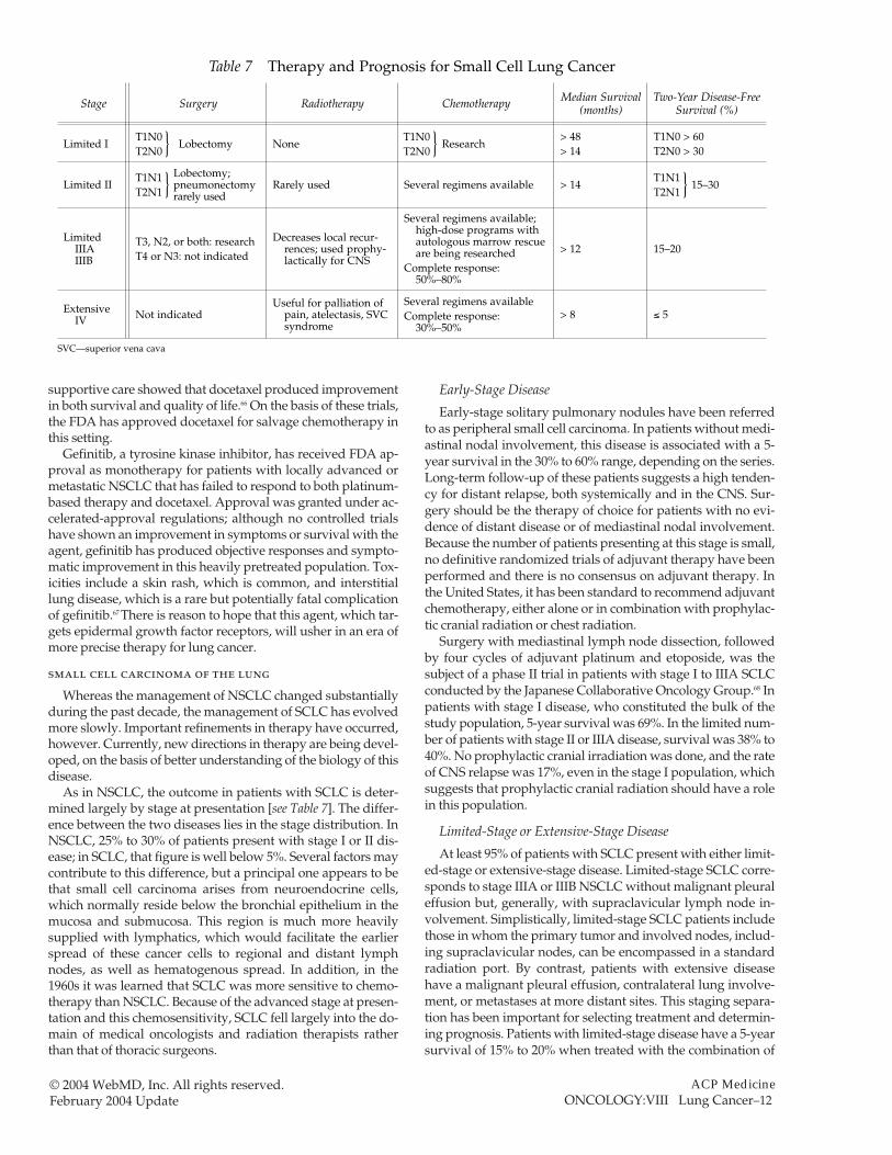
supportive care showed that docetaxel produced improvement
in both survival and quality of life.66 On the basis of these trials,
the FDA has approved docetaxel for salvage chemotherapy in
this setting.
Gefinitib, a tyrosine kinase inhibitor, has received FDA ap-
proval as monotherapy for patients with locally advanced or
metastatic NSCLC that has failed to respond to both platinum-
based therapy and docetaxel. Approval was granted under ac-
celerated-approval regulations; although no controlled trials
have shown an improvement in symptoms or survival with the
agent, gefinitib has produced objective responses and sympto-
matic improvement in this heavily pretreated population. Tox-
icities include a skin rash, which is common, and interstitial
lung disease, which is a rare but potentially fatal complication
of gefinitib.67 There is reason to hope that this agent, which tar-
gets epidermal growth factor receptors, will usher in an era of
more precise therapy for lung cancer.
small cell carcinoma of the lung
Whereas the management of NSCLC changed substantially
during the past decade, the management of SCLC has evolved
more slowly. Important refinements in therapy have occurred,
however. Currently, new directions in therapy are being devel-
oped, on the basis of better understanding of the biology of this
disease.
As in NSCLC, the outcome in patients with SCLC is deter-
mined largely by stage at presentation [see Table 7]. The differ-
ence between the two diseases lies in the stage distribution. In
NSCLC, 25% to 30% of patients present with stage I or II dis-
ease; in SCLC, that figure is well below 5%. Several factors may
contribute to this difference, but a principal one appears to be
that small cell carcinoma arises from neuroendocrine cells,
which normally reside below the bronchial epithelium in the
mucosa and submucosa. This region is much more heavily
supplied with lymphatics, which would facilitate the earlier
spread of these cancer cells to regional and distant lymph
nodes, as well as hematogenous spread. In addition, in the
1960s it was learned that SCLC was more sensitive to chemo-
therapy than NSCLC. Because of the advanced stage at presen-
tation and this chemosensitivity, SCLC fell largely into the do-
main of medical oncologists and radiation therapists rather
than that of thoracic surgeons.
Early-Stage Disease
Early-stage solitary pulmonary nodules have been referred
to as peripheral small cell carcinoma. In patients without medi-
astinal nodal involvement, this disease is associated with a 5-
year survival in the 30% to 60% range, depending on the series.
Long-term follow-up of these patients suggests a high tenden-
cy for distant relapse, both systemically and in the CNS. Sur-
gery should be the therapy of choice for patients with no evi-
dence of distant disease or of mediastinal nodal involvement.
Because the number of patients presenting at this stage is small,
no definitive randomized trials of adjuvant therapy have been
performed and there is no consensus on adjuvant therapy. In
the United States, it has been standard to recommend adjuvant
chemotherapy, either alone or in combination with prophylac-
tic cranial radiation or chest radiation.
Surgery with mediastinal lymph node dissection, followed
by four cycles of adjuvant platinum and etoposide, was the
subject of a phase II trial in patients with stage I to IIIA SCLC
conducted by the Japanese Collaborative Oncology Group.68 In
patients with stage I disease, who constituted the bulk of the
study population, 5-year survival was 69%. In the limited num-
ber of patients with stage II or IIIA disease, survival was 38% to
40%. No prophylactic cranial irradiation was done, and the rate
of CNS relapse was 17%, even in the stage I population, which
suggests that prophylactic cranial radiation should have a role
in this population.
Limited-Stage or Extensive-Stage Disease
At least 95% of patients with SCLC present with either limit-
ed-stage or extensive-stage disease. Limited-stage SCLC corre-
sponds to stage IIIA or IIIB NSCLC without malignant pleural
effusion but, generally, with supraclavicular lymph node in-
volvement. Simplistically, limited-stage SCLC patients include
those in whom the primary tumor and involved nodes, includ-
ing supraclavicular nodes, can be encompassed in a standard
radiation port. By contrast, patients with extensive disease
have a malignant pleural effusion, contralateral lung involve-
ment, or metastases at more distant sites. This staging separa-
tion has been important for selecting treatment and determin-
ing prognosis. Patients with limited-stage disease have a 5-year
survival of 15% to 20% when treated with the combination of
© 2004 WebMD, Inc. All rights reserved.
February 2004 Update
ACP MedicineONCOLOGY:VIII Lung Cancer–12
Radiotherapy
None
Rarely used
Decreases local recur-rences; used prophy-lactically for CNS
Useful for palliation ofpain, atelectasis, SVCsyndrome
Surgery
Lobectomy
Lobectomy;pneumonectomy rarely used
T3, N2, or both: research
T4 or N3: not indicated
Not indicated
Stage
Limited I
Limited II
LimitedIIIAIIIB
ExtensiveIV
Table 7—Therapy and Prognosis for Small Cell Lung Cancer
Two-Year Disease-FreeSurvival (%)
T1N0 > 60
T2N0 > 30
15–30
15–20
≤ 5
Chemotherapy
Research
Several regimens available
Several regimens available;high-dose programs withautologous marrow rescueare being researched
Complete response:50%–80%
Several regimens available
Complete response:30%–50%
Median Survival(months)
> 48
> 14
> 14
> 12
> 8
T1N1
T2N1 } T1N1
T2N1 }
T1N0
T2N0 } T1N0
T2N0 }
SVC—superior vena cava

chemotherapy and radiation. In contrast, patients with exten-
sive-stage disease generally have a 5-year survival of 1% to 2%
and have been treated primarily with chemotherapy, plus ad-
junctive radiation as needed. The distinction between limited-
and extensive-stage disease has largely been based on clinical
examination, as well as on the standard imaging studies of
chest CT through the adrenal glands, bone scan, and brain CT
or MRI. Of patients with no evidence of distant metastases on
these studies, an additional 5% will be found to have distant
disease by bone marrow evaluation. Currently, approximately
25% to 30% of patients with SCLC present with limited-stage
disease, and the remainder present with extensive-stage dis-
ease; however, with the development of more sophisticated
imaging techniques and the increased use of PET imaging,
some degree of stage migration is likely to occur.
Limited-stage disease Chemotherapy has been the main-
stay of treatment for limited-stage disease for the past 30 years.
For at least the past 10 years, the standard treatment has been
based on etoposide, either as a single agent or combined with
either cisplatin or carboplatin, generally for four to six cycles.
Most of the advances in the understanding of treatment of lim-
ited-stage SCLC have come from trials evaluating the role of
radiation therapy.69 These trials have shown that cure in limit-
ed-stage SCLC is associated with the use of full-dose combina-
tion chemotherapy, along with thoracic and cranial radiation
therapy. Thoracic radiation appears to be more beneficial if it is
done concurrently with chemotherapy, rather than sequential-
ly. Secondly, starting radiation therapy during the first few cy-
cles of chemotherapy appears to provide a survival advantage
over beginning radiation therapy at the end of chemotherapy.70
The explanation for this is that drug resistance may evolve
rapidly in small cell carcinoma, so in some patients, delaying
thoracic radiation even for a few months may result in escape
of drug-resistant cells from the lung and mediastinum to dis-
tant sites. Early intervention with radiation may result in the
destruction of these cells and an increase in the rate of cure.
However, as with NSCLC, the benefit of concurrent chemo-
therapy and radiation must be balanced against its higher tox-
icity (e.g., esophagitis).
Although clinical practice varies, most oncologists initiate
radiation therapy with chemotherapy cycle one or with cycles
two or three. The choice of timing depends on the clinical situa-
tion. The advantage of delaying radiation for one or two cycles
is that during this time the tumor mass may be significantly re-
duced, therefore easing the patient’s symptoms and often im-
proving performance status, so that the patient is better able to
cope with combination treatment. Most initial trials of concur-
rent therapy have used pretreatment tumor volumes to deter-
mine radiation fields. However, newer strategies of radiation
therapy will take advantage of chemotherapy-induced tumor
reduction and use higher doses of radiation delivered to a
smaller tumor volume. In one major trial, twice-daily radiation,
compared with once-daily treatment, has been associated with
improved surgical outcome,71 but twice-daily treatment is also
associated with significantly higher toxicity rates. The use of
twice-daily radiation can be considered in some patients with
high performance status. The standard dose of daily radiation
therapy that is given concurrently with chemotherapy has in-
creased from 45 Gy to 60 Gy.
Another significant advance in the understanding of SCLC
involves the role of prophylactic cranial irradiation. Early trials
that used prophylactic cranial irradiation along with chemo-
therapy reported a high incidence of both immediate and de-
layed neurologic toxicity. Studies have used prophylactic cra-
nial irradiation at lower dose fractions and have delayed its ini-
tiation until chemotherapy has been completed. These adjustments
seem to be associated with substantially diminished immediate
and delayed neurotoxicity. Moreover, a meta-analysis of the use
of prophylactic cranial irradiation from randomized clinical tri-
als (largely composed of patients with limited-stage disease) has
demonstrated not only a reduction in CNS relapse but also an
increase in long-term survival by approximately 5%.72
Several issues involving prophylactic cranial irradiation re-
main to be settled. One is its role in elderly patients, who ap-
pear to be at higher risk for neurotoxicity. Also, although trials
of SCLC have often included patients in complete remission
from either limited or extensive disease, most of the patients
have had limited disease. Therefore, the strength of the recom-
mendation for prophylactic cranial irradiation in patients in
complete remission from extensive-stage SCLC is less certain.
A number of trials have evaluated maintenance chemother-
apy or the addition of other treatments after chemoradiothera-
py for SCLC. Phase II studies of high-dose chemotherapy for
patients in remission have suggested an encouraging long-
term survival rate,73 and a potential long-term benefit has also
been reported in a small number of patients who received a
vaccine against a tumor ganglioside (BEC-2)74; the latter ap-
proach is currently the subject of a randomized phase III trial.
No benefit for these approaches has yet been proved, however,
so maintenance therapy after completion of standard therapy
cannot be recommended for SCLC patients, except in the con-
text of a clinical trial.
Extensive-stage disease Progress in the treatment of exten-
sive-stage SCLC over the past 10 years has been slower than
that of limited-stage SCLC.69 As with limited-stage disease,
treatment centers on chemotherapy. Although several different
chemotherapy combinations have been demonstrated to have
equivalent survival outcomes, etoposide-based regimens have
been shown to be superior to older regimens such as cy-
clophosphamide, doxorubicin, and vincristine. Secondly, most
clinical trials in SCLC have used cisplatin, but as noted above,
carboplatin has a better therapeutic ratio, is equally effective,
and causes significantly less nausea, vomiting, and neurotoxic-
ity. 75,76 Currently, however, cisplatin and carboplatin are used
with roughly equal frequency for SCLC in community practice
in the United States. Both agents are generally administered
every 3 to 4 weeks, depending on hematologic recovery. Al-
though the combination of platinum and etoposide remains
the most commonly used regimen in the United States, cis-
platin and irinotecan has been approved for treatment of SCLC
in Japan, and randomized trials of this combination are ongo-
ing in the United States.
Special populations Adaptation of treatment to special
populations is another very important principle in the manage-
ment of SCLC. Standard combination chemotherapy regimens,
which were developed for younger, healthier patients, pose a
greater risk of severe myelosuppression and life-threatening
complications in elderly patients with significant comorbid dis-
ease and in patients with poor performance status. Modified
regimens should be considered in such patients. If combination
therapy is employed, the use of carboplatin (dosed according
© 2004 WebMD, Inc. All rights reserved.
February 2004 Update
ACP MedicineONCOLOGY:VIII Lung Cancer–13

to renal function) rather than cisplatin may produce less toxici-
ty in this population. Although monotherapy with low-dose
etoposide is certainly a consideration, most elderly patients
should be considered for combination carboplatin and etopo-
side therapy if permitted by their level of functional illness and
degree of comorbid illness. Whether these patients should re-
ceive significantly reduced doses or near-standard doses plus
hematopoietic growth factor has not been well studied.
Relapse of disease Although SCLC typically responds to
therapy, the majority of patients with limited-stage disease and
nearly all patients with extensive-stage disease will experience
relapses. For limited-stage patients, the duration of remission
may range from months to years. For most patients with exten-
sive-stage disease, remission generally lasts only weeks to
months after completion of chemotherapy. In both groups of
patients, a major issue is the emergence of drug resistance,
which has severely hampered the benefit of second-line or so-
called salvage chemotherapy.
In this setting, a critical factor in choosing therapy is the time
to relapse. For patients who have had a prolonged remission
(longer than 1year), retreatment with the same regimen will of-
ten produce a response. On the other hand, in patients who fail
to respond to initial therapy (primary drug resistance), no ther-
apy has been convincingly shown to be beneficial, so these pa-
tients are best enrolled in experimental trials.
For patients who have had a remission of at least 2 months
after initial therapy, topotecan has been shown to produce a
significant improvement in disease-related symptoms—partic-
ularly dyspnea, but also anorexia, hoarseness, and fatigue—
and has been approved for second-line therapy for patients
with SCLC.77 The other commonly used approach in relapses of
SCLC has been prolonged courses of oral etoposide for 10 to 21
days per cycle. Other agents that have shown promise for sal-
vage therapy include paclitaxel, irinotecan, vinorelbine, and
gemcitabine.69,78 These agents, as well as more novel approach-
es, warrant further investigation.79
supportive care
Supportive care is important across all stages of both
NSCLC and SCLC. In patients with advanced or refractory dis-
ease, it often becomes the principal form of therapy. As the
ability of oncologists to deliver both chemotherapy and radia-
tion safely has improved, it is clear that one of the best support-
ive care approaches is to palliate disease-related symptoms
with chemotherapy, radiation, or both.
Over the past 10 years, management of pain, depression,
nausea, weight loss, and constipation in cancer patients has
been improved by the development of pharmacologic agents
with better efficacy and less toxicity [see CE:X Symptom Manage-ment in Palliative Medicine]. In patients who are receiving
myelosuppressive chemotherapy and so are at high risk for
neutropenic complications, the use of colony-stimulating fac-
tors has been demonstrated to substantially reduce the risk of
fever, neutropenia, and infection.80 How to identify the popula-
tions who are at highest risk and are thus candidates for prima-
ry prophylaxis with colony-stimulating factors is an area that
needs further investigation.
Anemia and fatigue are now appreciated as especially im-
portant concerns in patients with lung cancer.81 Analysis of the
1,748 lung cancer patients enrolled in the prospective trials of
recombinant human erythropoietin for cancer patients with
anemia has shown that amelioration of anemia led to signifi-
cant improvements in quality of life; these improvements ap-
plied to patients with SCLC or NSCLC receiving a variety of
chemotherapy regimens.82 Although all anemic cancer patients
may benefit from higher hemoglobin levels, those with lung
cancer may reap particular benefits because of dyspnea and fa-
tigue related to the cancer itself, as well as comorbid diseases
and the treatments used. In addition to substantially enhancing
quality of life, normalization of hemoglobin levels may also im-
prove therapeutic outcome, perhaps by improving tolerance to
treatment or by reducing tumor hypoxia. Studies evaluating
the role of hemoglobin maintenance in reducing cognitive dys-
function during treatment are ongoing.
treatment updates
The National Cancer Center Network (NCCN) provides ev-
idence-based, expert-panel guidelines for the management of
cancer, as well as supportive care guidelines. Clinicians are en-
couraged to review the NCCN Web site (http://www.nccn.org)
for annual updates of lung cancer management guidelines.
Prognosis
At present, the 5-year survival rate for lung cancer is approx-
imately 14%. The chance of survival is heavily dependent on
stage at presentation, however. For example, 5-year survival in
patients with stage I NSCLC is approximately 63%. Unfortu-
nately, more than 40% of all patients with NSCLC have stage
IV disease at diagnosis, and in this population the survival at 5
years is 1% or less. In patients with SCLC, 5-year survival
ranges from 30% to 60% for peripheral disease (stage I/II) to
1% to 3% for patients with extensive disease (stage IIIB/IV).
Unfortunately, approximately 65% to 70% of patients with
small cell lung cancer present with extensive disease.
The author has received grants or research support or has been a consultant ora member of the speakers’ bureaus of Abgenix, Inc.; Amgen, Inc.; AstraZenecaPharmaceuticals LP; Bristol-Myers Squibb Co; GlaxoSmithKline; ImCloneSystems Incorporated; Immunex Corporation; Eli Lilly and Company; OrthoBiotech Products, LP; QLT Inc.; and Schering-Plough Corporation during thepast 12 months.
References1. Jemal A, Murray T, Samuels A, et al: Cancer Statistics, 2003. CA Cancer J Clin 53:5,2003
2. Wynder El: The past, present, and future of the prevention of lung cancer. Ca Epi-demiol Biomarkers Prevent 7:735, 1998
3. Youth tobacco surveillance—United States, 2000. Centers for Disease Control.MMWR CDC Surveill Summ 50(4):1, 2001
4. Healton C, Messeri P, Reynolds J, et al: Tobacco use among middle and high schoolstudents—United States, 1999. MMWR Morb Mortal Wkly Rep 49(3):49, 2000
5. Halpern MT, Gillespie BW, Warner KE: Patterns of absolute risk of lung cancer mor-tality in former smokers. J Natl Cancer Inst 85:457, 1993
6. Shields PG: Molecular epidemiology of smoking and lung cancer. Oncogene 21:6870,2002
7. Vallyathan V, Green F, Ductaman B, et al: Roles of epidemiology, pathology, molec-ular biology, and biomarkers in the investigation of occupational lung cancer. J ToxicolEnviron Health B Crit Med 1:91, 1998
8. Neuberger JS, Gesell TF: Residential radon exposure and lung cancer: risk in non-smokers. Health Phys 83:1, 2002
9. Fong KM, Sekido Y, Minna JD: Molecular pathogenesis of lung cancer. J ThoracicCardiovasc Surg 118:1136, 1999
10. Carbone DP: The biology of lung cancer. Semin Oncol 24:388, 1997
11. Rozengurt E: Autocrine loops, signal transduction, and cell cycle abnormalities inthe molecular biology of lung cancer. Curr Opin Oncol 11:116, 1999
12. Schreiber G, Pitterle D, Young-Chul K, et al: Molecular genetic analysis of primarylung cancer and cancer metastatic to the lung. Anticancer Res 19:1109, 1999
© 2004 WebMD, Inc. All rights reserved.
February 2004 Update
ACP MedicineONCOLOGY:VIII Lung Cancer–14

13. Toma S, Raffo P, Isnardi L, et al: Retinoids in lung cancer chemoprevention andtreatment. Ann Oncol 10(suppl 5):S95, 1999
14. Omenn GS, Goodman GE, Thornquist MD, et al: Risk factors for lung cancer and forintervention effects in CARET, the Beta-Carotene and Rational Efficacy trial. J Natl Can-cer Inst 88:1550, 1996
15. Caraballoso M, Sacristan M, Serra C, et al: Drugs for preventing lung cancer inhealthy people. Cochrane Database Syst Rev (2):CD002141, 2003
16. Strauss GM: Randomized population trials and screening for lung cancer: breakingthe cure barrier. Cancer 89(11 suppl):2399, 2000
17. Henschke CI, Naidich DP, Yankelevitz DF, et al: Early Lung Cancer Action Project:initial findings on repeating screenings. Cancer 92:153, 2001
18. Swensen SJ, Jett JR, Sloan JA, et al: Screening for lung cancer with low dose spiralcomputed tomography. Am J Respir Crit Care Med 165:508, 2002
19. Mahadevia PJ, Fleisher LA, Frick KD, et al: Lung cancer screening with helical com-puted tomography in older adult smokers: a decision and cost-effectiveness analysis.JAMA 289:313, 2003
20. Lam S, Shibuya H: Early diagnosis of lung cancer. Clin Chest Med 20:53, 1999
21. NLST: National Lung Screening Trial. National Cancer Institute, 2003.http://www.cancer.gov/nlst
22. Patz EF, Erasmus JJ: Positron emission tomography imaging in lung cancer. ClinLung Cancer 1:42, 1999
23. van Tinteren H, Hoekstra OS, Smit EF, et al: Effectiveness of positron emission to-mography in the preoperative assessment of patients with suspected non-small-celllung cancer: the PLUS multicentre randomised trial. Lancet 359:1388, 2002
24. Mountain CF: A new international staging system for lung cancer. Chest 89(suppl4):225S, 1986
25. Mountain CF: Revisions in the international system for staging lung cancer. Chest111:1710, 1997
26. Mountain CF, Dresler CM: Regional lymph node classification for lung cancer stag-ing. Chest 111:1718, 1997
27. Ginsberg RJ: Multimodality treatment of resectable non–small cell lung cancer. ClinLung Cancer 1:194, 2000
28. Chemotherapy in non–small cell lung cancer: a meta-analysis using updated dataon individual patients from 52 randomized clinical trials. Non–Small Cell CollaborativeGroup. BMJ 311:899, 1995
29. Results of the randomized International Adjuvant Lung Cancer Trial (IALT): cis-platin-based chemotherapy (CT) vs. no CT in 1867 patients with resected non–small celllung cancer (NSCLC) (abstr 6). The IALT Investigators. Proc Am Soc Clin Oncol 22:2,2003
30. Pisters KMW, Ginsberg RJ, Putnam JB, et al: Induction paclitaxel & carboplatin (PC)in early stage non–small cell lung cancer (NSCLC): early results of a completed phase IItrial. Proc Am Soc Clin Oncol 18:1800, 1999
31. Sibley GS, Jamieson TA, Marks LB, et al: Radiotherapy alone for medically inopera-ble stage I non–small-cell lung cancer: the Duke experience. Int J Radiat Oncol Biol Phys40:149, 1998
32. Pass HI, Pogrebniak, Steinberg HW, et al: Randomized trial of neoadjuvant therapyfor lung cancer: interim analysis. Ann Thorac Surg 53:992, 1992
33. Roth JA, Fossella F, Komaki R, et al: A randomized trial comparing perioperativechemotherapy and surgery with surgery alone in resectable stage III non–small celllung cancer. J Natl Cancer Inst 86:673, 1994
34. Rosell R, Gomez-Codina J, Camps C, et al: A randomized trial comparing preopera-tive chemotherapy plus surgery with surgery alone in patients with non–small cell lungcancer. N Engl J Med 330:153, 1994
35. Albain KS, Scott CB, Rusch VR, et al: Phase III comparison of concurrent chemother-apy plus radiotherapy (CT/RT) and CT/RT followed by surgical resection of stage IIIA(pN2) non–small cell lung cancer (NSCLC): initial results from intergroup trial 0139 (abstr 2497). Proc Am Soc Clin Oncol 22:621, 2003
36. Effects of postoperative mediastinal radiation on completely resected stage II andstage III epidermoid cancer of the lung. The Lung Cancer Study Group. N Engl J Med315:1377, 1986
37. Martini N, Flehinger BJ, Zaman MB, et al: Results of resection in non–oat cell carci-noma of the lung with mediastinal lymph node metastases. Ann Surg 198:386, 1983
38. Postoperative radiotherapy in non–small-cell lung cancer: systematic review andmeta-analysis of individual patient data from nine randomized controlled trials. PORTMeta-analysis Trialists Group. Lancet 352:257, 1998
39. Keller SM, Adak S, Wagner H, et al: A randomized trial of postoperative adjuvanttherapy in patients with completely resected stage II or IIIa non–small cell lung cancer.N Engl J Med 343:1217, 2000
40. Perez CA, Pajak TF, Rubin P, et al: Long-term observations of the patterns of failurein patients with unresectable non–oat cell carcinoma of the lung treated with definitiveradiotherapy: report by the Radiation Therapy Oncology Group. Cancer 59:1874, 1987
41. Komaki R, Scott CB, Byhardt R, et al: Failure patterns by prognostic group deter-mined by recursive partitioning analysis (RPA) of 1547 patients on four radiation thera-py oncology group (RTOG) studies in inoperable non–small cell lung cancer (NSCLC).Int J Radiat Oncol Biol Phys 42:263, 1998
42. Continuous, hyperfractionated, accelerated radiotherapy (CHART) versus conven-tional radiotherapy in non–small cell lung cancer: mature data from the randomizedmulticentre trial. CHART Steering Committee. Radiother Oncol 52:137, 1999
43. Sause W, Kolesar P, Taylor S IV, et al: Final results of phase III trial in regionally ad-vanced unresectable non-small cell lung cancer: radiation therapy oncology group, East-ern Cooperative Oncology Group, and Southwest Oncology Group. Chest 117:358, 2000
44. Belani CP, Wang W, Johnson DH, et al: Induction chemotherapy followed by stan-dard thoracic radiotherapy vs. hyperfractionated accelerated radiotherapy (HART) forpatients with unresectable stage IIIA and B non-small cell lung cancer: phase III study ofthe Eastern Cooperative Oncology Group (abstr 2500). Proc Am Soc Clin Oncol 22:622,2003
45. Marks LB, Sibley G: The rationale and use of three-dimensional radiation treatmentplanning for lung cancer. Chest 116(6 suppl):539S, 1999
46. Clinical practice guidelines for the treatment of unresectable non-small cell lungcancer. American Society of Clinical Oncology. J Clin Oncol 15:2996, 1997
47. Vokes EE, Herndon JE II, Crawford J, et al: A randomized phase II study of gem-citabine or paclitaxel or vinorelbine with cisplatin and induction chemotherapy (IndCT) followed by concomitant chemoradiotherapy (XRT) for unresectable stage IIInon–small cell lung cancer (NSCLC) (CALGB study 9431). J Clin Oncol 20:4191, 2002
48. Furuse K, Fukuoka M, Kawahara M, et al: Phase III study of concurrent versus se-quential thoracic radiotherapy in combination with mitomycin, vindesine, and cisplatinin unresectable stage III non-small cell lung cancer. J Clin Oncol 17:2692, 1999
49. Fry WA, Phillips JL, Menck HR: Ten year survey of lung cancer treatment and sur-vival in hospitals in the United States: a national cancer data base report. Cancer86:1867, 1999
50. Johnson DH: Treatment strategies for metastatic non–small cell lung cancer. ClinLung Cancer 1:34, 1999
51. Klastersky J, Sculier JP, Lacroix H, et al: A randomized study comparing cisplatin orcarboplatin with etoposide in patients with advanced non-small cell lung cancer: Euro-pean Organization for Research and Treatment of Cancer Protocol 007861. J Clin Oncol8:1556, 1990
52. Rosell R, Gatzemeier U, Betticher DC, et al: Phase III randomised trial comparingpaclitaxel/carboplatin with paclitaxel/cisplatin in patients with advanced non-small-cell lung cancer: a cooperative multinational trial. Ann Oncol 13:1539, 2002
53. Le Chevalier T, Brisgand D, Douillard JY, et al: Randomized study of vinorelbineand cisplatin versus vindesine and cisplatin versus vinorelbine alone in advancednon–small cell lung cancer: results of a European multicenter trial including 612 pa-tients. J Clin Oncol 12:360, 1994
54. Wozniak AJ, Crowley JJ, Balcerzak SP, et al: Randomized trial comparing cisplatinwith cisplatin plus vinorelbine in the treatment of advanced non-small cell lung cancer:a Southwest Oncology Group study. J Clin Oncol 16:2495, 1998
55. Sandler A, Nemunaitis J, Dehnam C, et al: Phase III study of cisplatin (C) with orwithout gemcitabine (G) in patients with advanced non–small cell lung cancer (NSCLC)(abstr). Proc Am Soc Clin Onc 17:454a 1999
56. Bonomi PD, Kim K, Fairclough D, et al: Comparison of survival and quality of lifein advanced non–small cell lung cancer patients treated with two dose levels of pacli-taxel combined with cisplatin versus etoposide with cisplatin: results of an Eastern Co-operative Oncology Group trial. J Clin Oncol 18:623, 2000
57. Fossella F, Pereira JR, von Pawel J, et al: Randomized, multinational, phase III studyof docetaxel plus platinum combinations versus vinorelbine plus cisplatin for advancednon-small-cell lung cancer: the TAX 326 study group. J Clin Oncol 21:3016, 2003
58. Kelly K, Crowley J, Bunn P, et al: A randomized phase III trial of paclitaxel plus car-boplatin (PC) versus vinorelbine plus cisplatin (VC) in untreated advanced non–smallcell lung cancer (NSCLC): a Southwest Oncology Group (SWOG) trial. J Clin Oncol19:3210, 2001
59. Schiller JH, Harrington D, Belani CP, et al: Comparison of four chemotherapy regi-mens for advanced non–small cell lung cancer. N Engl J Med 346:92, 2002
60. Lilenbaum RC, Herndon J, List M, et al: Single-agent versus combinationchemotherapy in advanced non-small cell lung cancer: a meta-analysis and the Cancerand Leukemia Group B randomized trial of efficacy, quality of life, and cost efffective-ness (abstr 2). Proc Am Soc Clin Oncol 21:71a, 2002
61. Tong D, Gillick L, Hendrickson FR: The palliation of symptomatic osseous metas-tases: final results of the Study by the Radiation Therapy Oncology Group. Cancer50:893, 1982
62. Ellis PA, Smith IE, Hardy JR, et al: Symptom relief with MVP (mitomycin C, vin-blastine and cisplatin) chemotherapy in advanced nonsmall cell lung cancer. Br J Cancer71:366, 1995
63. Thatcher N, Hopwood P, Anderson H: Improving quality of life in patients withnonsmall cell lung cancer: research experience with gemcitabine. Eur J Cancer 33(suppl1):S8, 1997
64. Effects of vinorelbine on quality of life and survival of elderly patients with ad-vanced non–small cell lung cancer. The Elderly Lung Cancer Vinorelbine Italian StudyGroup. J Natl Cancer Inst 91:66, 1999
65. Randomized phase III trial of docetaxel versus vinorelbine or ifosfamide in patientswith advanced non-small-cell lung cancer previously treated with platinum-containingchemotherapy regimens. The TAX 320 Non–Small Cell Lung Cancer Study Group. JClin Oncol 18:2354, 2000
66. Shepherd FA, Dancey J, Ramlau R, et al: Prospective randomized trial of docetaxelversus best supportive care in patients with non-small-cell lung cancer previously treat-ed with platinum-based chemotherapy. J Clin Oncol 18:2095, 2000
67. Cohen MH, Williams GA, Sridhara R, et al: FDA drug approval summary: gefinitib(ZD1839) (Iressa) tablets. Oncologist 8:303, 2003
68. Oda M, Tsuchiya R, Suzuki K, et al: Patterns of failure in phase II trial of postopera-tive adjuvant cisplatin/etoposide (PE) in patients with completely resected stage I-IIIAsmall cell lung cancer (SCLC): the Japan Clinical Oncology Lung Cancer Study GroupTrial (JCOG9101) (abstr). Proc Am Soc Clin Onc 20:316a, 2001
69. Murray N: Small cell lung cancer at the millennium: radiotherapy innovations im-prove survival while new chemotherapy treatments remain unproven. Clin Lung Can-cer 1:181, 2000
© 2004 WebMD, Inc. All rights reserved.
February 2004 Update
ACP MedicineONCOLOGY:VIII Lung Cancer–15

70. Murray N, Coy P, Peter JL, et al: Importance of timing for thoracic irradiation in the
combined modality treatment of limited-stage small cell lung cancer. J Clin Oncol
11:336, 1993
71. Turrisi AT III, Kim K, Blum R, et al: Twice-daily compared with once-daily thoracic
radiotherapy in limited small-cell lung cancer treated concurrently with cisplatin and
etoposide. N Engl J Med 340:2002, 1999
72. Auperin A, Arriagada R, Pignon JP, et al: Prophylactic cranial irradiation for pa-
tients with small-cell lung cancer in complete remission. N Engl J Med 341:476, 1999
73. Elias A: Hematopoietic stem cell transplantation for small cell lung cancer. Chest
116(6 suppl), 531S, 1999
74. Grant SC, Kris MG, Houghton AN, et al: Long survival of patients with small cell
lung cancer after adjuvant treatment with the anti-idiotypic antibody BEC2 plus bacillus
Calmette-Guerin. Clin Cancer Res 5:1319, 1999
75. Ettinger DS: Carboplatin in the treatment of small cell lung cancer (SCLC). Lung
Cancer 18(suppl 2):132, 1997
76. Kosmidis PA, Samantas E, Fountzilas G, et al: Cisplatin/etoposide versus carbo-
platin/etoposide chemotherapy and irradiation in small cell lung cancer: a randomized
phase III study. Semin Oncol 21(suppl 6):23, 1994
77. Von Pawel J, Schiller JH, Shepherd FA, et al: Topotecan versus cyclophosphamide,
doxorubicin, and vincristine for the treatment of recurrent small-cell lung cancer. J Clin
Oncol 17:658, 1999
78. Noda K, Nishiwaki Y, Kawahara M, et al: Irinotecan plus cisplatin compared with
etoposide plus cisplatin for extensive small-cell lung cancer. N Engl J Med 346:85, 2002
79. Zangemeister-Wittke U, Stahel RA: Novel approaches to the treatment of small-cell
lung cancer. Cell Mol Life Sci 55:1585, 1999
80. Johnston E, Crawford J: Hematopoietic growth factors in the reduction of
chemotherapy toxicity. Semin Oncol 25:552, 1998
81. Groopman JE, Itri LM: Chemotherapy-induced anemia in adults: incidence and
treatment. J Natl Cancer Inst 91:1616, 1999
82. Crawford J, Demetri GD, Gabrilove JL, et al: Clinical benefits of epoetin alfa therapy
in patients with lung cancer. Clin Lung Cancer 3:180, 2002
AcknowledgmentThe author would like to acknowledge the Ruth and Herman Albert Thoracic Oncology
Program at Duke Comprehensive Cancer Center for support of this work and, in par-
ticular, Thomas A. D’Amico, M.D., Joseph A. Govert, M.D., Michael J. Kelly, M.D., and
Timothy Shafman, M.D., for their participation in the multidisciplinary development of
this chapter.
© 2004 WebMD, Inc. All rights reserved.
February 2004 Update
ACP MedicineONCOLOGY:VIII Lung Cancer–16