VIETNAM HEART INSTITUTE HANOI MEDICAL UNIVERSITYvnha.org.vn/upload/hoinghi/BAIBAOCAO HB1 English...
73
VIETNAM HEART INSTITUTE HANOI MEDICAL UNIVERSITY Truong Thanh Huong, MD, PhD. Echocardiography guides for congenital heart disease intervention
-
Upload
phungkhuong -
Category
Documents
-
view
222 -
download
0
Transcript of VIETNAM HEART INSTITUTE HANOI MEDICAL UNIVERSITYvnha.org.vn/upload/hoinghi/BAIBAOCAO HB1 English...
VIETNAM HEART INSTITUTE
HANOI MEDICAL UNIVERSITY
Truong Thanh Huong, MD, PhD.
Echocardiography guides for
congenital heart disease
intervention
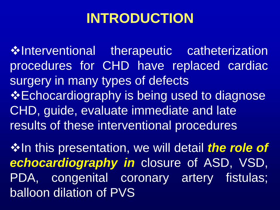
INTRODUCTION
Interventional therapeutic catheterization
procedures for CHD have replaced cardiac
surgery in many types of defects
Echocardiography is being used to diagnose
CHD, guide, evaluate immediate and late
results of these interventional procedures
In this presentation, we will detail the role of
echocardiography in closure of ASD, VSD,
PDA, congenital coronary artery fistulas;
balloon dilation of PVS
ATRIAL SEPTAL DEFECT
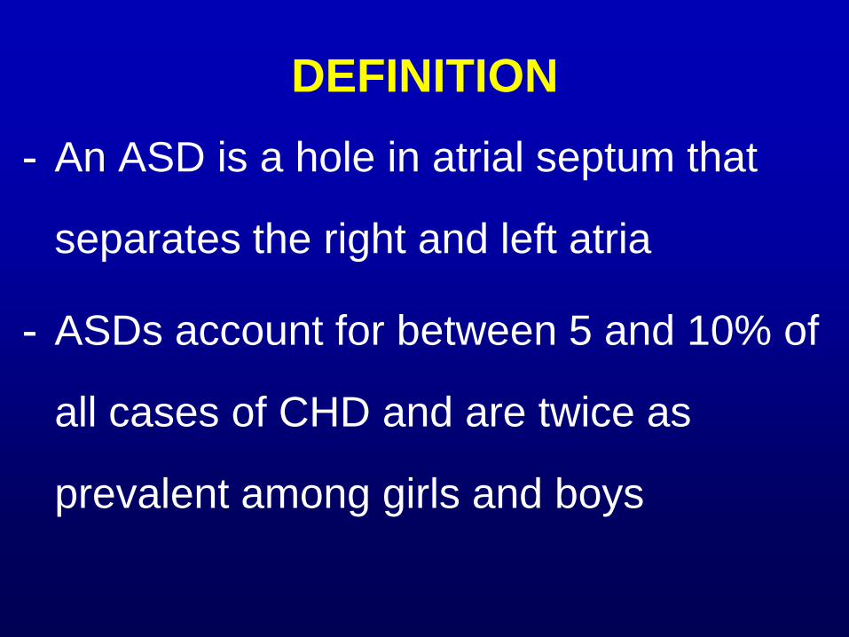
DEFINITION
- An ASD is a hole in atrial septum that
separates the right and left atria
- ASDs account for between 5 and 10% of
all cases of CHD and are twice as
prevalent among girls and boys
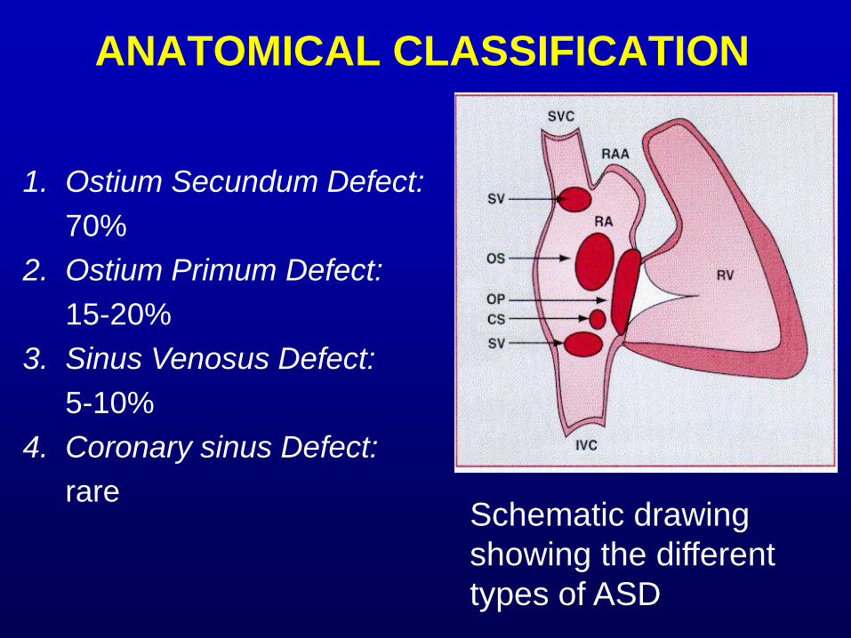
ANATOMICAL CLASSIFICATION
1. Ostium Secundum Defect:
70%
2. Ostium Primum Defect:
15-20%
3. Sinus Venosus Defect:
5-10%
4. Coronary sinus Defect:
rareSchematic drawing
showing the different
types of ASD
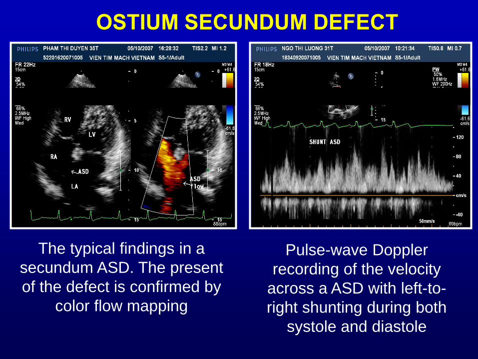
The typical findings in a
secundum ASD. The present
of the defect is confirmed by
color flow mapping
Pulse-wave Doppler
recording of the velocity
across a ASD with left-to-
right shunting during both
systole and diastole
Ostium secundum defect
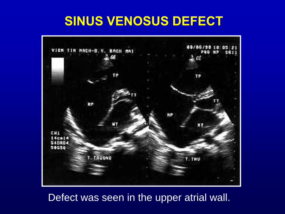
Sinus venosus defect
Defect was seen in the upper atrial wall.
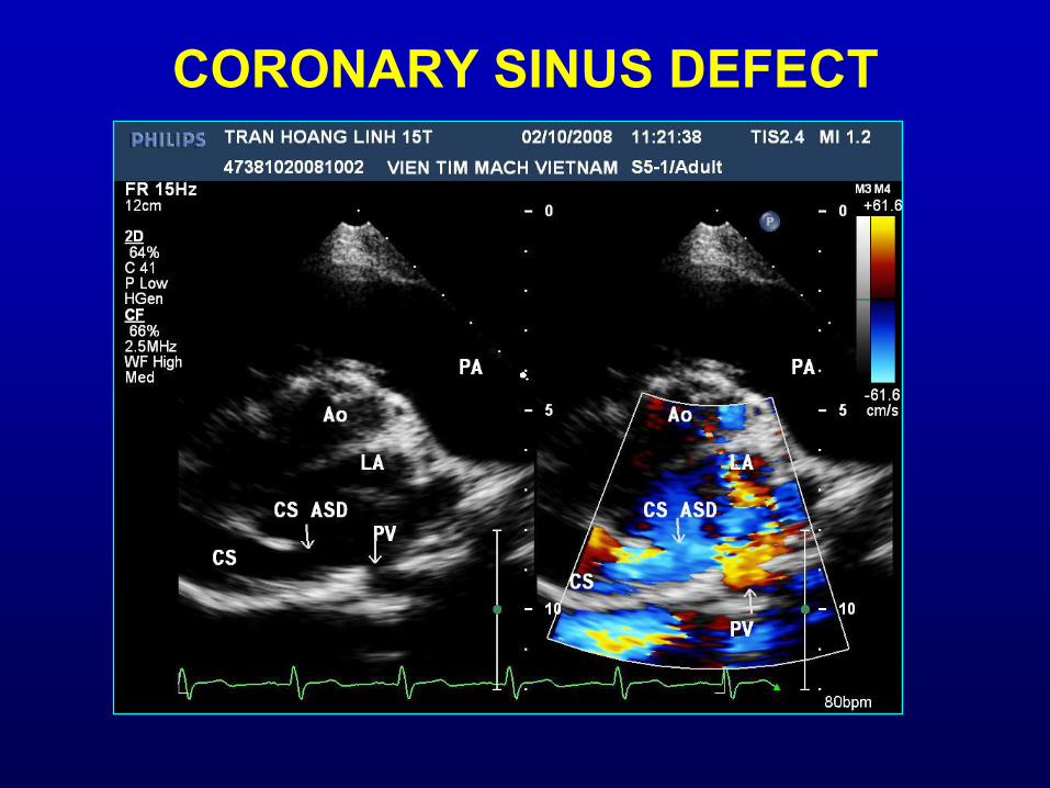
Coronary sinus defect
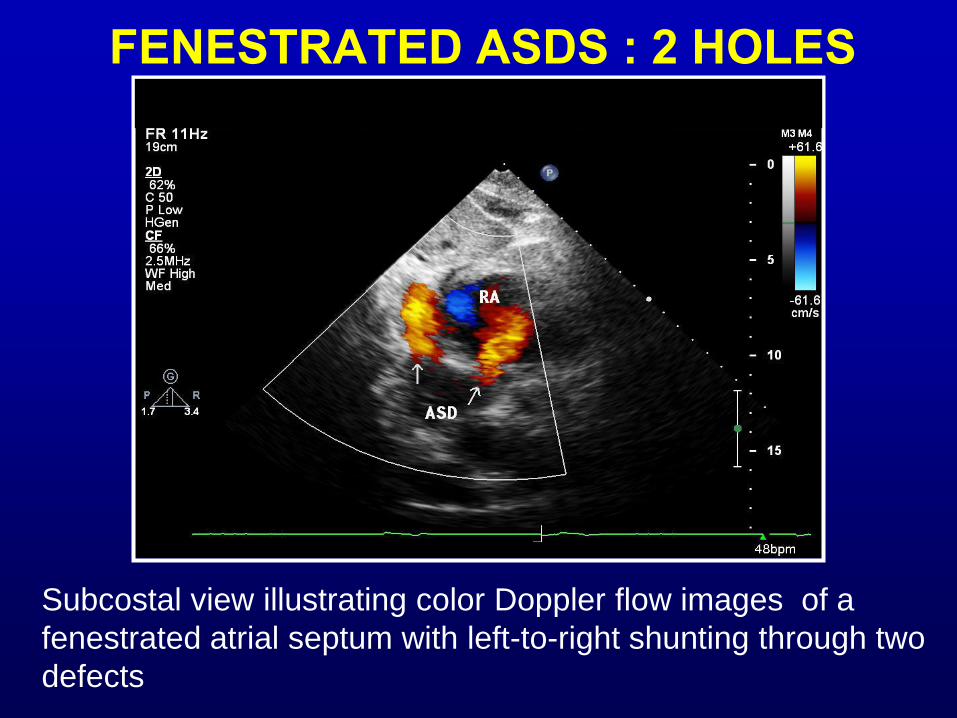
Fenestrated ASDs : 2 holes
Subcostal view illustrating color Doppler flow images of a
fenestrated atrial septum with left-to-right shunting through two
defects
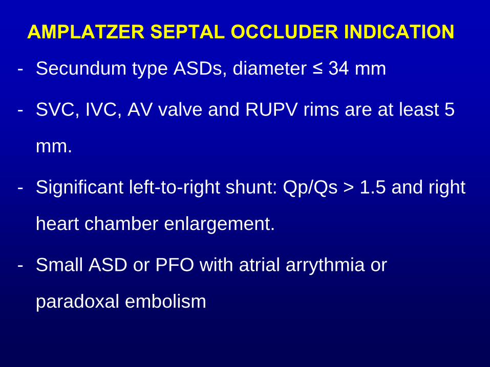
Amplatzer Septal Occluder Indication
- Secundum type ASDs, diameter ≤ 34 mm
- SVC, IVC, AV valve and RUPV rims are at least 5
mm.
- Significant left-to-right shunt: Qp/Qs > 1.5 and right
heart chamber enlargement.
- Small ASD or PFO with atrial arrythmia or
paradoxal embolism
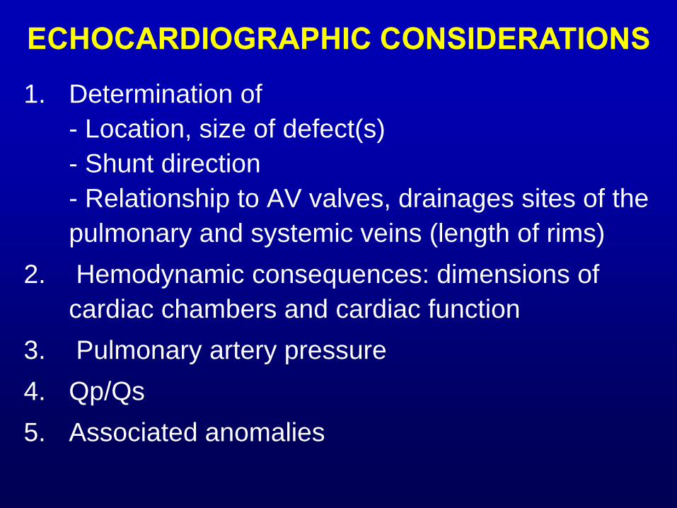
ECHOCARDIOGRAPHIC CONSIDERATIONS
1. Determination of
- Location, size of defect(s)
- Shunt direction
- Relationship to AV valves, drainages sites of the
pulmonary and systemic veins (length of rims)
2. Hemodynamic consequences: dimensions of
cardiac chambers and cardiac function
3. Pulmonary artery pressure
4. Qp/Qs
5. Associated anomalies

ASD sizing
Apical 4-chamber view of atrial septum. Measure of
the defect (dotted line) size is demonstrated
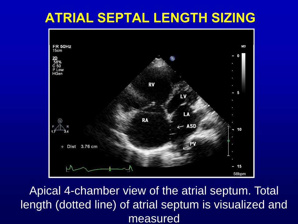
Atrial septal length sizing
Apical 4-chamber view of the atrial septum. Total
length (dotted line) of atrial septum is visualized and
measured
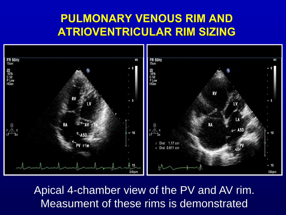
Pulmonary venous rim and
atrioventricular rim sizing
Apical 4-chamber view of the PV and AV rim.
Measument of these rims is demonstrated
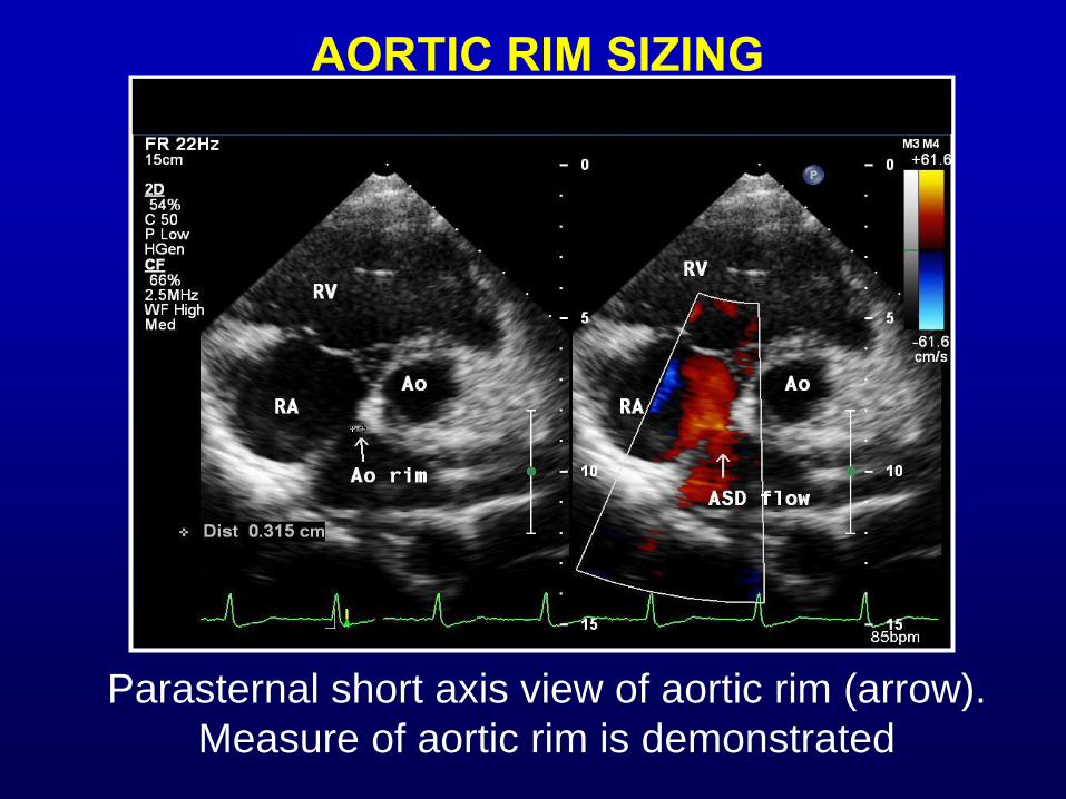
Aortic rim sizing
Parasternal short axis view of aortic rim (arrow).
Measure of aortic rim is demonstrated
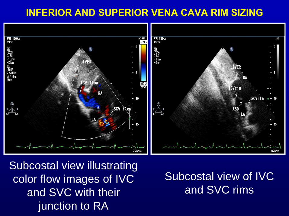
Inferior and superior vena cava rim sizing
Subcostal view illustrating
color flow images of IVC
and SVC with their
junction to RA
Subcostal view of IVC
and SVC rims
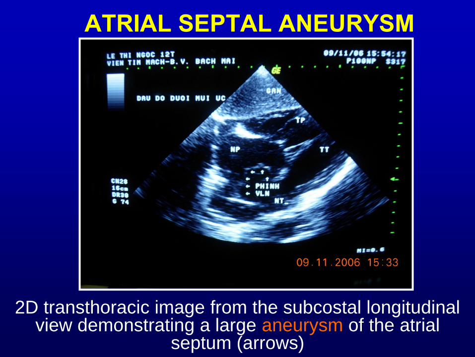
Atrial septal aneurysm
2D transthoracic image from the subcostal longitudinal view demonstrating a large aneurysm of the atrial
septum (arrows)
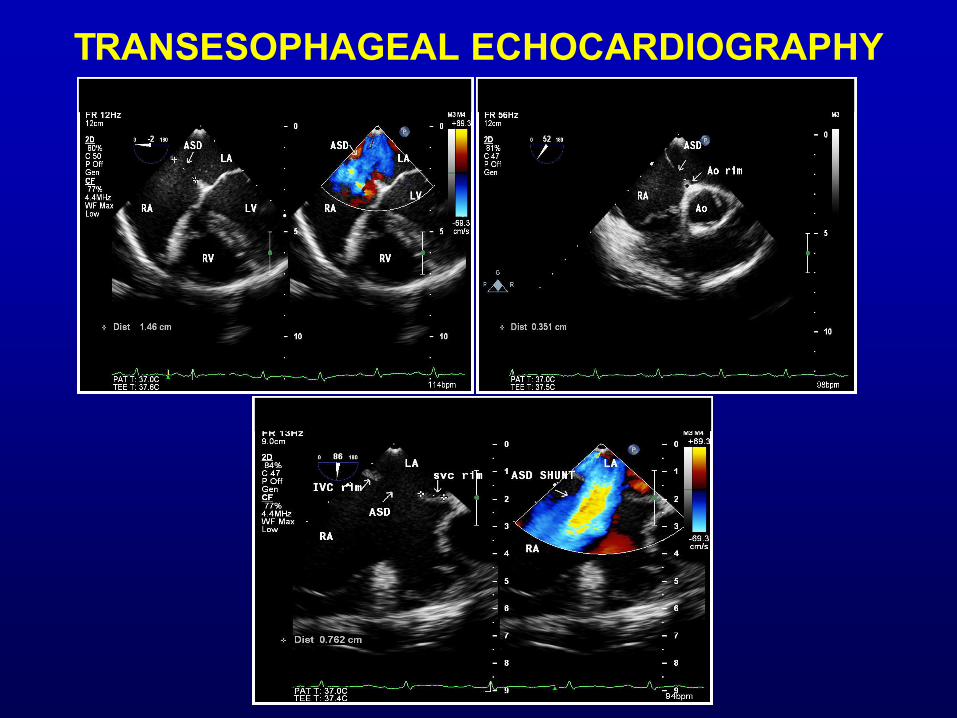
Transesophageal echocardiography
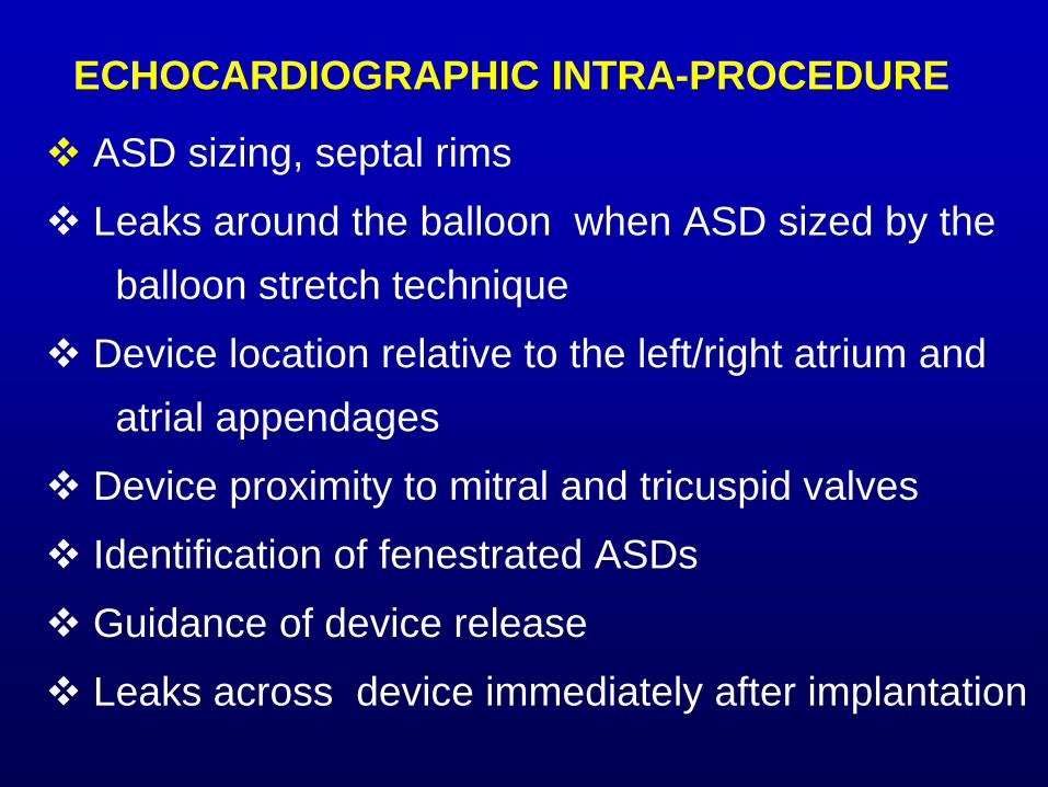
ECHOCARDIOGRAPHIC INTRA-PROCEDURE
ASD sizing, septal rims
Leaks around the balloon when ASD sized by the
balloon stretch technique
Device location relative to the left/right atrium and
atrial appendages
Device proximity to mitral and tricuspid valves
Identification of fenestrated ASDs
Guidance of device release
Leaks across device immediately after implantation
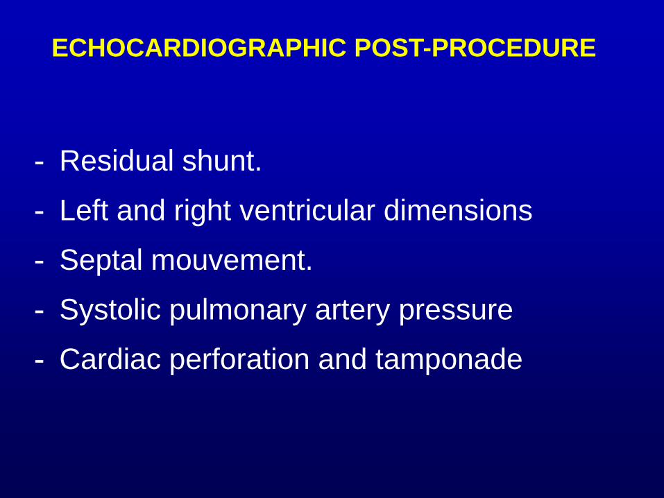
- Residual shunt.
- Left and right ventricular dimensions
- Septal mouvement.
- Systolic pulmonary artery pressure
- Cardiac perforation and tamponade
ECHOCARDIOGRAPHIC POST-PROCEDURE
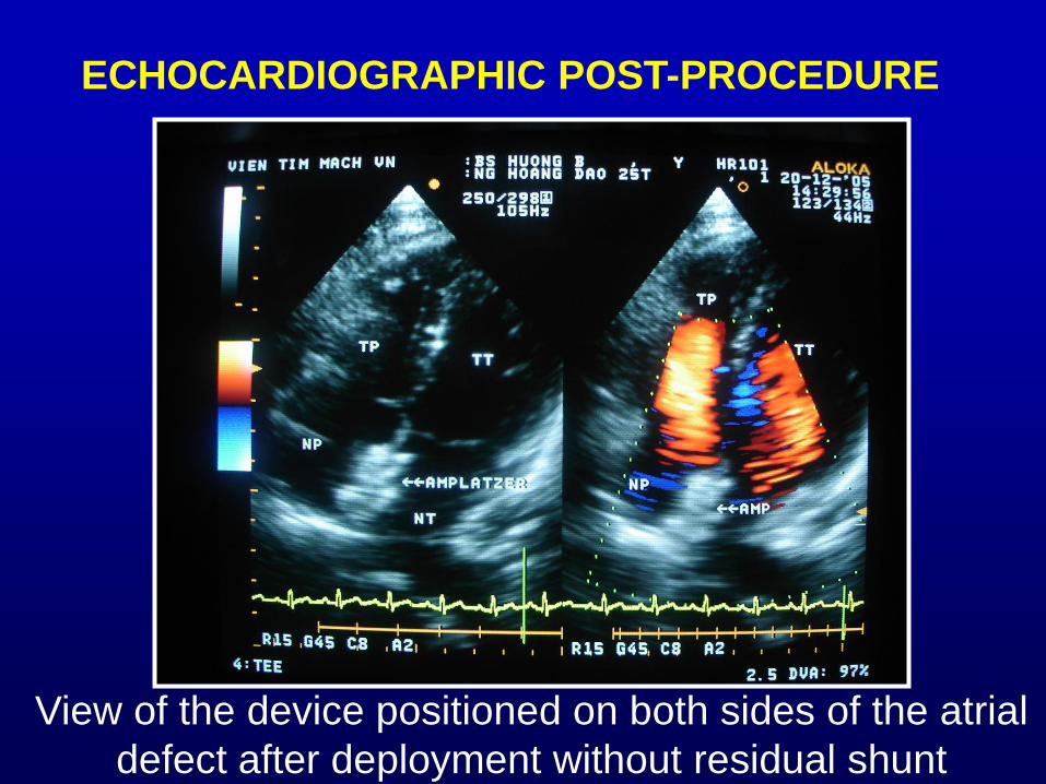
View of the device positioned on both sides of the atrial
defect after deployment without residual shunt
ECHOCARDIOGRAPHIC POST-PROCEDURE
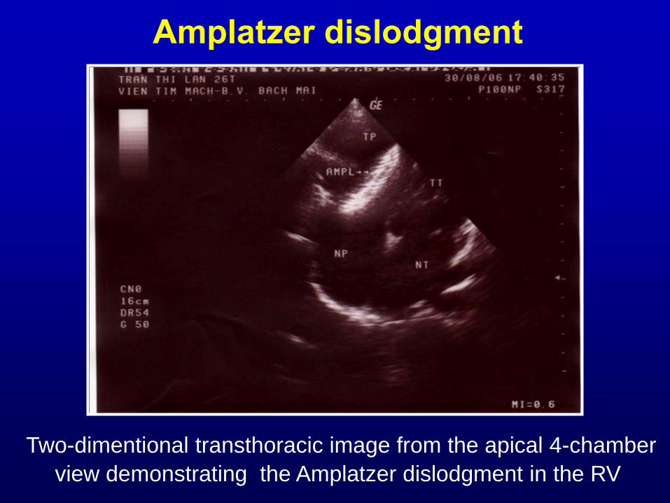
Two-dimentional transthoracic image from the apical 4-chamber
view demonstrating the Amplatzer dislodgment in the RV
Amplatzer dislodgment
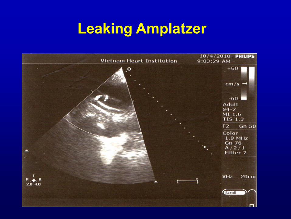
Leaking Amplatzer
VENTRICULAR SEPTAL
DEFECT
DEFINITION
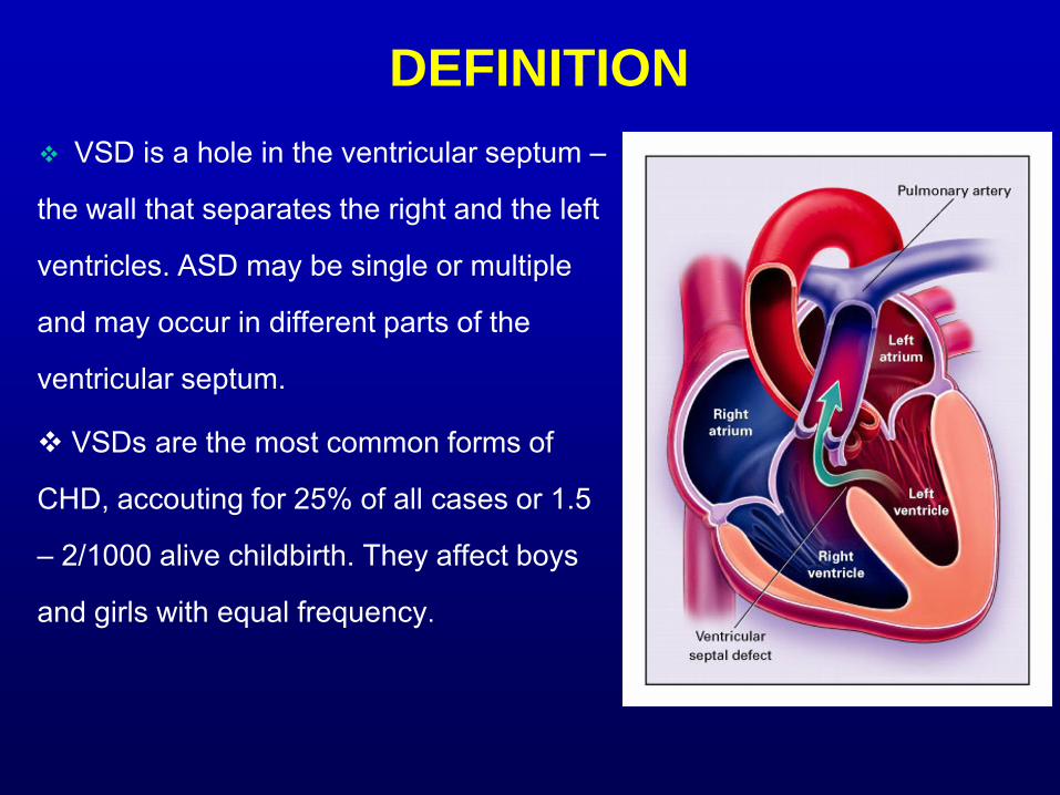
VSD is a hole in the ventricular septum –
the wall that separates the right and the left
ventricles. ASD may be single or multiple
and may occur in different parts of the
ventricular septum.
VSDs are the most common forms of
CHD, accouting for 25% of all cases or 1.5
– 2/1000 alive childbirth. They affect boys
and girls with equal frequency.
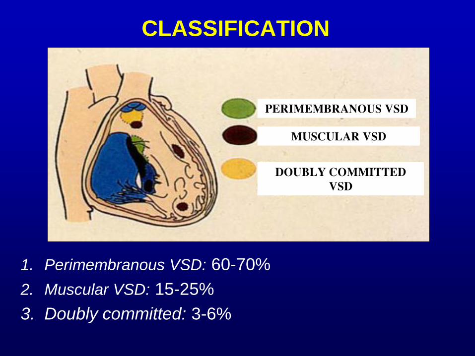
CLASSIFICATION
1. Perimembranous VSD: 60-70%
2. Muscular VSD: 15-25%
3. Doubly committed: 3-6%
Perimembranous VSD
Muscular VSD
Doubly committed
VSD
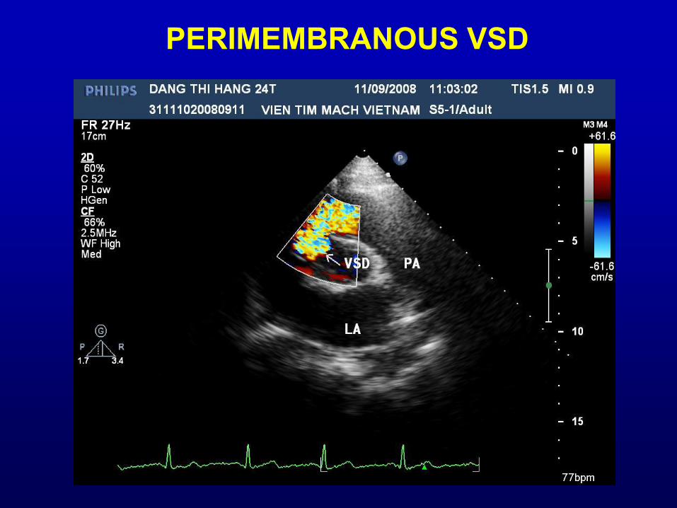
Perimembranous VSD
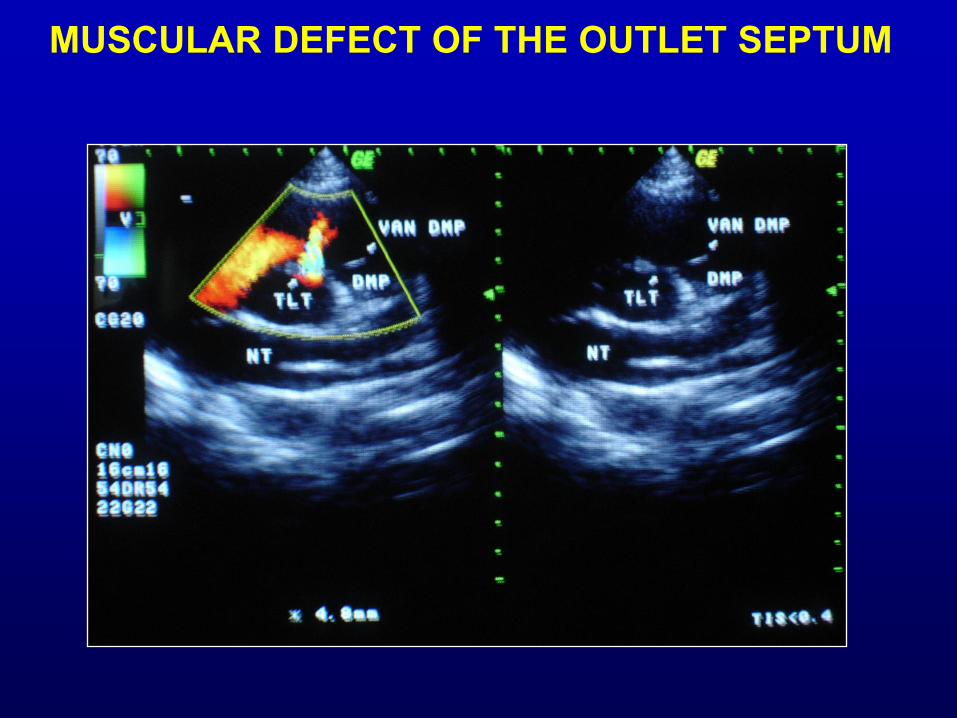
Muscular defect of the outlet septum
Muscular defect of the
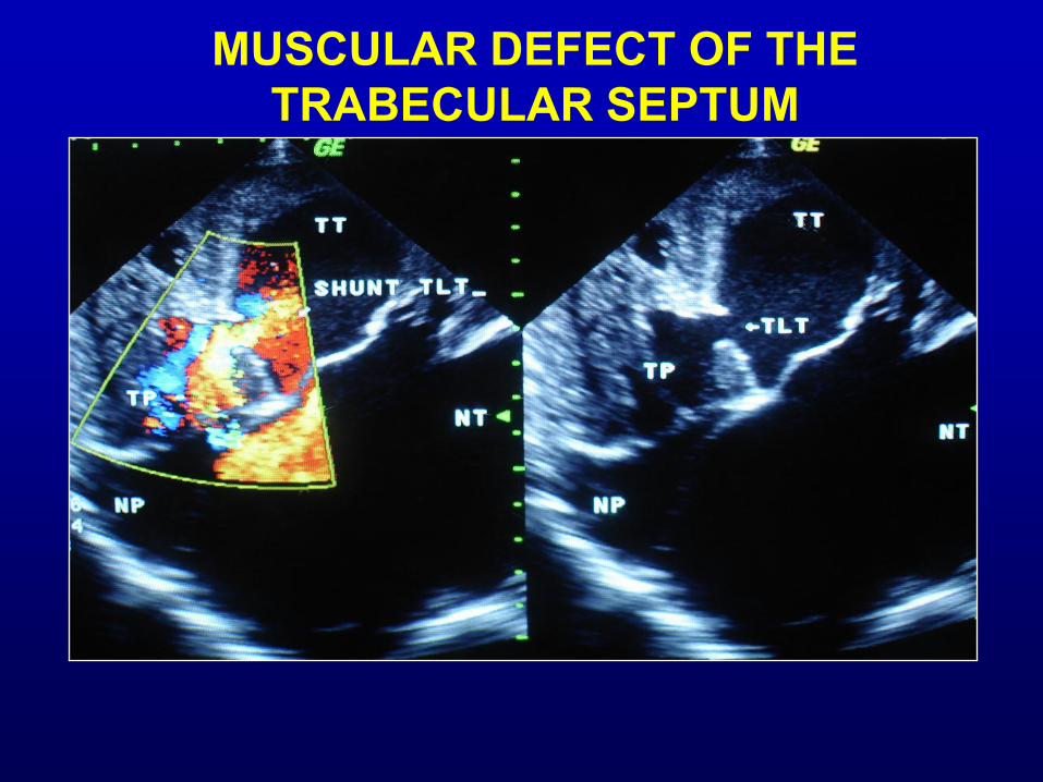
trabecular septum
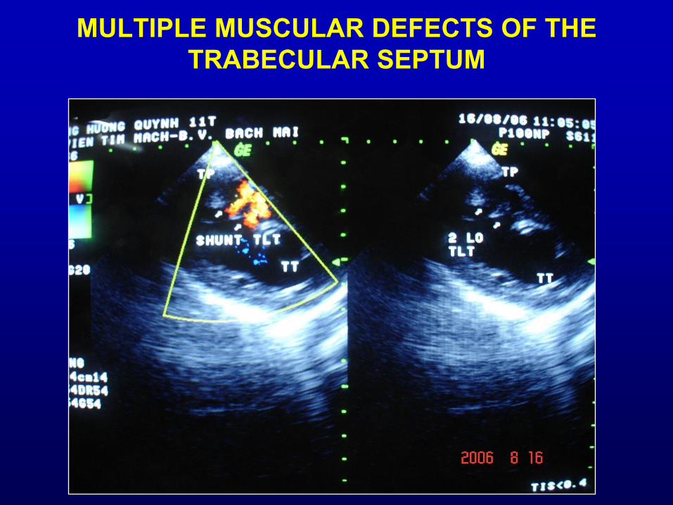
Multiple muscular defects of the
trabecular septum
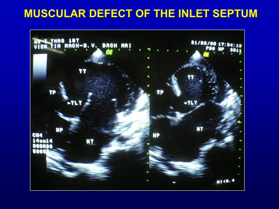
Muscular defect of the inlet septum
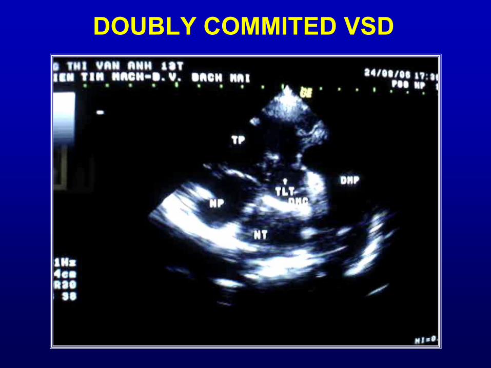
Doubly commited VSD
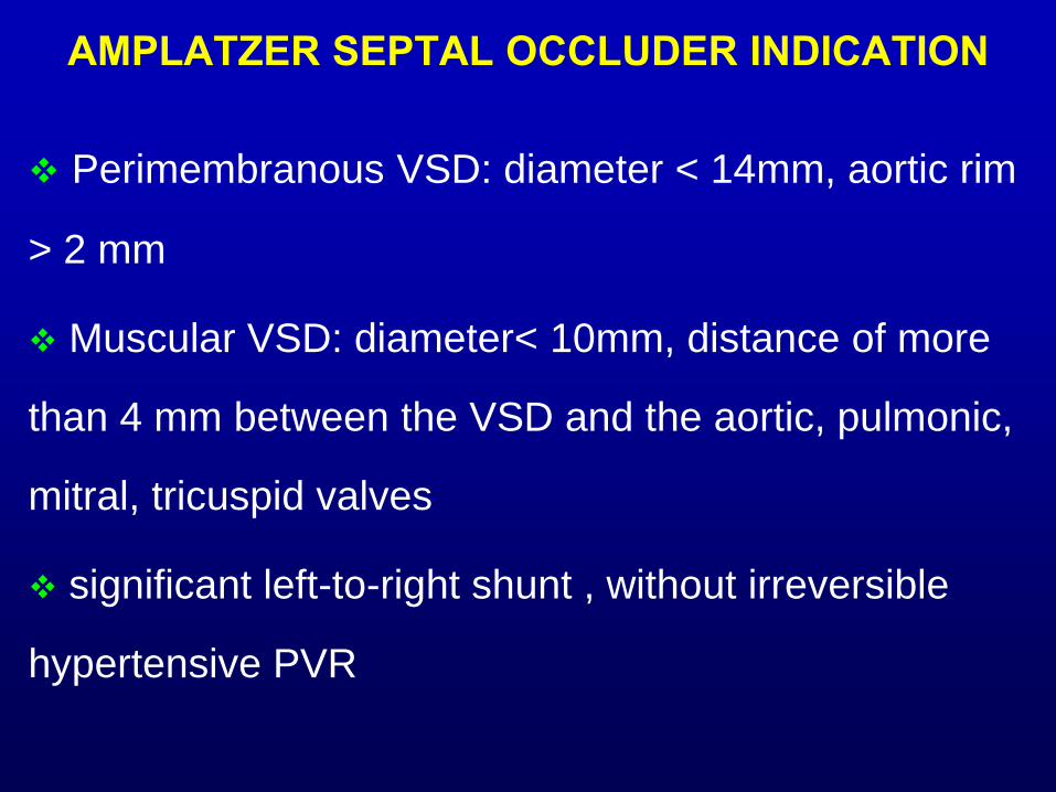
AMPLATZER SEPTAL OCCLUDER INDICATION
Perimembranous VSD: diameter < 14mm, aortic rim
> 2 mm
Muscular VSD: diameter< 10mm, distance of more
than 4 mm between the VSD and the aortic, pulmonic,
mitral, tricuspid valves
significant left-to-right shunt , without irreversible
hypertensive PVR
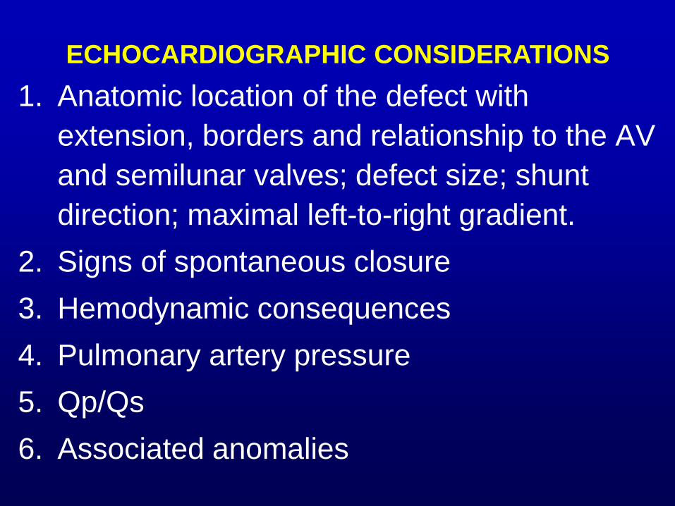
ECHOCARDIOGRAPHIC CONSIDERATIONS
1. Anatomic location of the defect with
extension, borders and relationship to the AV
and semilunar valves; defect size; shunt
direction; maximal left-to-right gradient.
2. Signs of spontaneous closure
3. Hemodynamic consequences
4. Pulmonary artery pressure
5. Qp/Qs
6. Associated anomalies
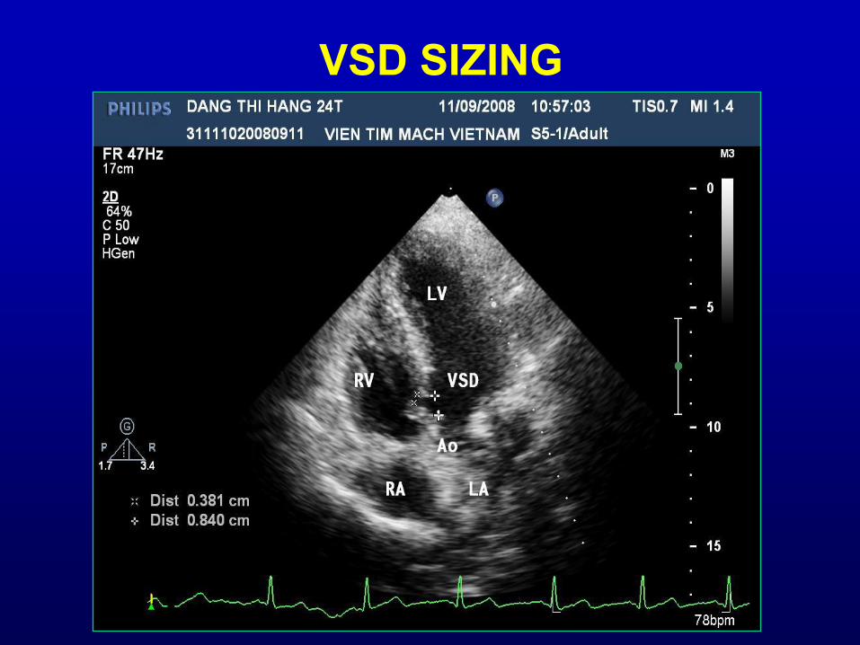
VSD sizing
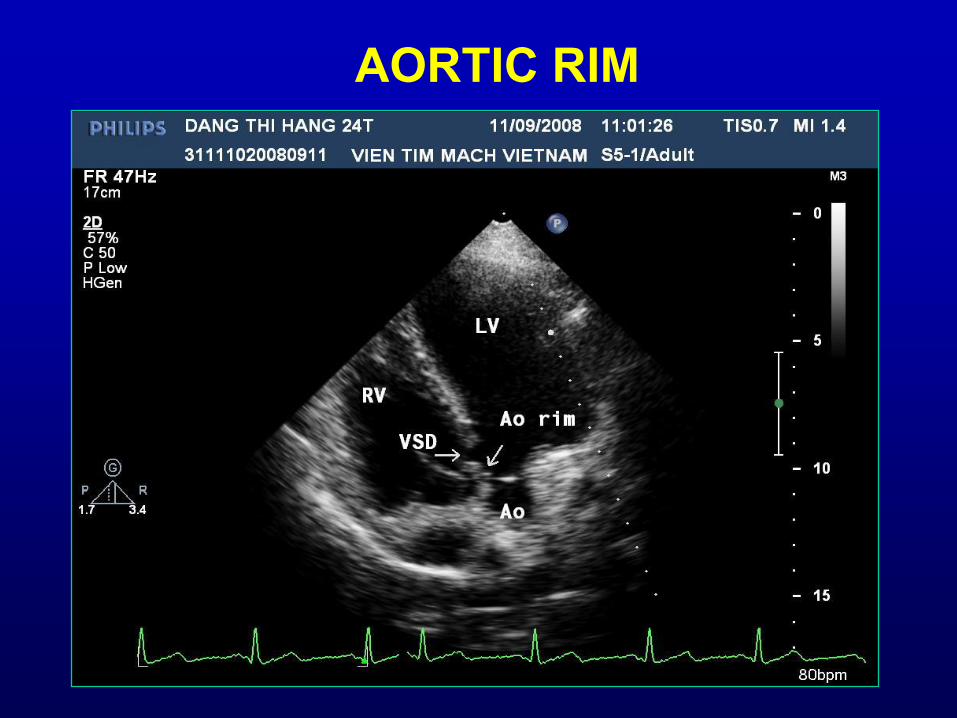
Aortic rim
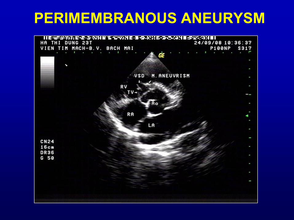
Perimembranous aneurysm
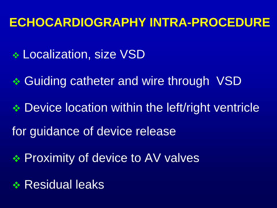
ECHOCARDIOGRAPHY INTRA-PROCEDURE
Localization, size VSD
Guiding catheter and wire through VSD
Device location within the left/right ventricle
for guidance of device release
Proximity of device to AV valves
Residual leaks
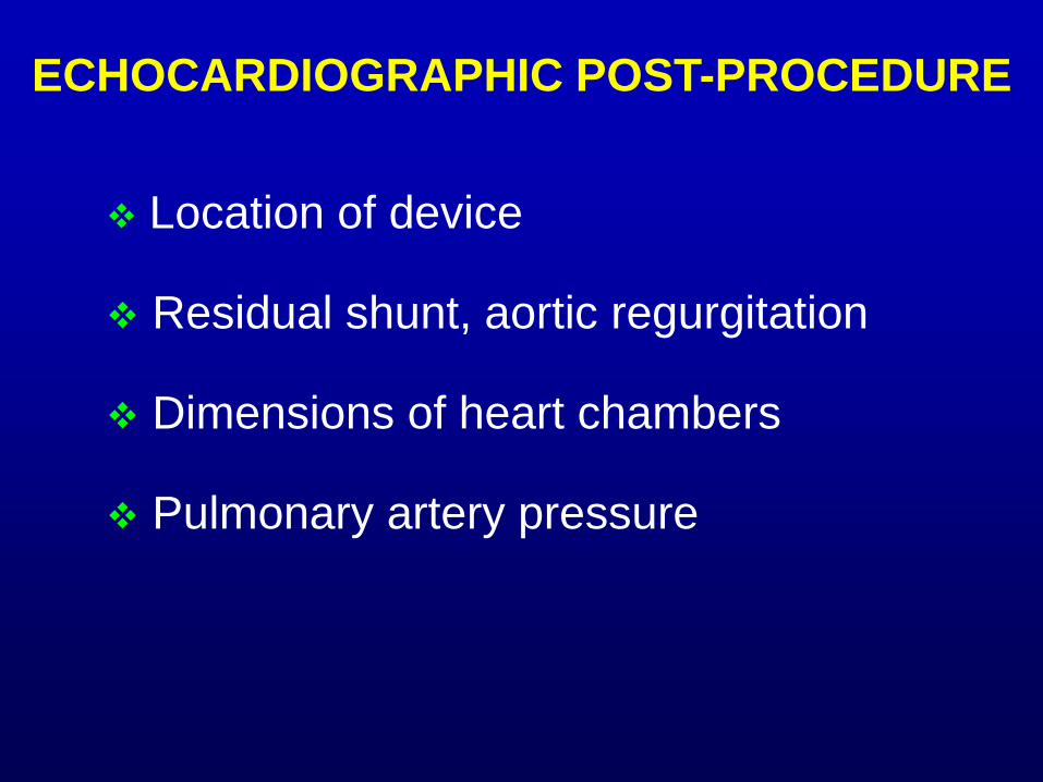
Location of device
Residual shunt, aortic regurgitation
Dimensions of heart chambers
Pulmonary artery pressure
ECHOCARDIOGRAPHIC POST-PROCEDURE
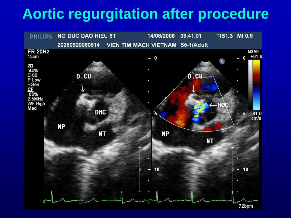
Aortic regurgitation after procedure
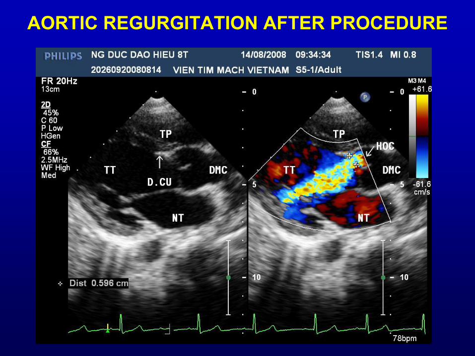
Aortic regurgitation after procedure
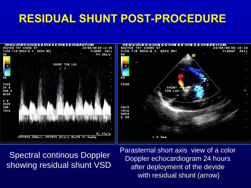
Residual Shunt post-procedure
Spectral continous Doppler
showing residual shunt VSD
Parasternal short axis view of a color
Doppler echocardiogram 24 hours
after deployment of the devide
with residual shunt (arrow)
PATENT DUCTUS
ARTERIOSUS
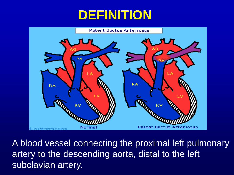
DEFINITION
A blood vessel connecting the proximal left pulmonary
artery to the descending aorta, distal to the left
subclavian artery.
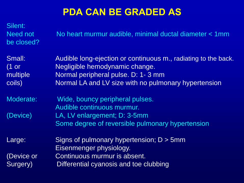
PDA can be graded as
Silent:
Need not No heart murmur audible, minimal ductal diameter < 1mm
be closed?
Small: Audible long-ejection or continuous m., radiating to the back.
(1 or Negligible hemodynamic change.
multiple Normal peripheral pulse. D: 1- 3 mm
coils) Normal LA and LV size with no pulmonary hypertension
Moderate: Wide, bouncy peripheral pulses.
Audible continuous murmur.
(Device) LA, LV enlargement; D: 3-5mm
Some degree of reversible pulmonary hypertension
Large: Signs of pulmonary hypertension; D > 5mm
Eisenmenger physiology.
(Device or Continuous murmur is absent.
Surgery) Differential cyanosis and toe clubbing
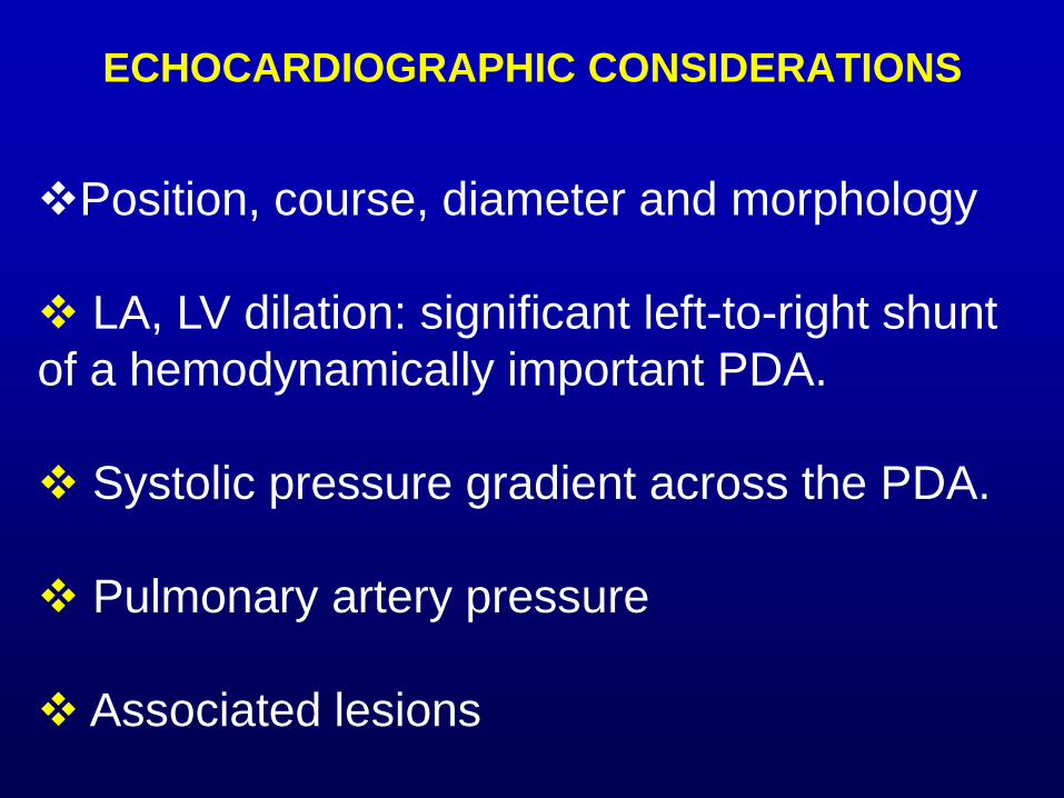
ECHOCARDIOGRAPHIC CONSIDERATIONS
Position, course, diameter and morphology
LA, LV dilation: significant left-to-right shunt
of a hemodynamically important PDA.
Systolic pressure gradient across the PDA.
Pulmonary artery pressure
Associated lesions
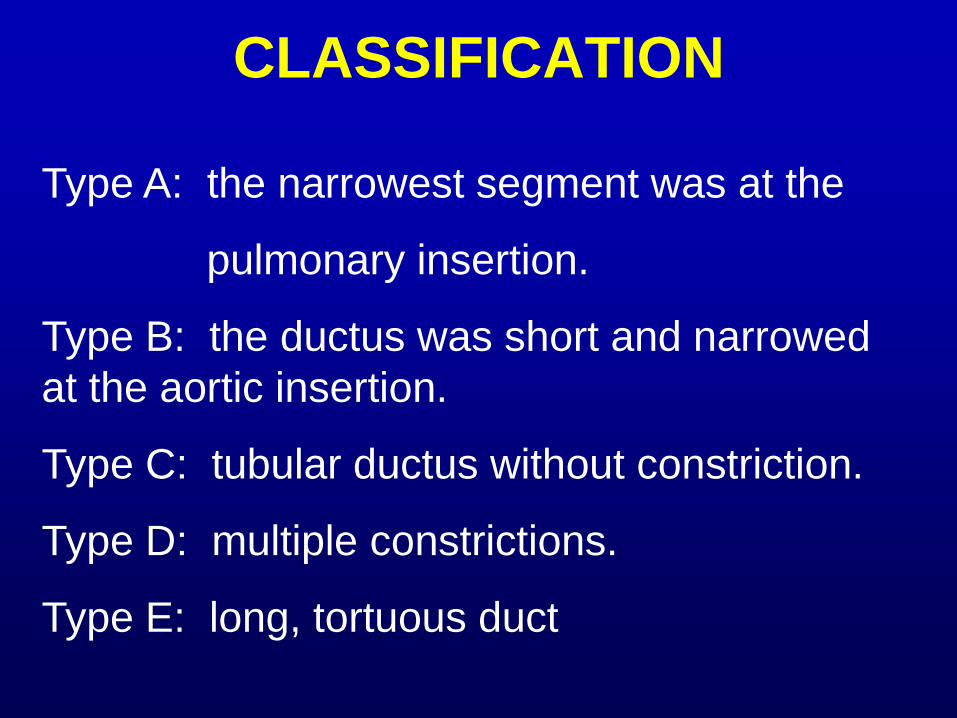
CLASSIFICATION
Type A: the narrowest segment was at the
pulmonary insertion.
Type B: the ductus was short and narrowed
at the aortic insertion.
Type C: tubular ductus without constriction.
Type D: multiple constrictions.
Type E: long, tortuous duct
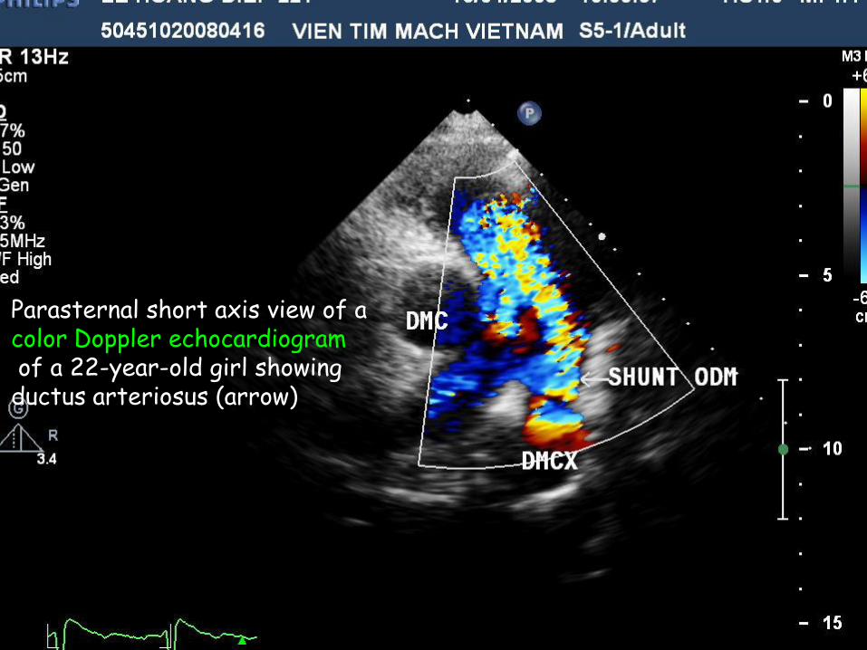
Parasternal short axis view of a color Doppler echocardiogramof a 22-year-old girl showingductus arteriosus (arrow)
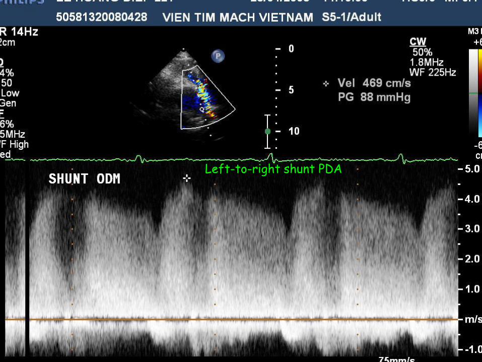
Left-to-right shunt PDA
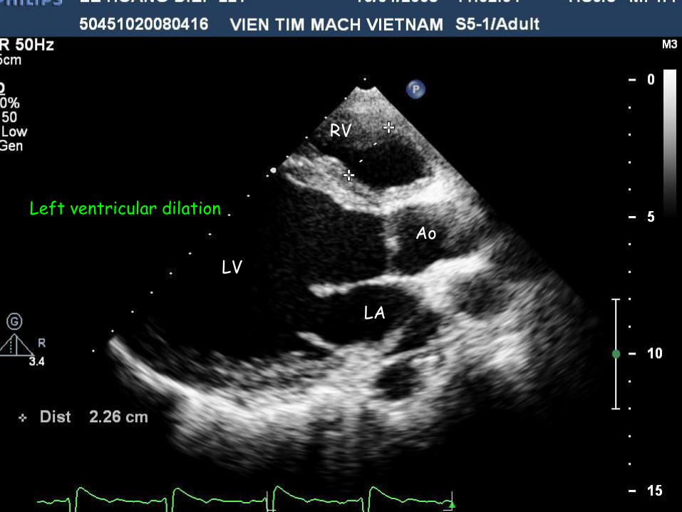
Left ventricular dilation
LV
RV
LA
Ao
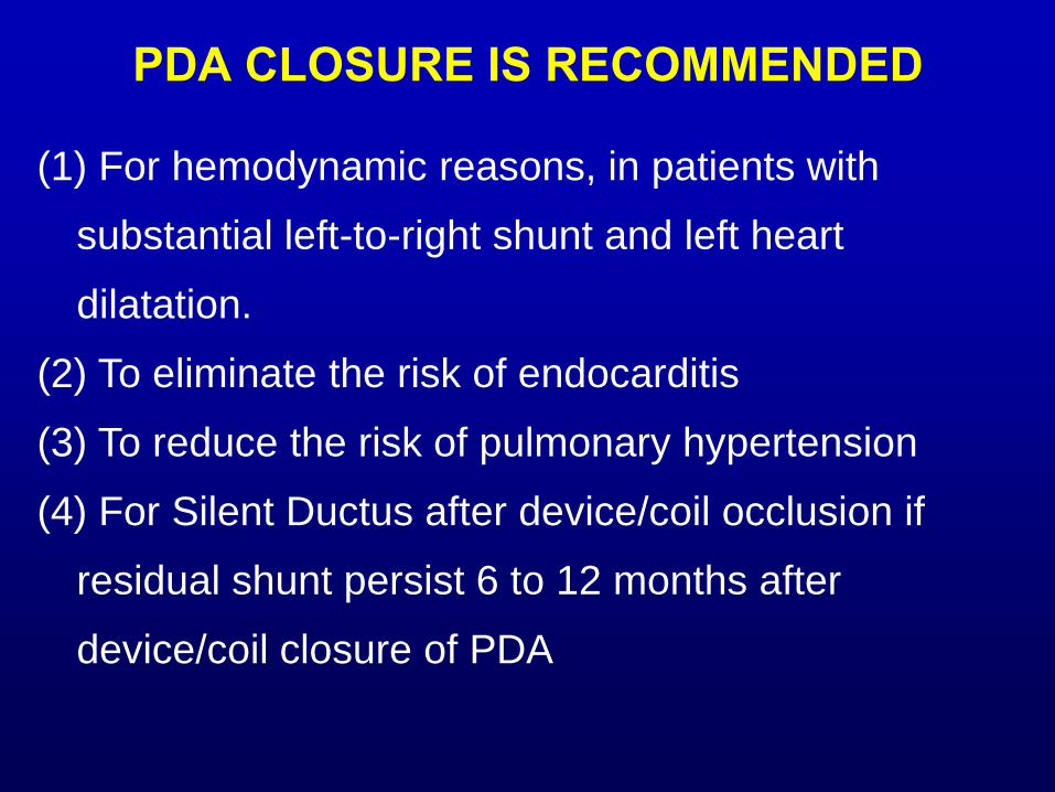
PDA closure is recommended
(1) For hemodynamic reasons, in patients with
substantial left-to-right shunt and left heart
dilatation.
(2) To eliminate the risk of endocarditis
(3) To reduce the risk of pulmonary hypertension
(4) For Silent Ductus after device/coil occlusion if
residual shunt persist 6 to 12 months after
device/coil closure of PDA
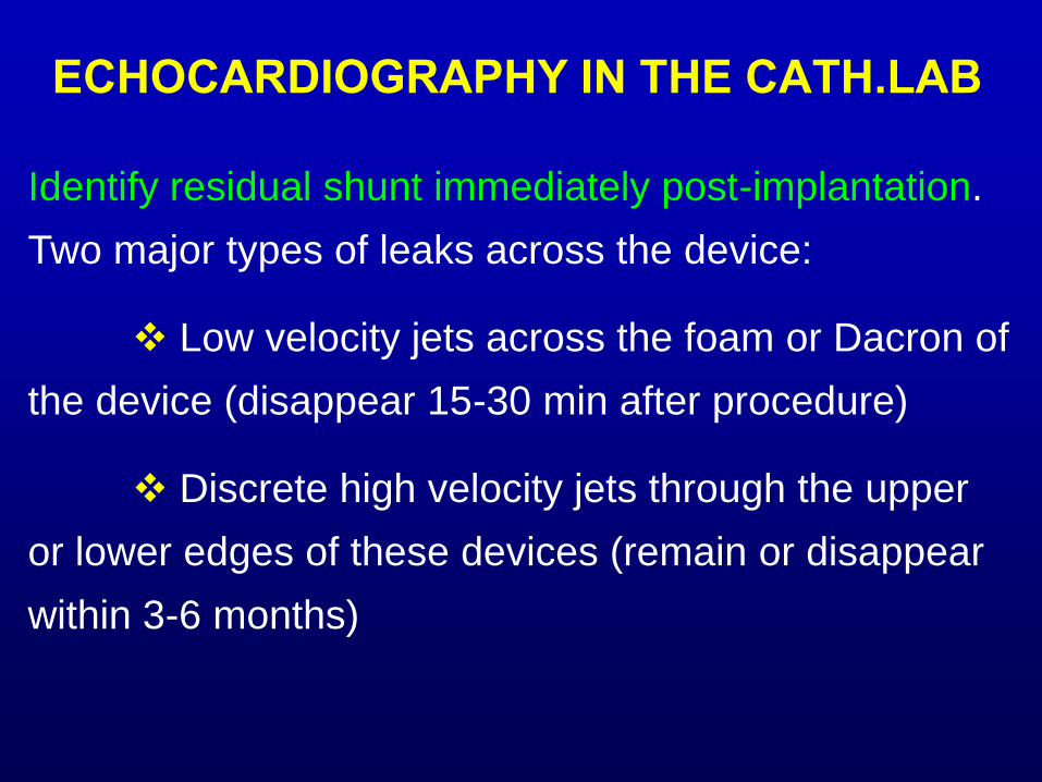
Echocardiography in the cath.lab
Identify residual shunt immediately post-implantation.
Two major types of leaks across the device:
Low velocity jets across the foam or Dacron of
the device (disappear 15-30 min after procedure)
Discrete high velocity jets through the upper
or lower edges of these devices (remain or disappear
within 3-6 months)
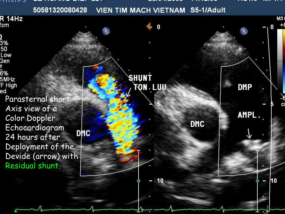
Parasternal shortAxis view of a Color Doppler Echocardiogram24 hours afterDeployment of theDevide (arrow) withResidual shunt.
VALVULAR PULMONARY
STENOSIS
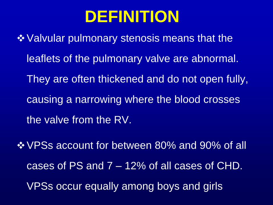
Valvular pulmonary stenosis means that the
leaflets of the pulmonary valve are abnormal.
They are often thickened and do not open fully,
causing a narrowing where the blood crosses
the valve from the RV.
VPSs account for between 80% and 90% of all
cases of PS and 7 – 12% of all cases of CHD.
VPSs occur equally among boys and girls
DEFINITION
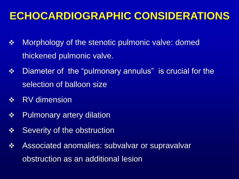
Morphology of the stenotic pulmonic valve: domed
thickened pulmonic valve.
Diameter of the “pulmonary annulus” is crucial for the
selection of balloon size
RV dimension
Pulmonary artery dilation
Severity of the obstruction
Associated anomalies: subvalvar or supravalvar
obstruction as an additional lesion
ECHOCARDIOGRAPHIC CONSIDERATIONS
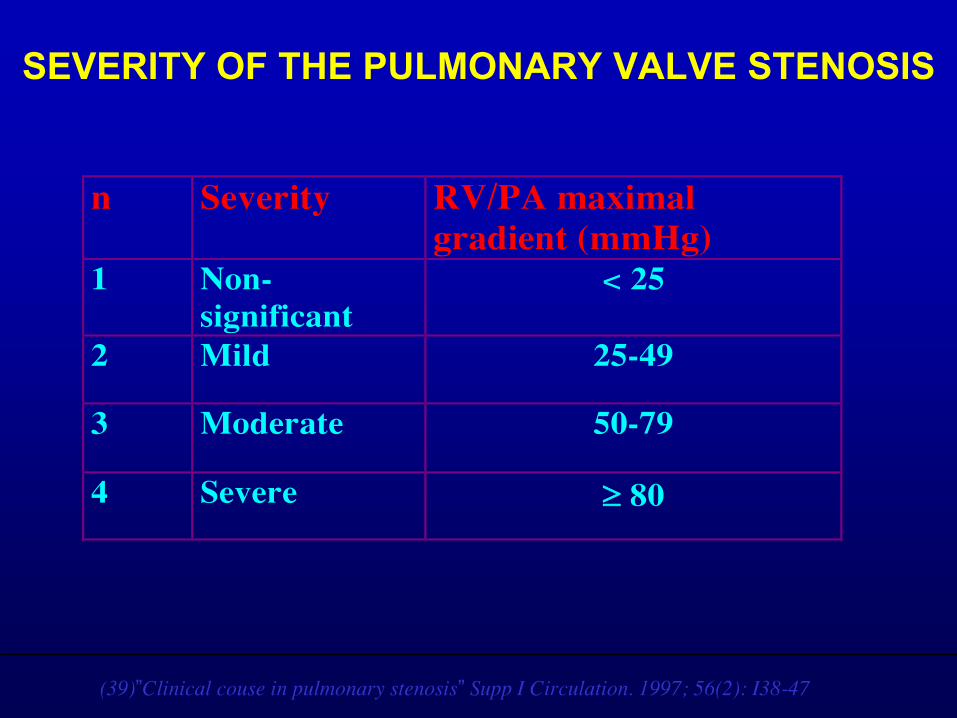
Severity of the pulmonary valve stenosis
(39)”Clinical couse in pulmonary stenosis” Supp I Circulation. 1997; 56(2): I38-47
n Severity RV/PA maximal
gradient (mmHg)
1 Non-
significant
< 25
2 Mild 25-49
3 Moderate 50-79
4 Severe 80
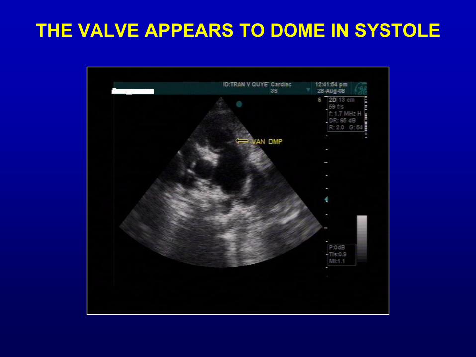
The valve appears to dome in systole
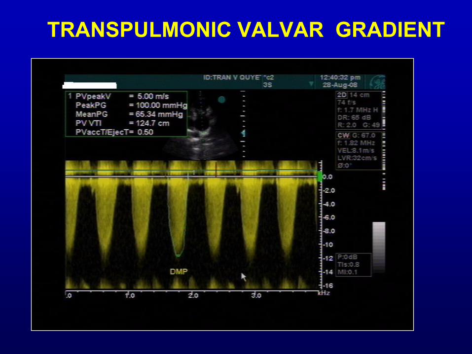
Transpulmonic valvar gradient
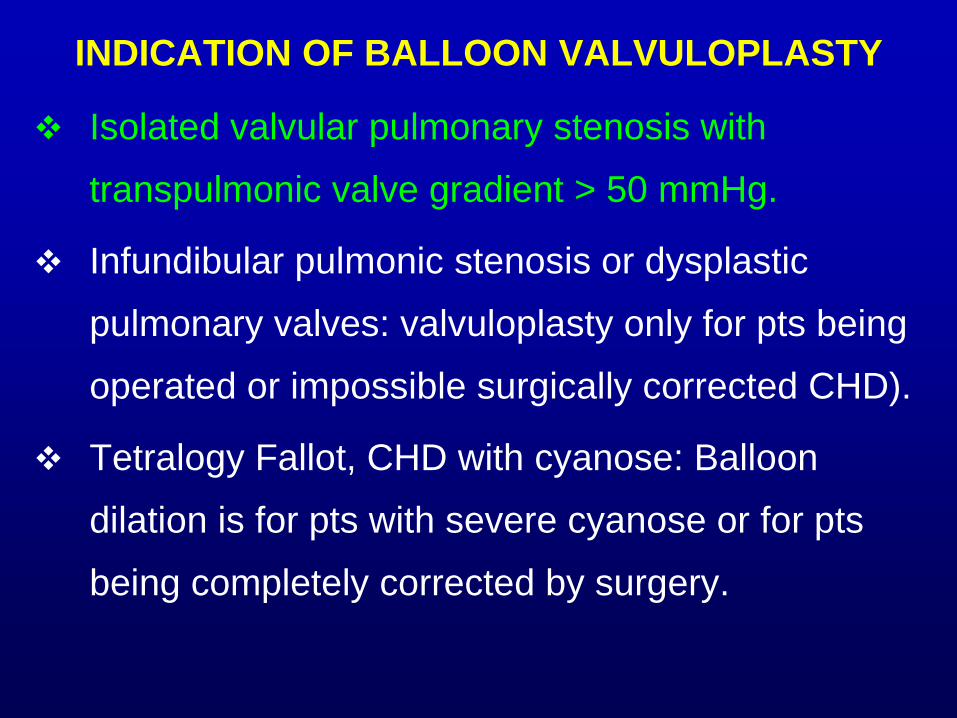
INDICATION OF BALLOON VALVULOPLASTY
Isolated valvular pulmonary stenosis with
transpulmonic valve gradient > 50 mmHg.
Infundibular pulmonic stenosis or dysplastic
pulmonary valves: valvuloplasty only for pts being
operated or impossible surgically corrected CHD).
Tetralogy Fallot, CHD with cyanose: Balloon
dilation is for pts with severe cyanose or for pts
being completely corrected by surgery.
Echocardiography in the cath.lab.
Echocardiography can identify if the
catheters centered across pulmonary
valves before inflating the balloon.
Residual obstruction and insufficiency
caused by balloon dilation procedure
The cusp perforation or avulsion
Echocardiography after procedure
- RV and LV dimensions, RV systolic pressure,
thickness of RV wall.
- Septal mouvement.
- Diameter of PA
- Transpulmonic and transinfundibular gradients
- Pulmonary regurgitation
CONGENITAL CORONARY
ARTERY FISTULA
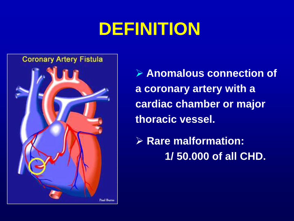
DEFINITION
Anomalous connection of
a coronary artery with a
cardiac chamber or major
thoracic vessel.
Rare malformation:
1/ 50.000 of all CHD.
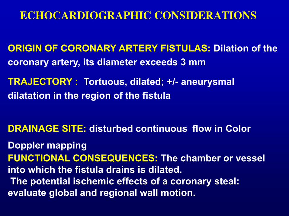
ORIGIN OF CORONARY ARTERY FISTULAS: Dilation of the
coronary artery, its diameter exceeds 3 mm
TRAJECTORY : Tortuous, dilated; +/- aneurysmal
dilatation in the region of the fistula
DRAINAGE SITE: disturbed continuous flow in Color
Doppler mapping
FUNCTIONAL CONSEQUENCES: The chamber or vessel
into which the fistula drains is dilated.
The potential ischemic effects of a coronary steal:
evaluate global and regional wall motion.
ECHOCARDIOGRAPHIC CONSIDERATIONS
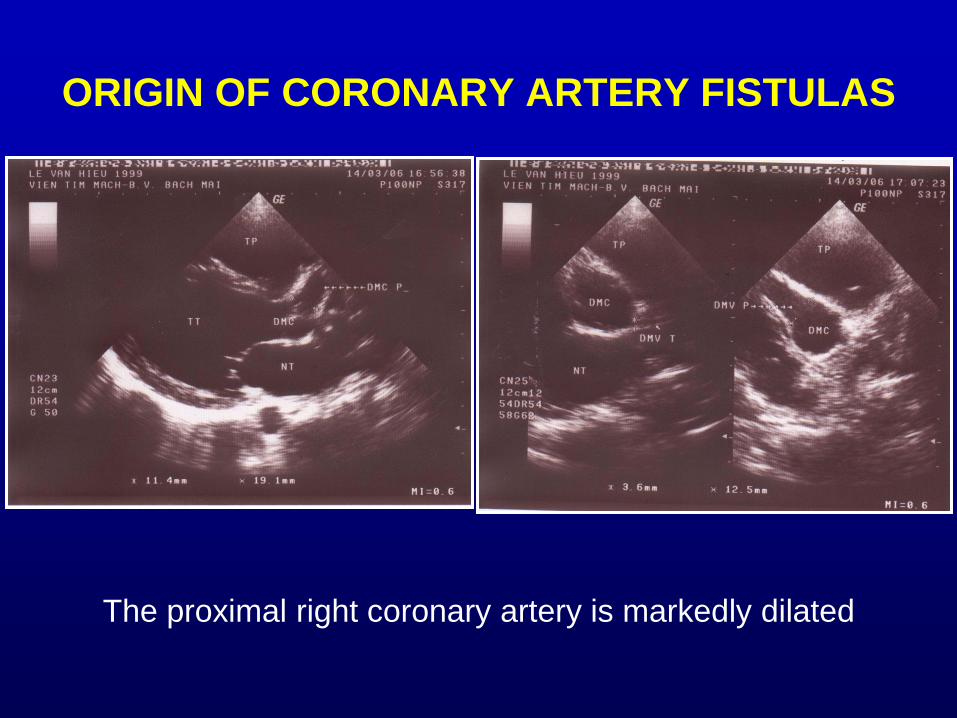
ORIGIN OF CORONARY ARTERY FISTULAS
The proximal right coronary artery is markedly dilated
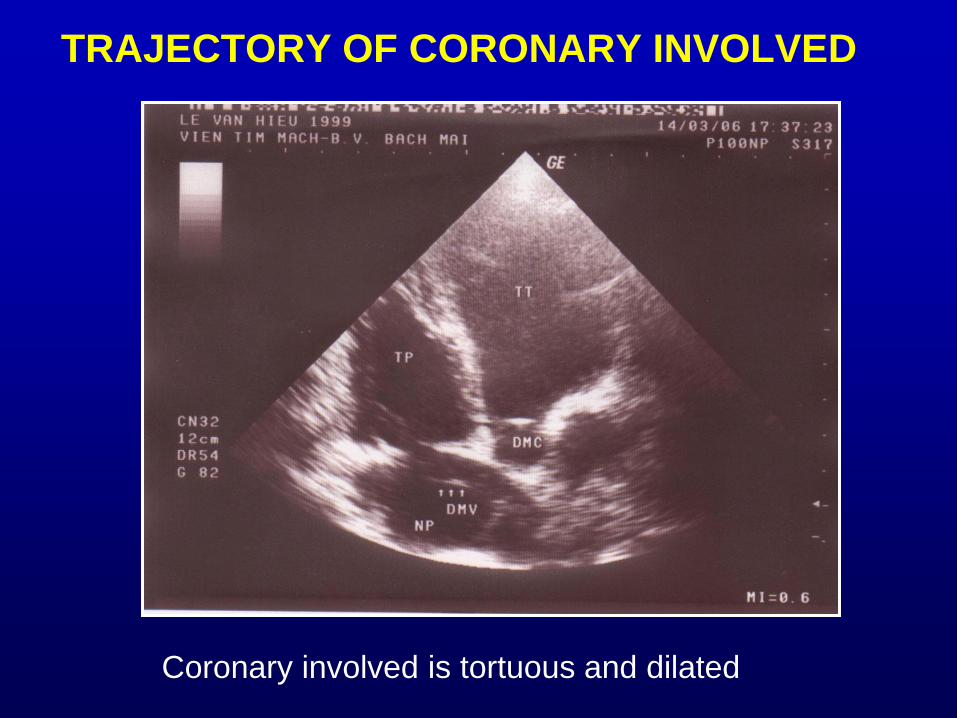
TRAJECTORY OF CORONARY INVOLVED
Coronary involved is tortuous and dilated
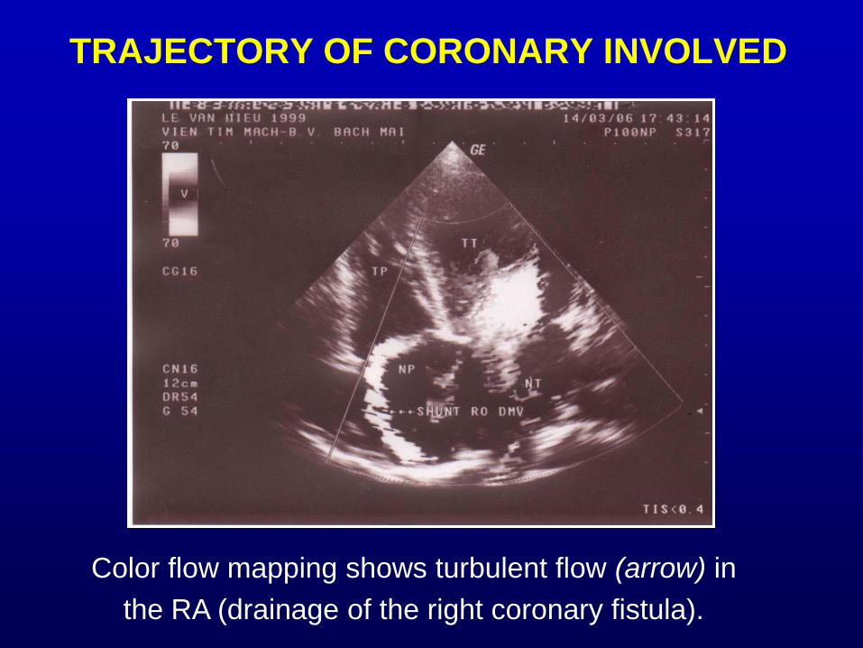
TRAJECTORY OF CORONARY INVOLVED
Color flow mapping shows turbulent flow (arrow) in
the RA (drainage of the right coronary fistula).
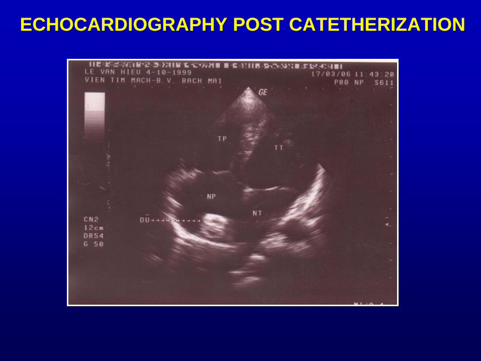
ECHOCARDIOGRAPHY POST CATETHERIZATION
OTHER MALFORMATIONS
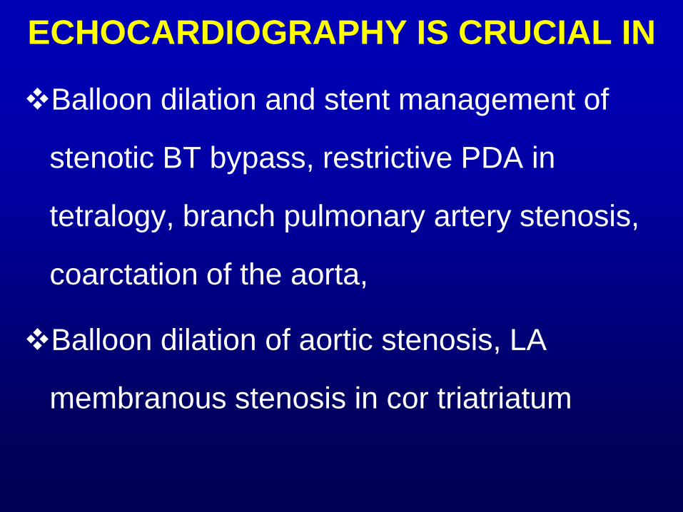
ECHOCARDIOGRAPHY IS CRUCIAL IN
Balloon dilation and stent management of
stenotic BT bypass, restrictive PDA in
tetralogy, branch pulmonary artery stenosis,
coarctation of the aorta,
Balloon dilation of aortic stenosis, LA
membranous stenosis in cor triatriatum
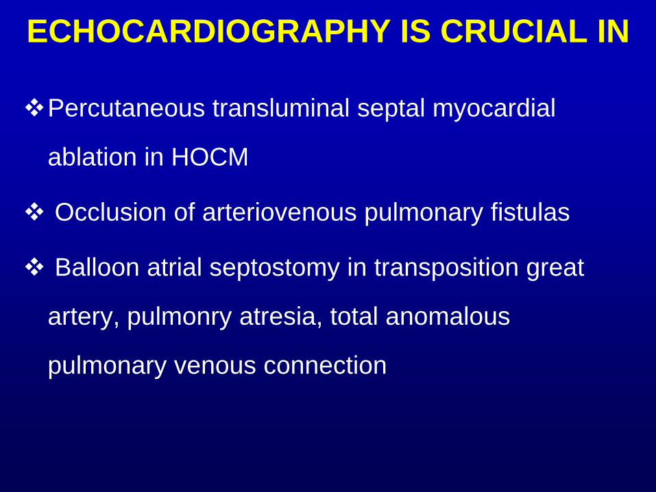
ECHOCARDIOGRAPHY IS CRUCIAL IN
Percutaneous transluminal septal myocardial
ablation in HOCM
Occlusion of arteriovenous pulmonary fistulas
Balloon atrial septostomy in transposition great
artery, pulmonry atresia, total anomalous
pulmonary venous connection


![[WeGO e-Government Program]City Paper Presentation : Hanoi(Vietnam)](https://static.fdocuments.us/doc/165x107/55c4c1a0bb61eb061e8b478e/wego-e-government-programcity-paper-presentation-hanoivietnam.jpg)