
Vg2Vd2 T Cell Receptor Recognition of Prenyl Pyrophosphates Is ...
15
of April 3, 2018. This information is current as CDRs Prenyl Pyrophosphates Is Dependent on All 2 T Cell Receptor Recognition of δ 2V γ V Hong Wang, Zhimei Fang and Craig T. Morita http://www.jimmunol.org/content/184/11/6209 doi: 10.4049/jimmunol.1000231 2010; 2010; 184:6209-6222; Prepublished online 5 May J Immunol Material Supplementary 1.DC1 http://www.jimmunol.org/content/suppl/2010/05/05/jimmunol.100023 References http://www.jimmunol.org/content/184/11/6209.full#ref-list-1 , 39 of which you can access for free at: cites 104 articles This article average * 4 weeks from acceptance to publication Fast Publication! • Every submission reviewed by practicing scientists No Triage! • from submission to initial decision Rapid Reviews! 30 days* • Submit online. ? The JI Why Subscription http://jimmunol.org/subscription is online at: The Journal of Immunology Information about subscribing to Permissions http://www.aai.org/About/Publications/JI/copyright.html Submit copyright permission requests at: Email Alerts http://jimmunol.org/alerts Receive free email-alerts when new articles cite this article. Sign up at: Print ISSN: 0022-1767 Online ISSN: 1550-6606. Immunologists, Inc. All rights reserved. Copyright © 2010 by The American Association of 1451 Rockville Pike, Suite 650, Rockville, MD 20852 The American Association of Immunologists, Inc., is published twice each month by The Journal of Immunology by guest on April 3, 2018 http://www.jimmunol.org/ Downloaded from by guest on April 3, 2018 http://www.jimmunol.org/ Downloaded from
Transcript of Vg2Vd2 T Cell Receptor Recognition of Prenyl Pyrophosphates Is ...
of April 3, 2018.This information is current as
CDRsPrenyl Pyrophosphates Is Dependent on All
2 T Cell Receptor Recognition ofδ2VγV
Hong Wang, Zhimei Fang and Craig T. Morita
http://www.jimmunol.org/content/184/11/6209doi: 10.4049/jimmunol.10002312010;
2010; 184:6209-6222; Prepublished online 5 MayJ Immunol
MaterialSupplementary
1.DC1http://www.jimmunol.org/content/suppl/2010/05/05/jimmunol.100023
Referenceshttp://www.jimmunol.org/content/184/11/6209.full#ref-list-1
, 39 of which you can access for free at: cites 104 articlesThis article
average*
4 weeks from acceptance to publicationFast Publication! •
Every submission reviewed by practicing scientistsNo Triage! •
from submission to initial decisionRapid Reviews! 30 days* •
Submit online. ?The JIWhy
Subscriptionhttp://jimmunol.org/subscription
is online at: The Journal of ImmunologyInformation about subscribing to
Permissionshttp://www.aai.org/About/Publications/JI/copyright.htmlSubmit copyright permission requests at:
Email Alertshttp://jimmunol.org/alertsReceive free email-alerts when new articles cite this article. Sign up at:
Print ISSN: 0022-1767 Online ISSN: 1550-6606. Immunologists, Inc. All rights reserved.Copyright © 2010 by The American Association of1451 Rockville Pike, Suite 650, Rockville, MD 20852The American Association of Immunologists, Inc.,
is published twice each month byThe Journal of Immunology
by guest on April 3, 2018
http://ww
w.jim
munol.org/
Dow
nloaded from
by guest on April 3, 2018
http://ww
w.jim
munol.org/
Dow
nloaded from

The Journal of Immunology
Vg2Vd2 T Cell Receptor Recognition of PrenylPyrophosphates Is Dependent on All CDRs
Hong Wang, Zhimei Fang, and Craig T. Morita
gd T cells differ from ab T cells in the Ags they recognize and their functions in immunity. Although most ab TCRs recognize
peptides presented by MHC class I or II, human gd T cells expressing Vg2Vd2 TCRs recognize nonpeptide prenyl pyrophos-
phates. To define the molecular basis for this recognition, the effect of mutations in the TCR CDR was assessed. Mutations in all
CDR loops altered recognition and cover a large footprint. Unlike murine gd TCR recognition of the MHC class Ib T22 protein,
there was no CDR3d motif required for recognition because only one residue is required. Instead, the length and sequence of
CDR3g was key. Although a prenyl pyrophosphate-binding site was defined by Lys109 in Jg1.2 and Arg51 in CDR2d, the area
outlined by critical mutations is much larger. These results show that prenyl pyrophosphate recognition is primarily by germline-
encoded regions of the gd TCR, allowing a high proportion of Vg2Vd2 TCRs to respond. This underscores its parallels to innate
immune receptors. Our results also provide strong evidence for the existence of an Ag-presenting molecule for prenyl py-
rophosphates. The Journal of Immunology, 2010, 184: 6209–6222.
Tcells can be divided into two distinct subsets, gd and ab,based on their expression of rearranging adaptive TCRs.gd T cells, mucosal-associated invariant ab T cells (1),
and invariant NK ab T cells (iNKTs) (2, 3) are innate lympho-cytes that bridge innate and adaptive immunity through theirrecognition of unconventional ligands. The major subset of humangd T cells expresses Vg2Vd2 TCR (also termed Vg9Vd2 orTRGV9TRDV2). Although a minor subset at birth (4), Vg2Vd2T cells expand between the ages of 1–10 y old (5). This expansionis not due to genetic causes because identical twins can differ intheir proportions of Vg2Vd2 T cells. Instead, a broad array ofbacterial and protozoal infections expand Vg2Vd2 T cells to largenumbers (.50% of blood T cells in some cases) (reviewed inRef. 6).The broad reactivity of Vg2Vd2 T cells to microbes and some
tumors was explained by the finding that nonpeptide prenylpyrophosphates, such as isopentenyl pyrophosphate (IPP), act asAgs for Vg2Vd2 T cells (7). Prenyl pyrophosphates are essentialmetabolites required by all organisms. The recognition of mi-crobes by Vg2Vd2 T cells is due to the preferential recognitionof (E)-4-hydroxy-3-methyl-but-2-enyl pyrophosphate (HMBPP)over IPP (HMBPP is 30,000-fold more active than IPP on
a molar basis) (8, 9). HMBPP is a metabolite in the 2-C-methyl-D-erythritol-4 phosphate pathway for isoprenoid biosynthesis.This pathway is used by most Eubacteria (including all myco-bacteria and Gram-negative rods) and Apicomplexan protozoa(the causative agents of malaria, toxoplasmosis, babesiosis, andcryptosporidiosis). Other stimulating compounds, such as bis-phosphonates (10, 11) and alkylamines (12), act by increasingcellular IPP levels by blocking farnesyl pyrophosphate (FPP)synthase in the mevalonate pathway (13, 14).Vg2Vd2 T cells function in both microbial and tumor immunity.
The early expansion of Vg2Vd2 T cells to prenyl pyrophosphatesresults in conversion to memory cells (C. Jin and C.T. Morita,unpublished observations) (15) capable of mounting adaptive re-sponses to Mycobacterium bovis bacillus Calmette-Guerin (BCG)(16) and other infections. As memory cells, Vg2Vd2 T cells canmount rapid responses to primary microbial infections with or-ganisms using the 2-C-methyl-D-erythritol-4 phosphate pathway(9). Activation of Vg2Vd2 T cells leads them to release Th1 cy-tokines, including IFN-g, TNF-a, and GM-CSF, but not IL-2 (17,18), and to secrete chemokines, such as MIP-1a (CCL3), MIP-1b(CCL4), lymphotactin (XCL1), and RANTES (CCL5) (19, 20).Vg2Vd2 T cells are potent killer cells and can lyse infected cells viaperforin and/or Fas-Fas ligand pathways (21) and kill releasedbacteria and protozoa through production of granulysin (21, 22) andthe cathelicidin LL-37 (23). Activated Vg2Vd2 T cells also killtumor cells from a variety of tissue origins through both TCR- andNK receptor-mediated recognition (reviewed in Ref. 6). Treatmentwith bisphosphonates and IL-2 activates Vg2Vd2 T cells, leading tostable disease or partial remissions in some patients with B cellmalignancies (24) or with metastatic prostate cancer (25). There arepresently several ongoing clinical trials examining immunotherapywith Vg2Vd2 T cells for a variety of cancers.Recognition of prenyl pyrophosphates is mediated by the Vg2Vd2
TCR because transfection of the Vg2Vd2 TCR to b2 Jurkat cellsconfers reactivity to prenyl pyrophosphates and Daudi and RPMI-8226 tumor cell lines (26). We and others proposed that reactivity tothe negatively charged prenyl pyrophosphates was dependent onpositively charged residues in a potential prenyl pyrophosphate-binding site located in Vg2Vd2 TCR (27, 28). Consistent with thishypothesis, reactivity was dependent on N-encoded amino acid
Division of Rheumatology, Department of Internal Medicine, Interdisciplinary Grad-uate Program in Immunology, University of Iowa College of Medicine, Iowa City, IA52242
Received for publication January 25, 2010. Accepted for publication March 31, 2010.
This work was supported by grants from the National Institutes of Health, NationalInstitute of Arthritis and Musculoskeletal and Skin Disease (AR45504), the NationalInstitute of Allergy and Infectious Diseases (Midwest Regional Center of Excellencefor Biodefense and Emerging Infectious Diseases Research, AI057160), and theNational Cancer Institute (CA113874) to C.T.M.
Address correspondence and reprint requests to Dr. Craig T. Morita, Division ofRheumatology, Department of Internal Medicine, Interdisciplinary Graduate Programin Immunology, University of Iowa College of Medicine, EMRB 400F, Iowa City, IA52242. E-mail address: [email protected]
The online version of this article contains supplemental material.
Abbreviations used in this paper: BSA, buried surface area; HMBPP, (E)-4-hydroxy-3-methyl-but-2-enyl pyrophosphate; iNKT, invariant NK ab T cell; IPP, isopentenylpyrophosphate.
Copyright� 2010 by The American Association of Immunologists, Inc. 0022-1767/10/$16.00
www.jimmunol.org/cgi/doi/10.4049/jimmunol.1000231
by guest on April 3, 2018
http://ww
w.jim
munol.org/
Dow
nloaded from

residues in the junctional segment of the Vg2 chain (29) as well aslysine residues in the Jg1.2 segment (also termed JgP), an arginineresidue in CDR2d, and an aliphatic amino acid residue in CDR3d(30, 31).Despite identification of a potential prenyl pyrophosphate-
binding site, there was no evidence for prenyl pyrophosphate bind-ing by the Vg2Vd2 TCR (28) or direct functional recognition (32).Instead, recognition of prenyl pyrophosphates required cell-cellcontact but neither Ag internalization nor processing (32, 33). Thisis similar to allergic CD4 and CD8 ab T cell recognition of smallnonpeptide drugs presented by MHC class I or class II molecules(34). In contrast to drug recognition, prenyl pyrophosphate Agrecognition is independent of classical MHC class I, MHC class II,b2-microglobulin, MICA/MICB, and CD1 molecules (32). More-over, photoaffinity analogs of prenyl pyrophosphates could formcovalent bonds to a putative presenting molecule distinct fromknown Ag-presenting molecules (35), and tetramers of macaqueVg2Vd2 TCR could bind to the surface of human but not mousecells only in the presence of HMBPP (36).The available studies suggest, but do not prove, that a pre-
senting molecule exists for prenyl pyrophosphates. To help clarifythis issue, we have studied the effect of alanine mutations inresidues in CDR1, CDR2, and CDR3 on prenyl pyrophosphaterecognition and analyzed CDR3 sequences of reactive Vg2Vd2T cell clones. We find that residues in all CDRs of the Vg2Vd2TCR can affect recognition, but no CDR3d motif is required forrecognition beyond the requirement for an aliphatic residue atposition 97. Instead, recognition is critically dependent on theVg2Jg1.2 chain, which is either invariant or has limited diversityin sequence and length. Given the large footprint of the requiredresidues, which surpasses the predicted prenyl pyrophosphate-binding site, it is likely that a presenting molecule is required forprenyl pyrophosphate Ags.
Materials and MethodsCell lines
The mutant Jurkat cell line, J.RT3-T3.5, was obtained from the AmericanType Culture Collection (Rockville, MD) and is a TCR b-negative variantof Jurkat that lacks TCR cell surface expression (37). J.RT3-T3.5 andJ.RT3-T3.5 transfectants were grown in RPMI 1640 supplemented with10% FCS, 10 mM HEPES, 10 mg/ml gentamycin, 5 3 1025 M 2-ME, andL-glutamine (Invitrogen, Carlsbad, CA). RPMI 8226 and Raji cells wereobtained from the American Type Culture Collection and maintained in themedium described above. The Va2 human fibroblast cell line was obtainedfrom Dr. Charles Stiles (Dana-Farber Cancer Institute) and maintained inDMEM with the additives listed above. Hygromycin B and G418 wereobtained from Invitrogen.
mAbs and Ags
mAbs reactive with the human Vg2Vd2 TCR used in this paper were asfollows: control mAb (P3), anti-pan TCRgd (anti-TCRd1), anti-Vg2(TigA, 7A5, 360, and 4D7), and anti-Vd2 (BB3, 4G6, 389, G1, andTiVd2). The specificity of these mAbs was previously reviewed (38).Prenyl pyrophosphates (IPP, dimethylallyl pyrophosphate, and FPP) andalkylamines were purchased from Sigma-Aldrich (St. Louis, MO).Phosphoantigens including monomethyl-, mono- ethyl-, phenethyl-, andisoamyl-pyrophosphate and the nucleotide-conjugated compound, ethyl-deoxyuridine triphosphate were synthesized as described (7, 38). Bro-mohydrin pyrophosphate, risedronate, and alendronate were provided byEric Oldfield (University of Illinois at Urbana-Champaign, Champaign,IL). HMBPP was synthesized as previously described (39). KM20 andKM22 are extracts of Escherichia coli strain lytB mutants that producea high level of HMBPP (9, 40).
cDNA cloning and mutagenesis of human Vg2Vd2 TCR
RNA was isolated from the human T cell clone DG.SF13 (Micro RNAisolation kit; Stratagene, La Jolla, CA) followed by cDNA synthesis usingSuperScript II reverse transcriptase and random hexamers (SuperScript first-
strand synthesis system for RT-PCR; Life Technologies, Gaithersburg, MD).PCR was done with Platinum Taq High Fidelity DNA polymerase (LifeTechnologies). PCR primers used to derive full-length Vg2Cg-chains were asdescribed previously (21). For the Vd2Cd-chain, the following primers wereused to introduce an XhoI restriction site into the 59 region and a BamHI siteinto the 39 region of the Vd2Cd-chain for cloning: 59-gggctcgagCAGG-CAGAAGGTGGTTGAGAG-39, Vd2 59 untranslated region; and 59-gggg-gatccGGAGTGTAGCTTCCTCAT-39, Vd2 39 untranslated region. TheVg2Jg1.2Cg1 PCR product was cloned into the pREP7 vector (Invitrogen)using the KpnI–XhoI sites. The Vd2Cd PCR product was cloned into thepREP9 vector (Invitrogen) using the XhoI–BamHI sites. For mutagenesis,TCR-g and TCR-d–chain cDNAs mutated by a single amino acid residuewere generated using QuikChange Site-Directed Mutagenesis Kit (Stra-tagene). To confirm the mutations, all Vg2Cg and Vd2Cdmutants were fullysequenced. Sequencing was done using an automated sequencer using thepREP forward and reverse primers along with the following reverse primers:Cg 39 UT, ATGGCCTCCTTGTGCCACCG; Cg internal, 59-TGTGTCG-TTAGTCTTCATGG-39; Cd 39 UT, GGAGTGTAGCTTCCTCATGC; andCd internal, 59-GACAATAGCAGGATCAAACT-39.
Derivation of Vg2Vd2 TCR transfectants
Human Vg2Vd2 TCR transfectants were derived by electroporation of theJurkat mutant J.RT3-T3.5, with unmutated or mutated DG.SF13 TCRg- and d-chain cDNAs, as described previously (41). Briefly, J.RT3-T3.5cells were grown at low density (1 to 2 3 105 cells/ml) prior to use,centrifuged while in log phase growth, and resuspended at 3.33 3 107/mlin PBS containing 10 mM HEPES. A total of 0.3 ml resuspended cells wasaliquoted into each electroporation cuvette; 20 mg each plasmid (pREP7-TCR-g and REP9-TCR-d) was added to the cells followed by incubationat room temperature for 10 min. The cells were electroporated (960 mF,250 V) using a Gene Pulser (Bio-Rad, Burlingame, CA) and incubated atroom temperature for an additional 10 min. The electroporated cells fromeach cuvette were washed twice in PBS, resuspended into 30 ml completemedia, plated in three 96-well round-bottom plates, and cultured at 37˚C.After 48 h, hygromycin B was added to 0.5 mg/ml. Transfectants survivinghygromycin B selection were then cultured in media containing both G418(1 mg/ml) and hygromycin B (0.5 mg/ml). After 2 to 3 wk of culture,transfectants were screened for IL-2 release in response to anti-TCRd1stimulation. Transfectants specifically responsive to anti-TCR stimulationwere expanded at low density and tested for their response to nonpeptideAgs. Multiple transfectants for each mutant that specifically released IL-2to anti-TCR stimulation were derived from separate transfections andtested to confirm each result (Supplemental Figs. 1, 2). Because defects inJurkat TCR signaling caused some TCR-expressing transfectants (despitehigh levels of TCR expression) to either not release IL-2 or to constitu-tively produce IL-2, only those mutants responding to anti-TCRd1 stim-ulation were analyzed.
IL-2 release and assay
Stimulation of TCR transfectants for IL-2 release was performed as de-scribed (38, 42). Briefly, 1 3 105 transfectants were cultured with theindicated half-log dilution of the anti-TCRd1 mAb, stimulatory com-pounds, or tumor cells in the presence of 13 105 glutaraldehyde-fixed Va2cells (except for tumor cells) and 10 ng/ml PMA. After 24 h, supernatantswere harvested and frozen at 220˚C. For IL-2 assays, the supernatantswere thawed and used at a one-eighth dilution to stimulate the proliferationof the IL-2–dependent cell line HT-2. The cultures were pulsed with 1 mCi[3H]thymidine (2 Ci/mmol) at 18 h and harvested 6 h later.
Flow cytometric analysis
TCR transfectants were analyzed by one- or two-color immunofluorescencepoststaining with the appropriate mAb as described (38). Cells were in-cubated with mouse mAbs on ice for 30 min, washed, and stained with PE-conjugated F(ab9)2 goat anti-mouse Ig antisera (Biosource, Camarillo, CA)for an additional 30 min on ice. After washing, the cells were resuspendedin FACS buffer containing propidium iodide and analyzed by flow cy-tometry. Flow cytometry was performed with an FACScan flow cytometer(BD Biosciences, Palo Alto, CA), and the data were analyzed using FlowJosoftware (Tree Star, Ashland, OR).
Modeling the human Vg2Vd2 TCR
The DG.SF13 TCR is expressed by a synovial Vg2Vd2 T cell clone isolatedfrom a patient with rheumatoid arthritis (43). This receptor was used forour previous transfection and mutagenesis experiments on the Vg2Vd2TCR (26, 29), and its sequence has been published (26). The sequence ofthe DG.SF13 TCR varies from the G115 TCR that was crystallized (28), as
6210 Vg2Vd2 TCR MUTATIONS AFFECTING ANTIGEN RECOGNITION
by guest on April 3, 2018
http://ww
w.jim
munol.org/
Dow
nloaded from
it has slightly different CDR3 regions. For comparison with other studies,numbering of amino acid residues is the same as used for G115 TCR.Vg2Vd2 T cell clone sequences listed in Supplemental Table I are fromRefs. 18, 27–29, 31, 38, 44–46. All figures were made with PyMOL X11Hybrid (DeLano Scientific, South San Francisco, CA). Electrostatic sur-face potential was calculated with the APBS PyMOL plugin (DeLanoScientific) (47). In silico mutations were made in PyMol using the Mu-tagenesis Wizard (DeLano Scientific). The contact residues for other TCRsfor MHC/CD1-peptide/lipid complexes were assessed for unconventionalTCRs (48, 49), for conventional ab TCRs specific for MHC class I\Ibcomplexes (50–65), and for conventional ab TCRs specific for MHC classII complexes (66–71). The TCR structures shown are taken from the TCR-ligand complex. The TCRs are identically scaled using a PyMol scriptkindly provided by Dr. DeLano but the diagonal orientation does notattempt to match the docking angles on their MHC/CD1 ligands. Contactresidues were colored as follows: those in CDR1 a/d are blue, CDR2 a/dare magenta, CDR3 a/d are yellow, CDR1 b/g are red, CDR2 b/g areorange, CDR3 b/g are green, and HV4 b/g are pink. Note that alternativeterminology for human g-chain exists: Vg2 is also termed Vg9(TRGV9); Jg1.1 is also termed JgP1 (TRGJP1), Jg1.2 is also termed JgP(TRGJP), Jg1.3 is also termed Jg1 (TRGJ1), Jg2.1 is also termed JgP2(TRGJP2), and Jg2.3 is also termed Jg2 (TRGJ2) (72). For discussions ofmurine gd TCR, we use the nomenclature of Heilig and Tonegawa (73).
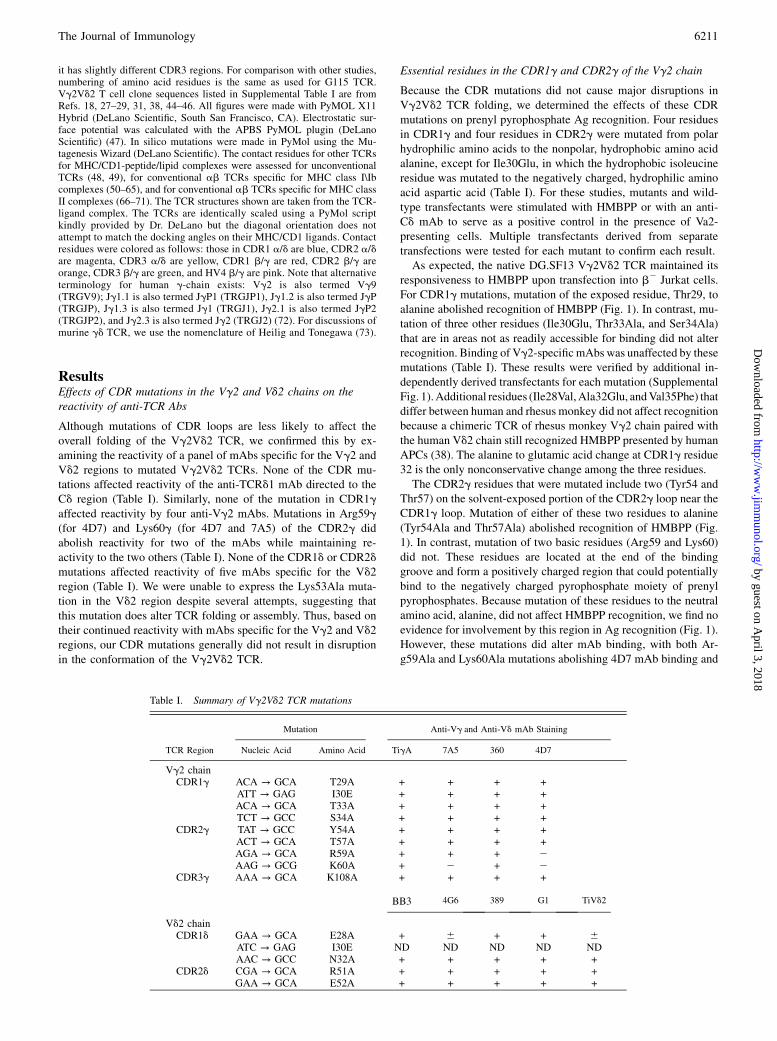
ResultsEffects of CDR mutations in the Vg2 and Vd2 chains on thereactivity of anti-TCR Abs
Although mutations of CDR loops are less likely to affect theoverall folding of the Vg2Vd2 TCR, we confirmed this by ex-amining the reactivity of a panel of mAbs specific for the Vg2 andVd2 regions to mutated Vg2Vd2 TCRs. None of the CDR mu-tations affected reactivity of the anti-TCRd1 mAb directed to theCd region (Table I). Similarly, none of the mutation in CDR1gaffected reactivity by four anti-Vg2 mAbs. Mutations in Arg59g(for 4D7) and Lys60g (for 4D7 and 7A5) of the CDR2g didabolish reactivity for two of the mAbs while maintaining re-activity to the two others (Table I). None of the CDR1d or CDR2dmutations affected reactivity of five mAbs specific for the Vd2region (Table I). We were unable to express the Lys53Ala muta-tion in the Vd2 region despite several attempts, suggesting thatthis mutation does alter TCR folding or assembly. Thus, based ontheir continued reactivity with mAbs specific for the Vg2 and Vd2regions, our CDR mutations generally did not result in disruptionin the conformation of the Vg2Vd2 TCR.
Essential residues in the CDR1g and CDR2g of the Vg2 chain
Because the CDR mutations did not cause major disruptions inVg2Vd2 TCR folding, we determined the effects of these CDRmutations on prenyl pyrophosphate Ag recognition. Four residuesin CDR1g and four residues in CDR2g were mutated from polarhydrophilic amino acids to the nonpolar, hydrophobic amino acidalanine, except for Ile30Glu, in which the hydrophobic isoleucineresidue was mutated to the negatively charged, hydrophilic aminoacid aspartic acid (Table I). For these studies, mutants and wild-type transfectants were stimulated with HMBPP or with an anti-Cd mAb to serve as a positive control in the presence of Va2-presenting cells. Multiple transfectants derived from separatetransfections were tested for each mutant to confirm each result.As expected, the native DG.SF13 Vg2Vd2 TCR maintained its
responsiveness to HMBPP upon transfection into b2 Jurkat cells.For CDR1g mutations, mutation of the exposed residue, Thr29, toalanine abolished recognition of HMBPP (Fig. 1). In contrast, mu-tation of three other residues (Ile30Glu, Thr33Ala, and Ser34Ala)that are in areas not as readily accessible for binding did not alterrecognition. Binding of Vg2-specificmAbs was unaffected by thesemutations (Table I). These results were verified by additional in-dependently derived transfectants for each mutation (SupplementalFig. 1).Additional residues (Ile28Val,Ala32Glu, andVal35Phe) thatdiffer between human and rhesus monkey did not affect recognitionbecause a chimeric TCR of rhesus monkey Vg2 chain paired withthe human Vd2 chain still recognized HMBPP presented by humanAPCs (38). The alanine to glutamic acid change at CDR1g residue32 is the only nonconservative change among the three residues.The CDR2g residues that were mutated include two (Tyr54 and
Thr57) on the solvent-exposed portion of the CDR2g loop near theCDR1g loop. Mutation of either of these two residues to alanine(Tyr54Ala and Thr57Ala) abolished recognition of HMBPP (Fig.1). In contrast, mutation of two basic residues (Arg59 and Lys60)did not. These residues are located at the end of the bindinggroove and form a positively charged region that could potentiallybind to the negatively charged pyrophosphate moiety of prenylpyrophosphates. Because mutation of these residues to the neutralamino acid, alanine, did not affect HMBPP recognition, we find noevidence for involvement by this region in Ag recognition (Fig. 1).However, these mutations did alter mAb binding, with both Ar-g59Ala and Lys60Ala mutations abolishing 4D7 mAb binding and
Table I. Summary of Vg2Vd2 TCR mutations
TCR Region
Mutation Anti-Vg and Anti-Vd mAb Staining
Nucleic Acid Amino Acid TigA 7A5 360 4D7
Vg2 chainCDR1g ACA → GCA T29A + + + +
ATT → GAG I30E + + + +ACA → GCA T33A + + + +TCT → GCC S34A + + + +
CDR2g TAT → GCC Y54A + + + +ACT → GCA T57A + + + +AGA → GCA R59A + + + 2AAG → GCG K60A + 2 + 2
CDR3g AAA → GCA K108A + + + +
BB3 4G6 389 G1 TiVd2
Vd2 chainCDR1d GAA → GCA E28A + 6 + + 6
ATC → GAG I30E ND ND ND ND NDAAC → GCC N32A + + + + +
CDR2d CGA → GCA R51A + + + + +GAA → GCA E52A + + + + +
The Journal of Immunology 6211
by guest on April 3, 2018
http://ww
w.jim
munol.org/
Dow
nloaded from

with Lys60Ala also abolishing 7A5 mAb binding. Binding by theTigA and 360 mAbs was unaffected (Table I). Differences be-tween human and rhesus monkey at residues Ser53Phe andArg59Lys are conservative changes and did not affect recognition.The serine to phenylalanine mutation is located in a recessed areaof the CDR2g loop.Thus, all of the mutations in CDR1g and CDR2g that alter
HMBPP recognition are located in highly accessible, solvent ex-posed areas of the CDR loops of the Vg2Vd2 TCR (28). Incontrast, mutations that did not affect HMBPP recognition werelocated in recessed, less solvent exposed areas. Importantly, allthree mutations that affected HMBPP response were distant (16–23 A) from the proposed prenyl pyrophosphate-binding area thatis composed mainly of residues from CDR3g and CDR2d.
Essential residues in the CDR1d and CDR2d region of the Vd2chain
To investigate the role of Vd-chain residues in nonpeptide Agrecognition, three residues in CDR1d and two residues in CDR2dwere selected for mutation (Fig. 2). One of the three mutations inCDR1d affected the HMBPP recognition. The residue mutated,Glu28, is highly accessible but spaced 19–22 A away from theproposed prenyl pyrophosphate-binding area. This mutation wasnonconservative, with an alanine being substituted for a negativelycharged glutamic acid residue (Fig. 2). The other mutations,Ile30Glu and Asn32Ala, did not affect recognition (Fig. 2).Two mutations in the CDR2d, Arg51Ala and Glu52Ala, both
affected recognition of HMBPP. The Arg51 and Glu52 residuesare located within the proposed prenyl pyrophosphate-bindingsite (within 4–10 A of Lys109 for Arg51 and 10–13 A for Glu52).The loss of reactivity by mutating Arg51 confirms an earlierstudy in which mutation of Arg51 to either alanine or glutamicacid abolished recognition (30). Changing one residue in CDR2doutside of this area, Asp54, to glycine did not affect recognitionbecause glycine is used at this position in the rhesus monkey Vd2chain, and a chimeric receptor of rhesus monkey Vd2 paired withhuman Vg2 still recognized prenyl pyrophosphates (38). Note thatthis residue is located 11–15 A away from the proposed bindingsite. These findings demonstrate that part but not all of CDR2dcontributes to prenyl pyrophosphate recognition.
Abrogation of Ag and tumor recognition by mutation of K108in CDR3g
We had earlier established the importance of the CDR3g in prenylpyrophosphate recognition by altering reactivity though mutationof its junctional region (29). We and others have proposed that
FIGURE 1. Recognition of HMBPP by Vg2Vd2 TCR transfectants
expressing mutant Vg2 chains. J.RT3-T3.5 b2 Jurkat cells were trans-
fected with unmutated or mutated DG.SF13 TCR-g cDNAs together with
the unmutated DG.SF13 TCR d-chain cDNA. After drug selection, anti-Cd
responsive transfectants were identified and stimulated with HMBPP in the
presence of Va2 APCs and 2.5 ng/ml PMA. The anti-Cd mAb (anti-
TCRd1) and HMBPP were added to the cultures stating at 2.15 mg/ml and
1000 nM, respectively, and serially diluted by half-log intervals. After
24 h, the culture supernatants were harvested and assayed for IL-2 activity
using the IL-2–dependent cell line HT2. Results from one transfectant are
shown for each mutation and are representative of the results obtained with
two to four other independently derived transfectants (Supplemental Fig.
1). Values shown are mean 6 SEM of duplicate or triplicate samples.
HMBPP reactivity was considered (++) if the maximum HMBPP response
was .40% of the control anti-Cd response, (+) if between 20–40% of the
control response, and (2) if ,10% of the control response.
FIGURE 2. Recognition of HMBPP by Vg2Vd2 TCR transfectants
expressing mutant Vd2 chains. J.RT3-T3.5 b2 Jurkat cells were trans-
fected with unmutated or mutated DG.SF13 TCR-d cDNAs together with
the unmutated DG.SF13 TCR g-chain cDNA. Culture conditions and the
IL-2 assay were as in Fig. 1. Results from one transfectant are shown for
each mutation and are representative of the results obtained with one to
four other independently derived transfectants (Supplemental Fig. 2).
6212 Vg2Vd2 TCR MUTATIONS AFFECTING ANTIGEN RECOGNITION
by guest on April 3, 2018
http://ww
w.jim
munol.org/
Dow
nloaded from

lysine residues in the Jg region of CDR3g combined with Arg51in CDR2d constitute a potential binding site for the pyrophosphateresidues of IPP and HMBPP because the positive charges on theamino groups are available for ionic bonding (27, 28, 30). Inaddition to prenyl pyrophosphates, there have been reports that themitochondrial protein F1-ATPase expressed on tumor cells can berecognized by the Vg2Vd2 TCR (74). To determine the impor-tance of this proposed site in Ag and tumor recognition, we mu-tated one of these residues, Lys108, in the Jg1.2 segment ofCDR3g to alanine.To study tumor reactivity reportedly due to F1-ATPase (74), we
determined reactivity of the wild-type Vg2Vd2 TCR and theLys108Ala TCR mutant for the stimulatory plasmacytoma, RPMI8226. Whereas the unmutated TCR transfectant responded to theRPMI 8226, but not to the control Raji cell line, mutation ofLys108 abrogated reactivity to RPMI 8226 (Fig. 3, right bottompanel).Similarly, mutation of Lys108 completely abrogated recognition
of HMBPP (Fig. 3) while preserving stimulation by an anti-CdmAb.
This mutation also abrogated direct stimulation by five other prenylpyrophosphates (Fig. 3). Other stimulatory compounds, such as bis-phosphonates and alkylamines, stimulate Vg2Vd2 T cells indirectlyby blocking the enzyme FPP synthase, causing increases in cellularIPP levels (13, 14). Consistent with IPP being the direct stimulatingAg, the Lys108Ala mutation also abrogated stimulation by six al-kylamines and two bisphosphonates (Fig. 3). These results confirman earlier report in which Lys108Ala, Lys108Glu, and Lys109Glumutations (but not Lys109Ala) abolished recognition of prenyl py-rophosphate, bisphosphonate, and alkylamine stimulatory com-pounds (30, 31).The loss of reactivity to the stimulatory compounds and the
tumor cell line by the Lys108Ala mutation suggests that there areshared structural features for these Ags (including the Ag expressedon the RPMI 8226 plasmacytoma).
Narrow length distribution of CDR3g in Vg2Jg1.2 chains butnot in Vg2Jg1.3/2.3 chains
To determine if there were any length restrictions or commonmotifs in the CDR3g junctional region of reactive Vg2Vd2 T cells,we examined reported sequences of 107 reactive and nonreactiveVg2Vd2 T cell clones (Supplemental Table I). The Jg1.2 segmentwas highly favored being used by 84 out of 90 (93%) reactiveclones. The length of CDR3g in Vg2Jg1.2 chains was also highlyrestricted, with 98% of T cell clones with CDR3g lengths of 62residues. There were no major differences in CDR3g length be-tween reactive and nonreactive clones (Supplemental Fig. 3).However, because TCR differences between reactive and non-reactive clones could be mapped to the CDR3d region in somecases (Table II), at least some of the Vg2 chains expressed bynonreactive clones would be reactive if paired with a reactive Vd2chain.Many clones (18 out of 84, 21%) used the invariant Vg2Jg1.2 aa
sequence (75) either due to germline gene rearrangement or theaddition of junctional residues with trimming of the germline Vg2and Jg1.2 segments (Supplemental Table I). This frequency of in-variant Vg2Jg1.2 chains is consistent with that reported for pe-ripheral blood Vg2Vd2 T cells (75). In most of the noninvariantVg2Jg1.2 chains, there were only one to two junctional residues(with a maximum of four) with corresponding losses in the germ-line Vg2 and Jg1.2 segments to preserve CDR3g length. Thus, allof the Vg2Jg1.2 chains had germline amino acid sequences formuch or all of the CDR3g region. When junctional residues wereadded, they favored basic lysine and arginine residues, with 25 outof 66 (38%) reactive Vg2Jg1.2 chains having at least one basicresidue added. Also, none of the TCRs had deletion of the Lys108or Lys109 residues. The glutamic acid located at position 105 is notrequired for reactivity because it is absent in some human Vg2Jg1.2chains (Supplemental Table I) and in rhesus monkey Vg2, which isable to recognize HMBPP presented by human APCs when pairedwith human Vd2 (Table II) (38).In contrast to Vg2Jg1.2 chains, a high proportion of Vg2Vd2
T cell clones using Vg2Jg1.3/2.3 chains were nonreactive toprenyl pyrophosphates (46% nonreactive versus 14% nonreactivefor Vg2Jg1.2 chains). Moreover, the Vg2Jg1.3/2.3 chains hadshorter chain lengths (6–14 residues versus 11–14 residues forVg2Jg1.2 chains) with higher variability in length. This shorterlength may be optimal for reactivity because a short Vg2Jg1.3chain (eight residues) using a GN junctional sequence paired withthe DG.SF13 Vd2 chain was responsive to HMBPP but a longermutant Vg2Jg1.2 (12 residues) chain, in which the GN residueswere substituted for a W residue, was unresponsive when pairedwith the same DG.SF13 Vd2 chain (Table II).
FIGURE 3. Critical role of K108 in the CDR3 of Vg2 in the recognition of
nonpeptide Ags and tumor cells. J.RT3-T3.5 b2 Jurkat cells were transfected
with the DG.SF13 TCR g-chain cDNA with a lysine (K) to alanine (A)
mutation at position 108 in the CDR3g region and the unmutated DG.SF13
TCR-d cDNA. The transfectant obtained was named K108A and compared
with an unmutated DG.SF13 TCR transfectant for stimulation by anti-Cd,
prenyl pyrophosphates, alkylamines, bisphosphonates (risedronate and
alendronate), HMBPP (in supernatants of theE. coli lytBmutants, KM20, and
KM22), bromohydrin pyrophosphate, and lymphoma cell lines (RPMI 8226
and Raji). Culture conditions and the IL-2 assay were as in Fig. 1.
The Journal of Immunology 6213
by guest on April 3, 2018
http://ww
w.jim
munol.org/
Dow
nloaded from

Conserved hydrophobic residue at position 97 but variableCDR3d length and sequence
CDR3d regions are the most variable junctional region in mammalsdue to the availability of multiple V, D, and J gene segments forrearrangement and due to the ability of multiple D segments to re-arrange in tandem and in any reading frame (76). In mice, theCDR3d region has been shown to be critical for gd T cell recogni-tion of T10 and T22MHC class Ib molecules. A 9 aa conserved loopmotif in the CDR3d region inserts into a cavity on the T10 moleculeand provides most of contact residues and 67% of the interface. Todetermine if a similar motif could be identified in the CDR3d regionof Vg2Vd2 TCR, we examined the sequences of 107 reactive andnonreactive Vg2Vd2 T cell clones (Supplemental Table I).Unlike its Vg2 chain partner, the length of the CDR3 of the Vd2
chain varied widely, ranging from 10 to 18 aas, while retaining re-
sponsiveness to prenyl pyrophosphates (Supplemental Fig. 3, Sup-
plemental Table I). Moreover, unlike Vg2 chains in which there are
critical residues in the Jg segment and a preference for Jg1.2, the
Vd2 chain can use Jd1, Jd2, or Jd3 and retain responsiveness despite
significantly different sequences and lengths. The longer and more
variable length of the CDR3d segment is a general characteristic of
d-chains (77).
Although the length of CDR3d is variable, there is a strong
preference for a hydrophobic residue at position 97. Ninety-two
percent of Vd2 chains (61 out of 71) from reactive T cell clones
used a hydrophobic residue at this position versus 53% (8 out of
15) of Vd2 chains from nonreactive T cell clones. No reactiveclone used proline, lysine, or arginine at this position (Supple-mental Table I). There are examples of clones with identical Vg2chains but different Vd2 chains in which loss of reactivity can becorrelated with a nonhydrophobic residue at position 97 (Table II).In these cases, the nonreactive Vg2Vd2 T cell clone has eithera polar serine amino acid at this position (for the nonreactive T22clone versus the reactive M6/M12 clones) or the kinked prolineamino acid (for the nonreactive I.7 clone compared with the re-active C.15 clone). Similarly, transfection of a Vg2 chain (g001)paired with a Vd2 chain (d255) with a proline at this position didnot confer reactivity to prenyl pyrophosphates, although two otherVd2 chains (d263 and d016) with hydrophobic residues did (31).Mutation of Leu97 to either alanine or serine abolished prenylreactivity (31), confirming its importance.Whereas position 97 was mainly restricted to hydrophobic amino
acids, the residues prior to and after 97 were quite diverse, in-
cluding both positively and negatively charged amino acids as well
Table II. Diversity of CDR3g or CDR3d sequences in Vg2Vd2 T cells expressing an identical Vg2 or Vd2 chain
Clone/Transfectant Vg2 N Jg Jg Vd2 (aa 97) N/D/N Jd Jd CDR3d Length Reactivity
AC2a LWEV QELGKKIK Jg1.2 CDT T GG SWDTRQM Jd3 13 +AC8a LWEV QELGKKIK Jg1.2 CDT W G S SWDTRQM Jd3 13 +G1b LWEV QELGKKIK Jg1.2 CD R V PP STGD TDKL Jd1 14 +G2b LWEV QELGKKIK Jg1.2 CDT V NADAEN DKL Jd1 13 +G3b LWEV QELGKKIK Jg1.2 CD A V LGDTSRP DKL Jd1 14 +G4b LWEV QELGKKIK Jg1.2 CD L V LGVNTG WDTRQM Jd3 16 +M3b LWEV QELGKKIK Jg1.2 CD W L LGDTV TDKL Jd1 13 +G5b LWEV QELGKKIK Jg1.2 CDT G GYAN WDTRQM Jd3 14 +G6b LWEV QELGKKIK Jg1.2 CDT V EA LTAQL Jd2 11 +M4b LWEV QELGKKIK Jg1.2 CD N W GDKM TAQL Jd2 12 +T2b LWEV QELGKKIK Jg1.2 CD N T GGYY SWDTRQM Jd3 15 +T3b LWEV QELGKKIK Jg1.2 CDT V LGDS SWDTRQM Jd3 15 +T4b LWEV QELGKKIK Jg1.2 CD I L GDAA LTAQ Jd2 12 +T5b LWEV QELGKKIK Jg1.2 CDT I LGDT WDTRQM Jd3 14 +T6b LWEV QELGKKIK Jg1.2 CDT L PLGAKGY DKL Jd1 14 +T7b LWEV QELGKKIK Jg1.2 CDT V SGDTH SWDTRQM Jd3 16 +
M12b LWEV ELGKKIK Jg1.2 CD S L LTSQLSLGDH TDKL Jd1 18 +M6b LWEV ELGKKIK Jg1.2 CD T G FRGGKDT WDTRQM Jd3 17 +T22b LWEV ELGKKIK Jg1.2 CD K S AGNP SWDTRQM Jd3 15 2
C.15 LWE A QELGKKIK Jg1.2 CDT L GSGGSAER TDKL Jd1 12 +I.7 LWE A QELGKKIK Jg1.2 CDT P GIGYT WDTRQM Jd3 15 2
g001/d263c LWEV QELGKKIK Jg1.2 CDT I LGD TDKL Jd1 10 +g001/d016c LWEV QELGKKIK Jg1.2 CDT V EGMRRNLLGERGSY TDKL Jd1 22 +g001/d255c LWEV QELGKKIK Jg1.2 CDT P MRAPDWGTLGN TDKL Jd1 19 2
PBLg/DGdd LWE GN YKK Jg1.3 CDT L VS TDKL Jd1 10 +mutDGg/DGdd LWE GN ELGKKIK Jg1.2 CDT L VS TDKL Jd1 10 2DG.SF13 LWE W ELGKKIK Jg1.2 CDT L VS TDKL Jd1 10 +DGg/monkeyde LWE W ELGKKIK Jg1.2 SD H I LEGGIRG TDKL Jd1 13 +Monkeyge/DGd LWEV Q QFGRKVK Jg1.2 CDT L VS TDKL Jd1 10 +
G115/G9f LWE AQ QELGKKIK Jg1.2 CD T L GMGGEY TDKL Jd1 13 +
Horizontal dividers separate groups of TCRs that share either identical Vg2 or Vd2 chains but that differ in the other chain and, in some cases, in prenyl pyrophosphatereactivity. Position 97 in the Vd2 chains is bolded and its position indicated in the column title. Likely sequence differences causing loss of reactivity are underlined. Note thatalternative names for Vg2, Jg1.2, and Jg1.3 are Vg9 (TRGV9), JgP (TRGJP), and Jg1 (TRGJ1).
aSequences from T cell clones (18).bSequences from T cell clones, reactivity reported in Ref. 44.cSequences from T cell transfectants, reactivity reported in Ref. 31.dSequences from T cell transfectants, reactivity reported in Ref. 29.eSequences from T cell transfectants, reactivity reported in Ref. 38.fSequences from G115/G9 T cell clone used for crystal structure, reactivity reported in Ref. 28.
6214 Vg2Vd2 TCR MUTATIONS AFFECTING ANTIGEN RECOGNITION
by guest on April 3, 2018
http://ww
w.jim
munol.org/
Dow
nloaded from

as proline (Supplemental Table I). Moreover, there were no other
clearly defined motifs in the N-, Dd-, or Jd-encoded portions of
CDR3d that are required for reactivity. Thus, beyond a strong
preference for a hydrophobic residue at position 97, no other motif
can be found within CDR3d that correlates with reactivity. Taken
together, the diverse length and amino acid composition of
CDR3d from reactive Vg2Vd2 T cell clones suggests that optimal
prenyl pyrophosphate reactivity requires a hydrophobic residue at
position 97 but is tolerant to a wide variety of sequences in and
lengths of the N, D, and J regions.
Model of Vg2Vd2 TCR contact residues in relation to theproposed prenyl pyrophosphate-binding site
The structures of human G115 Vg2Vd2 TCR (Fig. 4) and murineG8 Vg4Va/d11.3 TCR show more protrusions and clefts in thebinding face of the TCR (48, 78) compared with the flatter sur-faces of ab TCRs specific for peptide-MHC class I/II complexes.The CDRs of the Vg2Vd2 TCR protrude (Fig. 4A). CDR3g andCDR3d are especially prominent, creating a cleft between the twoCDRs. There are two positively charged regions in this cleft(shaded blue) (Figs. 4D, 4F, 6), one positioned near the base ofCDR3d formed by Lys109g from Jg1.2 and Arg51d from CDR2dand a second located on the other side of the cleft formed byArg59g and Lys60g from CDR2g.Mutation of residues in the second positively charged region
formed by Arg59g and Lys60g from CDR2g, although abolishingbinding by some anti-Vg2 mAbs (Table I), did not affect prenyl
pyrophosphate recognition (Table III), arguing against the in-volvement of this second positively charged region. In contrast,alanine mutation of Lys108 (Fig. 3) and Lys109 (30) that is lo-cated in the first positively charged region closest to the Leu97residue in CDR3d completely abrogated recognition of all prenylpyrophosphates studied. Alanine mutation of Lys108 also abol-ished bisphosphonate and alkylamine recognition. Importantly,recognition of the plasmacytoma, RPMI 8226, was also lost. Thisresult suggests that the Ag recognized by Vg2Vd2 T cells inRPMI 8226 is IPP or another prenyl pyrophosphate and not F1-ATPase because these two Ags have grossly different structuresand would be expected to use different contact residues. However,F1-ATPase could function as a presenting molecule, although wehave found no evidence for this (data not shown).This first positively charged region is positioned immediately
adjacent to the conserved hydrophobic amino acid at position 97 ofCDR3d and has potential contributions from Lys108g, Lys109g,and Lys111g from Jg1.2 and Arg51d from CDR2d (Fig. 4D, 4F).However, in silico mutagenesis and calculation of the surface po-tential suggests that most of the positive potential in the region isfrom Arg51d with some contribution from Lys109g, but little or nocontribution from Lys108g and Lys111g (Fig. 5). Consistent withthis analysis, mutation of Lys109g to alanine reduced but did notabolish prenyl pyrophosphate recognition (31), whereas mutationof Arg51d to alanine completely abolished recognition. We nowshow that the acidic amino acid Glu52d, from CDR2d that is nearbut outside the binding pocket, is also required for recognition. The
Table III. Effect of Vg2Vd2 TCR mutations and rhesus monkey polymorphisms on prenyl pyrophosphatereactivity
TCR Region
PolymorphismBetween Human andRhesus Monkeya
Mutation of HumanVg2Vd2 TCR
Prenyl PyrophosphateReactivity Reference
Vg2 chainCDR1g I28V + (38)
T29A 2 Current paperI30E + Current paper
A32E + (38)T33A + Current paperS34A + Current paper
V35I + (38)CDR2g S53F + (38)
Y54A 2 Current paperT57A 2 Current paper
R59K + (38)R59A + Current paperK60A + Current paper
CDR3g W104GN 2 (29)E105Q + (38)F106L + (38)K108R + (38)
K108A 2 Current paper and (31)K108E 2 (30)K109A + (31)K109E 2 (30)
Vd2 chainCDR1d E28A 2 Current paper
I30E + Current paperG31S + (38)
CDR2d R51A 2 Current paper and (31)R51E 2 (31)E52A 2 Current paper
D54G + (38)CDR3d C94S + (38)
L97A 2 (31)L97S 2 (31)
aReactivity of rhesusmonkeyVgene segmentswas assessed by pairingwith the corresponding partner humanVgene segmentsand transfecting into J.RT3-T3.5 b2 Jurkat followed by stimulation with Ags presented by human APCs as described (38).
The Journal of Immunology 6215
by guest on April 3, 2018
http://ww
w.jim
munol.org/
Dow
nloaded from

area is a potential prenyl pyrophosphate-binding site because the
positively charged amino acids can form ionic bonds to the nega-tively charged pyrophosphate moiety on IPP or HMBPP. The al-
kenyl chain could be positioned over the hydrophobic amino acid atposition 97. Most proteins that bind phosphate(s) use either this
mechanism of binding or use negatively charged amino acids toform an ionic bond to one of the positive charges on a divalent
cation, leaving the second free to bind to the negative charges on thephosphate group. However, there is no evidence for direct high-
affinity binding of prenyl pyrophosphates to theVg2Vd2 TCR either
by equilibrium dialysis (C.T. Morita, unpublished observations) orsoaking Vg2Vd2 TCR crystals in IPP or HMBPP (28).When the mutated CDR residues are localized on the Vg2Vd2
TCR structure (critical residues colored red and noncritical resi-dues colored green, Table III), a potential binding footprint of theTCR to its ligand is delineated (Fig. 4C, 4E). This area is muchlarger than the size of IPP or HMBPP (IPP and HMBPP are 10.3 Aand 10.9 A in extended conformation, respectively) and encom-passes more than just the prenyl pyrophosphate-binding site (Fig.4B, 4D, 4F). Moreover, although both abolish recognition, Thr29in CDR1g is 34 A from Glu28 in CDR1d. Similarly, Lys109 inCDR3g is 15.3 A from Thr29 in CDR1g and 20.1 A from Glu28in CDR1d; again, much larger distances than the size of a prenylpyrophosphate. Thus, the footprint of Vg2Vd2 TCR residues re-quired for prenyl pyrophosphate recognition is significantly largerthan the proposed prenyl pyrophosphate-binding site.
DiscussionIn this study, we show that mutations in all CDRs of the Vg2Vd2TCR can affect recognition of prenyl pyrophosphates. Mutation ofhighly accessible amino acids in CDR1g, CDR2g, and CDR1dloops all can abolish recognition, whereas mutations localized onthe sides of the loops do not. Unlike TCR recognition by the twoother unconventional innate T cells in which the contact residuesare defined, there is no evidence that an extended Da/d or Ja/damino acid motif is required for recognition. Instead, there isa preference for a single hydrophobic, aliphatic amino acid atposition 97 of CDR3d as previously reported (31, 79), but norestriction on: 1) the type of amino acid immediately preceding orfollowing position 97; 2) the overall length of the CDR3d loop; or3) other sequences in the D/N or J portions of the CDR3d loop. Incontrast, the length of CDR3g shows low variability, basic aminoacids are preferred in its N region, and the Jg1.2 segment ispreferentially used. Basic amino acids (Lys108 and Lys109) in theJg segment, the basic Arg51 and acidic Glu52 residues in theCDR2d, and the aliphatic amino acid at position 97 in CDR3d areall important for recognition and are located in or near a proposedprenyl pyrophosphate-binding site. Taken together, these resultsshow that a large portion of the Vg2Vd2 TCR is required forprenyl pyrophosphate recognition.How well do alanine mutations predict residues in T cell Ag
receptors that contact Ag or their presenting molecule? In threestudies, ab TCR contact residues for peptide-MHC class I ligands
FIGURE 4. Recognition of prenyl pyrophosphates
by human Vg2Vd2 TCR is dependent on all CDRs. A,
Top-down view of the Vg2Vd2 TCR with CDRs for
the g- and d-chains labeled. CDR residues are colored
such that those in CDR1d are blue, CDR1d are ma-
genta, CDR3d are yellow, HV4d are cyan, CDR1g are
red, CDR2g are orange, CDR3g are green, and HV4g
are pink. B, Top-down view of the Vg2Vd2 TCR with
basic amino acids colored blue and acidic amino acids
colored red. IPP is shown in the potential prenyl py-
rophosphate-binding site for size comparison. C, Top-
down view of critical amino acid residues that disrupt
prenyl pyrophosphate recognition are colored red on
the Vg2Vd2 TCR, whereas noncritical amino acid
residues are colored green. D, Top-down view of the
surface potential of the Vg2Vd2 TCR (colored from
red [28 kT] to blue [+8 kT]) reveals two positively
charged potential binding sites. E, Side view of critical
amino acid residues that disrupt prenyl pyrophosphate
recognition are colored red on the Vg2Vd2 TCR,
whereas noncritical amino acid residues are colored
green. F, Side view of the surface potential of the
Vg2Vd2 TCR (colored as in D).
6216 Vg2Vd2 TCR MUTATIONS AFFECTING ANTIGEN RECOGNITION
by guest on April 3, 2018
http://ww
w.jim
munol.org/
Dow
nloaded from

were determined from crystal structures. CDR contact and non-
contact residues were then mutated to alanine and the mutant
TCRs tested for binding to their respective peptide-MHC class I
ligands. Mutation of contact residues to alanine reduced MHC/
peptide ligand binding for 81% (17 out of 21) of the contact
residues in the LC13 TCR (80), for 70% (7 out of 10) in the 2C
TCR (81), and for 59% (13/22) in the JM22 TCR (82). Noncontact
residues in CDRs containing contact residues also decreased
binding, albeit, as might be expected, at lower rates (39%, 7 out of
18, for the LC13 TCR and 60%, 9 out of 15, for the 2C TCR).
Importantly, none of 12 mutations in the a and b hypervariable
four-regions of the 2C TCR affected binding (none were contact
residues). Most mutations that grossly affected structure were not
expressed (only 1 out of 40 in LC13 (80), 9 out of 51 in 2C (81),
and 0 out of 12 in JM22) (82). Thus, most mutations (70% overall)
in CDR contact residues greatly reduce binding, whereas muta-
tions in HV4 loops that did not contain contact residues had no
effect. From these results, we would predict that mutations that
abolish recognition likely identify either contact residues for the
Ag/presenting molecule or noncontact residues in loops that
contain contact residues. Our finding that residues well outside the
proposed prenyl pyrophosphate-binding site affect recognition
suggests that a larger molecule binds and presents these small
molecules.
Unconventional innate versus conventional peptide-MHC TCRrecognition by T cells
Although expressing rearranging TCRs, innate T cells, such as gdT cells and iNKTs, likely play roles that more closely resemblethose of innate myeloid cells. In keeping with their unique roles inthe immune system, innate T cells commonly express TCRs thatuse germline encoded portions of the TCR to either recognizenonpeptide Ags presented by MHC class Ib or CD1 molecules orto directly recognize MHC class I-like molecules. Many gd T cellpopulations, such as murine dendritic epidermal gd T cells (83),express TCR with invariant receptors. ab iNKTs that recognizeself and foreign glycolipids presented by CD1d also expressa semi-invariant TCR composed of a conserved VaJa-chainpaired with a limited set of Vb-chains (84).There are only two examples of unconventional TCR recognition
by innate T cells that have been characterized at the molecular levelto compare with Vg2Vd2 TCR recognition of prenyl pyro-phosphates. The only example for gd T cells is the structure ofa murine gd TCR, G8, bound to the MHC class Ib molecule T22.In this study, a W...EGYEL motif in an extended CDR3d loop (85)contacts a disordered region at the end of the a2 helix of the T22molecule from the side (48). The contact residues of the G8 TCRare shown in Fig. 6 (top middle panel). Eighty-nine percent of thegd TCR interface with T22 is contributed by the Vd-chain, with
FIGURE 5. Surface potential effect of in
silico alanine mutations of basic residues in the
Vg2Vd2 TCR. Basic residues were mutated to
alanine in silico using PyMol (DeLano Scien-
tific), and the surface potential of the mutated
TCRs were calculated using the APBS plugin
in PyMol. The TCRs are oriented with the
g-chains on the top left and the d-chains on
the bottom right of the panels. Top-down views
of the surface potential of the Vg2Vd2 TCR are
shown (colored from red [28 kT] to blue [+8
kT]. The two positively charged regions are
circled. A, Surface potential for the unmutated
Vg2Vd2 TCR. B–F, Effect on surface potential
of alanine replacement of R59A and K60Ag
(B), R51Ad (C), K109Ag (D), K108Ag (E),
and K111Ag (F).
The Journal of Immunology 6217
by guest on April 3, 2018
http://ww
w.jim
munol.org/
Dow
nloaded from

67% contributed by the CDR3d loop. Underscoring the impor-tance of the CDR3d loop, swapping of this region for the CDR3aof an ab TCR is sufficient to confer recognition of T22 (86). Allof the gd T cells that recognized T22 have the W...EGYEL se-quence motif in CDR3d. Importantly, this motif is commonlyderived from the germline-encoded Dd2 gene segment and ispresent on ∼0.85% of nonselected murine Vd2 chains; this mayaccount for the high frequency of T22-reactive gd T cells (1/50–1/1000) in unimmunized mice (85).The other example of unconventional innate TCR recognition is
human and murine iNKT ab TCR recognition of glycolipidspresented by CD1d (49, 87). iNKT TCRs bind CD1d/a-GalCer ina top-down orientation paralleling the a helixes of CD1d. Again,CDR3a, which is the equivalent of CDR3d, plays an importantrole contributing 52% of the buried surface area (BSA) (Fig. 6, topleft panel). Only the Ja18 region of CDR3a contacts the CD1d-lipid complex, explaining the use of this J region by the invariantVa24 chain of the iNKT TCR. Like the G8 gd TCR in whichCDR3g contains only two contact residues, CDR3b contains onlyone contact residue that contributes only 6% of the BSA. Instead,the germline-encoded CDR2b contributes 28% of the BSA. Whenthe contact residues in the iNKT TCR were mutated to alanine,four out of seven CDR3a loop residues and two out of four
CDR2b abolished recognition, whereas three other residues inother CDRs had no or little effect (88). Thus, despite large dif-ferences in the docking orientation of the iNKT ab TCR and theG8 gd TCR for their ligands, unconventional recognition by bothinvolves primarily germline-encoded regions in CDR3a/d, withcritical contributions from the germline-encoded CDR2b in iNKTTCR.In contrast to these examples, our study shows a different mode
of recognition for the Vg2Vd2 TCR that does not rely as heavily onthe CDR3a/d region. Instead, CDR3g appears to be the criticalregion for prenyl pyrophosphate recognition. Mutation of eitherLys108 or Lys109 in the conserved Jg1.2 region abolishes recog-nition as does altering the CDR3g length/sequence (29). Moreover,20–30% of adult Vg2Vd2 T cells express an invariant Vg2Jg1.2sequence, with the rest exhibiting little differences in CDR3glength while favoring basic residues in the junctional region.Conservation of CDR3g length may be required to maintain thecorrect positioning of the Jg1.2 region in the putative prenyl py-rophosphate-binding site (Figs. 4B, 5). We find that the Arg51d andGlu52d residues in CDR2d are also required for recognition andconfirm that there is a strong preference for an aliphatic hydro-phobic amino acid (generally leucine, isoleucine, or valine, butnot proline) at position 97 in CDR3d but find few restrictions
FIGURE 6. Potential contact residues of the
Vg2Vd2 TCR for a putative Ag presenting
molecule-prenyl pyrophosphate complex differs
from other unconventional TCRs and conven-
tional TCRs specific for MHC-peptide com-
plexes. The TCRs are oriented with the
b/g-chains on the top left and the a/d-chains on
the bottom right of the panels. Contact residues
are colored such that those in CDR2a/d are
blue, CDR2a/d are magenta, CDR3a/d are
yellow, HV4a/d are cyan, CDR1b/g are red,
CDR2b/g are orange, and CDR3b/g are green.
The diagonal orientation does not attempt to
match the docking angle on the MHC/CD1 li-
gand. Top panels, Putative contact residue
footprint of the Vg2Vd2 TCR based on the ef-
fects of point mutations on human Vg2Vd2 T
cell reactivity to nonpeptide Ag (top left panel).
Note the potential large contribution of germ-
line-encoded regions of CDR3g compared with
only a minor contribution by L97 in CDR3d. In
contrast, the G8 gd TCR (top middle panel),
that is specific for the T22 MHC class Ib mol-
ecule, predominantly uses CDR3d for recogni-
tion. The iNKT ab TCR (top left panel) binding
to the CD1d-a-GalCer complex also pre-
dominantly uses the CDR3a region (which
corresponds to CDR3d). Middle panels, Con-
ventional ab TCRs specific for MHC class I-
foreign peptide complexes generally use both
CDR3a and CDR3b for MHC-peptide recog-
nition in addition to CDR1 and CDR2. Bottom
panels, Conventional ab TCRs specific for
MHC class II/peptide complexes are similar to
MHC class I/peptide-specific ab TCRs and in-
volve extensive CDR3a and CDR3b contacts in
addition to CDR1 and CDR2 contacts. Addi-
tional examples of ab TCR recognition of
MHC-peptide complexes are shown in Supple-
mental Figs. 4, 5.
6218 Vg2Vd2 TCR MUTATIONS AFFECTING ANTIGEN RECOGNITION
by guest on April 3, 2018
http://ww
w.jim
munol.org/
Dow
nloaded from

on CDR3d length or sequence. All of these critical residues clusterin or around an area of positive surface potential due to Arg51d and,to a lesser extent, Lys109g, as determined by in silico mutation(Fig. 5).In addition to this site, we now show that critical residues are also
present in exposed areas of CDR1g and CDR2g as well as CDR1d.These residues are distant (15.3 A for Thr29 in CDR1g, 15.2 A forTyr54 in CDR2g, and 20.1 A for Glu28 in CDR1d) from theproposed binding site, making it unlikely that they would directlyinfluence prenyl pyrophosphate binding. Instead, we would pro-pose that these residues contact a presenting molecule for theprenyl pyrophosphates similar to ab TCRs contacting peptide-MHC class I or class II complexes.The potential footprint of the Vg2Vd2 TCR on the putative Ag-
presenting molecule shows more similarities than differences withthe footprints of conventional ab TCR that bind to MHC class I orclass II/foreign peptide complexes (89). Like the Vg2Vd2 TCR,most antimicrobial ab TCRs contact their ligands using all sixCDRs, although their relative energetic contributions to bindingvary (contact residues of TCRs specific for peptide-MHC class Iand class II are shown in Fig. 6 and Supplemental Figs. 4, 5). Themain exceptions are A6 and B7 TCRs specific for HLA-A2-TAX(57) (Supplemental Fig. 4).Generally, CDR1a and CDR2a are centered over the MHC a2
helix, and CDR1b and CDR2b are centered over the MHC a1helix in a diagonal orientation. The CDR3a region is centered onthe N-terminal end of the peptide, and the CDR3b region iscentered on the C-terminal end, but both regions can also contactMHC. Centering the diverse CDR3 regions over the peptide al-lows for the recognition of an array of sequences. Autoimmunerecognition of self-peptides represents a major exception to thediagonal orientation of ab TCRs docking to their ligands becauseautoimmune TCRs may bind with unusual topologies (90) andmay shift to docking over the extreme N terminus of the peptide(91). Although the docking orientation of the Vg2Vd2 TCR andthe structure of the putative presenting molecule is unknown, thesimilarities between the footprint of antimicrobial ab TCRs andthe footprint of the Vg2Vd2 TCR would suggest that a presentingmolecule is required.Vg2Vd2 TCR recognition also differs from antimicrobial ab
TCR recognition in important ways. In contrast to antimicrobialab TCRs in which most have multiple contact residues in CDR3a(Fig. 6, Supplemental Figs. 4, 5), the CDR3d region (equivalent toCDR3a) makes a limited contribution to recognition because theonly preference found was for an aliphatic residue at position 97.Moreover, Vg2Vd2 recognition is primarily though the germline-encoded gene segments, CDR1g, CDR2g, CDR1d, and CDR2dregions, coupled with Vg2Jg1.2 chains that are either invariant(lacking N nucleotide additions) or with limited diversity in se-quence and length.Given the large footprint for Vg2Vd2 TCR, we speculate that
there is a presenting molecule for prenyl pyrophosphate Ags.Because there is no evidence for genetic restriction in Vg2Vd2TCR recognition, we would predict that the putative presentingmolecule has little or no polymorphisms in contact regions for theTCR. Additionally, because human tumor cells of diverse originand different hematopoietic APCs present Ag, the putative pre-senting molecule would need to be widely expressed much likeMHC class I molecules. Although prenyl pyrophosphate reactivityis preserved in both New World and Old World monkeys (92, 93),rodent and guinea pig gd T cells (94) are unable to respond toprenyl pyrophosphate Ags nor can rodent, hamster, or other spe-cies APCs present prenyl pyrophosphate Ags to human Vg2Vd2T cells (35, 95). There may exist ruminant gd T cells with prenyl
pyrophosphate reactivity, although this has not been definitivelyestablished (96). Consistent with the lack of reactivity by mostother species, no other nonprimate species has homologous Vgenes for the Vg2 and Vd2 gene segments. Despite the reactivityof monkeys to prenyl pyrophosphates, both Vero and COS-7 celllines (95) (derived from African green monkeys) are unable topresent to human Vg2Vd2 T cells. Thus, there may be xenogeneicdifferences between African green monkeys and humans in theputative presenting molecules. Finally, we would predict that theputative presenting molecule has a structure homologous to MHCclass I or class II like all other nonclassical presenting moleculesand ligands (CD1, MR1, and MICA/MICB) so far discovered,although none of the known MHC class Ib molecules are viablecandidates. It also is possible that the presentation molecule be-longs to a new class of presenting molecules, such as an enzyme,a heat shock protein, or an Ig superfamily protein, that is not re-lated in structure to MHC class I/II molecules.Presentation of prenyl pyrophosphates by a presenting molecule
is also supported by functional and TCR binding studies. Rec-ognition of prenyl pyrophosphates requires cell-cell contact forVg2Vd2 T cell activation as assessed by intracellular calcium flux(32) and TNF-a release (33) that is identical to the contact re-quirement for ab T cell recognition of MHC/peptide complexesbut distinct from ligand recognition by hormone and neurotrans-mitter receptors. Also, there is no evidence for direct recognitionof prenyl pyrophosphates because attempts to soak IPP andHMBPP into Vg2Vd2 TCR crystals failed (28), and no binding ofprenyl pyrophosphates to soluble Vg2Vd2 TCRs was noted inequilibrium dialysis or microcalorimetry experiments (C.T. Mor-ita, unpublished observations). Similarly, a monkey Vg2Vd2 TCRtetramer does not directly bind HMBPP but will bind to APCwhen the APCs are incubated with HMBPP or infected with M.bovis bacillus Calmette-Guerin (36). This binding was observedwith human and monkey but not mouse, rat, or pig cells, dem-onstrating specificity for primate APCs identical to the results onpresentation of prenyl pyrophosphates (33, 97), bisphosphonates(95), alkylamines (97), and photoaffinity prenyl pyrophosphateanalogs (35). Binding was abolished by protease treatment of thecell surface, strongly suggesting that the surface binding moleculewas a protein (36). Finally, photoaffinity analogs of prenyl py-rophosphates are able to covalently crosslink to protein(s) on theAPC surface for presentation, and this binding could be specifi-cally inhibited by an inactive prenyl pyrophosphate analog (35).Taken together, these findings provide additional evidence for theexistence of a cell surface-presenting molecule for prenyl pyro-phosphate Ags.As discussed above, recognition of a-GalCer/CD1d by the
iNKT TCR is primarily through germline-encoded invariantVa24Ja18 CDR3a and CDR2b regions (49). However, for otherlipids, iNKT recognition also requires residues in CDR1a in ad-dition to the CDR3a and CDR2b regions. Again, these residues areencoded by germline gene segments (98). Thus, the basic orien-tation and framework for binding of both human and mouseiNKTs to their respective CD1d molecules and to different lipids isconserved such that others have proposed that the iNKT receptorcan be considered to have characteristics of a pattern-recognitionreceptor (98). However, the fine specificity and binding affinity ofiNKT TCR can be affected by the Vb-chain partner and its CDR3bsequence (87, 99, 100), allowing iNKT TCR to distinguish be-tween a number of different lipids. Similar differences in the CDR3sequences of the Vg2Vd2 TCR also likely affect prenyl pyro-phosphate recognition.Although Vg2Vd2 TCRs appear to function like pattern-
recognition receptors (101), as proposed for iNKT TCRs (98), this
The Journal of Immunology 6219
by guest on April 3, 2018
http://ww
w.jim
munol.org/
Dow
nloaded from
is not to assert that there is no selection on Vg2Vd2 TCRs duringdevelopment or Ag stimulation, which is a hallmark of adaptiveimmune recognition. Vg2Vd2 TCR selection clearly occurs be-cause Vg2Vd2 T cells using the Jg1.2 region and having an ali-phatic amino acid at position 97 are preferentially expandedin vitro (31, 102) and overrepresented in adult Vg2Vd2 T cells ascompared with fetal/thymic Vg2Vd2 T cells (79). However, evenin fetuses and neonates, there is a very high proportion of Vg2Vd2T cells that respond to prenyl pyrophosphates with two out of twofetal liver and at least three out of eight cord blood Vg2Vd2 T cellclones responding (9; data not shown). Also, Davadou et al. (44)found that 22 out of 25 (88%) PHA-derived thymic Vg2Vd2T cells using the Vg2Jg1.2 chain responded to prenyl py-rophosphates and to Daudi. Overall, 30 out of 38 (79%) of thymicVg2Vd2 T cell clones responded (44). In all of these cases, se-lection by exogenous Ags is unlikely. Therefore, although pe-ripheral selection is likely responsible for the near universalreactivity of Vg2Vd2 T cells that is found in adults, a high pro-portion of Vg2Vd2 T cells start out reactive, probably due toconstraints in gene rearrangement (103).Based on our results, we would predict that most of the rec-
ognition is mediated by germline-encoded elements of the Vg2Vd2TCR and that there is a presenting molecule for prenyl pyro-phosphate Ags. Although only three examples of unconventionalrecognition have now been molecularly defined, all share the useof certain germline elements of the TCR for recognition, the in-variant Va14Ja24 gene segments for lipid/CD1d recognition (49),a Dd motif for T10/T22 (48, 85), and, as noted by others (79, 103),the Vg2Jg1.2 chain and the Vd2 gene segment for prenyl py-rophosphates. We speculate that unconventional innate gd and abTCRs containing invariant V chains, highly biased V gene usage,or conserved CDR3 motifs are likely to bind their ligands usingthe invariant or biased portion of the TCR. Moreover, the portionsof invariant chains, biased V genes, or conserved motifs of innateTCRs that contact the ligands seem to favor germline-encodedsegments as also suggested by Born and O’Brien (104). Thus,innate TCRs with a single reactivity may have diverse CDR3 re-gions like Vg2Vd2 T cells but would contact their ligands througheither germline-encoded CDR1/2 regions in one or both chains orthrough germline-encoded D and/or J regions in CDR3s. However,unlike T10-specific Vg2Vd2 TCR and iNKT TCR, in situations inwhich both chains are invariant (like the murine dendritic epi-dermal Vg5Vd1 TCR and the murine Vg6Vd1 TCR), both CDR3regions would likely be involved in binding. This type of recog-nition allows the majority of a given population of gd T cells toreact to their Ag either because they express monoclonal TCRs orbecause the diversity in the CDR3 regions is of less importance.
AcknowledgmentsWe thank K. Ness, G. Workalemahu, and C. Jin for critical review of this
manuscript.
DisclosuresThe authors have no financial conflicts of interest.
References1. Gapin, L. 2009. Where do MAIT cells fit in the family of unconventional
T cells? PLoS Biol. 7: e70.2. Cerundolo, V., J. D. Silk, S. H. Masri, and M. Salio. 2009. Harnessing invariant
NKT cells in vaccination strategies. Nat. Rev. Immunol. 9: 28–38.3. Kronenberg, M., and I. Engel. 2007. On the road: progress in finding the unique
pathway of invariantNKTcell differentiation.Curr.Opin. Immunol.19: 186–193.4. Morita, C. T., C. M. Parker, M. B. Brenner, and H. Band. 1994. TCR usage and
functional capabilities of human gd T cells at birth. J. Immunol. 153: 3979–
3988.
5. Parker, C. M., V. Groh, H. Band, S. A. Porcelli, C. Morita, M. Fabbi, D. Glass,J. L. Strominger, and M. B. Brenner. 1990. Evidence for extrathymic changes inthe T cell receptor g/d repertoire. J. Exp. Med. 171: 1597–1612.
6. Morita, C. T., C. Jin, G. Sarikonda, and H. Wang. 2007. Nonpeptide antigens,presentation mechanisms, and immunological memory of human Vg2Vd2T cells: discriminating friend from foe through the recognition of prenyl py-rophosphate antigens. Immunol. Rev. 215: 59–76.
7. Tanaka, Y., C. T. Morita, Y. Tanaka, E. Nieves, M. B. Brenner, andB. R. Bloom. 1995. Natural and synthetic non-peptide antigens recognized byhuman gd T cells. Nature 375: 155–158.
8. Hintz, M., A. Reichenberg, B. Altincicek, U. Bahr, R. M. Gschwind, A.-K. Kollas,E. Beck, J. Wiesner, M. Eberl, and H. Jomaa. 2001. Identification of (E)-4-hydroxy-3-methyl-but-2-enyl pyrophosphate as a major activator for human gdT cells in Escherichia coli. FEBS Lett. 509: 317–322.
9. Puan, K.-J., C. Jin, H. Wang, G. Sarikonda, A. M. Raker, H. K. Lee,M. I. Samuelson, E. Marker-Hermann, L. Pasa-Tolic, E. Nieves, et al. 2007.Preferential recognition of a microbial metabolite by human Vg2Vd2 T cells.Int. Immunol. 19: 657–673.
10. Kunzmann, V., E. Bauer, and M. Wilhelm. 1999. g/d T-cell stimulation bypamidronate. N. Engl. J. Med. 340: 737–738.
11. Sanders, J.M., S.Ghosh, J.M.W. Chan, G.Meints, H.Wang,A.M.Raker, Y. Song,A. Colantino, A. Burzynska, P. Kafarski, et al. 2004. Quantitative structure-activityrelationships for gd T cell activation by bisphosphonates. J. Med. Chem. 47: 375–384.
12. Bukowski, J. F., C. T. Morita, and M. B. Brenner. 1999. Human gd T cellsrecognize alkylamines derived from microbes, edible plants, and tea: im-plications for innate immunity. Immunity 11: 57–65.
13. Gober, H. J., M. Kistowska, L. Angman, P. Jeno, L. Mori, and G. De Libero.2003. Human T cell receptor gd cells recognize endogenous mevalonate me-tabolites in tumor cells. J. Exp. Med. 197: 163–168.
14. Thompson, K., J. Rojas-Navea, and M. J. Rogers. 2006. Alkylamines causeVg9Vd2 T-cell activation and proliferation by inhibiting the mevalonatepathway. Blood 107: 651–654.
15. De Rosa, S. C., J. P. Andrus, S. P. Perfetto, J. J. Mantovani, L. A. Herzenberg,L. A. Herzenberg, and M. Roederer. 2004. Ontogeny of gd T cells in humans.J. Immunol. 172: 1637–1645.
16. Shen, Y., D. Zhou, L. Qiu, X. Lai, M. Simon, L. Shen, Z. Kou, Q. Wang, L. Jiang,J. Estep, et al. 2002. Adaptive immune response of Vg2Vd2+ T cells duringmycobacterial infections. Science 295: 2255–2258.
17. Garcıa, V. E., P. A. Sieling, J.-H. Gong, P. F. Barnes, K. Uyemura, Y. Tanaka,B. R. Bloom, C. T. Morita, and R. L. Modlin. 1997. Single-cell cytokineanalysis of gd T cell responses to nonpeptide mycobacterial antigens. J. Im-munol. 159: 1328–1335.
18. Morita, C. T., S. Verma, P. Aparicio, C. Martinez, H. Spits, and M. B. Brenner.1991. Functionally distinct subsets of human g/d T cells. Eur. J. Immunol. 21:2999–3007.
19. Cipriani, B., G. Borsellino, F. Poccia, R. Placido, D. Tramonti, S. Bach,L. Battistini, and C. F. Brosnan. 2000. Activation of C-C b-chemokines inhuman peripheral blood gd T cells by isopentenyl pyrophosphate and regulationby cytokines. Blood 95: 39–47.
20. Tikhonov, I., C. O. Deetz, R. Paca, S. Berg, V. Lukyanenko, J. K. Lim, andC. D. Pauza. 2006. Human Vg2Vd2 T cells contain cytoplasmic RANTES. Int.Immunol. 18: 1243–1251.
21. Spada, F. M., E. P. Grant, P. J. Peters, M. Sugita, A. Melian, D. S. Leslie,H. K. Lee, E. van Donselaar, D. A. Hanson, A. M. Krensky, et al. 2000. Self-recognition of CD1 by g/d T cells: implications for innate immunity. J. Exp.Med. 191: 937–948.
22. Dieli, F., M. Troye-Blomberg, J. Ivanyi, J. J. Fournie, A. M. Krensky,M. Bonneville, M. A. Peyrat, N. Caccamo, G. Sireci, and A. Salerno. 2001.Granulysin-dependent killing of intracellular and extracellular Mycobacteriumtuberculosis by Vg9/Vd2 T lymphocytes. J. Infect. Dis. 184: 1082–1085.
23. Dudal, S., C. Turriere, S. Bessoles, P. Fontes, F. Sanchez, J. Liautard,J. P. Liautard, and V. Lafont. 2006. Release of LL-37 by activated humanVg9Vd2 T cells: a microbicidal weapon against Brucella suis. J. Immunol. 177:5533–5539.
24. Wilhelm, M., V. Kunzmann, S. Eckstein, P. Reimer, F. Weissinger, T. Ruediger,and H.-P. Tony. 2003. gd T cells for immune therapy of patients with lymphoidmalignancies. Blood 102: 200–206.
25. Dieli, F., D. Vermijlen, F. Fulfaro, N. Caccamo, S. Meraviglia, G. Cicero,A. Roberts, S. Buccheri, M. D’Asaro, N. Gebbia, et al. 2007. Targeting humangd T cells with zoledronate and interleukin-2 for immunotherapy of hormone-refractory prostate cancer. Cancer Res. 67: 7450–7457.
26. Bukowski, J. F., C. T. Morita, Y. Tanaka, B. R. Bloom, M. B. Brenner, andH. Band. 1995. Vg2Vd2 TCR-dependent recognition of non-peptide antigensand Daudi cells analyzed by TCR gene transfer. J. Immunol. 154: 998–1006.
27. Morita, C. T., H. K. Lee, H. Wang, H. Li, R. A. Mariuzza, and Y. Tanaka. 2001.Structural features of nonpeptide prenyl pyrophosphates that determine theirantigenicity for human gd T cells. J. Immunol. 167: 36–41.
28. Allison, T. J., C. C. Winter, J. J. Fournie, M. Bonneville, and D. N. Garboczi.2001. Structure of a human gd T-cell antigen receptor. Nature 411: 820–824.
29. Bukowski, J. F., C. T. Morita, H. Band, and M. B. Brenner. 1998. Crucial role ofTCRg chain junctional region in prenyl pyrophosphate antigen recognition bygd T cells. J. Immunol. 161: 286–293.
30. Miyagawa, F., Y. Tanaka, S. Yamashita, B. Mikami, K. Danno, M. Uehara, andN. Minato. 2001. Essential contribution of germline-encoded lysine residues inJg1.2 segment to the recognition of nonpeptide antigens by human gd T cells. J.Immunol. 167: 6773–6779.
6220 Vg2Vd2 TCR MUTATIONS AFFECTING ANTIGEN RECOGNITION
by guest on April 3, 2018
http://ww
w.jim
munol.org/
Dow
nloaded from

31. Yamashita, S., Y. Tanaka, M. Harazaki, B. Mikami, and N. Minato. 2003.Recognition mechanism of non-peptide antigens by human gd T cells. Int.Immunol. 15: 1301–1307.
32. Morita, C. T., E. M. Beckman, J. F. Bukowski, Y. Tanaka, H. Band,B. R. Bloom, D. E. Golan, and M. B. Brenner. 1995. Direct presentation ofnonpeptide prenyl pyrophosphate antigens to human gd T cells. Immunity 3:495–507.
33. Lang, F., M. A. Peyrat, P. Constant, F. Davodeau, J. David-Ameline, Y. Poquet,H. Vie, J. J. Fournie, and M. Bonneville. 1995. Early activation of humanVg9Vd2 T cell broad cytotoxicity and TNF production by nonpeptidic myco-bacterial ligands. J. Immunol. 154: 5986–5994.
34. Pichler, W. J., A. Beeler, M. Keller, M. Lerch, S. Posadas, D. Schmid,Z. Spanou, A. Zawodniak, and B. Gerber. 2006. Pharmacological interaction ofdrugs with immune receptors: the p-i concept. Allergol. Int. 55: 17–25.
35. Sarikonda, G., H. Wang, K.-J. Puan, X.-H. Liu, H. K. Lee, Y. Song,M. D. Distefano, E. Oldfield, G. D. Prestwich, and C. T. Morita. 2008. Photo-affinity antigens for human gd T cells. J. Immunol. 181: 7738–7750.
36. Wei, H., D. Huang, X. Lai, M. Chen, W. Zhong, R. Wang, and Z. W. Chen.2008. Definition of APC presentation of phosphoantigen (E)-4-hydroxy-3-methyl-but-2-enyl pyrophosphate to Vg2Vd2 TCR. J. Immunol. 181: 4798–4806.
37. Saito, T., A. Weiss, J. Miller, M. A. Norcross, and R. N. Germain. 1987.Specific antigen-Ia activation of transfected human T cells expressing murineTi ab-human T3 receptor complexes. Nature 325: 125–130.
38. Wang, H., H. K. Lee, J. F. Bukowski, H. Li, R. A.Mariuzza, Z.W. Chen, K.-H. Nam,and C. T. Morita. 2003. Conservation of nonpeptide antigen recognition by rhesusmonkey Vg2Vd2 T cells. J. Immunol. 170: 3696–3706.
39. Giner, J.-L. 2002. A convenient synthesis of (E)-4-hydroxy-3-methyl-2-butenylpyrophosphate and its [4-13C]-labeled form. Tetrahedron Lett. 43: 5457–5459.
40. Puan, K.-J., H. Wang, T. Dairi, T. Kuzuyama, and C. T. Morita. 2005. fldA is anessential gene required in the 2-C-methyl-D-erythritol 4-phosphate pathway forisoprenoid biosynthesis. FEBS Lett. 579: 3802–3806.
41. Grant, E. P., M. Degano, J. P. Rosat, S. Stenger, R. L. Modlin, I. A. Wilson,S. A. Porcelli, and M. B. Brenner. 1999. Molecular recognition of lipid antigensby T cell receptors. J. Exp. Med. 189: 195–205.
42. Morita, C. T., H. Li, J. G. Lamphear, R. R. Rich, J. D. Fraser, R. A. Mariuzza,and H. K. Lee. 2001. Superantigen recognition by gd T cells: SEA recognitionsite for human Vg2 T cell receptors. Immunity 14: 331–344.
43. Tanaka, Y., S. Sano, E. Nieves, G. De Libero, D. Rosa, R. L. Modlin,M. B. Brenner, B. R. Bloom, and C. T. Morita. 1994. Nonpeptide ligands forhuman gd T cells. Proc. Natl. Acad. Sci. USA 91: 8175–8179.
44. Davodeau, F., M.-A. Peyrat, M.-M. Hallet, J. Gaschet, I. Houde, R. Vivien,H. Vie, and M. Bonneville. 1993. Close correlation between Daudi and my-cobacterial antigen recognition by human gd T cells and expression of V9JPC1g/V2DJCd-encoded T cell receptors. J. Immunol. 151: 1214–1223.
45. De Libero, G., G. Casorati, C. Giachino, C. Carbonara, N. Migone,P. Matzinger, and A. Lanzavecchia. 1991. Selection by two powerful antigensmay account for the presence of the major population of human peripheral g/dT cells. J. Exp. Med. 173: 1311–1322.
46. Bender, A., B. Heckl-Ostreicher, E. J. M. Grondal, and D. Kabelitz. 1993.Clonal specificity of human gd T cells: Vg9+ T-cell clones frequently recognizePlasmodium falciparum merozoites, Mycobacterium tuberculosis, and group-Astreptococci. Int. Arch. Allergy Immunol. 100: 12–18.
47. Baker, N. A., D. Sept, S. Joseph, M. J. Holst, and J. A. McCammon. 2001.Electrostatics of nanosystems: application to microtubules and the ribosome.Proc. Natl. Acad. Sci. USA 98: 10037–10041.
48. Adams, E. J., Y. H. Chien, and K. C. Garcia. 2005. Structure of a gd T cellreceptor in complex with the nonclassical MHC T22. Science 308: 227–231.
49. Borg, N. A., K. S. Wun, L. Kjer-Nielsen, M. C. Wilce, D. G. Pellicci, R. Koh,G. S. Besra, M. Bharadwaj, D. I. Godfrey, J. McCluskey, and J. Rossjohn. 2007.CD1d-lipid-antigen recognition by the semi-invariant NKT T-cell receptor.Nature 448: 44–49.
50. Tynan, F. E., H. H. Reid, L. Kjer-Nielsen, J. J. Miles, M. C. Wilce, L. Kostenko,N. A. Borg, N. A. Williamson, T. Beddoe, A. W. Purcell, et al. 2007. A T cellreceptor flattens a bulged antigenic peptide presented by a major histocom-patibility complex class I molecule. Nat. Immunol. 8: 268–276.
51. Gagnon, S. J., O. Y. Borbulevych, R. L. Davis-Harrison, R. V. Turner,M. Damirjian, A. Wojnarowicz, W. E. Biddison, and B. M. Baker. 2006. T cellreceptor recognition via cooperative conformational plasticity. J. Mol. Biol.363: 228–243.
52. Garboczi, D. N., P. Ghosh, U. Utz, Q. R. Fan, W. E. Biddison, and D. C. Wiley.1996. Structure of the complex between human T-cell receptor, viral peptideand HLA-A2. Nature 384: 134–141.
53. Kjer-Nielsen, L., C. S. Clements, A. W. Purcell, A. G. Brooks, J. C. Whisstock,S. R. Burrows, J. McCluskey, and J. Rossjohn. 2003. A structural basis for theselection of dominant ab T cell receptors in antiviral immunity. Immunity 18:53–64.
54. Garcia, K. C., M. Degano, L. R. Pease, M. Huang, P. A. Peterson, L. Teyton,and I. A. Wilson. 1998. Structural basis of plasticity in T cell receptor recog-nition of a self peptide-MHC antigen. Science 279: 1166–1172.
55. Colf, L. A., A. J. Bankovich, N. A. Hanick, N. A. Bowerman, L. L. Jones,D. M. Kranz, and K. C. Garcia. 2007. How a single T cell receptor recognizesboth self and foreign MHC. Cell 129: 135–146.
56. Buslepp, J., H. Wang, W. E. Biddison, E. Appella, and E. J. Collins. 2003. Acorrelation between TCR Va docking on MHC and CD8 dependence: im-plications for T cell selection. Immunity 19: 595–606.
57. Ding, Y. H., K. J. Smith, D. N. Garboczi, U. Utz, W. E. Biddison, andD. C. Wiley. 1998. Two human T cell receptors bind in a similar diagonal modeto the HLA-A2/Tax peptide complex using different TCR amino acids. Im-munity 8: 403–411.
58. Stewart-Jones, G. B., A. J. McMichael, J. I. Bell, D. I. Stuart, and E. Y. Jones.2003. A structural basis for immunodominant human T cell receptor recogni-tion. Nat. Immunol. 4: 657–663.
59. Reiser, J. B., C. Gregoire, C. Darnault, T. Mosser, A. Guimezanes,A. M. Schmitt-Verhulst, J. C. Fontecilla-Camps, G. Mazza, B. Malissen, andD. Housset. 2002. A T cell receptor CDR3b loop undergoes conformationalchanges of unprecedented magnitude upon binding to a peptide/MHC class Icomplex. Immunity 16: 345–354.
60. Hoare, H. L., L. C. Sullivan, G. Pietra, C. S. Clements, E. J. Lee, L. K. Ely,T. Beddoe, M. Falco, L. Kjer-Nielsen, H. H. Reid, et al. 2006. Structural basisfor a major histocompatibility complex class Ib-restricted T cell response. Nat.Immunol. 7: 256–264.
61. Tynan, F. E., S. R. Burrows, A. M. Buckle, C. S. Clements, N. A. Borg,J. J. Miles, T. Beddoe, J. C. Whisstock, M. C. Wilce, S. L. Silins, et al. 2005.T cell receptor recognition of a ‘super-bulged’ major histocompatibility com-plex class I-bound peptide. Nat. Immunol. 6: 1114–1122.
62. Reiser, J. B., C. Darnault, C. Gregoire, T. Mosser, G. Mazza, A. Kearney,P. A. van der Merwe, J. C. Fontecilla-Camps, D. Housset, and B. Malissen.2003. CDR3 loop flexibility contributes to the degeneracy of TCR recognition.Nat. Immunol. 4: 241–247.
63. Reiser, J. B., C. Darnault, A. Guimezanes, C. Gregoire, T. Mosser,A. M. Schmitt-Verhulst, J. C. Fontecilla-Camps, B. Malissen, D. Housset, andG. Mazza. 2000. Crystal structure of a T cell receptor bound to an allogeneicMHC molecule. Nat. Immunol. 1: 291–297.
64. Mazza, C., N. Auphan-Anezin, C. Gregoire, A. Guimezanes, C. Kellenberger,A. Roussel, A. Kearney, P. A. van der Merwe, A. M. Schmitt-Verhulst, andB. Malissen. 2007. How much can a T-cell antigen receptor adapt to structurallydistinct antigenic peptides? EMBO J. 26: 1972–1983.
65. Gras, S., S. R. Burrows, L. Kjer-Nielsen, C. S. Clements, Y. C. Liu,L. C. Sullivan, M. J. Bell, A. G. Brooks, A. W. Purcell, J. McCluskey, andJ. Rossjohn. 2009. The shaping of T cell receptor recognition by self-tolerance.Immunity 30: 193–203.
66. Reinherz, E. L., K. Tan, L. Tang, P. Kern, J. Liu, Y. Xiong, R. E. Hussey,A. Smolyar, B. Hare, R. Zhang, et al. 1999. The crystal structure of a T cellreceptor in complex with peptide and MHC class II. Science 286: 1913–1921.
67. Hennecke, J., A. Carfi, and D. C. Wiley. 2000. Structure of a covalently sta-bilized complex of a human ab T-cell receptor, influenza HA peptide and MHCclass II molecule, HLA-DR1. EMBO J. 19: 5611–5624.
68. Maynard, J., K. Petersson, D. H. Wilson, E. J. Adams, S. E. Blondelle,M. J. Boulanger, D. B. Wilson, and K. C. Garcia. 2005. Structure of an auto-immune T cell receptor complexed with class II peptide-MHC: insights intoMHC bias and antigen specificity. Immunity 22: 81–92.
69. Li, Y., Y. Huang, J. Lue, J. A. Quandt, R. Martin, and R. A. Mariuzza. 2005.Structure of a human autoimmune TCR bound to a myelin basic protein self-peptide and a multiple sclerosis-associated MHC class II molecule. EMBO J.24: 2968–2979.
70. Deng, L., R. J. Langley, P. H. Brown, G. Xu, L. Teng, Q. Wang, M. I. Gonzales,G. G. Callender, M. I. Nishimura, S. L. Topalian, and R. A. Mariuzza. 2007.Structural basis for the recognition of mutant self by a tumor-specific, MHCclass II-restricted T cell receptor. Nat. Immunol. 8: 398–408.
71. Dai, S., E. S. Huseby, K. Rubtsova, J. Scott-Browne, F. Crawford,W. A. Macdonald, P. Marrack, and J. W. Kappler. 2008. Crossreactive T cellsspotlight the germline rules for ab T cell-receptor interactions with MHCmolecules. Immunity 28: 324–334.
72. Brenner, M. B., J. L. Strominger, and M. S. Krangel. 1988. The gd T cell re-ceptor. Adv. Immunol. 43: 133–192.
73. Heilig, J. S., and S. Tonegawa. 1986. Diversity of murine g genes and ex-pression in fetal and adult T lymphocytes. Nature 322: 836–840.
74. Scotet, E., L. O. Martinez, E. Grant, R. Barbaras, P. Jeno, M. Guiraud,B. Monsarrat, X. Saulquin, S. Maillet, J. P. Esteve, et al. 2005. Tumor recog-nition following Vg9Vd2 T cell receptor interactions with a surface F1-AT-Pase-related structure and apolipoprotein A-I. Immunity 22: 71–80.
75. Delfau, M.-H., A. J. Hance, D. Lecossier, E. Vilmer, and B. Grandchamp. 1992.Restricted diversity of Vg9-JP rearrangements in unstimulated human g/dT lymphocytes. Eur. J. Immunol. 22: 2437–2443.
76. Davis, M. M., and P. J. Bjorkman. 1988. T-cell antigen receptor genes andT-cell recognition. Nature 334: 395–402.
77. Rock, E. P., P. R. Sibbald, M. M. Davis, and Y. H. Chien. 1994. CDR3 length inantigen-specific immune receptors. J. Exp. Med. 179: 323–328.
78. Allison, T. J., and D. N. Garboczi. 2002. Structure of gd T cell receptors andtheir recognition of non-peptide antigens. Mol. Immunol. 38: 1051–1061.
79. Davodeau, F., M. A. Peyrat, M. M. Hallet, I. Houde, H. Vie, and M. Bonneville.1993. Peripheral selection of antigen receptor junctional features in a majorhuman gd subset. Eur. J. Immunol. 23: 804–808.
80. Borg, N. A., L. K. Ely, T. Beddoe, W. A. Macdonald, H. H. Reid,C. S. Clements, A. W. Purcell, L. Kjer-Nielsen, J. J. Miles, S. R. Burrows, et al.2005. The CDR3 regions of an immunodominant T cell receptor dictate the‘energetic landscape’ of peptide-MHC recognition. Nat. Immunol. 6: 171–180.
81. Manning, T. C., C. J. Schlueter, T. C. Brodnicki, E. A. Parke, J. A. Speir,K. C. Garcia, L. Teyton, I. A. Wilson, and D. M. Kranz. 1998. Alanine scanningmutagenesis of an ab T cell receptor: mapping the energy of antigen recog-nition. Immunity 8: 413–425.
The Journal of Immunology 6221
by guest on April 3, 2018
http://ww
w.jim
munol.org/
Dow
nloaded from

82. Ishizuka, J., G. B. E. Stewart-Jones, A. van der Merwe, J. I. Bell,A. J. McMichael, and E. Y. Jones. 2008. The structural dynamics and energeticsof an immunodominant T cell receptor are programmed by its Vb domain.Immunity 28: 171–182.
83. Havran, W. L., Y. H. Chien, and J. P. Allison. 1991. Recognition of self antigensby skin-derived T cells with invariant g d antigen receptors. Science 252: 1430–1432.
84. Porcelli, S., C. E. Yockey, M. B. Brenner, and S. P. Balk. 1993. Analysis ofT cell antigen receptor (TCR) expression by human peripheral blood CD4-8-
a/b T cells demonstrates preferential use of several Vb genes and an invariantTCR a chain. J. Exp. Med. 178: 1–16.
85. Shin, S., R. El-Diwany, S. Schaffert, E. J. Adams, K. C. Garcia, P. Pereira, andY.-H. Chien. 2005. Antigen recognition determinants of gd T cell receptors.Science 308: 252–255.
86. Adams, E. J., P. Strop, S. Shin, Y. H. Chien, and K. C. Garcia. 2008. An au-tonomous CDR3d is sufficient for recognition of the nonclassical MHC class Imolecules T10 and T22 by gd T cells. Nat. Immunol. 9: 777–784.
87. Pellicci, D. G., O. Patel, L. Kjer-Nielsen, S. S. Pang, L. C. Sullivan,K. Kyparissoudis, A. G. Brooks, H. H. Reid, S. Gras, I. S. Lucet, et al. 2009.Differential recognition of CD1d-a-galactosyl ceramide by the Vb8.2 and Vb 7semi-invariant NKT T cell receptors. Immunity 31: 47–59.
88. Wun, K. S., N. A. Borg, L. Kjer-Nielsen, T. Beddoe, R. Koh, S. K. Richardson,M. Thakur, A. R. Howell, J. P. Scott-Browne, L. Gapin, et al. 2008. A minimalbinding footprint on CD1d-glycolipid is a basis for selection of the uniquehuman NKT TCR. J. Exp. Med. 205: 939–949.
89. Godfrey, D. I., J. Rossjohn, and J. McCluskey. 2008. The fidelity, occasionalpromiscuity, and versatility of T cell receptor recognition. Immunity 28: 304–314.
90. Hahn, M., M. J. Nicholson, J. Pyrdol, and K. W. Wucherpfennig. 2005. Un-conventional topology of self peptide-major histocompatibility complex bind-ing by a human autoimmune T cell receptor. Nat. Immunol. 6: 490–496.
91. Deng, L., and R. A. Mariuzza. 2007. Recognition of self-peptide-MHC com-plexes by autoimmune T-cell receptors. Trends Biochem. Sci. 32: 500–508.
92. Sicard, H., S. Ingoure, B. Luciani, C. Serraz, J.-J. Fournie, M. Bonneville,J. Tiollier, and F. Romagne. 2005. In vivo immunomanipulation of Vg9Vd2T cells with a synthetic phosphoantigen in a preclinical nonhuman primatemodel. J. Immunol. 175: 5471–5480.
93. Daubenberger, C. A., M. Salomon, W. Vecino, B. Hubner, H. Troll,R. Rodriques, M. E. Patarroyo, and G. Pluschke. 2001. Functional and struc-
tural similarity of Vg9Vd2 T cells in humans and Aotus monkeys, a primateinfection model for Plasmodium falciparum malaria. J. Immunol. 167: 6421–6430.
94. Xiong, X., C. T. Morita, J. F. Bukowski, M. B. Brenner, and C. C. Dascher.2004. Identification of guinea pig gd T cells and characterization during pul-monary tuberculosis. Vet. Immunol. Immunopathol. 102: 33–44.
95. Kato, Y., Y. Tanaka, H. Tanaka, S. Yamashita, and N. Minato. 2003. Re-quirement of species-specific interactions for the activation of human gd T cellsby pamidronate. J. Immunol. 170: 3608–3613.
96. Welsh, M. D., H. E. Kennedy, A. J. Smyth, R. M. Girvin, P. Andersen, andJ. M. Pollock. 2002. Responses of bovine WC1+ gd T cells to protein andnonprotein antigens of Mycobacterium bovis. Infect. Immun. 70: 6114–6120.
97. Green, A. E., A. Lissina, S. L. Hutchinson, R. E. Hewitt, B. Temple, D. James,J. M. Boulter, D. A. Price, and A. K. Sewell. 2004. Recognition of nonpeptideantigens by human Vg9Vd2 T cells requires contact with cells of human origin.Clin. Exp. Immunol. 136: 472–482.
98. Scott-Browne, J. P., J. L. Matsuda, T. Mallevaey, J. White, N. A. Borg,J. McCluskey, J. Rossjohn, J. Kappler, P. Marrack, and L. Gapin. 2007.Germline-encoded recognition of diverse glycolipids by natural killer T cells.Nat. Immunol. 8: 1105–1113.
99. Mallevaey, T., J. P. Scott-Browne, J. L. Matsuda, M. H. Young, D. G. Pellicci,O. Patel, M. Thakur, L. Kjer-Nielsen, S. K. Richardson, V. Cerundolo, et al.2009. T cell receptor CDR2b and CDR3b loops collaborate functionally toshape the iNKT cell repertoire. Immunity 31: 60–71.
100. Florence, W. C., C. Xia, L. E. Gordy, W. Chen, Y. Zhang, J. Scott-Browne,Y. Kinjo, K. O. Yu, S. Keshipeddy, D. G. Pellicci, et al. 2009. Adaptability ofthe semi-invariant natural killer T-cell receptor towards structurally diverseCD1d-restricted ligands. EMBO J. 28: 3579–3590.
101. Morita, C. T., R. A. Mariuzza, and M. B. Brenner. 2000. Antigen recognition byhuman gd T cells: pattern recognition by the adaptive immune system. SpringerSemin. Immunopathol. 22: 191–217.
102. Evans, P. S., P. J. Enders, C. Yin, T. J. Ruckwardt, M. Malkovsky, andC. D. Pauza. 2001. In vitro stimulation with a non-peptidic alkylphosphateexpands cells expressing Vg2-Jg1.2/Vd2 T-cell receptors. Immunology 104:19–27.
103. Chien, Y.-H., and M. Bonneville. 2006. gd T cell receptors. Cell. Mol. Life Sci.63: 2089–2094.
104. Born, W. K., and R. L. O’Brien. 2009. Antigen-restricted gd T-cell receptors?Arch. Immunol. Ther. Exp. (Warsz.) 57: 129–135.
6222 Vg2Vd2 TCR MUTATIONS AFFECTING ANTIGEN RECOGNITION
by guest on April 3, 2018
http://ww
w.jim
munol.org/
Dow
nloaded from


![Inositol Pyrophosphates: Energetic, Omnipresent and ...J. Indian Inst. Sci. | VOL 97:1 | 23]40 March 2017| journal.iisc.ernet.in 1 3 Inositol Pyrophosphates: Energetic, Omnipresent](https://static.fdocuments.us/doc/165x107/603319ed228e525579130834/inositol-pyrophosphates-energetic-omnipresent-and-j-indian-inst-sci-vol.jpg)


![Study of Estrogen Receptor, Progesterone Receptor, …...[CANCER RESEARCH 49,4298-4304, August 1. 1989] Study of Estrogen Receptor, Progesterone Receptor, and the Estrogen-regulated](https://static.fdocuments.us/doc/165x107/5f95792bbdbd5e0915333803/study-of-estrogen-receptor-progesterone-receptor-cancer-research-494298-4304.jpg)
