Trace elements in the Allende meteorite-IV. Amoeboid olivine
Upload
vidur-s-singhCategory
view
218download
0
8/7/2019 VESICULAR TRANSPORT & AMOEBOID MOVEMENT2
http://slidepdf.com/reader/full/vesicular-transport-amoeboid-movement2 1/48
MEMBERS:
Maryann Andrew
Joanne Beejaimal
Jessica Bruzon-SurreyAllincia Michaud
Stephan Muridall
Herawattie Persaud
Swarastie PersaudJoshua Ramah
Drohinath Singh
Aartie Somwaru
Jomane Thompson

8/7/2019 VESICULAR TRANSPORT & AMOEBOID MOVEMENT2
http://slidepdf.com/reader/full/vesicular-transport-amoeboid-movement2 2/48

8/7/2019 VESICULAR TRANSPORT & AMOEBOID MOVEMENT2
http://slidepdf.com/reader/full/vesicular-transport-amoeboid-movement2 3/48
� Cytosis is a special kind of transport mechanism which involves themovement of water and other molecules into (endocytosis) and out of
(exocytosis) a cell.
� During this movement all molecules must pass through the cell·s plasma
membrane.
�� By far the most important type of movement that occurs in the body is
that of the muscle cells, however there are other types of movements
that play important roles in the human organism e.g amoeboid
movement, which is the movement of an entire cell in
� relation to its surroundings. It receives its name from amebae and thefact that they move in this manner.
� This presentation seeks to study the characteristics of these processes in
recognition of their importance in the body.

8/7/2019 VESICULAR TRANSPORT & AMOEBOID MOVEMENT2
http://slidepdf.com/reader/full/vesicular-transport-amoeboid-movement2 4/48
� Cell membrane described as a lipid bilayer.
� Double layer of lipid one molecule thick.
� Basic bilayer composed of phospholipids molecules.
� Phosphate end hydrophilic and fatty acid end hydrophobic.
� The hydrophobic portion of the phospholipids has a tendency to
attach to one another in the middle of the membrane, since
these molecules repel water, while the hydrophilic portion
constitutes the inner and outer surfaces of the cell membrane.

8/7/2019 VESICULAR TRANSPORT & AMOEBOID MOVEMENT2
http://slidepdf.com/reader/full/vesicular-transport-amoeboid-movement2 5/48
� The layer in the middle of the membrane is impermeable to
water soluble substance such as glucose and urea. This portion
can however be penetrated by oxygen, carbon dioxide and
alcohol (fat soluble substances).

8/7/2019 VESICULAR TRANSPORT & AMOEBOID MOVEMENT2
http://slidepdf.com/reader/full/vesicular-transport-amoeboid-movement2 6/48

8/7/2019 VESICULAR TRANSPORT & AMOEBOID MOVEMENT2
http://slidepdf.com/reader/full/vesicular-transport-amoeboid-movement2 7/48

8/7/2019 VESICULAR TRANSPORT & AMOEBOID MOVEMENT2
http://slidepdf.com/reader/full/vesicular-transport-amoeboid-movement2 8/48
1. Integral Proteins
Penetrates throughout the entire membrane. Provides channel
for water soluble substances. Take part in active transport
(carry molecule in opposite direction to natural transport). Acts
as receptors, enzymes.
2. Peripheral Proteins
Attaches to surface of plasma membrane. Functions entirely as
enzymes/controller of transport through cells in membrane
pores.

8/7/2019 VESICULAR TRANSPORT & AMOEBOID MOVEMENT2
http://slidepdf.com/reader/full/vesicular-transport-amoeboid-movement2 9/48
Glycocalyx-loose carbohydrate coat outside cell membrane.
Function
1. Has negative electrical charge, giving cell an overall
negative charge.2. Cell adhesion.
3. Receptor for binding hormone such as insulin

8/7/2019 VESICULAR TRANSPORT & AMOEBOID MOVEMENT2
http://slidepdf.com/reader/full/vesicular-transport-amoeboid-movement2 10/48
� Cholesterol, a form of lipids that are dissolved in the bilayer of
the membrane and determines the permeability and fluidity of
the membrane as well.

8/7/2019 VESICULAR TRANSPORT & AMOEBOID MOVEMENT2
http://slidepdf.com/reader/full/vesicular-transport-amoeboid-movement2 11/48

8/7/2019 VESICULAR TRANSPORT & AMOEBOID MOVEMENT2
http://slidepdf.com/reader/full/vesicular-transport-amoeboid-movement2 12/48
� Endocytosis is the process by which extracellular substances are
imported into the cell via transport vesicles because it cannot
cross the hydrophobic plasma membrane. This process requires
a lot of energy in the form of adenosine triphosphate (ATP), the
chemical compound used as energy in the majority of cells.
� There are two main forms of endocytosis, namely
phagocytosis and pinocytosis.

8/7/2019 VESICULAR TRANSPORT & AMOEBOID MOVEMENT2
http://slidepdf.com/reader/full/vesicular-transport-amoeboid-movement2 13/48
� The basic difference between these two processes can be easily
distinguished from their names.
� Pinocytosis (cell drinking) involves movement of small particles
into the cell from the extracellular fluid (ECF) whereas� Phagocytosis (cell eating) involves the movement of large
particles into the cell from the ECF.

8/7/2019 VESICULAR TRANSPORT & AMOEBOID MOVEMENT2
http://slidepdf.com/reader/full/vesicular-transport-amoeboid-movement2 14/48
� Both phagocytosis and pinocytosis take place by the same basic
mechanism which occurs as follows:
1. On the cell membrane there are receptors found in coated pits
for the substance which is to be imported into the cell.2. Beneath the pits, on the inside of the membrane are fibrillar
proteins called clathrin.When the receptors bind to the
particle, a series of other processes are activated.
3. First there is an invagination of the pit, with the fibrillar
proteins working to wrap the edges of the pit around the
product, enclosing it along with some ECF.

8/7/2019 VESICULAR TRANSPORT & AMOEBOID MOVEMENT2
http://slidepdf.com/reader/full/vesicular-transport-amoeboid-movement2 15/48
4. Once completely invaginated, contractile proteins, actin
and myosin, cause the vesicle to be ´pinchedµ off the cell
membrane, forming either a pinocytic or phagocytic
vesicle. Energy in the form of ATP is required.
5. The receptors are now removed from the vesicle to be
sent back to the surface to be recycled. The new vesicle
now contains no receptors.6. The new vesicle then fuses with a lysosome and forms a
secondary lysosome. The enzymes of the lysosome then
hydrolyse the particles which are subsequently released
into the cytoplasm of the cell.

8/7/2019 VESICULAR TRANSPORT & AMOEBOID MOVEMENT2
http://slidepdf.com/reader/full/vesicular-transport-amoeboid-movement2 16/48

8/7/2019 VESICULAR TRANSPORT & AMOEBOID MOVEMENT2
http://slidepdf.com/reader/full/vesicular-transport-amoeboid-movement2 17/48
� Phagocytosis is the process by which certain living cellstermed ´phagocytesµ ingest or engulf other cells or
particles.
� The phagocyte may be a free-living one-celled organism,
such as an amoeba, or one of the body cells, such as a
leukocyte (white blood cell).
� In some organisms, such as amoebas and sponges,
phagocytosis is a means of feeding; in higher organisms
phagocytosis is chiefly a defensive reaction against
infection and invasion of the body by foreign substances.

8/7/2019 VESICULAR TRANSPORT & AMOEBOID MOVEMENT2
http://slidepdf.com/reader/full/vesicular-transport-amoeboid-movement2 18/48
� Phagocytes include neutrophils, monocytes, macrophages,
dendritic cells, mast cells, fibroblasts, and erythrocytes.� Phagocytosis mainly occurs in tissue macrophages and
neutrophils.
� The particles commonly phagocytosed by leukocytes
include bacteria, dead tissue cells, protozoa, various dustparticles, pigments, and other minute foreign bodies.
� It is initiated when these foreign bodies bind with the
surface receptors in the coated pits.

8/7/2019 VESICULAR TRANSPORT & AMOEBOID MOVEMENT2
http://slidepdf.com/reader/full/vesicular-transport-amoeboid-movement2 19/48

8/7/2019 VESICULAR TRANSPORT & AMOEBOID MOVEMENT2
http://slidepdf.com/reader/full/vesicular-transport-amoeboid-movement2 20/48
� The process of phagocytosis is an immune response.
� Macrophages are termed antigen presenting cells, as they take
antigen to T cells to be killed.
� Lymphokines are involved in the immune response, which areproduced by lymphocytes and play a role in attracting other
immune cells to an affected area.

8/7/2019 VESICULAR TRANSPORT & AMOEBOID MOVEMENT2
http://slidepdf.com/reader/full/vesicular-transport-amoeboid-movement2 21/48

8/7/2019 VESICULAR TRANSPORT & AMOEBOID MOVEMENT2
http://slidepdf.com/reader/full/vesicular-transport-amoeboid-movement2 22/48
� Macrophages found in the lungs can phagocytose particles
trapped in the alveoli.
� If the particle is digestible, the digestive products are released
into the lymph.� Otherwise, the macrophage will form a capsule around the
particle resulting in its degradation.

8/7/2019 VESICULAR TRANSPORT & AMOEBOID MOVEMENT2
http://slidepdf.com/reader/full/vesicular-transport-amoeboid-movement2 23/48
� In the proximal tubule, the reabsorption of large molecules
such as proteins occurs.
In this process:
� The protein attaches to the brush border of the luminalmembrane.
� This portion of the membrane then invaginates until it iscompletely pinched off and a vesicle is formed containing theprotein.
� Once inside the cell, the protein is digested into its constituentamino acids, which are reabsorbed through the basolateral
membrane into the interstitial fluid.
NOTE: This requires energy, it is considered a form of activetransport.

8/7/2019 VESICULAR TRANSPORT & AMOEBOID MOVEMENT2
http://slidepdf.com/reader/full/vesicular-transport-amoeboid-movement2 24/48
8/7/2019 VESICULAR TRANSPORT & AMOEBOID MOVEMENT2
http://slidepdf.com/reader/full/vesicular-transport-amoeboid-movement2 25/48
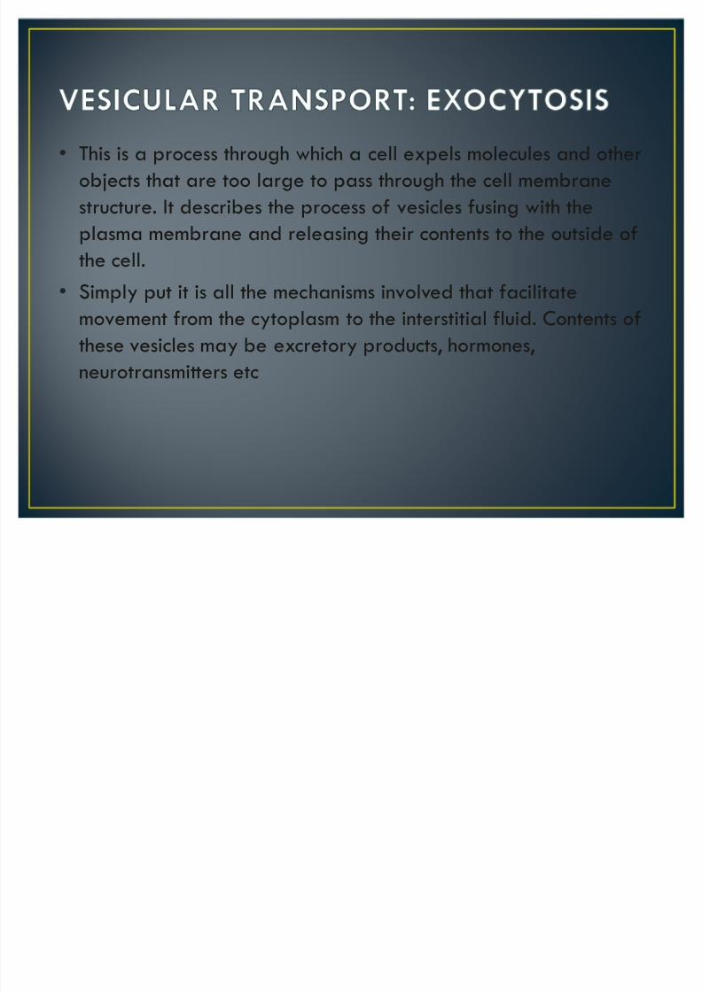
� This is a process through which a cell expels molecules and other
objects that are too large to pass through the cell membrane
structure. It describes the process of vesicles fusing with the
plasma membrane and releasing their contents to the outside of
the cell.
� Simply put it is all the mechanisms involved that facilitate
movement from the cytoplasm to the interstitial fluid. Contents of
these vesicles may be excretory products, hormones,
neurotransmitters etc

8/7/2019 VESICULAR TRANSPORT & AMOEBOID MOVEMENT2
http://slidepdf.com/reader/full/vesicular-transport-amoeboid-movement2 26/48
Constitutive Secretion
In constitutive secretion the protein is sorted into vesicles in the Golgi apparatus
and moves directly to the cell surface membrane. This results in the release of
soluble proteins to the exterior.
Regulated Secretion
Regulated secretion requires an external signal,
a specific sorting signal coat as well as
intracellular calcium.

8/7/2019 VESICULAR TRANSPORT & AMOEBOID MOVEMENT2
http://slidepdf.com/reader/full/vesicular-transport-amoeboid-movement2 27/48

8/7/2019 VESICULAR TRANSPORT & AMOEBOID MOVEMENT2
http://slidepdf.com/reader/full/vesicular-transport-amoeboid-movement2 28/48
Exocytosis occurs in five stages:
1. Vesicle trafficking ² vesicle containing excretory products istransported through the cytoplasm towards the cellmembrane i.e. towards point of deposition
2.Vesicle tethering ² vesicle is suspended and pulled towardpoint of expulsion
3. Vesicle docking ² vesicle comes into contact with the cellmembrane, where it begins to chemically and physicallymerge with the cell membrane proteins.
4. Vesicle priming ² In those cells where chemical transmitters arebeing released, this step involves the chemicalpreparations for the last step of exocytosis.

8/7/2019 VESICULAR TRANSPORT & AMOEBOID MOVEMENT2
http://slidepdf.com/reader/full/vesicular-transport-amoeboid-movement2 29/48
� 5. Vesicle fusion - Proteins from the walls of the vesicle
merge with the cell membrane and breach, pushing the
vesicle contents out of the cell and into the interstitialfluid.

8/7/2019 VESICULAR TRANSPORT & AMOEBOID MOVEMENT2
http://slidepdf.com/reader/full/vesicular-transport-amoeboid-movement2 30/48
Diagram showing process of exocytosis

8/7/2019 VESICULAR TRANSPORT & AMOEBOID MOVEMENT2
http://slidepdf.com/reader/full/vesicular-transport-amoeboid-movement2 31/48
Many cellular processes involve exocytosis. Examples ofthese processes within the human body are:
� Hypothalamic hormone release
� Secretion of proteins like enzymes, peptide hormonesand antibodies from cells.
� Release of neurotransmitters from presynaptic neuronse.g. acetylcholine release
� Acrosome reaction during fertilization� Recycling of plasma membrane bound receptors
� Insulin release from pancreatic cells

8/7/2019 VESICULAR TRANSPORT & AMOEBOID MOVEMENT2
http://slidepdf.com/reader/full/vesicular-transport-amoeboid-movement2 32/48
� Acetylcholine is a neurotransmitter necessary for muscular
functions.
� It is secreted in cytoplasm of terminal boutons then absorbed
into synaptic vesicles.� Dense bars present on neural membrane have protein particles
which act as voltage-gated calcium channels.
� These channels open when there is an action potential and allow
Ca2+ to diffuse from synaptic space into the bouton
� Ca2+ attracts the vesicles drawing them to the membrane with
which they fuse and are then exocytosed.

8/7/2019 VESICULAR TRANSPORT & AMOEBOID MOVEMENT2
http://slidepdf.com/reader/full/vesicular-transport-amoeboid-movement2 33/48
Diagram showing release of acetylcholine at a neuromuscular
junction.
8/7/2019 VESICULAR TRANSPORT & AMOEBOID MOVEMENT2
http://slidepdf.com/reader/full/vesicular-transport-amoeboid-movement2 34/48
Factors that play a regulatory role in exocytosis include:
� Nitric oxide
� Calcium concentration
� Glucose concentration [in the case of insulin secretion]
8/7/2019 VESICULAR TRANSPORT & AMOEBOID MOVEMENT2
http://slidepdf.com/reader/full/vesicular-transport-amoeboid-movement2 35/48

8/7/2019 VESICULAR TRANSPORT & AMOEBOID MOVEMENT2
http://slidepdf.com/reader/full/vesicular-transport-amoeboid-movement2 36/48

8/7/2019 VESICULAR TRANSPORT & AMOEBOID MOVEMENT2
http://slidepdf.com/reader/full/vesicular-transport-amoeboid-movement2 37/48
� Cellular movement is a cyclical process which is driven primarily
by actin polymerization and acto-myosin contractility.
� Cells in the body exhibit three main types of movements: amoeboid
ciliary
muscular.
8/7/2019 VESICULAR TRANSPORT & AMOEBOID MOVEMENT2
http://slidepdf.com/reader/full/vesicular-transport-amoeboid-movement2 38/48
� Ciliary Movement: occurs in most of our internal tubular organs
which are lined by ciliated epithelium.
For example:� The coordinated movements of cilia in the trachea help us in
removing dust particles and some of the foreign substances
inhaled along with the atmospheric air.
� Passage of ova through the female reproductive tract is alsofacilitated by the ciliary movement.

8/7/2019 VESICULAR TRANSPORT & AMOEBOID MOVEMENT2
http://slidepdf.com/reader/full/vesicular-transport-amoeboid-movement2 39/48

8/7/2019 VESICULAR TRANSPORT & AMOEBOID MOVEMENT2
http://slidepdf.com/reader/full/vesicular-transport-amoeboid-movement2 40/48
� Muscular Movement:Movement of our limbs, jaws,tongue, etc, require muscularmovement. The contractileproperty of muscles are effectively
used for locomotion and othermovements by human beings andmajority of multicellular organisms

8/7/2019 VESICULAR TRANSPORT & AMOEBOID MOVEMENT2
http://slidepdf.com/reader/full/vesicular-transport-amoeboid-movement2 41/48
� A moeboid movement is brought about by
reversible changes in the actin filaments of the cell'scytoskeleton.
� Cross-linking of these filaments by other proteinscreates a three-dimensional network with gel-likeproperties of the cell.
Key steps in amoeboid locomotion are:� protrusion of the leading edge of the cell,
� adhesion of the leading edge and release at the cellbody and rear
� cytoskeletal contraction to pull the cell forward.

8/7/2019 VESICULAR TRANSPORT & AMOEBOID MOVEMENT2
http://slidepdf.com/reader/full/vesicular-transport-amoeboid-movement2 42/48
� A s a cell moves towards a substrate, it experiencesexternal forces, which include the viscous force or
resistance from the surrounding medium andcell-substrate interaction forces, and internal forcesthat are generated by the cytoskeleton.
� The cytoskeleton is the essential component increating these motility-driving forces, and incoordinating the entire process of movement.
� The cell propels the membrane forward by orienting and reorganizing (growing) the actinnetwork at its leading edge.
� A s the leading edge begins to protrude, adhesionmolecules gathered in the extending region helpattach the leading edge to the substrate.

8/7/2019 VESICULAR TRANSPORT & AMOEBOID MOVEMENT2
http://slidepdf.com/reader/full/vesicular-transport-amoeboid-movement2 43/48
� Cell-substrate attachments are created at theleading edge when actin bundles link thecytoskeleton to the substrate at certain sites via
adhesion molecules
� A s the cell continues to adhere at the leading edge,it releases from the cell body and rear of the cell,possibly by the disassembly or contraction of its
attachments (actin bundles).
� At the rear the receptors are pulled away from theirligands. This leads to the formation of new endocytoticvesicles.
� Within the cell, these vesicles stream toward thepseudopodial end of the cell, where they are used toform still new membrane for the pseudopodium.

8/7/2019 VESICULAR TRANSPORT & AMOEBOID MOVEMENT2
http://slidepdf.com/reader/full/vesicular-transport-amoeboid-movement2 44/48
� The rest of the cell is pulled forward, mainly by contractile forces that are produced by myosinmotors sliding on actin filaments, which are in the
cell body and at the rear.

8/7/2019 VESICULAR TRANSPORT & AMOEBOID MOVEMENT2
http://slidepdf.com/reader/full/vesicular-transport-amoeboid-movement2 45/48
8/7/2019 VESICULAR TRANSPORT & AMOEBOID MOVEMENT2
http://slidepdf.com/reader/full/vesicular-transport-amoeboid-movement2 46/48
� White Blood Cells exhibit ameboid movement when they
move out of blood into tissues as tissue macrophages.
� Fibroblasts utilize ameboid movement to repair damaged
tissues.
� Embryonic cells also use ameboid locomotion to migrate from
their sites of origin to other areas.
� Germinal cells of the skin move towards a cut area by this
type of locomotion.

8/7/2019 VESICULAR TRANSPORT & AMOEBOID MOVEMENT2
http://slidepdf.com/reader/full/vesicular-transport-amoeboid-movement2 47/48
� Chemotaxis is the most essential initiator of Ameboid movement.
� Most of the cells that exhibit ameboid locomotion move toward
the source of a chemotactic substance; that is, from an area
of lower concentration toward an area of higher concentration;
which is called positive chemotaxis. On the other hand, some
cells move away from the source, which is called negative
chemotaxis.
� The side of the cell most exposed to the chemotactic substance
develops changes in the membrane that causes protrusion of the
pseudopodium.

8/7/2019 VESICULAR TRANSPORT & AMOEBOID MOVEMENT2
http://slidepdf.com/reader/full/vesicular-transport-amoeboid-movement2 48/48
� Ananthakrishnan R, Ehrlicher A. The Forces Behind Cell Movement. Int J Biol
Sci 2007; 3:303-317. Available from http://www.biolsci.org/v03p0303.htm
� Guyton Textbook of Medical Physiology; 11th Edition
� www.britannica.com/EBchecked/topic/454919/phagocytosis
� http://www.bookrags.com/research/nutrition-and-nutrient-transport-to-wap/
� http://www.answers.com/topic/pinocytosis
� www.newworldencyclopedia.org/entry/Phagocytosis
� http://www.biology-online.org/dictionary/Pinocytosis
� http://www.medterms.com/script/main/art.asp?articlekey=14505