
Vertical relations - University of Babylon · Vertical relations Vertical relation: It is the...
16
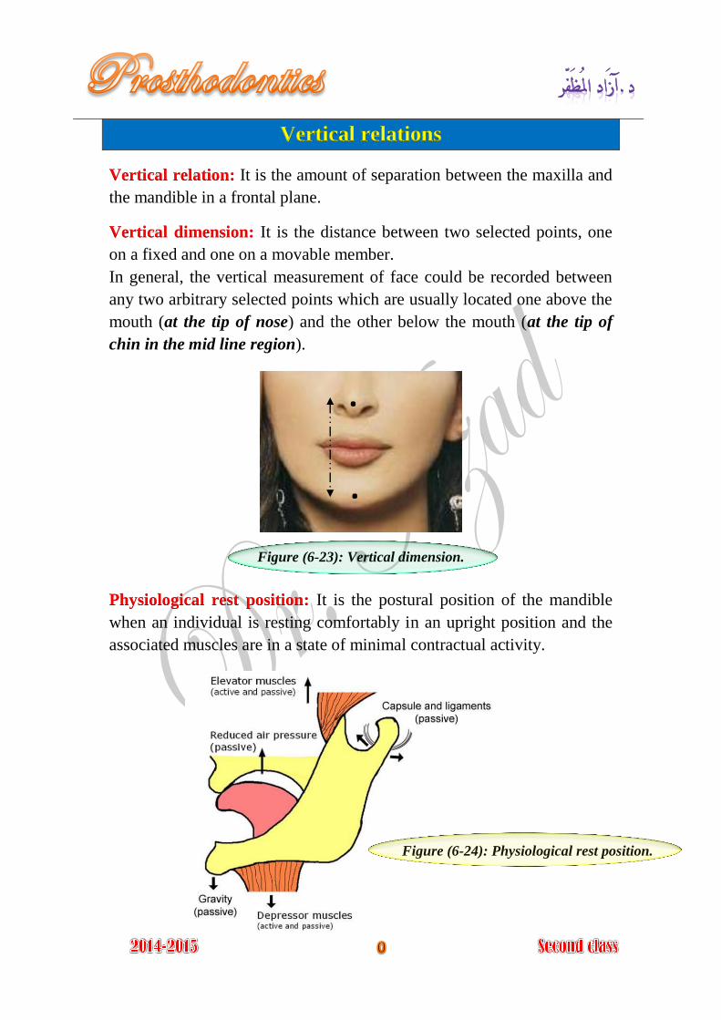
Vertical relations It is the amount of separation between the maxilla and Vertical relation: the mandible in a frontal plane. It is the distance between two selected points, one Vertical dimension: on a fixed and one on a movable member. In general, the vertical measurement of face could be recorded between any two arbitrary selected points which are usually located one above the mouth (at the tip of nose) and the other below the mouth (at the tip of chin in the mid line region). Figure (6-23): Vertical dimension. It is the postural position of the mandible Physiological rest position: when an individual is resting comfortably in an upright position and the associated muscles are in a state of minimal contractual activity. Figure (6-24): Physiological rest position. . .
Transcript of Vertical relations - University of Babylon · Vertical relations Vertical relation: It is the...
Vertical relations
It is the amount of separation between the maxilla and Vertical relation:
the mandible in a frontal plane.
It is the distance between two selected points, one Vertical dimension:
on a fixed and one on a movable member.
In general, the vertical measurement of face could be recorded between
any two arbitrary selected points which are usually located one above the
mouth (at the tip of nose) and the other below the mouth (at the tip of
chin in the mid line region).
Figure (6-23): Vertical dimension.
It is the postural position of the mandible Physiological rest position:
when an individual is resting comfortably in an upright position and the
associated muscles are in a state of minimal contractual activity.
Figure (6-24): Physiological rest position.
.
.
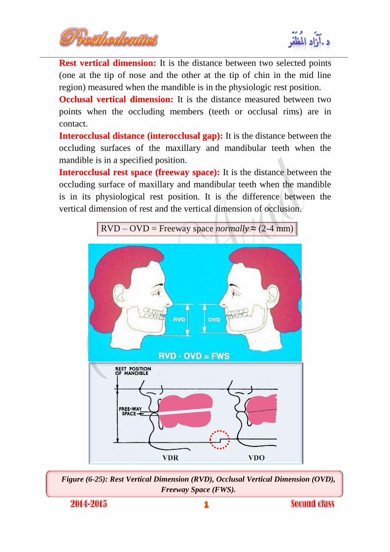
It is the distance between two selected points Rest vertical dimension:
(one at the tip of nose and the other at the tip of chin in the mid line
region) measured when the mandible is in the physiologic rest position.
It is the distance measured between two Occlusal vertical dimension:
points when the occluding members (teeth or occlusal rims) are in
contact.
It is the distance between the Interocclusal distance (interocclusal gap):
occluding surfaces of the maxillary and mandibular teeth when the
mandible is in a specified position.
It is the distance between the Interocclusal rest space (freeway space):
occluding surface of maxillary and mandibular teeth when the mandible
is in its physiological rest position. It is the difference between the
vertical dimension of rest and the vertical dimension of occlusion.
RVD – OVD = Freeway space normally ≈ (2-4 mm)
Figure (6-25): Rest Vertical Dimension (RVD), Occlusal Vertical Dimension (OVD),
Freeway Space (FWS).
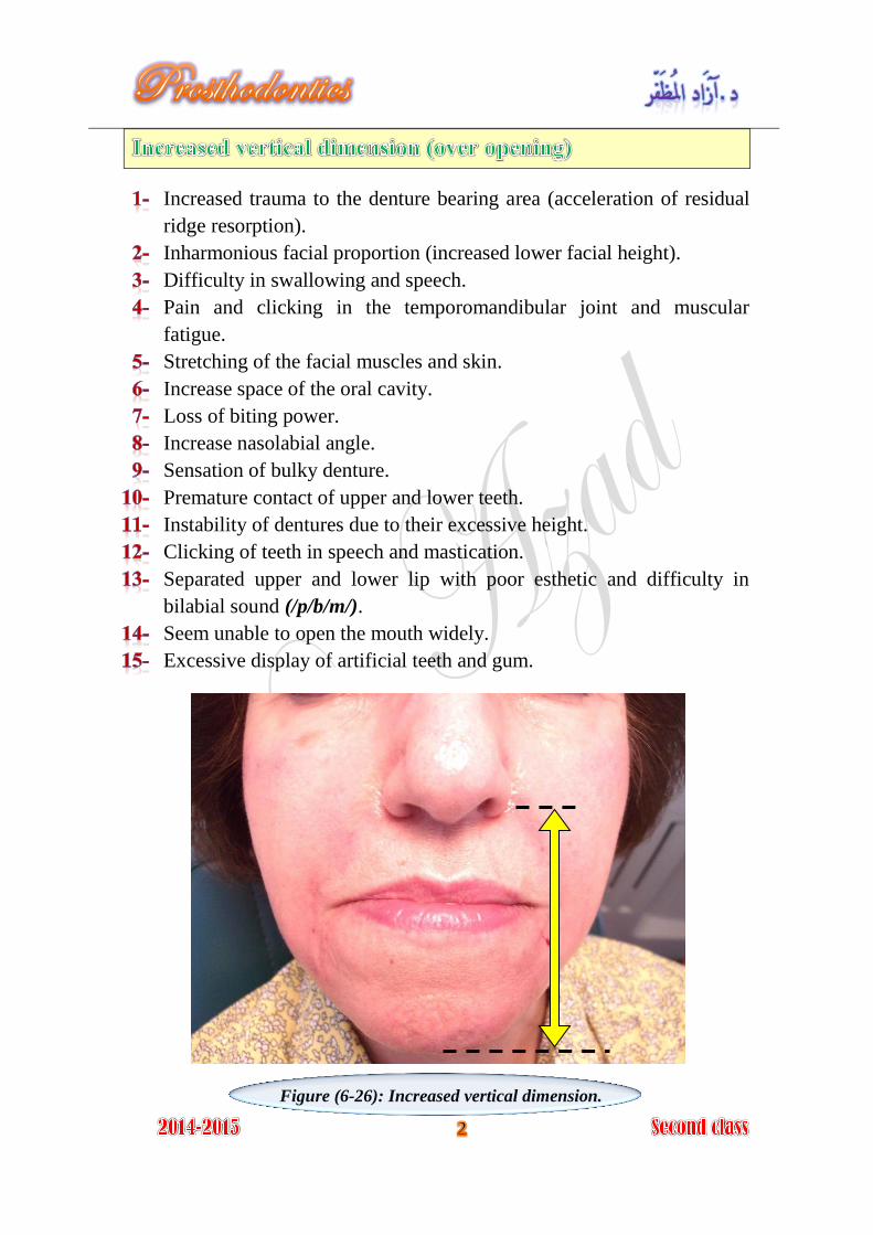
Increased trauma to the denture bearing area (acceleration of residual
ridge resorption).
Inharmonious facial proportion (increased lower facial height).
Difficulty in swallowing and speech.
Pain and clicking in the temporomandibular joint and muscular
fatigue.
Stretching of the facial muscles and skin.
Increase space of the oral cavity.
Loss of biting power.
Increase nasolabial angle.
Sensation of bulky denture.
Premature contact of upper and lower teeth.
Instability of dentures due to their excessive height.
Clicking of teeth in speech and mastication.
Separated upper and lower lip with poor esthetic and difficulty in
bilabial sound (/p/b/m/).
Seem unable to open the mouth widely.
Excessive display of artificial teeth and gum.
Figure (6-26): Increased vertical dimension.
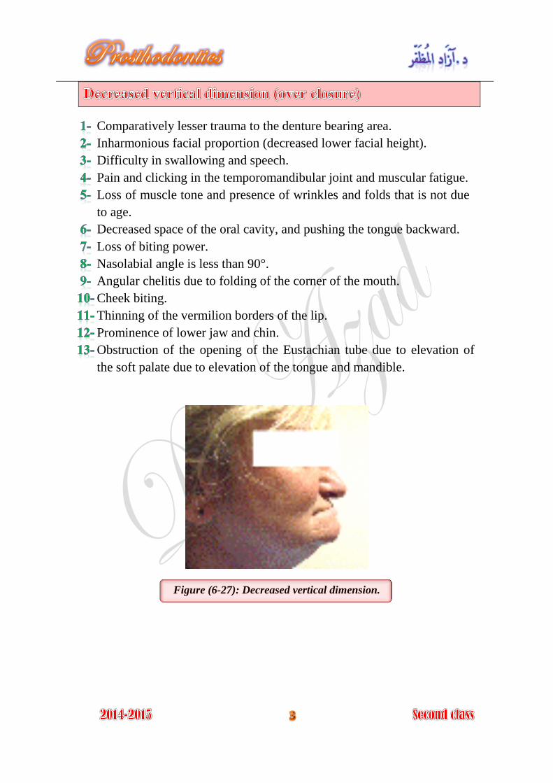
Comparatively lesser trauma to the denture bearing area.
Inharmonious facial proportion (decreased lower facial height).
Difficulty in swallowing and speech.
Pain and clicking in the temporomandibular joint and muscular fatigue.
Loss of muscle tone and presence of wrinkles and folds that is not due
to age.
Decreased space of the oral cavity, and pushing the tongue backward.
Loss of biting power.
Nasolabial angle is less than 90°.
Angular chelitis due to folding of the corner of the mouth.
Cheek biting.
Thinning of the vermilion borders of the lip.
Prominence of lower jaw and chin.
Obstruction of the opening of the Eustachian tube due to elevation of
the soft palate due to elevation of the tongue and mandible.
Figure (6-27): Decreased vertical dimension.
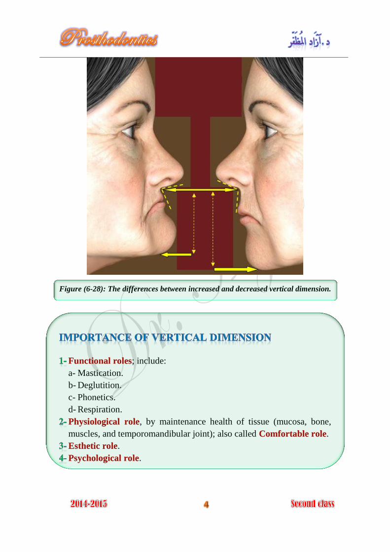
Figure (6-28): The differences between increased and decreased vertical dimension.
; include: Functional roles
a- Mastication.
b- Deglutition.
c- Phonetics.
d- Respiration.
, by maintenance health of tissue (mucosa, bone, Physiological role
muscles, and temporomandibular joint); also called . Comfortable role
. Esthetic role
. Psychological role
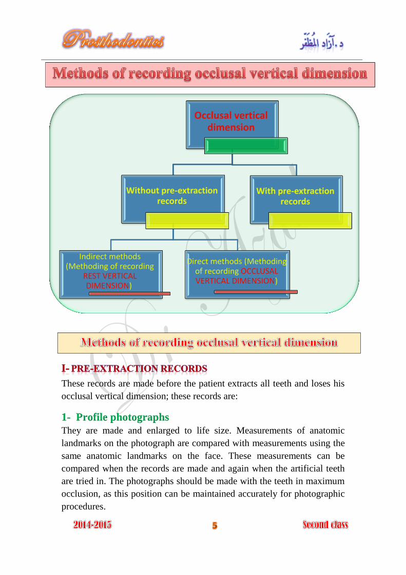
These records are made before the patient extracts all teeth and loses his
occlusal vertical dimension; these records are:
1- Profile photographs
They are made and enlarged to life size. Measurements of anatomic
landmarks on the photograph are compared with measurements using the
same anatomic landmarks on the face. These measurements can be
compared when the records are made and again when the artificial teeth
are tried in. The photographs should be made with the teeth in maximum
occlusion, as this position can be maintained accurately for photographic
procedures.
Occlusal vertical dimension
Without pre-extraction records
Indirect methods (Methoding of recording
REST VERTICAL DIMENSION)
Direct methods (Methoding of recording OCCLUSAL VERTICAL DIMENSION)
With pre-extraction records
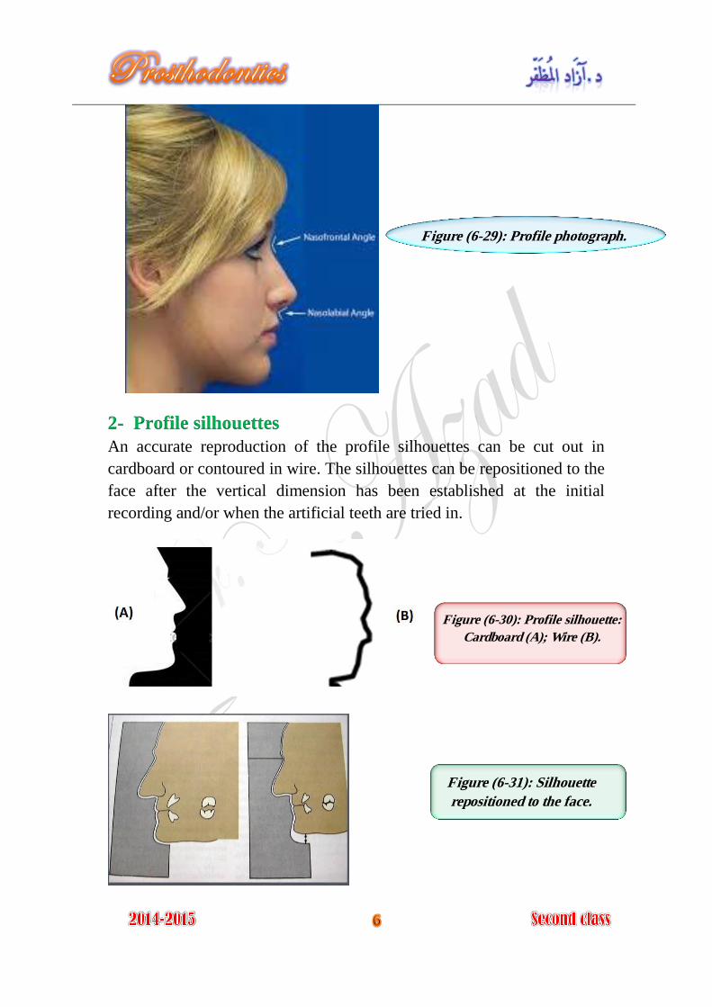
2- Profile silhouettes
An accurate reproduction of the profile silhouettes can be cut out in
cardboard or contoured in wire. The silhouettes can be repositioned to the
face after the vertical dimension has been established at the initial
recording and/or when the artificial teeth are tried in.
Figure (6-29): Profile photograph.
Figure (6-30): Profile silhouette:
Cardboard (A); Wire (B).
Figure (6-31): Silhouette
repositioned to the face.
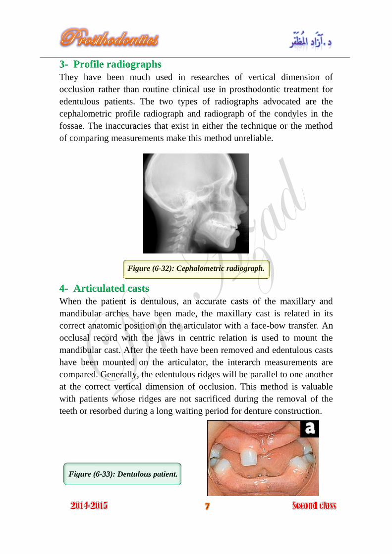
3- Profile radiographs
They have been much used in researches of vertical dimension of
occlusion rather than routine clinical use in prosthodontic treatment for
edentulous patients. The two types of radiographs advocated are the
cephalometric profile radiograph and radiograph of the condyles in the
fossae. The inaccuracies that exist in either the technique or the method
of comparing measurements make this method unreliable.
Figure (6-32): Cephalometric radiograph.
4- Articulated casts
When the patient is dentulous, an accurate casts of the maxillary and
mandibular arches have been made, the maxillary cast is related in its
correct anatomic position on the articulator with a face-bow transfer. An
occlusal record with the jaws in centric relation is used to mount the
mandibular cast. After the teeth have been removed and edentulous casts
have been mounted on the articulator, the interarch measurements are
compared. Generally, the edentulous ridges will be parallel to one another
at the correct vertical dimension of occlusion. This method is valuable
with patients whose ridges are not sacrificed during the removal of the
teeth or resorbed during a long waiting period for denture construction.
Figure (6-33): Dentulous patient.
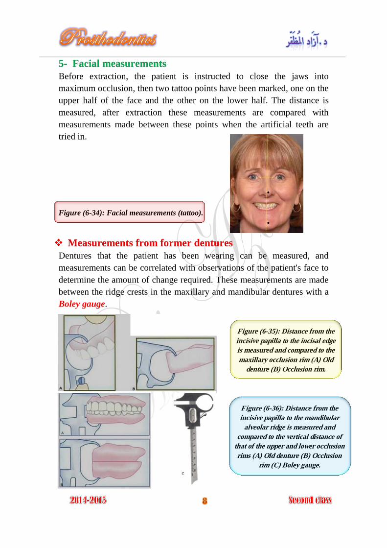
5- Facial measurements
Before extraction, the patient is instructed to close the jaws into
maximum occlusion, then two tattoo points have been marked, one on the
upper half of the face and the other on the lower half. The distance is
measured, after extraction these measurements are compared with
measurements made between these points when the artificial teeth are
tried in.
Figure (6-34): Facial measurements (tattoo).
Measurements from former dentures
Dentures that the patient has been wearing can be measured, and
measurements can be correlated with observations of the patient's face to
determine the amount of change required. These measurements are made
between the ridge crests in the maxillary and mandibular dentures with a
Boley gauge.
.
.
Figure (6-35): Distance from the
incisive papilla to the incisal edge
is measured and compared to the
maxillary occlusion rim (A) Old
denture (B) Occlusion rim.
Figure (6-36): Distance from the
incisive papilla to the mandibular
alveolar ridge is measured and
compared to the vertical distance of
that of the upper and lower occlusion
rims (A) Old denture (B) Occlusion
rim (C) Boley gauge.
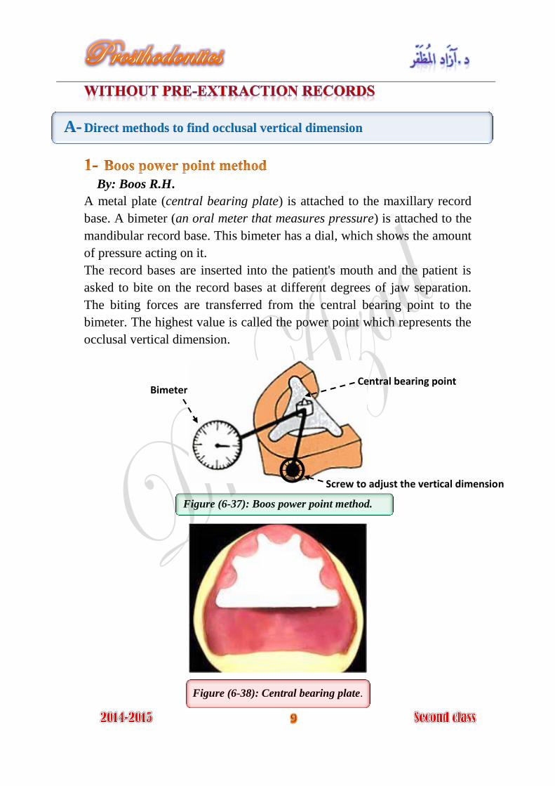
A- Direct methods to find occlusal vertical dimension
By: Boos R.H.
A metal plate (central bearing plate) is attached to the maxillary record
base. A bimeter (an oral meter that measures pressure) is attached to the
mandibular record base. This bimeter has a dial, which shows the amount
of pressure acting on it.
The record bases are inserted into the patient's mouth and the patient is
asked to bite on the record bases at different degrees of jaw separation.
The biting forces are transferred from the central bearing point to the
bimeter. The highest value is called the power point which represents the
occlusal vertical dimension.
Figure (6-37): Boos power point method.
Figure (6-38): Central bearing plate.
Bimeter
Screw to adjust the vertical dimension
Central bearing point
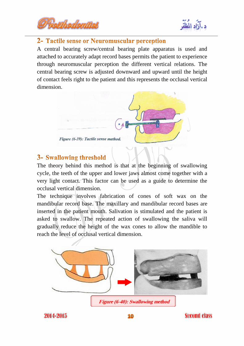
A central bearing screw/central bearing plate apparatus is used and
attached to accurately adapt record bases permits the patient to experience
through neuromuscular perception the different vertical relations. The
central bearing screw is adjusted downward and upward until the height
of contact feels right to the patient and this represents the occlusal vertical
dimension.
The theory behind this method is that at the beginning of swallowing
cycle, the teeth of the upper and lower jaws almost come together with a
very light contact. This factor can be used as a guide to determine the
occlusal vertical dimension.
The technique involves fabrication of cones of soft wax on the
mandibular record base. The maxillary and mandibular record bases are
inserted in the patient mouth. Salivation is stimulated and the patient is
asked to swallow. The repeated action of swallowing the saliva will
gradually reduce the height of the wax cones to allow the mandible to
reach the level of occlusal vertical dimension.
Figure (6-40): Swallowing method

Figure (6-41): Beginning of swallowing cycle (very light teeth contact).
By: Silverman M. Meyer
Silverman's closest speaking space: It is the minimal amount of
interocclusal space between the upper and lower teeth when sounds like
ch, s, and j are pronounced. There is 1-2 mm clearance between teeth
when observed from the profile and frontal view.
Phonetic tests of the vertical dimension include listening to speech sound
production and observing the relationships of teeth during speech. The
production of ch, s, and j sounds brings the anterior teeth closest together
without contact.
If the distance is too large, it means that too small a vertical dimension of
occlusion may have been established.
If the anterior teeth touch when these sounds are made, the vertical
dimension is probably too great.
Figure (6-42):
Silverman's closest speaking space.
Bolus
B- Indirect methods to find occlusal vertical dimension
(methods of recording rest vertical dimension)
1- Facial measurements
Instruct the patient to stand or sit comfortably upright with eyes looking
straight ahead at some object which is on the same level. Insert the
maxillary record base with the attached contoured occlusion rim. With an
indelible marker, place a point of reference on the end of the patient's
nose and another on the point of the chin. The patient is asked to perform
functional movements like wetting his lips and swallowing, and to relax
his shoulders (this is done to relax the supra- and infrahyoid muscles).
When the mandible drops to the rest position, the distance between the
points of reference is measured. Repeat this procedure until the
measurements are consistent. Such measurements are helpful but cannot
be considered as absolute.
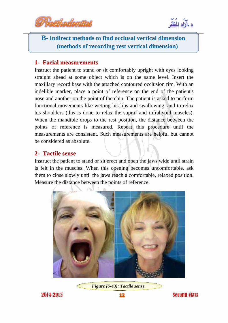
2- Tactile sense
Instruct the patient to stand or sit erect and open the jaws wide until strain
is felt in the muscles. When this opening becomes uncomfortable, ask
them to close slowly until the jaws reach a comfortable, relaxed position.
Measure the distance between the points of reference.
Figure (6-43): Tactile sense.

3- Phonetics
Ask the patient to repeat pronounce the letter m a certain numbers of
times, like repeat the name Emma until they are aware of the contacting
of the lips as the first syllabus em is pronounced. When patient have
rehearsed this procedure, ask that they stop all jaw movement when the
lips touch. At this time measure between the two points of reference.
Figure (6-44):
/m/ sound
4- Facial expression
The experienced dentist may notice the relaxed facial expression when
the patient's jaws are at rest.
The following facial features indicate that the jaw is in its physiological
rest position:
The upper and lower lips should be even anteroposteriorly and in slight
contact in a single plane. The skin around the eyes and over the chin
should be relaxed; it should not be stretched, shiny, or excessively
wrinkled. The nostrils are relaxed and breathing should be unobstructed.
These evidences of rest position of the maxillomandibular musculature
are the indications for recording a measurement of the vertical dimension
of rest.
5- Anatomical landmarks (Willis method)
The Willis guide is designed to measure the distance from the pupils of
the eyes to the corner of the mouth and the distance from the anterior
nasal spine to the lower border of the mandible. When these
measurements are equal, the jaws are considered at rest. Its accuracy is
questionable in patients with facial asymmetry.
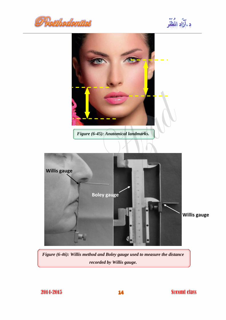
Figure (6-45): Anatomical landmarks.
Figure (6-46): Willis method and Boley gauge used to measure the distance
recorded by Willis gauge.
Boley gauge
Willis gauge
Willis gauge
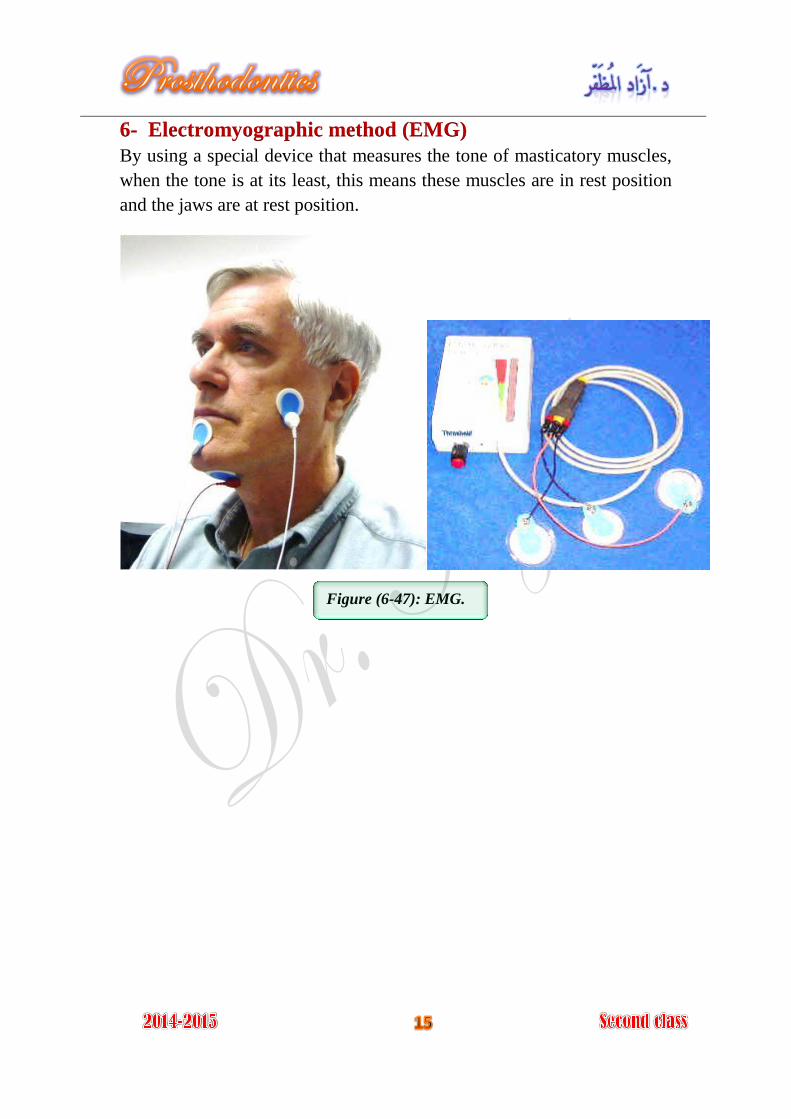
6- Electromyographic method (EMG)
By using a special device that measures the tone of masticatory muscles,
when the tone is at its least, this means these muscles are in rest position
and the jaws are at rest position.
Figure (6-47): EMG.


![Jaw Relations and Mandibular Positions [36-37]€¦ · Jaw Relations • Jaw Relation : It refers to the position of the mandible relative to the position of the maxilla. Centric](https://static.fdocuments.us/doc/165x107/5e9704f8e960161a1a7aa3b7/jaw-relations-and-mandibular-positions-36-37-jaw-relations-a-jaw-relation-.jpg)


