Vdl lablines volume 19 issue 01
16
T he concept of “production livestock veterinary medi- cine” — care that shifts focus from strictly clinical medicine to a multidisciplinary focus on the farmer’s management and ultimately profit — has spread only slowly through veterinary academia. The vision of the new CSU Field Investigation Unit is to actively bring all the university’s multidisciplinary veterinary resources to bear on production problems faced by Colorado livestock producers, identify underlying production or disease issues and solve them with research-based strategies. A collaboration of personnel from the agriculture school’s Department of Animal Sciences, the veterinary college’s Department of Clinical Sciences and the Veteri- nary Diagnostic Laboratories, the unit invites livestock owners or attending veterinarians to request assistance with a disease problem or production issue they feel is beyond the scope of locally available resources. The inves- tigative unit will be available at all times, with points of contact available in each of the participating departments. Once a problem is presented and defined, a team will be assembled from the unit roster that best employs the expertise available. The Field Investigation Unit will aim not only to resolve the current problem as presented, but also to formulate comprehensive plans and recommen- dations for future, preventive use. “We understand livestock production is complex,” said VDL Director Barb Powers, in announcing the new consultation service during the Colorado Cattlemen’s Association 2014 Mid-Winter Conference. “To be suc- cessful, producers simultaneously pursue best practices in systems management, nutrition, disease prevention and other aspects of health and husbandry. Those of us at CSU want to support producers by considering prob- lems and solutions in a very comprehensive way.” In one of its first cases, Field Investigation Unit mem- bers took part in a conference call with a commercial cow/calf producer whose young cows and heifers were failing to conceive, while older cows had few fertility problems. He feared an infectious disease. But as team members asked questions and discussed the problem with the producer, they determined the infertility was likely rooted not in infection, but in nutri- tion and management. They suggested new approaches to pasturing and calving, improved rations and vitamin and trace-mineral forage testing. Members of the Field Investigation Unit will also follow up with the Colorado rancher during the next calving season to ensure the fer- tility trouble has resolved. The new unit satisfies the mission of the VDL and the broader university on several levels. It will contribute to the continued vitality of the livestock sector of Colorado’s $41 billion annual agricultural industry. It will further improve both timeliness and quality of service received by the client. It will further outreach and student involve- ment. And it will point to a need for further research into areas of livestock production in the state and region. Diagnostic news and trends from the Colorado State University Veterinary Diagnostic Laboratories Volume 19, Number 1 Spring/Summer 2014 Food Animal Production Medicine New Field Investigation Unit Puts Mulitidisciplinary Medicine to Work V ETERINARY DIAGNOSTIC LABORATORIES TO REACH THE FIELD INVESTIGATIVE UNIT n Veterinary Diagnostic Laboratories • Charlie Davis (970) 297-0370 (970) 689-1632 charlie.davis @colostate.edu • Barb Powers (970) 297-1285 (970) 221-5729 barb.powers @colostate.edu n Livestock Medicine, James L. Voss Veterinary Teaching Hospital • (970) 297-5000 Extension 1 during business hours Extension 2 after business hours n Department of Animal Sciences • Jason Ahola (970) 491-3312 jason.ahola @colostate.edu • John Wagner (970) 491-2174 john.wagner @colostate.edu — Charlie Davis, DVM, CSU VDL Lab Coordinator
-
Upload
rach-timmons -
Category
Documents
-
view
231 -
download
2
description
Â
Transcript of Vdl lablines volume 19 issue 01
The concept of “production livestock veterinary medi-
cine” — care that shifts focus from strictly clinical
medicine to a multidisciplinary focus on the farmer’s
management and ultimately profit — has spread only
slowly through veterinary academia. The vision of the
new CSU Field Investigation Unit is to actively bring all
the university’s multidisciplinary veterinary resources to
bear on production problems faced by Colorado livestock
producers, identify underlying production or disease
issues and solve them with research-based strategies.
A collaboration of personnel from the agriculture
school’s Department of Animal Sciences, the veterinary
college’s Department of Clinical Sciences and the Veteri-
nary Diagnostic Laboratories, the unit invites livestock
owners or attending veterinarians to request assistance
with a disease problem or production issue they feel is
beyond the scope of locally available resources. The inves-
tigative unit will be available at all times, with points of
contact available in each of the participating departments.
Once a problem is presented and defined, a team will
be assembled from the unit roster that best employs the
expertise available. The Field Investigation Unit will aim
not only to resolve the current problem as presented, but
also to formulate comprehensive plans and recommen-
dations for future, preventive use.
“We understand livestock production is complex,”
said VDL Director Barb Powers, in announcing the new
consultation service during the Colorado Cattlemen’s
Association 2014 Mid-Winter Conference. “To be suc-
cessful, producers simultaneously pursue best practices
in systems management, nutrition, disease prevention
and other aspects of health and husbandry. Those of us
at CSU want to support producers by considering prob-
lems and solutions in a very comprehensive way.”
In one of its first cases, Field Investigation Unit mem-
bers took part in a conference call with a commercial
cow/calf producer whose young cows and heifers were
failing to conceive, while older cows had few fertility
problems. He feared an infectious disease.
But as team members asked questions and discussed
the problem with the producer, they determined the
infertility was likely rooted not in infection, but in nutri-
tion and management. They suggested new approaches
to pasturing and calving, improved rations and vitamin
and trace-mineral forage testing. Members of the Field
Investigation Unit will also follow up with the Colorado
rancher during the next calving season to ensure the fer-
tility trouble has resolved.
The new unit satisfies the mission of the VDL and the
broader university on several levels. It will contribute to
the continued vitality of the livestock sector of Colorado’s
$41 billion annual agricultural industry. It will further
improve both timeliness and quality of service received
by the client. It will further outreach and student involve-
ment. And it will point to a need for further research into
areas of livestock production in the state and region.
Diagnostic news and trends from the Colorado State University Veterinary Diagnostic Laboratories Volume 19, Number 1 Spring/Summer 2014
Food Animal Production Medicine
New Field Investigation Unit Puts Mulitidisciplinary Medicine to Work
Veterinary Diagnostic Laboratories
TO Reach The field invesTigaTive uniT
n veterinary diagnostic laboratories• Charlie Davis
(970) 297-0370 (970) 689-1632 charlie.davis @colostate.edu
• Barb Powers (970) 297-1285 (970) 221-5729 barb.powers @colostate.edu
n Livestock Medicine, James L. Voss Veterinary Teaching Hospital• (970) 297-5000
Extension 1 during business hours extension 2 after business hours
n Department of Animal Sciences• Jason Ahola
(970) 491-3312 jason.ahola @colostate.edu
• John Wagner (970) 491-2174 john.wagner @colostate.edu
— Charlie Davis, DVM, CSU VDL Lab Coordinator

2 Volume 19, Number 1
RefeRences
1. Valli VE, San Myint M, Barthel A, et al. Classification of canine malignant lymphomas according to the World Health Organization criteria. Vet Pathol 2011;48:198-211.
2. Sato M, Kanemoto H, Kagawa Y, et al. Evaluation of the prognostic significance of BCL6 gene expression in canine high-grade B-cell lymphoma. Vet J 2012;191:108-114.
3. Flood-Knapik KE, Durham AC, Gregor TP, et al. Clinical, histopathological and immunohistochemical characterization of canine indolent lymphoma. Vet Comp Oncol 2013;11:272-286.
4. Williams MJ, Avery AC, Lana SE, et al. Canine lymphoproliferative disease characterized by lymphocytosis: immunophenotypic markers of prognosis. J Vet Intern Med 2008;22:596-601.
5. Avery PR, Burton J, Bromberek JL, et al. Flow cytometric characterization and clinical outcome of CD4+ T-Cell lymphoma in dogs: 67 cases. J Vet Intern Med. 2014 Mar-Apr;28(2):538-46.
6. Seelig DM, Avery PR, Webb T, et al. Canine T-zone lymphoma: unique immunophenotypic features, outcome and population characteristics.J Vet Intern Med. 2014 May-Jun;28(3):878-86.
Canine Oncology Innovations
What Lymphoma is this, and Why does it Matter?It has become common to divide LPD, or lympho-
proliferative disorders — the collective term for
lymphoma and leukemia — into “B cell” and “T cell,”
with the understanding that T cell lymphomas have a
worse prognosis. This idea, however, needs to be re-
examined.
Proper classification of LPD can greatly facilitate
decisions about whether to treat LPD — some owners
may decide not to treat in the face of a poor progno-
sis. Some indolent LPD can be monitored without
therapy. Proper classification of LPD can also guide, in
some cases, the type of treatment chosen.
The World Health Organization recognizes over 50
different types of LPD in people, and a study in 2011
indicates that many of these types can be found in
dogs.1 Such diversity occurs because each type of lym-
phoma arises from a lymphocyte at a separate stage of
differentiation.
Different types of lymphoma can have dramatically
different outcomes and treatment options. Three of
the most common types of B cell LPD are diffuse large
B cell lymphoma, marginal zone lymphoma and B cell
chronic lymphocytic leukemia (CLL), or small cell lym-
phoma. Diffuse large B cell lymphoma has a median
survival of approximately one year when treated with
CHOP chemotherapy.2 Marginal zone lymphoma,
however, has a median survival of two years3 and B cell
CLL has a median survival of greater than three years.4
Both diseases can be treated with prednisone and chlo-
rambucil, rather than a more aggressive protocol.3
T-cell lymphomas are also highly heterogeneous.
Take, for example, two different types of T cell lym-
phomas shown in the chart above — although many
additional forms exist. The first, peripheral T cell lym-
phoma, has an overall median survival of 150 days
when dogs are treated with multi-drug therapy.5 This
type of lymphoma is particularly common in Boxers
with a median age of 7 years, and it often features
hypercalcemia or a mediastinal mass. It is most likely
derived from T cells at an early stage of development.
The second type of lymphoma is called T zone lym-
phoma. Four in 10 T zone cases are in golden retrievers
with a median age of 10.6 This form of T cell lym-
phoma has an indolent course, with patients often not
Pros Cons Cost
Flow cytometry
3 Sample is easy to obtain without anesthesia3 Assay is objective3 Can provide prognostic information for
many, but not all types of LPD in dogs3 Can be carried out on blood without a
lymph node aspirate if the CBC indicates lymphocytosis or blasts in the blood
7 Cannot distinguish certain types of B cell lymphomas (diffuse large B cell versus marginal zone)
7 Cells must be alive, so samples need to be shipped overnight on ice
7 Offered by restricted number of labs (CSU Clinical Immunology is the largest provider of this service)
$115
Histology/ Immuno-histochemistry
3 Formalin fixation means sample does not require special shipping or handling
3 Can classify more lymphoma subtypes than flow cytometry
3 Available from most reference labs
7 Usually requires sedation/anesthesia, so can be more expensive
7 More subjective than flow cytometry
$114
— Anne Avery, DVM, PhD, CSU Associate Professor of Microbiology, Immunology and Pathology
The high heterogeneity of T-cell lymphomas leads to differing progression in the disease and different choices in treatment.
� Peripheral T-Cell
� T-Zone
100%
80%
60%
40%
20%
250 500 750 1000 1250 Days survival
Which test do I choose?
Percent Survival Rate
Spring/Summer 2014 3 LABLINES
requiring chemotherapy for months. When dogs show
clinical signs and chemotherapy is instituted, some
data indicate prednisone and chlorambucil is as effec-
tive as more expensive and aggressive protocols.3
TESTInG OPTIOnSHow can you tell what type of LPD is afflicting your
patient? Two tests can help classify a dog’s LPD:
n Flow cytometry, which is performed on a lymph
node aspirate or blood using fluorescent labeled
antibodies to different cell surface proteins. Flow
cytometry identifies different types of lymphoma
by the constellation of proteins cells express on the
surface.
n Histology/immunohistochemistry, which is per-
formed on a biopsy and includes staining for B and
T cell subsets. Histology identifies different types of
lymphoma by their cell size, morphology and the
architecture of the malignant node.
The choice of test depends on the owner’s circumstances
and where the primary disease manifests. Since initial
diagnosis is often made from a fine-needle aspirate, con-
sultation with clinical pathology can be helpful before
deciding what test to submit for further classification.
Food Animal Production Medicine
High-Country Calf MortalityProducer reports from ranches over 2,438 meters
altitude in southwest Colorado suggest mortality
of preweaned beef calves may be substantially higher
than the national average, despite a history of select-
ing herd sires adapted to low pulmonary pressure.
Diagnostic investigations have been limited due to
the extensive mountainous terrain over which these
calves are grazed with their dams. The objective of our
study was to determine the causes of calf mortality on
five high-altitude ranches in Colorado that have been
selectively breeding sires with low pulmonary pres-
sure of less than 45 mm Hg for over 20 years. Calves
were followed from spring branding at 6 weeks old
to fall weaning at 7 months old. We recorded clinical
signs, took blood samples from sick calves, performed
postmortem examinations and submitted select tissue
samples for aerobic culture or histopathology.
On the principal study ranch, 9.6 percent, or 59 of
612 calves, that were branded in the spring either died
or were presumed dead by weaning in the fall. In total,
28 necropsies were performed. Half of those had lesions
consistent with pulmonary hypertension and right-side
heart failure, and half died from bronchopneumonia.
Remodeling of the pulmonary arterial system, indicative
of pulmonary hypertension, was evident in the former
and to varying degrees in the latter.
There is a need to better characterize the additional
risk factors that complicate pulmonary arterial pres-
sure testing of herd sires as a strategy to control pul-
monary hypertension.
© “Spring Time in the Rockies,” Bo Isogna TheLightningMan.com. Some rights reserved. Used under CC BY-NC 2.0.
— Dan Gould, DVM, PhD, CSU Microbiology, Immunology and Pathology Professor Emeritus; Joe Neary, MA, MS, VetMB, CSU Integrated Livestock Management.
To discuss a case of suspected LPD, call the clinical immunology lab at (970) 491-1170. You can also find more information at csu-cvmbs.colostate.edu/academics/mip/ci-lab
Since an initial LPD diagnosis is often made from a fine-needle aspirate, consultation with clinical pathology can be helpful before deciding what test to submit for further classification.© School of Veterinary Medicine and Science, University
of Nottingham. Some rights reserved. Used under CC BY-NC-SA 2.0.
Lymph Node
Lyphoblasticleukemia/lymphoma
Bone Marrow Myeloma
Difuse large B-cell lymphomaSurvival: 1 year
B-cell chroniclymphocytic leukemiaSurvival: 3 years
Marginal zone lymphomaSurvival: 2 years
4 Volume 19, Number 1
Chemistry and Toxicology
As Livestock Producers Dip into Low-Quality Forages, Beware Nitrate Even with the continuing drought in Southeastern
Colorado, greater than 99 percent of the total 1,376
forage samples CSU VDL’s Rocky Ford laboratory ana-
lyzed for nitrate concentrations over the last two years
could be fed to livestock either as is or by diluting with
low nitrate forages. That leaves less than 1 percent
considered not safe to feed.
Although this pattern remained constant through
the first two months of 2014, since the first of March
we have received samples of corn stalks, sudan hay,
oat hay and pigweed that contained from 17,000 ppm
nitrate to over 60,000 ppm nitrate. This pattern is con-
tinuing, with more samples measuring in the 5000 to
10,000 ppm range than in the first two months of 2014.
It seems this pattern is unlikely to change anytime
soon, as producers have exhausted their forage supplies
and are forced to feed very poor quality forages.
Therefore, producers should not assume forages are
safe for use without testing. The consequences can be
costly. Just in the last month we have seen cases of nitrate
poisoning with multiple death losses and abortions
from cows that exhibited clinical illness. In one instance,
the cattle were turned into a pasture heavily contami-
nated with pigweed which contained more than 60,000
ppm nitrate. A similar incident occurred when cows
were fed hay comprised primarily of pigweed that had
19,000 ppm nitrate. Six cows died in less than 12 hours
after they were fed recently purchased sudan hay. The
cows’ ocular fluid contained more than 50 ppm nitrate,
confirming nitrate poisoning as the cause of death.
TESTInG REMAInS THE BEST STRATEGYTesting forages for nitrate content is still the only way to
try to prevent such losses from occurring.
When to test. Nitrate levels in plants increase during
periods of plant stress and impaired growth. A wide vari-
ety of plants including many common grasses, winter
forages, hay and crop residues are known to accumulate
toxic levels of nitrate, especially after heavy applications
of nitrogen fertilizers. Herbicide application can increase
the level of nitrate in row crops and weeds. It is not
unusual for sorghum sudan hay grown on heavily fertil-
ized crop fields to contain greater than 6000 ppm nitrate
even when adequately irrigated. Nitrate poisoning is
most likely to occur when cattle are in poor body condi-
tion and being maintained on low energy diets. Adverse
weather conditions potentiate nitrate poisoning.
What to test. Sorghum sudan hybrids, millet, oat hay,
corn and milo stalks and a multitude of weeds — pri-
marily kochi, pigweed and thistles — comprise the
bulk of the nitrate testing done at the Rocky Ford
CVM VDL. The effects of nitrates in feed and water
are additive, and both sources should be considered.
High nitrate forages should not be fed when cattle are
exposed to high nitrate water.
How to test. Quite often we receive a single plant
from a field or sample of hay from one bale to test for
nitrate. This practice is discouraged, as an extremely
wide variation in nitrate levels can occur throughout
a particular field. Kansas State University reported that
in a field of drought-stressed sudan, some bales con-
tained more the twice the average nitrate level for the
entire field. When taking pre-harvest samples, several
samples taken at the level of expected harvest (do not
include roots) should be taken from throughout the
field. When there are major differences in plant growth
within a field, it is best to divide the field into areas
according to growth and test them individually so
areas with plants high in nitrate can be identified and
managed accordingly. With post-harvest samples, core
samples from a number of bales should be combined
and analyzed. Results from single grab samples taken
from the exterior of one or two bales should not be
relied on to represent the entire lot.
Which animals to target. Although nitrate poisoning
has occurred in cattle consuming forages with less than
5000 ppm nitrate, the general recommendation is that
forages with up to 10,000 ppm, or 1 percent, nitrate are
considered safe to feed to non-pregnant cattle that have
been acclimated to the high nitrate feed. Cattle in poor
body condition consuming low energy diets are most
susceptible to nitrate poisoning. Sheep are less susceptible
than cattle. Nitrate poisoning has also been reported in
goats and alpacas. Although a few references consider
horses to be as susceptible to nitrates as ruminants, other
reports state that no harmful effects occurred in preg-
nant and open mares consuming forages containing
from 25,000 to 35,000 ppm nitrate over a period of sev-
eral months. Others have reported no ill effects in horses
consuming high nitrate forages that killed cows fed the
— Gene Niles, DVM, DABVT, Director, CSU VDL Rocky Ford Branch
References:n Toxic Plants of north
America; Burrows and Tyrl, Iowa State Press, 2001
n Clinical Veterinary Toxicology; Plumlee; Mosby; 2004; p 127-130
n Cattlemen’s Day 1990; progress report 592; Agriculture Experiment Station, Kansas State University
n nitrate and nitrite Toxicity in horses; news: Kentucky Equine Research
n nitrates in Livestock Feeding; nebGuide, G1779; University of nebraska Lincoln
n Alpaca plant poisoning: nitrate-nitrite and possible cyanide; Australian Veterinary Journal Vol 87, no 3, March 2009, p 113-115
© AgriLife Today. “Feeding protein cubes.” Some rights reserved. Used under CC BY-NC-ND 2.0. .
Spring/Summer 2014 5 LABLINES
same hay. A Kentucky Equine Research News publication
lists 20,000 ppm as the level of concern for horses. High
nitrate forages do not pose a risk to other non-ruminants.
How to interpret and use results. Results are reported
in a variety of ways — in either percentages or as parts
per million and in units of either nitrate, nitrate-nitro-
gen or potassium-nitrate. To correctly interpret nitrate
results, be sure the units of measurement on the labo-
ratory report match the reference chart in use:
n Percentage times 10,000 equals ppm; ppm divided
by 10,000 equals percentage.
n Ppm nitrate-nitrogen times 4.4 equals ppm nitrate.
n Ppm nitrate times by 0.23 equals ppm nitrate-nitrogen.
n Ppm potassium nitrate times 1.63 equals ppm nitrate.
Recommendations for the maximum safe level of
nitrate in forages fed to pregnant cows range from 4400
ppm to 9300 ppm. Many references recommend forages
with nitrate levels ranging from 5000 to 9300 ppm be
limited to less than 50 percent of the dry matter for preg-
nant cows, while others consider these levels safe if cows
are acclimated to the high nitrate. Controlled studies have
failed to demonstrate abortions in cows that do not show
clinical signs of illness, but it’s best to keep nitrate levels in
forages fed to pregnant cows below 5000 ppm.
Cattle can adapt to increased nitrate levels in their
diets in as little as seven days. Once acclimated, cattle
should remain on a constant level of nitrate, as accli-
mation can be lost as quickly as it is gained.
1All plants contain nitrate, but under stress certain forages — corn, sorghum, oats, soybeans, millet,
sudan and sorghum/sudan hybrids — can accumulate toxic levels. Pigweed, Canadian thistle, kochia, ragweed and other weeds also tend to accumulate nitrate.
2nitrate content varies widely throughout the plant but is greatest in the lower third of the stalk.
3Concentration is usually high in young plants and decreases as the plant matures. However, at high levels
of soil nitrate or under conditions of stress, content may be high at maturity. Highest levels occur just before flowering and decline rapidly after pollination and seed formation.
4Abrupt setbacks to growth, like drought or freezing, may result in high nitrate. Lack of sunlight, temperature
extremes or hail damage can also increase levels.
5 Levels increase immediately after a drought-breaking rain; therefore, harvest should be delayed for a week.
6When silage is made from high nitrate forages, anaerobic fermentation converts nitrate to ammonia,
significantly reducing the nitrate content. Levels in properly ensiled forages can decrease by 30 percent to 60 percent over a month or two. Forages with significantly elevated nitrate levels at harvest should be retested before fed.
7Green chop should be fed as soon as possible. Storage heating can convert nitrate to nitrite — the toxic agent
in nitrate poisoning — increasing potential toxicity up to ten times.
8To analyze harvested hay or silage, take a composite sample from six to eight different bales or locations in
the silo. Cored samples are preferred on bales.
9The effects of nitrate levels in forage, feed and water are additive. Livestock water containing 1,000 ppm
nitrate can contribute to nitrate poisoning even when feed contains only moderate levels.
10High energy feeds and gradual introduction to high nitrate feeds will increase tolerance. Healthy
animals have a higher resistance than ill or poorly nourished animals.
10 FACTORS RELATED TO nITRATE TOxICITY
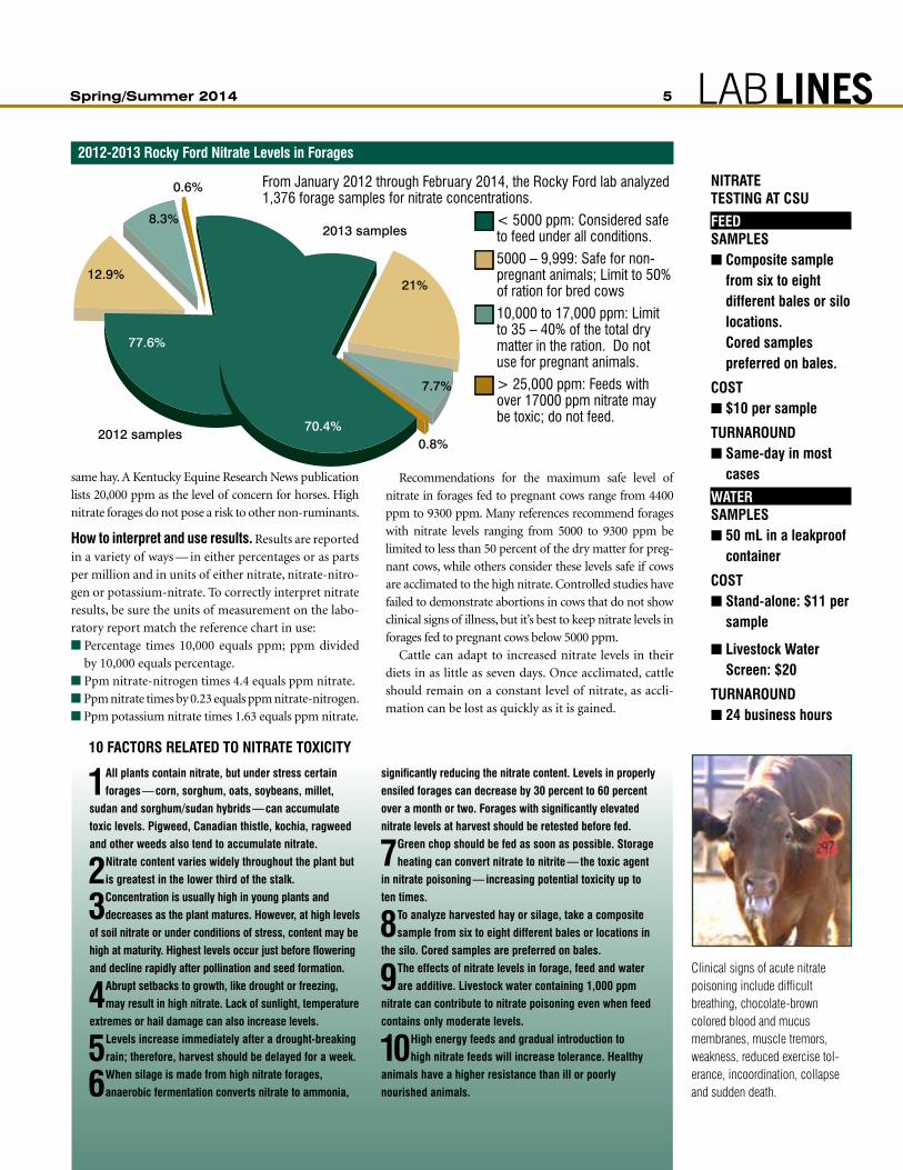
2012 samples
77.6%
12.9%
8.3%
0.6%
2013 samples
70.4%
21%
7.7%
0.8%
From January 2012 through February 2014, the Rocky Ford lab analyzed 1,376 forage samples for nitrate concentrations.
n < 5000 ppm: Considered safe to feed under all conditions.
n 5000 – 9,999: Safe for non- pregnant animals; Limit to 50% of ration for bred cows
n 10,000 to 17,000 ppm: Limit to 35 – 40% of the total dry matter in the ration. Do not use for pregnant animals.
n > 25,000 ppm: Feeds with over 17000 ppm nitrate may be toxic; do not feed.
2012-2013 Rocky Ford nitrate Levels in Forages
Clinical signs of acute nitrate poisoning include difficult breathing, chocolate-brown colored blood and mucus membranes, muscle tremors, weakness, reduced exercise tol-erance, incoordination, collapse and sudden death.
niTRaTe TesTing aT csu
feedSAMPLESn Composite sample
from six to eight different bales or silo locations. Cored samples preferred on bales.
cOsTn $10 per sample
TuRnaROundn Same-day in most
cases
WATERSAMPLESn 50 mL in a leakproof
container
cOsTn Stand-alone: $11 per
sample
n Livestock Water screen: $20
TuRnaROundn 24 business hours

6 Volume 19, Number 1
CSU VDL in the Field: Case Study
Sodium Intoxication in a EweA 4,500-head commercial sheep ranch in south-
eastern Wyoming presented a 4-year-old, lac-
tating crossbreed ewe for evaluation of six hours of
recumbency and obtunded mentation. A second ewe
showing similar signs earlier that day had died without
treatment, but the carcass was not available for exami-
nation. The ewe had given birth to two healthy lambs
17 days prior to illness onset, one of 30 ewes and 54
lambs that had been moved into a 10-acre, fenced grass
pasture the previous evening.
The ewes and lambs had been coralled for about 14
days before being pastured. While in the corral, they
were provided ad lib grass hay and water pumped into
troughs from a nearby stream. The pasture contained a
mixture of native grasses and shrubs, and access to the
same stream was provided at a corner of the pasture. A
tub of loose salt, not supplemented with trace minerals
or medications, was present in the center of the pasture.
A fenced-in shed was present in the center of the pas-
ture; the ewes and lambs had been gathered the previous
night and confined within the shed during the previous
night. Inspection of the pasture revealed no toxic plants
or materials. The ewes were immunized annually for
tetanus and Clostridium perfringens C and D, with the
most recent booster administered two months before.
Physical examination revealed a recumbent, thin,
severely obtunded ewe with a normal heart rate, respi-
ratory rate and rectal temperature. Hydration status,
mucous membrane color, and capillary refill time were
normal. Neurologic examination revealed fascicula-
tions of the facial muscles and variable, bilateral nys-
tagmus. Vision, pupillary light responses, muscle tone
and spinal reflexes were normal.
The ewe’s venous blood sample glucose concentra-
tion, as determined by a portable glucometer, was 404
mg/dl, compared to a normal of 70 to 100. A sample of
cerebrospinal fluid was obtained by lumbosacral punc-
ture. Differential diagnoses included head trauma, lis-
teriosis, bacterial meningitis, rabies, salt intoxication
or water deprivation, and type D enterotoxemia. Dexa-
methasone at 0.1 mg/kg IV, oxytetracycline at 11 mg/
kg IV and 20 cc SC Clostridium perfringens types C and
D antitoxin were administered. The ewe died about
one hour after treatment.
Brain-tissue fluorescent antibody testing for rabies
virus was negative. Gross necropsy revealed multiple
pulmonary and mediastinal abscesses suggestive of
caseous lymphadenitis. Serum biochemical analysis
revealed hypernatremia (serum sodium concentra-
tion, 195 mEq/l; normal, 142-152), hyperchloridemia
(152 mEq/l; normal, 103-113), hyperglycemia (serum
glucose concentration, 419 mg/dl) with normal serum
calcium and magnesium concentrations. Serum
activities of hepatic and muscle enzymes were within
normal limits. Analysis of cerebral spinal fluid revealed
no cytologic abnormalities; however, the sodium con-
centration of the fluid was 195 mEq/l (normal, < 160),
suggesting salt intoxication.
Histopathologic examination of cerebrum, cerebel-
— Dave Van Metre, DVM, DACVIM, CSU Animal Population Health Institute Professor
Spring/Summer 2014 7 LABLINES
CSU VDL in the Field: Disease Updates
Canine PheochromocytomasPheochromocytoma associated catecholamine-
induced cardiomyopathy is a well-recognized
entity in man and has been described in mice and non-
human primates. But it has not yet been identified or
described in dogs.
In our retrospective study, we identified nine dogs
with histologically confirmed pheochromocytomas
and concurrent cardiovascular pathology observed
histologically (n=6), echocardiographically (n=4) or
electrocardiographically (n=5).
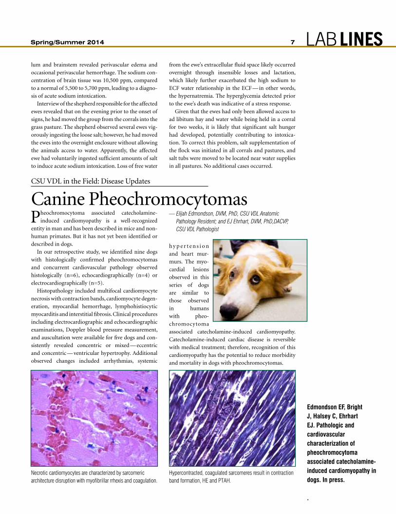
Histopathology included multifocal cardiomyocyte
necrosis with contraction bands, cardiomyocyte degen-
eration, myocardial hemorrhage, lymphohistiocytic
myocarditis and interstitial fibrosis. Clinical procedures
including electrocardiographic and echocardiographic
examinations, Doppler blood pressure measurement,
and auscultation were available for five dogs and con-
sistently revealed concentric or mixed — eccentric
and concentric — ventricular hypertrophy. Additional
observed changes included arrhythmias, systemic
h y p e r t e n s i o n
and heart mur-
murs. The myo-
cardial lesions
observed in this
series of dogs
are similar to
those observed
in humans
with pheo-
chromocytoma
associated catecholamine-induced cardiomyopathy.
Catecholamine-induced cardiac disease is reversible
with medical treatment; therefore, recognition of this
cardiomyopathy has the potential to reduce morbidity
and mortality in dogs with pheochromocytomas.
Edmondson EF, Bright J, Halsey C, Ehrhart EJ. Pathologic and cardiovascular characterization of pheochromocytoma associated catecholamine-induced cardiomyopathy in dogs. In press.
.
— Elijah Edmondson, DVM, PhD, CSU VDL Anatomic Pathology Resident; and EJ Ehrhart, DVM, PhD,DACVP, CSU VDL Pathologist
lum and brainstem revealed perivascular edema and
occasional perivascular hemorrhage. The sodium con-
centration of brain tissue was 10,500 ppm, compared
to a normal of 5,500 to 5,700 ppm, leading to a diagno-
sis of acute sodium intoxication.
Interview of the shepherd responsible for the affected
ewes revealed that on the evening prior to the onset of
signs, he had moved the group from the corrals into the
grass pasture. The shepherd observed several ewes vig-
orously ingesting the loose salt; however, he had moved
the ewes into the overnight enclosure without allowing
the animals access to water. Apparently, the affected
ewe had voluntarily ingested sufficient amounts of salt
to induce acute sodium intoxication. Loss of free water
from the ewe’s extracellular fluid space likely occurred
overnight through insensible losses and lactation,
which likely further exacerbated the high sodium to
ECF water relationship in the ECF — in other words,
the hypernatremia. The hyperglycemia detected prior
to the ewe’s death was indicative of a stress response.
Given that the ewes had only been allowed access to
ad libitum hay and water while being held in a corral
for two weeks, it is likely that significant salt hunger
had developed, potentially contributing to intoxica-
tion. To correct this problem, salt supplementation of
the flock was initiated in all corrals and pastures, and
salt tubs were moved to be located near water supplies
in all pastures. No additional cases occurred.
Necrotic cardiomyocytes are characterized by sarcomeric architecture disruption with myofibrillar rrhexis and coagulation.
Hypercontracted, coagulated sarcomeres result in contraction band formation, HE and PTAH.

8 Volume 19, Number 1
Exotics Medicine
Rickettsia in an Emperor Scorpion?A n adult male emperor scorpion was received for
necropsy from an arachnid collection in which
two scorpions and one tarantula had recently died due
to necrotizing, bacterial enteritis. This scorpion pre-
sented clinically with a brief, two-day history of marked
listlessness and weakness, including inability to lift
the tail. Upon
death, the
coelom was
opened and
swabbed for in-house aerobic culture, which was nega-
tive for growth. The body was placed in formalin for
fixation and further prosection.
The most notable histopathologic finding was large
numbers of pale, granular phagocytic-like cells filling
the hemolymph in all sections and multifocally infiltrat-
ing the fat bodies and skeletal muscle, particularly of the
mesosoma, or mid-section. Phagocytic cells were stained
for micro-organisms using a panel of histochemical stains.
Granular intra-histiocytic material was strongly acid fast
positive, highlighting numerous small intra-cytoplasmic
rods. Intracytoplasmic material was non-Gram staining
and silver-negative. Systemic granulomatosis with pos-
sible intracytoplasmic organisms was diagnosed.
Using the University of Minnesota Veterinary Diag-
nostic Laboratory’s transmission electron microscopy to
produce high quality detailed images, we confirmed the
phagocytic cells to be of macrophage lineage. The gran-
ular intracytoplasmic material consisted of numerous
cytoplasmic vesicles containing tightly packed bacilliform
organisms of about 350 by 60 nanometers. Although
initially these acid-fast organisms were thought to be
most consistent with Mycobacterium, Mycobacteria
are significantly larger, at generally 1 to 4 micrometers.
A differential etiologic diagnosis included intra-his-
tiocytic bacilliform virus infection. Bacilliform viruses
include Ascovirus, Baculovirus, and Nudivirus species.
These large viruses are histologically similar in size and
morphology to those we detected; however, bacilliform
viruses should be closely associated with the nucleus,
forming intra-nuclear inclusion bodies at certain stages,
as well as replicative phases that occur in close association
with the nucleus. Our macrophage examination, in con-
trast, showed these organisms proliferated within intracy-
toplasmic vesicles with no nuclear association.
Mycobacterium PCR on formalin-fixed paraffin
embedded tissue with phagocytic cells submitted to
Washington State University’s diagnostic lab was uni-
versally negative. However, when PCR primers for other
small, intracellular and possible acid-fast positive bacteria
were then employed, results using Rickettsia sp. primers
were strongly positive. Additional speciation of this pos-
sible Rickettsia organism is currently underway. This is
thought to be a novel Rickettsia species infecting a scor-
pion, an arachnid for which rickettsial disease has not
been previously described.
EXOTICS
(Clockwise from top)n Pale, granular phagocytic-like cells filled the hemolymph
in all sections and multifocally infiltrated the fat bodies and skeletal muscle, particularly of the mesosoma.
n Granular intracytoplasmic material consisted of numerous cytoplasmic vesicles containing tightly packed bacilliform organisms of about 350 by 60 nanometers.
n Granular intra-histiocytic material was strongly acid fast positive, highlighting numerous small intra-cytoplasmic rods.
— Sushan Han, DVM, PhD, DACVP, CSU VDL Pathologist; Aníbal Armién DVM, MS, PhD, DACVP
Spring/Summer 2014 9 LABLINESCSU VDL In Press
A Roundup of VDL Faculty ResearchBurgess BA, noyes nR, Bolte DS, Hyatt DR, Van Metre DC, Morley PS. Rapid Salmonella detec-tion in experimentally-inoculated equine faecal and veterinary hospital environmental samples using commercially available lateral flow immunoassays. Equine Vet J. 2014 Feb 9. doi: 10.1111/evj.12234.VDL Bacteriology Section Head Doreene Hyatt partic-
ipated in a Department of Clinical Sciences study that
first inoculated horse feces and environmental swabs
from the floor of a building
next door to the teaching hos-
pital with a Salmonella enterica
serotype Typhimurium from a
former equine patient, and then
tested them using one of two
commercial rapid-diagnostic
tests kits marketed for S. enterica
detection in meat, poultry and chicken feed. The cheap
and quick lateral-flown immunoassays use antibodies
specific for surface antigens of Salmonella and colloidal
gold-antibody conjugates incorporated into test strips.
Their study showed the strips could reliably detect
S. enterica within 18 hours, indicating they may be
useful for rapid point-of-care testing to allow manag-
ers to implement measures more effectively to decrease
animal and zoonotic infections in hospitals, racetracks,
shows and other high-risk areas where horses congre-
gate and shed the bacteria.
Hoon-Hanks LL, Regan D, Dubey JP, Carol Porter M, Duncan CG. Hepatic neosporosis in a dog treated for pemphigus foliaceus. J Vet Diagn Invest. 2013 nov;25(6):807-10. doi: 10.1177/1040638713507257.VDL Pathologist Colleen Duncan led this case study
on a 4-year-old spayed Border Collie diagnosed three
months prior with pemphigus foliaceus, receiving com-
bination immunosuppressive therapy, and presented
for progressive lethargy, inappetence and weakness,
which subsequently led to elective humane euthanasia.
Gross postmortem examination revealed a diffusely
pale tan to slightly yellow, enlarged, markedly friable
liver with an enhanced reticular pattern. Histologically,
the hepatic changes consisted of multifocal to coalesc-
ing areas of severe vacuolar degeneration, numerous
coalescing foci of hepatocellular necrosis, and myriad
intra- and extracellular protozoa that reacted immu-
nohistochemically with polyclonal antibodies to Neos-
pora caninum, and not Toxoplasma gondii. Neosporosis
in the current case is thought to be due to reactivation
of latent N. caninum occurring with the administra-
tion of glucocorticoid therapy. The severe complica-
tion in the present case highlights the importance of
early detection and mitigation of common infections
in immunosuppressed animals.
newkirk KM, Hendrix DV, Anis EA, Rohrbach BW, Ehrhart EJ, Lyons JA, Kania SA. Detec-tion of papillomavirus in equine periocular and penile squamous cell carcinoma. J Vet Diagn Invest. 2014 Jan;26(1):131-5. doi: 10.1177/1040638713511618.In this study, VDL Pathologist E.J. Ehrhart helped re-
examine 23 equine penile squamous cell carcinoma
cases from three teaching hospital between 1996 and
2012, using PCR analysis to detect amplified DNA for
papillomavirus. Their findings that 43 percent of the
penile carcinomas yielded the viral DNA — demon-
strating 99 percent homology to EcPV-2 E1 gene over
670 — while none of the 42 periocular carcinomas did
suggests the possibility for differing pathogeneses for
the two entities. Although a combination of factors,
including ultraviolet radiation, lack of pigmentation
and papillomavirus, have been implicated in equine
squamous cell carcinoma, their findings suggest squa-
mous cell carcinomas have differing pathogeneses and,
therefore, may respond differently to treatment.
Pepin KM, Spackman E, Brown JD, Pabilonia KL, Garber LP, et al. Using quantitative disease dynamics as a tool for guiding response to avian influenza in poultry in the United States of America. Prev Vet Med. 2014 Mar 1;113(4):376-397. doi: 10.1016/j.prevetmed.2013.11.011.In this multi-institutional review, Kristy Pabilonia,
VDL Avian Diagnostics and BSL3 Operations Section
Head contributes to the discussion of where and how
quantitative data could be employed to better predict
and control an outbreak of avian flu should it infect
the U.S. commercial poultry flock. Dynamic modeling,
backed by standardized, rich data, could be useful in
both prevention and response to any outbreak, includ-
ing:
n Quantifying where and when wild bird hosts and
poultry may interact and spread the virus.
n Understanding how the structure of the U.S. poultry
industry might affect transmission.
n Quantifying the processes responsible for how avian
flu might spread between poultry operations.
n Validating current policy-decision tools.
VDL Pathologist Chad Frank took part in a 2013 alpaca case study describing what the authors believe to be the first case reported in a camelid species of rhabdomyosarcoma, a soft-tissue sarcoma rarely encountered in veterinary medicine.Source: Goncarovs-Gran KO, Frank CB, Baird AN, Couetil LL, Ramos-Vara JA. Pathology in practice. J Am Vet Med Assoc. 2013 Oct 15;243(8):1113-5. doi: 10.2460/javma.243.8.1113.
10 Volume 19, Number 1
CSU VDL in the Field: Disease Updates
Looking for some Pre-Paid Feline Histology? We Have a Proposal for YouThe Veterinary Diagnostic Lab has begun a coop-
erative project with the dentistry section of the
teaching hospital to evaluate the presence of Pasteu-
rella multocida serotypes A and B and their toxins in
oral specimens of cats with chronic gingivostomatitis.
The role of various viruses in this condition has been
previously investigated, but little work has been done
on bacteria. If Pasteurella and its toxins were found to
play a role in this disease condition, there might be new
therapeutic possibilities for affected cats.
To conduct this research, we need samples for
microbiological and histologic evaluation from cats
with gingivitis and caudal stomatitis. The cost of all
the testing will be covered by the researchers. However,
submissions must meet certain requirements to be eli-
gible to participate in the project:
n Cats need to be over 1 year of age
n They cannot have FIV or FeLV infection, renal dis-
ease, liver disease or other known systemic diseases
that may affect the oral cavity.
n Cats would need to have had no antibiotics, no ste-
roids and no immune-modulating drugs within the
two weeks before samples are collected.
n Depomedrol (methylprednisolone) must be avoided
for four weeks before sampling.
n Historical information will be needed from the sub-
mitting veterinarian and from the cat owner.
n Tissue for microbiology will need to be sent in spe-
cial media that will be provided.
n Tissue for histopathology in formalin is also needed.
When submitting amputation specimens, the Tissue Trimming Section encourages you to share information about the limb, including:n Which limb is submitted?n Where is the lesion within the limb?n Are you interested in skin margins?n Most importantly, are pins and plates
remaining in the submitted limb? The trimming steps include using a band saw to permit bone viewing. If the band saw blade hits plates and pins, it is dangerous for both
the saw blade and our employees. Sharing this information allows us to process the limb in a safe and timely fashion.
FOR YOUR SAKE, AnD OURS, DOn’T nEGLECT HISTORY On AMPUTATIOn SPECIMEnS
Think a case may be useful to this study? Please contact:n Roxanne MacLellan
(970) 297-4178 [email protected]
n Patricia Cole (970) 297-5127 Patricia.cole@ colostate.edu.
— Pat Cole, DVM, PhD, DACVP, CSU VDL Pathologist
Spring/Summer 2014 11 LABLINESEnvironmental Testing
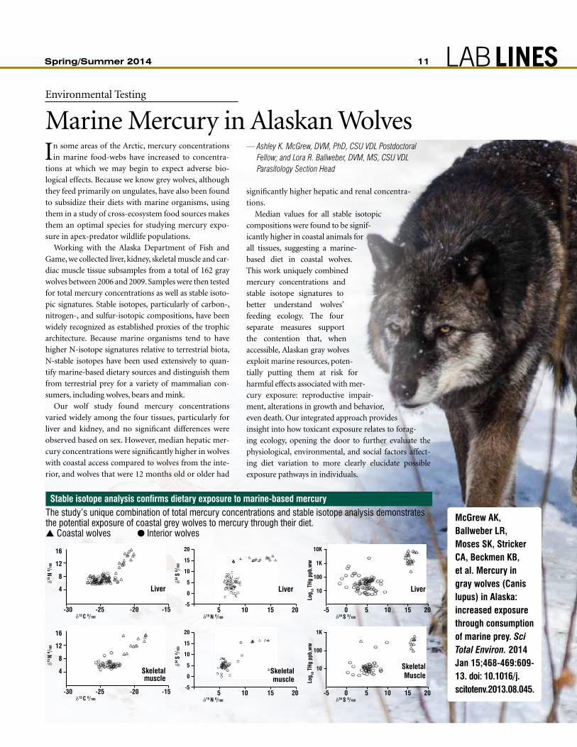
Marine Mercury in Alaskan WolvesIn some areas of the Arctic, mercury concentrations
in marine food-webs have increased to concentra-
tions at which we may begin to expect adverse bio-
logical effects. Because we know grey wolves, although
they feed primarily on ungulates, have also been found
to subsidize their diets with marine organisms, using
them in a study of cross-ecosystem food sources makes
them an optimal species for studying mercury expo-
sure in apex-predator wildlife populations.
Working with the Alaska Department of Fish and
Game, we collected liver, kidney, skeletal muscle and car-
diac muscle tissue subsamples from a total of 162 gray
wolves between 2006 and 2009. Samples were then tested
for total mercury concentrations as well as stable isoto-
pic signatures. Stable isotopes, particularly of carbon-,
nitrogen-, and sulfur-isotopic compositions, have been
widely recognized as established proxies of the trophic
architecture. Because marine organisms tend to have
higher N-isotope signatures relative to terrestrial biota,
N-stable isotopes have been used extensively to quan-
tify marine-based dietary sources and distinguish them
from terrestrial prey for a variety of mammalian con-
sumers, including wolves, bears and mink.
Our wolf study found mercury concentrations
varied widely among the four tissues, particularly for
liver and kidney, and no significant differences were
observed based on sex. However, median hepatic mer-
cury concentrations were significantly higher in wolves
with coastal access compared to wolves from the inte-
rior, and wolves that were 12 months old or older had
significantly higher hepatic and renal concentra-
tions.
Median values for all stable isotopic
compositions were found to be signif-
icantly higher in coastal animals for
all tissues, suggesting a marine-
based diet in coastal wolves.
This work uniquely combined
mercury concentrations and
stable isotope signatures to
better understand wolves’
feeding ecology. The four
separate measures support
the contention that, when
accessible, Alaskan gray wolves
exploit marine resources, poten-
tially putting them at risk for
harmful effects associated with mer-
cury exposure: reproductive impair-
ment, alterations in growth and behavior,
even death. Our integrated approach provides
insight into how toxicant exposure relates to forag-
ing ecology, opening the door to further evaluate the
physiological, environmental, and social factors affect-
ing diet variation to more clearly elucidate possible
exposure pathways in individuals.
— Ashley K. McGrew, DVM, PhD, CSU VDL Postdoctoral Fellow; and Lora R. Ballweber, DVM, MS, CSU VDL Parasitology Section Head
A
B
-5
0
5
10
15
20
0 5 10 15 20
34
15
-5
0
5
10
15
20
0 5 10 15 20
34
15
Skeletalmuscle
20
15
10
5
0
-5 5 10 15 20
δ15 N 0/100
δ34 S
0 /100
A
B
-5
0
5
10
15
20
0 5 10 15 20
34
15
-5
0
5
10
15
20
0 5 10 15 20
34
15
Liver
20
15
10
5
0
-5 5 10 15 20
δ15 N 0/100
δ34 S
0 /100
SkeletalMuscle
1K
100
10
-5 0 5 10 15 20
Log
10 T
Hg p
pb,w
w
δ34 S 0/100
Liver
10K
1K
100
10
-5 0 5 10 15 20
Log
10 T
Hg p
pb,w
w
δ34 S 0/100
Skeletalmuscle
16
12
8
4
-30 -25 -20 -15
δ15 N
0 /100
δ13 C 0/100
Liver
16
12
8
4
-30 -25 -20 -15
δ15 N
0 /100
δ13 C 0/100
The study’s unique combination of total mercury concentrations and stable isotope analysis demonstrates the potential exposure of coastal grey wolves to mercury through their diet.s Coastal wolves l Interior wolves
Stable isotope analysis confirms dietary exposure to marine-based mercury
McGrew AK, Ballweber LR, Moses SK, Stricker CA, Beckmen KB, et al. Mercury in gray wolves (Canis lupus) in Alaska: increased exposure through consumption of marine prey. Sci Total Environ. 2014 Jan 15;468-469:609-13. doi: 10.1016/ j.scitotenv. 2013. 08.045.

12 Volume 19, Number 1
Lab Updates
Fifty Years of Diagnostic ExpertiseDwayne Hamar’s educational journey began in
a one-room schoolhouse in Central Nebraska
where he had two other classmates, at the most. While
his foray into academia had humble beginnings, he has
made a dignified name for himself at Colorado State
University as an associate professor in the College of
Veterinary Medicine and Biomedical Sciences.
Hamar was first hired into the microbiology and
pathology department and began doing research in
the diagnostic lab where he still works today. His
work with the Veterinary Diagnostic Lab used to take
up only about eight to 10 hours of his time per week;
he now supervises a few technicians, and the lab as a
whole employs over 100 people.
Hamar obtained his undergraduate degree in chem-
istry from what was then known as Kearney State Col-
lege, now University of Nebraska at Kearney. He grew
up on a farm with four younger brothers and sisters.
“Something that was unique about my education —
we didn’t have kindergarten,” he recalls. “I started first
grade when I was five. I was two months past 17 when I
graduated from high school; two months past 21 when
I graduated from college. So when your dad tells you
when you get out of high school that ‘if you’re not in
college by fall, you’re on your own,’ I don’t think it gives
you much choice but to go to college.”
PROPEnSITY FOR BIOCHEMISTRYHamar’s natural propensity for chemistry paired with
his interest in biology and livestock led him to the field
of biochemistry. He obtained his master’s and doctor-
ate from the University of Nebraska, focusing specifi-
cally on the metabolism of ruminant animals.
Hamar’s role with CSU evolved with advancements
in the field of diagnostic medicine. Three years after he
started working here in 1964, he was asked to teach bio-
chemistry to veterinary students, because his education
provided him with a good scientific base in physiology,
bacteriology, basic chemistry and biochemistry.
He spent a year teaching biochemistry in Iran in the
1970s, but moved back once the possibility of a revolu-
tion became more of a reality. He also spent 12 years
advising students in the College of Veterinary Medi-
cine, an experience that he enjoyed because of the con-
tact it gave him with students and faculty.
lOTs Of changesNowadays, Hamar supervises the chemistry and toxi-
cology lab and works as the quality manager for the
entire diagnostic lab. He has seen a lot of changes, at
the university and within the field.
“The College of Veterinary Medicine has been very
progressive in terms of being a leader within the coun-
try in their approach to veterinary medicine and the
requirements for admissions,” Hamar said. “The types
of students that are admitted and the kind of educa-
tion they get has seen a lot of change. Part of that is just
a change in society over that period of time.”
Advances in the field of diagnostic medicine have
led to an increase in specificity and precision. And
while that also means veterinary medicine has gotten
more expensive, as has human medicine, it brings
about better diagnoses that serve both a scientific and
pragmatic purpose.
fOund his nicheIn his 50 years with the University, Hamar has had
many diverse roles and experiences. Having truly
found his niche working in the diagnostic lab, he has
been able to fully explore his strengths.
“It’s important to understand that different people
have different strengths,” he said. “We made a decision
in the department a few years back that the strength of
the unit was a component of each individual; that each
individual didn’t need to have their strengths in the
same place because then the whole department wasn’t
strong … and so that’s the way it is.”
“It’s been a fun ride,” he said of his experience
with the university which has been a definitive aspect
throughout the course of his life.
Spring/Summer 2014 13 LABLINES
Leigh Cooper joins the Veteri-
nary Diagnostic Lab as lab sup-
port, working in both sample
receiving and the virology lab.
Originally from New Mexico, she
came to Fort Collins to attend
Colorado State and recently
graduated with a bachelor of sci-
ence degree in equine science. In
her free time she enjoys volunteering at the Colorado
Horse Rescue and hiking with her dog.
Tracy Toberman lived in eastern
Colorado until age eight, when
her family moved to the South.
She attended East Carolina Uni-
versity and graduated with a
degree in biology. After college,
she moved to Charleston, S.C.,
and worked at a small-animal
veterinary hospital, where she
realized she wanted to make working in veterinary med-
icine her longterm career. In her spare time, she enjoys
hiking, sports and spending time with her dogs.
David Rutherford, originally
from Rochester, N.Y., got his
bachelor of science degree in
biotechnology from Syracuse’s
College of Environmental Sci-
ence and Forestry. He moved
to Colorado in September 2013
to be closer to his girlfriend,
whom he now lives with along
with their dog, Mishawaka. He enjoys hiking, camp-
ing, the great outdoors and classic rock.
Tom Davis was raised in Detroit
but made several moves through
Grand Rapids, Southern Ohio,
Phoenix, Seattle and Hawaii. Start-
ing as a technician in Michigan’s
health department, he returned to
graduate school at Miami Univer-
sity in Ohio and then worked his
way through several commercial
and governmental labs to acting chief of the Arizona State
Laboratory. Before coming to Fort Collins, where he now
works in the Chemistry and Toxicology Section, he was
half owner of a laboratory quality-assurance consulting
business which performed third-party quality assurance,
training and lab audits for clients including the U.S. Navy
and the University of Hawaii. He has no children but has
two dogs that he treats like kids.
Kevin Daniels joins the lab in
chemistry and toxicology sup-
port. Originally from Colorado
Springs, he graduated from Uni-
versity of Colorado at Colorado
Springs in May 2012 with a bach-
elors degree in chemistry. He
came to Fort Collins in Novem-
ber to work with the VDL and
is excited at the opportunity to be involved with the
impactful work done here. In his spare time, he reports
he enjoys the great outdoors and being a terrible chef.
Danielle Goranson is the VDL
office’s newest administrative
assistant. She grew up in Peetz,
Colo., and earned her degree in
political science from CSU. After
graduation, Danielle pursued a
career path in finance and cus-
tomer service, but hungered for
a more positive and intellectual
environment. When not working, Danielle enjoys hiking,
camping, dancing, traveling and general tomfoolery.
Seth Martinez, a Littleton native,
is a senior undergraduate CSU
microbiology student. Until he
graduates and moves to nurs-
ing school to pursue a career as a
registered nurse, he will be work-
ing in the VDL tissue-trimming
lab. When not in school, he likes
to explore new hiking trails and
enjoys the Colorado wilderness, rock climbing and skiing.
Jason Williams joins the VDL
as parasitology technician. Born
and raised in Amelia County, Va.,
Jason moved to Colorado in 2006
to attend Colorado State, where
he graduated in 2011 with a bach-
elor’s degree in microbiology. His
hobbies include hunting, fishing
and spending time with his pugs.
Get to Know the Laboratory
New Members Join the Lab Team

14 Volume 19, Number 1
Guardians of Public Health
National Network Funding UpdateThe recent Farm Bill authorized
up to $15 million annually to
fund the National Animal Health
Laboratory Network (NAHLN).
The state and federal partnership
is of extreme importance to the
nation’s agriculture industry and
food supply. NAHLN monitors for
the potential incursion of foreign
animal disease like foot and mouth
disease, performs surveillance test-
ing for bovine spongiform enceph-
alopathy, scrapie, chronic wasting
disease, avian influence and classi-
cal swine fever, as well as monitors
for emerging new diseases such
as the recently identified porcine
epidemic diarrhea virus outbreak.
NAHLN network labs have been
providing USDA diagnostic data
to better track this costly disease
to determine where it came from,
where it is spreading, and the best
control practices.
Now that NAHLN has been reau-
thorized, funding must be appro-
priated. The House Agricultural
Committee proposed total funding
of a little over $11 million annu-
ally. Meanwhile, the Senate’s ver-
sion of the appropriation proposed
a separate and distinct line item for
the network. The appropriations
request has yet to pass the full Senate
and House floor to be finalized.
While still short of our final goal
of $30 million annually for the
NAHLN, the funding is a step in the
right direction. We thank our fed-
eral legislators and animal industry
partners for their support. AAVLD
will continue to work with Congress
to develop funding levels to allow
for NAHLN’s full functionality.
— Barbara Powers, DVM, PhD, DACVP, CSU VDL Director
POSTERSn Seung Yoo: Modulation of coagulation and
fibrinolysis by carbon monoxide and nitric oxide in dogs: a thromboelastographic analysis.
n Dan Regan: Losartan repurposed as novel monocyte migration inhibitor for treatment of cancer metastasis.
n Elijah Edmundson: Histopathologic characterization of catecholamine induced cardiomyopathy in dogs with pheochromocytomas.
PRESEnTATIOnS n Deanna Dailey: Dog1 Is A Sensitive And
Specific Immunohistochemical Marker For Diagnosis Of Canine Gastrointestinal Stromal Tumors (Gists)
n Elijah Edmundson: Characterization of the tumor spectrum in HZE ion irradiated outbred mice.
n Brendan Podell: A Guinea Pig Model of Type 2 Diabetes as a Risk Factor for Tuberculosis.
n April White: Mystery cytology case: Impression smear from an inguinal mass.
n Seung Yoo: A horse of a different color.
AWARDSn Seung Yoo and Elijah Edmundson: Graduate
Student/Resident ACVP Travel Awards.n Seung Yoo: CL Davis Student Scholarship
Award.n Elijah Edmundson: ACVP Young Investigator
Poster Competition Award, Experimental Disease and Industrial and Toxicologic Pathology category, second place.
n Chuck Halsey: ACVP Young Investigator Poster Competition Award, Experimental Disease and Industrial and Toxicologic Pathology category, first place.
CSU RESIDEnT PARTICIPATIOn AnD AWARDS AT ACVP
CSU residents were well represented at the 2013 Annual Concurrent Meeting of the American College of Veterinary Pathologists and the American Society for Veterinary Clinical Pathology, nov. 16 to 20 in Montreal:

Spring/Summer 2014 15 LABLINES
Parasitology Section Head Lora Ballweber will attend the
American Association of Veterinary Parasitologists annual
meeting in Denver July 26 through 29. Look for her also at
Oklahoma State’s Veterinary College Fall Conference, Nov. 13
and 14 in Stillwater, and the National Animal Health Laboratory
Network/American Association of Veterinary Laboratory
Diagnosticians Quality Management System Training, Aug. 5
to 8 in Ames, Iowa. She will also attend the Shelter Medicine Training Day, Oct. 23 in Colorado Springs.
Come meet Ballweber along with VDL Director Barb Powers, Western Slope Lab Director Don Kitchen, Rocky Ford Lab Director
Gene Niles, Pathologists Tawfik Aboellail and Gary Mason,
Avian Diagnostics and BSL3 Operations Section Head Kristy Pabilonia, Chemistry and Toxicology Section Head Dwayne Hamar, Bacteriology Section Head Doreene Hyatt and Virology
Section Head Hana Van Campen at this year’s 57th annual
meeting of the American Association of Veterinary Laboratory Diagnosticians, Oct. 16 through 22, in Kansas City.
Pabilonia will also be at the National Poultry Improvement Plan Biennial Conference, July 10 to 12 in Charlotte.
Niles, Ballweber and Pabilonia will be at the fall meeting of the
Colorado Veterinary Medical Association, Sept. 18 to 21 in
Loveland, as will Van Campen, who will be helping with the Ag
Animal section of CVMA Fall meeting Sept 18-21.
Kristy Pabilonia will attend the annual meeting of the American Veterinary Medical Association, July 26 through 29 in Denver,
along with Hana Van Campen, who will be volunteering there.
VDL Pathologist Gary Mason will also be there, speaking during
the small-ruminant sessions.
VDL Pathologist E.J. Ehrhart will be at the second annual
Comparative Ocular Pathology Society annual meeting, Sept. 17
through 19 in Colorado Springs.
VDL Pathologist Colleen Duncan travels to Alaska in July to
capture and tag sea lions, where she will also work with VDL
Pathologist Terry Spraker on a fur-seal study. He also plans to
attend the Marine Mammals of the Holarctic Conference in St.
Petersburg, Russia, in September and the Alaska Marine Science Symposium in January in Anchorage.
Van Campen will be attending the American Association of Veterinary Laboratory Diagnosticians Executive Board Meeting
in Denver on July 31.
Rocky Ford Lab Director Niles will be at the Academy of Veterinary Consultants meeting in Denver, July 31 to Aug. 2.
Look for Lab Coordinator Charlie Davis at the Colorado Wool Growers Association convention July 16 and 17 in Montrose, Colo.
CSU VDL On THE ROAD: UPCOMInG COnFEREnCES, SYMPOSIA AnD APPEARAnCES
Lab Updates
Faculty Acheivement RecognizedCSU Veterinary Diagnostic Lab
Director Barb Powers received
the university’s Oliver P. Pennock
Distinguished Service Award in
May. Paying tribute to a distin-
guished professor of civil engi-
neering in the 1920s, the award
recognizes faculty members who
have made meritorious achieve-
ment over five or more years.
Powers was honored for advanc-
ing veterinary pathology through
education, service and research
since coming to CSU in 1981 as a
PhD candidate and pathology resi-
dent and moving up to head the
lab in 1997.
“She has been the driving force
that resulted in the building of a
new facility and a national repu-
tation,” said Gregg Dean, head of
the Department of Microbiology,
Immunology, and Pathology. “She
is a powerful advocate for animal
diagnostics, Colorado agriculture
and the veterinary community.”
“It may be my name on the
plaque,” Powers says, “but this
award recognizes the distinguished
achievement our entire team and
organization has made in the last
15 years. I am humbled to receive
it on their behalf.”
MASOn MERITORIOUS TEACHERAssociate Professor of Microbiology,
Immunology and Pathology Gary
Mason received the 2014 Zoetis Dis-
tinguished Teaching Award May 15.
Nominated and voted upon by vet-
erinary students, the award honors
faculty who have made outstanding
achievement in teaching.
p
BARBARA POWERS, DVM, PHD, DACVP
diRecTOR
Update from the Director
Nonprofit OrganizationUS Postage
PAIDFort Collins, Colorado 80523
Permit Number 19
inside This issueinside This issueREGULAR COLUMnS
Veterinary Diagnostic Laboratories College of Veterinary Medicine and Biomedical Sciences Fort Collins, CO 80523-1644
PRODUCTIOn MEDICInE . . .p. 1new field investigation unit puts multidisciplinary medicine to work.
CAnInE OnCOLOGY . . . . . p. 2It’s time to re-examine how we divide lympho-proliferative disorders. Here’s why.
TOxICOLOGY. . . . . . . . . . . p. 4Low-quality forages spell increased nitrate toxicity.
ExOTICS MEDICInE. . . . . . p. 8VDL faculty report on a novel case of Rickettsia in a scorpion.
EnVIROnMEnT . . . . . . . . p. 11Arctic wolf study puts new perspective on mercury risk.
LAB UPDATES . . . . . . . . . p. 12For a half century, VDL’s Dwayne Hamar has been involved in many diverse roles with CSU’s VDL.Lymph Node
Lyphoblasticleukomia/lymphoma
Bone Marrow Myeloma
Difuse large B-cell lymphomaSurvival: 1 year
B-cell chroniclymphocytic leukemiaSurvival: 3 years
Marginal zone lymphomaSurvival: 2 years
In this issue of LabLines, we highlight the
new Field Investigation Unit that was
introduced early in 2014. This unit is off
to a vigorous start, with multiple investi-
gations already concluded or ongoing.
Inside you will also find many articles
on a variety of issues affecting multiple
species, new discoveries, and interesting
cases. In addition, there is an opportu-
nity to participate in a new study. We
introduce many new staff members who
have started over the last six months as
well as honoring Dr. Dwayne Hamar for
his 50 Years of Service at Colorado State University,
mostly within our laboratories. Our pathology resi-
dents and faculty have received numerous awards this
year as well. See inside for details!
We have just released our
Annual Report for the year 2013.
Inside you’ll find more than 60
pages highlighted the annual
achievements of the diagnostic
lab system, from scientific papers
published by faculty to funded
and ongoing investigations we’re
undertaking, to accession sum-
maries and a wealth of diagnostic results
summaries that give a unique insight in to
the health situation of Colorado’s animal
population. The entire report is available
on our web site, under the Regulations and
Resources tab. A limited hard copies are also
available; call us if you have comments or
wish to receive a hard copy.
In January, we met with our External
Advisory Committee, as we do annually,
and reviewed our new five-year strategic
plan. There are many new internal and exter-
nal goals within this plan; many of these are
already well underway, or are even completed. Also This
September, we will be holding the president’s reception
of the annual Colorado Veterinary Medical Association
Convention in the Diagnostic Medicine Center and
look forward to an interactive reception with many
of you. Later in October, we hope to see many of you
at the American Association of Veterinary Laboratory
Diagnosticians annual meeting in Missouri.
Best wishes,
Barbara E. Powers, DVM, PhD, DACVP, Director
n CASE STUDY p. 6 Ewe sodium intoxication.
n VDL In PRESS p. 9 new research round-up.
n DISEASE UPDATES p. 10 Have a cat gingivitis case?
n LAB UPDATE p. 13 Meet new staff members.
n LAB UPDATE p. 15 Faculty acheivements.