Vasodilator Stress Perfusion CMR Imaging Is Feasible and ...
20
Vasodilator Stress Perfusion CMR Imaging Is Feasible and Prognostic in Obese Patients The Harvard community has made this article openly available. Please share how this access benefits you. Your story matters Citation Shah, Ravi V., Bobak Heydari, Otavio Coelho-Filho, Siddique A. Abbasi, Jiazhuo H. Feng, Tomas G. Neilan, Sanjeev Francis, et al. 2014. “Vasodilator Stress Perfusion CMR Imaging Is Feasible and Prognostic in Obese Patients.” JACC: Cardiovascular Imaging 7 (5) (May): 462–472. doi:10.1016/j.jcmg.2013.11.011. Published Version 10.1016/j.jcmg.2013.11.011 Citable link http://nrs.harvard.edu/urn-3:HUL.InstRepos:32415212 Terms of Use This article was downloaded from Harvard University’s DASH repository, and is made available under the terms and conditions applicable to Other Posted Material, as set forth at http:// nrs.harvard.edu/urn-3:HUL.InstRepos:dash.current.terms-of- use#LAA
Transcript of Vasodilator Stress Perfusion CMR Imaging Is Feasible and ...
Vasodilator Stress PerfusionCMR Imaging Is Feasible andPrognostic in Obese Patients
The Harvard community has made thisarticle openly available. Please share howthis access benefits you. Your story matters
Citation Shah, Ravi V., Bobak Heydari, Otavio Coelho-Filho, Siddique A.Abbasi, Jiazhuo H. Feng, Tomas G. Neilan, Sanjeev Francis, et al.2014. “Vasodilator Stress Perfusion CMR Imaging Is Feasible andPrognostic in Obese Patients.” JACC: Cardiovascular Imaging 7 (5)(May): 462–472. doi:10.1016/j.jcmg.2013.11.011.
Published Version 10.1016/j.jcmg.2013.11.011
Citable link http://nrs.harvard.edu/urn-3:HUL.InstRepos:32415212
Terms of Use This article was downloaded from Harvard University’s DASHrepository, and is made available under the terms and conditionsapplicable to Other Posted Material, as set forth at http://nrs.harvard.edu/urn-3:HUL.InstRepos:dash.current.terms-of-use#LAA
Vasodilator Stress Perfusion CMR Imaging Is Feasible andPrognostic in Obese Patients
Ravi V. Shah, MD*,†, Bobak Heydari, MD, MPH*, Otavio Coelho-Filho, MD, MPH‡, SiddiqueA. Abbasi, MD*, Jiazhuo H. Feng, BS*, Tomas G. Neilan, MD*,†, Sanjeev Francis, MD†, RonBlankstein, MD*, Michael Steigner, MD*, Michael Jerosch-Herold, PhD*, and Raymond Y.Kwong, MD, MPH*
*Noninvasive Cardiovascular Imaging Section, Cardiovascular Division, Department of Medicineand Department of Radiology, Brigham and Women's Hospital, Harvard Medical School, Boston,Massachusetts
†Division of Cardiology, Massachusetts General Hospital, Harvard Medical School, Boston,Massachusetts
‡Cardiology Division, State University of Campinas (UNICAMP), Campinas, São Paulo, Brazil
Abstract
Objectives—This study sought to determine feasibility and prognostic performance of stress
cardiac magnetic resonance (CMR) in obese patients (body mass index [BMI] ≥30 kg/m2).
Background—Current stress imaging methods remain limited in obese patients. Given the
impact of the obesity epidemic on cardiovascular disease, alternative methods to effectively risk
stratify obese patients are needed.
Methods—Consecutive patients with a BMI ≥30 kg/m2 referred for vasodilating stress CMR
were followed for major adverse cardiovascular events (MACE), defined as cardiac death or
nonfatal myocardial infarction. Univariable and multivariable Cox regressions for MACE were
performed to determine the prognostic association of inducible ischemia or late gadolinium
enhancement (LGE) by CMR beyond traditional clinical risk indexes.
Results—Of 285 obese patients, 272 (95%) completed the CMR protocol, and among these, 255
(94%) achieved diagnostic imaging quality. Mean BMI was 35.4 ± 4.8 kg/m2, with a maximum
weight of 200 kg. Reasons for failure to complete CMR included claustrophobia (n = 4),
intolerance to stress agent (n = 4), poor gating (n = 4), and declining participation (n = 1).
Sedation was required in 19 patients (7%; 2 patients with intravenous sedation). Sixteen patients
required scanning by a 70-cm-bore system (6%). Patients without inducible ischemia or LGE
experienced a substantially lower annual rate of MACE (0.3% vs. 6.3% for those with ischemia
and 6.7% for those with ischemia and LGE). Median follow-up of the cohort was 2.1 years. In a
© 2014 by the American College of Cardiology Foundation Published by Elsevier Inc.
Reprint requests and correspondence: Dr. Raymond Y. Kwong, Brigham and Women's Hospital, Harvard Medical School, 75 FrancisStreet, Boston, Massachusetts 02115, [email protected] other authors have reported that they have no relationships relevant to the contents of this paper to disclose. Drs. Shah and Heydaricontributed equally to this work.
NIH Public AccessAuthor ManuscriptJACC Cardiovasc Imaging. Author manuscript; available in PMC 2014 July 24.
Published in final edited form as:JACC Cardiovasc Imaging. 2014 May ; 7(5): 462–472. doi:10.1016/j.jcmg.2013.11.011.
NIH
-PA
Author M
anuscriptN
IH-P
A A
uthor Manuscript
NIH
-PA
Author M
anuscript
multivariable stepwise Cox regression including clinical characteristics and CMR indexes,
inducible ischemia (hazard ratio 7.5; 95% confidence interval: 2.0 to 28.0; p = 0.002) remained
independently associated with MACE. When patients with early coronary revascularization
(within 90 days of CMR) were censored on the day of revascularization, both presence of
inducible ischemia and ischemia extent per segment maintained a strong association with MACE.
Conclusions—Stress CMR is feasible and effective in prognosticating obese patients, with a
very low negative event rate in patients without ischemia or infarction.
Keywords
cardiac magnetic resonance; obesity; stress testing
Obesity (body mass index [BMI] ≥30 kg/m2) affects more than 1 in 3 Americans (1), with
an increased risk of coronary artery disease (CAD) independent of traditional coronary risk
factors (2,3). Given the explosion in obesity and obesity-related cardiovascular and
metabolic disease, the ability to risk stratify obese patients for adverse cardiovascular events
is paramount. Despite their widespread use in a general referral population, both stress
echocardiography and nuclear perfusion have important technical challenges in obese
patients. Stress echocardiography is limited by operator dependence of acquisition and poor
acoustic windows as a result of body habitus, and its impact on long-term prognosis remains
unclear. As the mainstay of current clinical practice, single-photon emission computed
tomography (SPECT) nuclear perfusion imaging may be hampered by attenuation artifacts
and reduced signal-to-noise ratio, limiting diagnostic accuracy in obese patients (4–9). The
prognostic impact of a negative nuclear scan may be associated with BMI. In one study, a
normal perfusion scan was associated with a nearly 1.7% risk of death at 1 year (9).
Although positron emission tomography (PET) overcomes many of these challenges, as a
result of superior diagnostic and prognostic performance in obese patients (10), ionizing
radiation, radiotracer availability, and cost pose important limitations to its widespread
adoption.
Stress cardiac magnetic resonance (CMR) can assess ventricular function, stress and rest
perfusion, and viability within a single examination (11). Relative to alternative techniques,
stress CMR has high spatial and temporal resolution and is not limited by acoustic windows
or image acquisition. However, concerns over feasibility in obese patients because of
claustrophobia and safety monitoring have limited the adoption of stress CMR in this
important, burgeoning population. To address these fundamental concerns, we sought to
determine the feasibility of stress CMR in obese patients and the prognostic impact of stress
CMR results on cardiovascular events.
Methods
Study population
We identified patients with a BMI ≥30 kg/m2 from consecutive patients ≥18 years of age
referred to Brigham and Women's Hospital between December 2001 and August 2011 for
the assessment of suspected myocardial ischemia. Exclusion criteria consisted of known
hypersensitivity to gadolinium-based contrast agents, glomerular filtration rate <30 ml/min/
Shah et al. Page 2
JACC Cardiovasc Imaging. Author manuscript; available in PMC 2014 July 24.
NIH
-PA
Author M
anuscriptN
IH-P
A A
uthor Manuscript
NIH
-PA
Author M
anuscript

1.73 m2, pregnancy, weight >250 kg (because of table limitations), acute myocardial
infarction (MI), hemodynamic instability, presence of metallic hazards, and history of
asthma or bronchospastic disease. Patients with atrial fibrillation at the time of CMR were
included in the study. Clinical data were collected by medical history and clinical
examination on the day of stress CMR by a trained physician using standardized criteria.
Our institutional review board approved follow-up of clinical events, and all participating
patients gave written informed consent for stress CMR.
Cardiac magnetic resonance
Patients were scanned with a 60-cm–bore, 1.5-T magnet (Signa CV/i, GE Medical Systems,
Milwaukee, Wisconsin) with an 8-element phased-array coil before 2006 and with a 3.0-T
scanner (60-cm–bore Magnetom Trio [TIM system], Siemens, Erlangen, Germany) with a
16-element phased array coil after 2006. With the availability of a wide-bore scanner (70-cm
bore diameter, 3.0-T Magnetom Verio, Siemens) in 2007, this scanner was used when
requested by the referral clinician, significant patient history of claustrophobia from pre-
CMR screening, or failure to perform CMR at either 60-cm–bore scanners. Oral sedation
(diazepam 0.5 to 1.0 mg) was given 15 to 20 min before CMR examination where
necessary. Intravenous conscious sedation was offered if oral sedation failed to control
claustrophobic symptoms (both via short-acting benzodiazepine, intravenous or oral; or
fentanyl, intravenous). The stress CMR protocol was optimized for obese patients via: 1)
lateral placement of 1 of the electrocardiographic leads to improve gating signal; 2)
increasing field of view with real-time adjustment to prevent wrap artifact; and 3) dynamic
gradient echo shimming around cardiac structures before cine steady-state free precession
imaging. All patients were instructed to refrain from caffeine for 24 h and to fast for at least
6 h before the CMR examination. Our CMR protocol included localizers, cine function,
stress and rest perfusion, and late gadolinium enhancement (LGE) imaging. A 12-lead
electrocardiogram was performed both before and following CMR examination. Cine
images were acquired in both short-axis (contiguous 8-mm slices) and long-axis (4-chamber,
2-chamber, and 3-chamber) views of the left ventricle with steady-state free precession cine
imaging (repetition time 3.4 ms; echo time 1.2 ms; matrix 256 × 256; field of view 34 to 40
cm).
Vasodilator stress was achieved using either intravenous adenosine (Adenoscan, Astellas
Pharma US, Deerfield, Illinois) (n = 149 [58%]) infused at a rate of 140 μg/kg/min over 6
min or regadenoson (Lexiscan, Astellas Pharma US) (n = 106 [42%]) as an intravenous
bolus of 0.4 mg, hand-injected over 10 s. Myocardial perfusion images for both stress and
rest were acquired with a saturation-prepared, single-shot spoiled gradient echo sequence
(repetition time 6 ms; echo time 2.3 ms; slice thickness 8 mm) during bolus injection of 0.1
mmol/kg intravenous gadolinium (Magnevist, Bayer HealthCare, Wayne, New Jersey) at 4
to 5 ml/s, followed by a saline flush. Myocardial perfusion images were acquired in 3 short-
axis (basal, mid, and apical) and 1 long-axis (4-chamber) view (for acquisitions on the
Siemens scanners). LGE was performed using a standard inversion recovery gradient echo
sequence in short- and long-axis slices 10 to 15 min after rest perfusion imaging (with total
gadolinium dose 0.20 mmol/kg, with appropriate TI time to null normal myocardium). Vital
signs, electrocardiography, and respiration were monitored contin uously throughout the
Shah et al. Page 3
JACC Cardiovasc Imaging. Author manuscript; available in PMC 2014 July 24.
NIH
-PA
Author M
anuscriptN
IH-P
A A
uthor Manuscript
NIH
-PA
Author M
anuscript

CMR scan. Blood pressure was measured with noninvasive sphygmomanometry before and
following administration of the vasodilator agent. All images were acquired at end-
expiration with electrocardiographic gating (with provisional pulse gating for
electrocardiographic gating failure).
CMR analysis
Perfusion images were interpreted by 2 experienced observers (R.Y.K. and B.H.) blinded to
clinical and follow-up data. CMR images were analyzed by standard post-processing
techniques on an offline workstation (Mass Clinical 7.4, Medis, Leiden, the Netherlands).
Ventricular volumes, function, and mass were quantified by previously published techniques
(12). Cine wall motion was assessed in a 17-segment American Heart Association model,
and perfusion imaging was assessed in a 16-segment model (13). Inducible ischemia was
defined as any stress perfusion defect that: 1) remained dark and persisted for at least 3
phases beyond peak contrast enhancement; 2) was at least 1 pixel in thickness; and 3)
followed a coronary distribution. These criteria are similar to multicenter published studies
(14,15). Inducible ischemia was defined as the presence of any segmental subendocardial
stress perfusion defect in the absence of LGE in the same segment. Therefore, segments
with LGE and matching stress perfusion defects were not counted as “inducible ischemia.”
LGE was evaluated qualitatively (presence or absence) and quantitatively using a signal
intensity threshold of ≥2 SD above remote normal myocardium, as previously published
(12).
Clinical outcomes
Patient follow-up was obtained through review of the electronic medical record, followed by
a mailed standard questionnaire or scripted telephone interview by trained study personnel
(when medical records were not available). Mortality was confirmed by the Social Security
Death Index. Our primary outcome was a composite of cardiac death or nonfatal MI, defined
as major adverse cardiac events (MACE). Our secondary outcome was a composite of all-
cause mortality or nonfatal MI. Nonfatal MI was defined as a new clinical presentation of
chest pain or dyspnea with an elevation of cardiac biomarkers (>99th percentile of the upper
limits of normal) in a temporal pattern consistent with an acute MI (16).
Statistics
Parametric continuous data were expressed as means with SD, whereas nonparametrically
distributed continuous data were presented as medians with interquartile range (IQR).
Categorical variables were expressed as counts with percentages. Comparison between
groups was performed with a 2-sample Student t test for continuous, normal data, and
Wilcoxon rank sum test for continuous, non-normal data. The Fisher exact test was used to
compare stratified categorical data. MACE-free survival stratified by the presence or
absence of inducible ischemia was estimated by the Kaplan-Meier method and compared
using a log-rank test. Univariable Cox regression models were used to estimate unadjusted
hazard of MACE for clinical and CMR characteristics. The independent prognostic value of
inducible ischemia for MACE was evaluated using a multivariable Cox model constructed
by inclusion of significant covariates on univariable screen in a stepwise selection algorithm
Shah et al. Page 4
JACC Cardiovasc Imaging. Author manuscript; available in PMC 2014 July 24.
NIH
-PA
Author M
anuscriptN
IH-P
A A
uthor Manuscript
NIH
-PA
Author M
anuscript

(p < 0.05 for model entry and retention). All clinical covariates and CMR indexes (left
ventricular and right ventricular function, mass, volumes, wall motion, and perfusion results)
were included as potential covariates in the multivariable model. We assessed for the
presence of collinearity amongst covariates in the final multivariable model. The
proportional hazards assumption for the multivariable model was evaluated quantitatively by
including time-dependent covariates in the final multivariable Cox model. All statistical
analyses were performed using SAS version 9.2 (SAS Institute, Cary, North Carolina). A 2-
tailed p < 0.05 was considered statistically significant.
Results
Feasibility of stress CMR in obese patients
A total of 285 patients fulfilled the inclusion criteria for the study. Thirteen patients (4.6%)
were unable to complete the stress CMR study (4 patients were claustrophobic, 4 patients
were intolerant of the stress agent, 4 patients had poor gating, and 1 patient declined
participation) (Fig. 1). Another 17 patients (6.0%) had nondiagnostic imaging quality, with
the remaining 255 patients comprising the study cohort. One hundred thirty-one patients
were scanned on the 1.5-T scanner before August 2006. Nineteen (6.7%) patients required
sedation for the CMR scan, among them 2 patients requiring intravenous conscious sedation
(because oral agents were not effective). Sixteen (5.6%) required scanning using a wide-
bore scanner, among them 4 patients requiring oral sedation and 1 requiring intravenous
sedation. f the overall population, 24 (8%) had atrial fibrillation, and 3 of 24 had
nondiagnostic perfusion studies and were excluded from the study.
Baseline characteristics
Patient and CMR characteristics for the entire study cohort and stratified by inducible
ischemia are shown in Tables 1 and 2, respectively. Primary indications for CMR included
suspicion of cardiac ischemia in 80% and evaluation of suspected cardiac dyspnea in 20%.
The overall study cohort (mean age 56 years, 40% female) had a mean BMI of 35.4 ± 4.8
kg/m2. The maximum weight successfully scanned was 200 kg (440 lbs). The majority of
patients were in sinus rhythm (92%), whereas 21 patients were in atrial fibrillation during
stress CMR. Patients with no evidence of inducible ischemia by CMR had a higher BMI
(35.8 ± 5.3 kg/m2 vs. 34.4 ± 3.4 kg/m2, p < 0.05), and lower prevalence of history of heart
failure (14% vs. 27%, p < 0.05), symptomatic angina (44% vs. 22%, p < 0.001), prior MI
(7% vs. 36%, p < 0.0001), or prior coronary artery bypass surgery (2% vs. 16%, p < 0.001).
The overall study cohort had a median left ventricular ejection fraction (LVEF) of 62%
(IQR: 16%), left ventricular mass index of 58 g/m2 (IQR: 24 g/m2), and right ventricular
ejection fraction of 55% (IQR: 12%). Patients with no evidence of inducible ischemia on
CMR had a significantly greater LVEF (63% vs. 57%, p < 0.001), lower left ventricular end-
systolic volume index (29 ml/m2 vs. 34 ml/m2, p < 0.05), and less frequent resting wall
motion abnormalities (14% vs. 47%, p < 0.0001). In those patients with evidence of
infarction by LGE imaging, infarct size was not different with respect to the presence or
absence of inducible ischemia.
Shah et al. Page 5
JACC Cardiovasc Imaging. Author manuscript; available in PMC 2014 July 24.
NIH
-PA
Author M
anuscriptN
IH-P
A A
uthor Manuscript
NIH
-PA
Author M
anuscript

Clinical outcomes
Median follow-up was 2.1 (IQR: 2.8 years) years, with no loss to follow-up in the patients
studied. In the entire cohort, there were 19 deaths of any cause (7 cardiovascular [37%]). In
addition, 7 patients experienced nonfatal MI during study follow-up. For the whole-study
cohort, annualized event rates were 2.2% for MACE, 4.0% for a composite of all-cause
mortality and MI, and 3.1% for all-cause mortality alone. The annualized event rates for
MACE and composite of all-cause mortality and MI by presence of inducible ischemia and
LGE are shown in Figure 2. Patients with no evidence of inducible ischemia or infarction by
LGE had a very low annualized rate of MACE (0.3%). Patients with LGE without inducible
ischemia experienced a higher annualized rate of MACE (2.4%), which was significantly
lower than those patients with presence of inducible ischemia, but no evidence of LGE
(2.4% vs. 6.3%, p < 0.0001). The annualized rate of MACE for patients with both inducible
ischemia and LGE was highest, at 6.7%.
Prognostic value of stress CMR in obese patients
Univariable analysis of patient and CMR characteristics for association with MACE, and all-
cause mortality/nonfatal MI are displayed in Table 3. Age, history of diabetes, prior MI,
LVEF, presence of resting wall motion abnormalities, LGE, and inducible ischemia were all
significantly associated with MACE in univariable Cox models. Kaplan-Meier event-free
survival curves for MACE (primary outcome) or all-cause mortality/nonfatal MI (secondary
outcome) stratified by inducible ischemia are shown in Figure 3. Obese patients with
inducible ischemia had a significantly lower event-free survival for both primary and
secondary outcomes as compared with patients without inducible ischemia (p < 0.0001 and
p < 0.005, respectively). This relationship held when patients with LGE were excluded (Fig.
4). In a multivariable stepwise Cox regression including clinical characteristics and CMR
indexes, inducible ischemia (hazard ratio [HR]: 7.5; 95% confidence interval [CI]: 2.0 to
28.0; p = 0.002) and resting ST abnormalities (HR: 3.2; 95% CI: 1.1 to 9.5; p = 0.04)
remained independently associated with MACE. None of the time-dependent covariates
included in the final multivariable Cox model were significant, suggesting that the
proportionality assumption was not violated.
Censoring of patients who underwent early coronary revascularization
Of the 255 patients in the study, 32 patients underwent early coronary revascularization
(within 90 days of stress CMR). In the 223 patients remaining in the cohort after exclusion
of these patients, there were 9 MACE events (5 nonfatal MI and 4 cardiac deaths). When
these patients were censored on the day of the early revascularization, both presence of
inducible ischemia and ischemia extent per segment remained highly associated with MACE
(HR: 9.81; 95% CI: 2.74 to 35.19; p = 0.0005 and HR: 1.15; 95% CI: 1.07 to 1.23; p =
0.0001, respectively).
Discussion
In this cohort of 285 obese patients clinically referred for stress CMR, we found that stress
CMR produced diagnostic quality imaging in more than 89% of patients. Despite lingering
concerns over claustrophobia, only 7% of obese patients required sedation to achieve a
Shah et al. Page 6
JACC Cardiovasc Imaging. Author manuscript; available in PMC 2014 July 24.
NIH
-PA
Author M
anuscriptN
IH-P
A A
uthor Manuscript
NIH
-PA
Author M
anuscript
diagnostic CMR. CMR inducible ischemia was a strong, independent predictor of MACE in
multivariable models, with lack of inducible ischemia or LGE associated with a low annual
MACE rate of 0.3%. Collectively, these results suggest that stress CMR is highly feasible
and has excellent prognostic utility in obese patients.
Cardiovascular disease in obese patients represents a unique challenge for clinicians,
especially in light of technical limitations inherent to current, widely utilized methods of risk
stratification (e.g., nuclear, echocardiographic, treadmill stress testing, or computed
tomography angiography). Despite the growing at-risk obese population, current literature
examining the prognostic importance of stress echocardiography and nuclear imaging in this
population remains limited. Although a negative dobutamine stress echocardiogram carries
an annualized rate of cardiac MACE of 1.3% in a general referral population (17), the
prognostic performance of dobutamine stress echocardiography in obese patients is not
widely known. With regard to nuclear imaging, Elhendy et al. (18) reported on the
prognostic importance and feasibility of dobutamine 99mTc-tetrofosmin SPECT imaging
without attenuation correction in 267 obese patients (mean BMI 37 kg/m2) with a similar
risk profile as our study. These authors demonstrated significant breast or diaphragmatic
attenuation in 40 patients (16%) leading to non-diagnostic stress perfusion in 6 (2%). This
technical limitation is not experienced with stress perfusion CMR. At a mean follow-up of
5.5 years, 22 cardiac deaths were observed, with an annualized rate of cardiac death and MI
of 1% in patients with normal perfusion, significantly higher than observed with stress
CMR. Importantly, abnormal nuclear stress perfusion was a strong univariate and
multivariate predictor of MACE in this study. Although the authors suggest that attenuation
correction may have improved stratification of risk in obese patients, the high annual MACE
rate and the frequent presence of imaging artifacts with SPECT methods may more
accurately reflect real-world clinical practice. Finally, computed tomography angiography
(CTA) has emerged as a useful modality to assess for obstructive CAD, with high diagnostic
accuracy (8,19). Indeed, in a study of more than 13,000 patients, BMI was strongly
associated with the extent of obstructive CAD at CTA (20). Despite these excellent results,
however, CTA requires radiation exposure, and its diagnostic performance may be limited
by a low signal-to-noise ratio in the obese population.
The prognostic importance of negative nuclear myocardial perfusion imaging has been
shown to be associated with BMI. In a retrospective study of 433 patients with a
significantly higher BMI (47.3 kg/m2) with exercise or vasodilator 99mTc SPECT, Duvall et
al. (9) reported that despite having only 2% poor-quality scans, patients with normal
perfusion had up to a 3.7% risk of death at 2 years, and 1.7% risk of death at 1 year, higher
than stress perfusion CMR in our study. Importantly, 82Rb-PET has also shown high
diagnostic and prognostic performance in obese patients. In a landmark study of 134 obese
patients referred for clinically indicated dipyridamole 82Rb-PET, patients with normal PET
perfusion had a 0% cardiac event rate at a mean 3.1-year follow-up (10), significantly better
than SPECT imaging. These findings are comparable to our reported results for stress
perfusion CMR. In addition to ischemia, however, the improvements in spatial resolution
with CMR allow detection of small subendocardial MI that is prognostically important,
without ionizing radiation (21).
Shah et al. Page 7
JACC Cardiovasc Imaging. Author manuscript; available in PMC 2014 July 24.
NIH
-PA
Author M
anuscriptN
IH-P
A A
uthor Manuscript
NIH
-PA
Author M
anuscript

Our results in an obese population are in keeping with recent large reports in overall referral
populations attesting to the prognostic role of stress CMR (22,23), suggesting the
generalizability of standard perfusion techniques to obese patients. In a recent large study
involving dobutamine stress CMR in 3,138 patients, Kelle et al. (23) describe
complementary prognostic information from wall motion abnormalities and LGE, though
data on average BMI in this cohort was not provided. These results have been confirmed in a
large recent meta-analysis (22). Our work extends the aggregate data in stress CMR by
demonstrating its prognostic utility specifically in patients with obesity, in whom imaging
methods are limited, with potentially nonspecific results (21). Moreover, we also confirm in
an obese population that inducible ischemia and LGE by stress perfusion CMR are robust
markers of risk even in those patients without a clinical history of prior infarction, in
keeping with prior reports suggesting the prognostic importance of LGE in patients with
clinically unrecognized MI (24,25). In addition, as technical developments increasing the
field strength possible with open magnetic resonance imaging systems emerge, the
possibility of further application of stress perfusion CMR in obese patients may become
more generalizable.
Study limitations
The results of our study must be taken in the context of its design. The relatively low event
rate in our study reduces the number of clinical and CMR parameters that may be evaluated
in multivariable modeling. Despite the low number of events, this event rate and our patient
risk profile are comparable to similar studies in the nuclear literature. Even with this low
overall rate of MACE, the absence of inducible ischemia and LGE still identified patients
with a dramatically low rate of MACE. Over the 10-year study period, both 1.5-and 3.0-T
field strengths were used for stress perfusion imaging. It should be noted that the proportion
of nondiagnostic scans were similar at 6% between 1.5- and 3.0-T. Therefore, although our
imaging protocol was tailored to obese patients, the methods used to optimize cine,
perfusion, and LGE imaging were found to be highly transportable across scanner platforms
and field strengths, suggesting this was not a significant limitation to generalizability. In
addition, given the increasing adoption of 3.0-T scanners worldwide for CMR, it is likely
that image quality and standardization will continue to improve. Although we report in the
current study that a negative CMR is associated with a very low annualized rate of cardiac
events, comparison of prognostic utility of CMR between obese and nonobese patients is not
possible on the basis of the current evidence. Finally, although the determination of a
threshold extent of inducible ischemia is a clinically important question, the limited number
of events in this select population does not allow us to calculate this threshold, although
other studies in nuclear and stress CMR have examined this important question (26,27).
Conclusions
Stress CMR is feasible in an obese population, and absence of ischemia or myocardial scar
is associated with very low MACE in long-term follow-up. With a growing population of
obese patients at significant risk for cardiovascular disease, these results suggest the use of
stress CMR to stratify risk in symptomatic obese patients with suspected CAD may be
warranted.
Shah et al. Page 8
JACC Cardiovasc Imaging. Author manuscript; available in PMC 2014 July 24.
NIH
-PA
Author M
anuscriptN
IH-P
A A
uthor Manuscript
NIH
-PA
Author M
anuscript
Acknowledgments
Dr. Shah is supported by an American Heart Association Post-Doctoral Fellowship Award (11POST000002) and atraining grant from the Heart Failure National Institutes of Health Clinical Research Network (U01-HL084877). Dr.Heydari is supported by a Clinical Fellowship Award from the Alberta Heritage Foundation for Medical Research.Dr. Kwong is supported by National Institutes of Health grant RO1-HL091157. Dr. Kwong receives researchsupport from Astellas Pharma US.
References
1. Ogden CL, Carroll MD, Curtin LR, McDowell MA, Tabak CJ, Flegal KM. Prevalence ofoverweight and obesity in the United States, 1999-2004. JAMA. 2006; 295:1549–55. [PubMed:16595758]
2. Bogers RP, Bemelmans WJ, Hoogenveen RT, et al. for the BMI-CHD Collaboration Investigators.Association of overweight with increased risk of coronary heart disease partly independent of bloodpressure and cholesterol levels: a meta-analysis of 21 cohort studies including more than 300 000persons. Arch Intern Med. 2007; 167:1720–8. [PubMed: 17846390]
3. Whitlock G, Lewington S, Sherliker P, et al. Prospective Studies Collaboration. Body-mass indexand cause-specific mortality in 900 000 adults: collaborative analyses of 57 prospective studies.Lancet. 2009; 373:1083–96. [PubMed: 19299006]
4. DePuey EG 3rd. How to detect and avoid myocardial perfusion SPECT artifacts. J Nucl Med. 1994;35:699–702. [PubMed: 8151397]
5. Dunn JP, Huizinga MM, See R, Irani WN. Choice of imaging modality in the assessment ofcoronary artery disease risk in extreme obesity. Obesity (Silver Spring). 2010; 18:1–6. [PubMed:19461587]
6. Einstein AJ, Weiner SD, Bernheim A, et al. Multiple testing, cumulative radiation dose, and clinicalindications in patients undergoing myocardial perfusion imaging. JAMA. 2010; 304:2137–44.[PubMed: 21078807]
7. Thompson RC, Heller GV, Johnson LL, et al. Value of attenuation correction on ECG-gated SPECTmyocardial perfusion imaging related to body mass index. J Nucl Cardiol. 2005; 12:195–202.[PubMed: 15812374]
8. Shaw LJ, Hausleiter J, Achenbach S, et al. Coronary computed tomographic angiography as agatekeeper to invasive diagnostic and surgical procedures: results from the multicenter CONFIRM(Coronary CT Angiography Evaluation for Clinical Outcomes: an International Multicenter)registry. J Am Coll Cardiol. 2012; 60:2103–14. [PubMed: 23083780]
9. Duvall WL, Croft LB, Corriel JS, et al. SPECT myocardial perfusion imaging in morbidly obesepatients: image quality, hemodynamic response to pharmacologic stress, and diagnostic andprognostic value. J Nucl Cardiol. 2006; 13:202–9. [PubMed: 16580956]
10. Yoshinaga K, Chow BJ, Williams K, et al. What is the prognostic value of myocardial perfusionimaging using rubidium-82 positron emission tomography? J Am Coll Cardiol. 2006; 48:1029–39.[PubMed: 16949498]
11. Heydari B, Jerosch-Herold M, Kwong RY. Assessment of myocardial ischemia withcardiovascular magnetic resonance. Prog Cardiovasc Dis. 2011; 54:191–203. [PubMed: 22014487]
12. Yan AT, Shayne AJ, Brown KA, et al. Characterization of the peri-infarct zone by contrast-enhanced cardiac magnetic resonance imaging is a powerful predictor of post-myocardialinfarction mortality. Circulation. 2006; 114:32–9. [PubMed: 16801462]
13. Cerqueira MD, Weissman NJ, Dilsizian V, et al. Standardized myocardial segmentation andnomenclature for tomographic imaging of the heart. A statement for healthcare professionals fromthe Cardiac Imaging Committee of the Council on Clinical Cardiology of the American HeartAssociation. Circulation. 2002; 105:539–42. [PubMed: 11815441]
14. Schwitter J, Wacker CM, Wilke N, et al. MR-IMPACT II: Magnetic Resonance Imaging forMyocardial Perfusion Assessment in Coronary artery disease Trial: perfusion-cardiac magneticresonance vs. single-photon emission computed tomography for the detection of coronary arterydisease: a comparative multicentre, multivendor trial. Eur Heart J. 2013; 34:775–81. [PubMed:22390914]
Shah et al. Page 9
JACC Cardiovasc Imaging. Author manuscript; available in PMC 2014 July 24.
NIH
-PA
Author M
anuscriptN
IH-P
A A
uthor Manuscript
NIH
-PA
Author M
anuscript

15. Schwitter J, Wacker CM, van Rossum AC, et al. MR-IMPACT: comparison of perfusion-cardiacmagnetic resonance with single-photon emission computed tomography for the detection ofcoronary artery disease in a multicentre, multivendor, randomized trial. Eur Heart J. 2008; 29:480–9. [PubMed: 18208849]
16. Thygesen K, Alpert JS, White HD. Universal definition of myocardial infarction. J Am CollCardiol. 2007; 50:2173–95. [PubMed: 18036459]
17. Poldermans D, Fioretti PM, Boersma E, et al. Long-term prognostic value of dobutamine-atropinestress echocardiography in 1737 patients with known or suspected coronary artery disease: asingle-center experience. Circulation. 1999; 99:757–62. [PubMed: 9989960]
18. Elhendy A, Schinkel AF, van Domburg RT, et al. Prognostic stratification of obese patients bystress 99mtc-tetrofosmin myocardial perfusion imaging. J Nucl Med. 2006; 47:1302–6. [PubMed:16883009]
19. Miller JM, Rochitte CE, Dewey M, et al. Diagnostic performance of coronary angiography by 64-row CT. New Engl J Med. 2008; 359:2324–36. [PubMed: 19038879]
20. Labounty TM, Gomez MJ, Achenbach S, et al. Body mass index and the prevalence, severity, andrisk of coronary artery disease: an international multicentre study of 13,874 patients. Eur HeartJournal Cardiovasc Imaging. 2013; 14:456–63.
21. Wagner A, Mahrholdt H, Holly TA, et al. Contrast-enhanced MRI and routine single photonemission computed tomography (SPECT) perfusion imaging for detection of subendocardialmyocardial infarcts: an imaging study. Lancet. 2003; 361:374–9. [PubMed: 12573373]
22. Lipinski MJ, McVey CM, Berger JS, Kramer CM, Salerno M. Prognostic value of stress cardiacmagnetic resonance imaging in patients with known or suspected coronary artery disease: asystematic review and meta-analysis. J Am Coll Cardiol. 2013; 62:826–38. [PubMed: 23727209]
23. Kelle S, Nagel E, Voss A, et al. A bicenter cardiovascular magnetic resonance prognosis studyfocusing on dobutamine wall motion and late gadolinium enhancement in 3,138 consecutivepatients. J Am Coll Cardiol. 2013; 61:2310–2. [PubMed: 23563138]
24. Schelbert EB, Cao JJ, Sigurdsson S, et al. Prevalence and prognosis of unrecognized myocardialinfarction determined by cardiac magnetic resonance in older adults. JAMA. 2012; 308:890–6.[PubMed: 22948699]
25. Kwong RY, Chan AK, Brown KA, et al. Impact of unrecognized myocardial scar detected bycardiac magnetic resonance imaging on event-free survival in patients presenting with signs orsymptoms of coronary artery disease. Circulation. 2006; 113:2733–43. [PubMed: 16754804]
26. Shaw LJ, Berman DS, Maron DJ, et al. Optimal medical therapy with or without percutaneouscoronary intervention to reduce ischemic burden: results from the Clinical Outcomes UtilizingRevascularization and Aggressive Drug Evaluation (COURAGE) trial nuclear substudy.Circulation. 2008; 117:1283–91. [PubMed: 18268144]
27. Shah R, Heydari B, Coelho-Filho O, et al. Stress cardiac magnetic resonance imaging provideseffective cardiac risk reclassification in patients with known or suspected stable coronary arterydisease. Circulation. 2013; 128:605–14. [PubMed: 23804252]
Abbreviations and Acronyms
BMI body mass index
CAD coronary artery disease
CTA computed tomography angiography
CI confidence interval
CMR cardiac magnetic resonance
HR hazardratio
IQR interquartile range
Shah et al. Page 10
JACC Cardiovasc Imaging. Author manuscript; available in PMC 2014 July 24.
NIH
-PA
Author M
anuscriptN
IH-P
A A
uthor Manuscript
NIH
-PA
Author M
anuscript
LGE late gadolinium enhancement
LVEF leftventricular ejection fraction
MACE major adverse cardiac events
MI myocardial infarction
PET positron emission tomography
SPECT single-photon emissioncomputed tomography
Shah et al. Page 11
JACC Cardiovasc Imaging. Author manuscript; available in PMC 2014 July 24.
NIH
-PA
Author M
anuscriptN
IH-P
A A
uthor Manuscript
NIH
-PA
Author M
anuscript

Figure 1. Feasibility of CMR in Obese PatientsThe pie chart illustrates the feasibility of performing stress perfusion cardiac magnetic
resonance (CMR) in the retrospective cohort of obese patients. A total of 285 patients were
referred for CMR, 13 (4.6%) of whom failed largely as the result of claustrophobia,
intolerance to vasodilator agent, and poor electrocardiographic gating. Of the remaining 272
patients, 255 (94%) were found by 2 experienced CMR observers to have images of
diagnostic quality. Sedation to perform CMR was required in 19 (6.7%) patients.
Shah et al. Page 12
JACC Cardiovasc Imaging. Author manuscript; available in PMC 2014 July 24.
NIH
-PA
Author M
anuscriptN
IH-P
A A
uthor Manuscript
NIH
-PA
Author M
anuscript

Figure 2. Annualized Rate of MACE and Composite of All-Cause Mortality and MI Stratified byInducible Ischemia and Presence of LGEAnnual event rates for the entire study cohort (N = 255) of major adverse cardiac events
(MACE) (cardiovascular [CV] death and myocardial infarction [MI]) and composite of all-
cause mortality are shown stratified by presence of inducible ischemia and late gadolinium
enhancement (LGE) by stress perfusion cardiac magnetic resonance.
Shah et al. Page 13
JACC Cardiovasc Imaging. Author manuscript; available in PMC 2014 July 24.
NIH
-PA
Author M
anuscriptN
IH-P
A A
uthor Manuscript
NIH
-PA
Author M
anuscript

Figure 3. Kaplan-Meier Survival Curves Stratified by Presence of Inducible IschemiaKaplan-Meier survival curves for MACE (CV death and MI) stratified by presence of
inducible ischemia by stress perfusion CMR. Abbreviations as in Figures 1 and 2.
Shah et al. Page 14
JACC Cardiovasc Imaging. Author manuscript; available in PMC 2014 July 24.
NIH
-PA
Author M
anuscriptN
IH-P
A A
uthor Manuscript
NIH
-PA
Author M
anuscript

Figure 4. Kaplan-Meier Survival Curves for Patients With No LGE, Stratified by Presence ofInducible IschemiaKaplan-Meier survival curves of patients with no evidence of prior MI determined by any
presence of subendocardial LGE on CMR for MACE (CV death and MI), stratified by
presence of inducible ischemia. Abbreviations as in Figures 1 and 2.
Shah et al. Page 15
JACC Cardiovasc Imaging. Author manuscript; available in PMC 2014 July 24.
NIH
-PA
Author M
anuscriptN
IH-P
A A
uthor Manuscript
NIH
-PA
Author M
anuscript

NIH
-PA
Author M
anuscriptN
IH-P
A A
uthor Manuscript
NIH
-PA
Author M
anuscript
Shah et al. Page 16
Table 1Patient Characteristics for All Patients and Patients With and Without InducibleIschemia
Inducible Ischemia − (n = 180) Inducible Ischemia + (n = 75) p Value
Age, yrs, median (IQR) 54 (15) 61 (15) <0.0001
Male 78 (43) 23 (31) 0.07
Body mass index, kg/m2, median (IQR) 34 (7) 34 (5) <0.05
Past history
Diabetes mellitus 51 (28) 31 (41) 0.06
Hypertension 115 (64) 56 (75) 0.14
Current or previous smoking 36 (20) 22 (29) 0.10
Hypercholesterolemia 95 (53) 55 (73) <0.01
Prior myocardial infarction 12 (7) 27 (36) <0.0001
Prior PCI 21 (12) 25 (33) <0.001
Prior CABG surgery 3 (2) 12 (16) <0.0001
Prior congestive heart failure 26 (14) 20 (27) <0.05
Symptomatic angina 40 (22) 33 (44) <0.001
Medications on presentation
Beta-blocker 92 (51) 53 (71) <0.01
Diuretic therapy 48 (27) 32 (43) <0.05
Aspirin 82 (46) 57 (76) <0.0001
ACE inhibitor 94 (52) 48 (64) 0.10
Statin 83 (46) 57 (76) <0.0001
Calcium channel blocker 37 (21) 11 (15) 0.30
Nitroglycerin 14 (8) 21 (28) <0.0001
Physical examination and electrocardiographic features
Systolic BP, mm Hg, median (IQR) 135 (22) 129 (31) 0.01
Diastolic BP, mm Hg, median (IQR) 76 (17) 70 (18) 0.05
Heart rate, beats/min, median (IQR) 68 (20) 71 (17) 0.36
Presence of pathologic Q wave 13 (7) 17 (23) <0.01
Left bundle branch block 12 (7) 4 (5) 0.78
Left ventricular hypertrophy 14 (8) 5 (7) 1.0
Nonsinus rhythm 13 (7) 8 (11) 0.46
Resting ST-segment deviation 15 (8) 21 (28) <0.001
T-wave inversion 31 (17) 22 (29) 0.06
Values are median (IQR) or n (%).
+ = with; − = without; ACE = angiotensin-converting enzyme; BP = blood pressure; CABG = coronary artery bypass surgery CMR = cardiacmagnetic resonance; IQR = interquartile range; LGE = late gadolinium enhancement; LV = left ventricle; PCI = percutaneous coronaryintervention; RV = right ventricle.
JACC Cardiovasc Imaging. Author manuscript; available in PMC 2014 July 24.
NIH
-PA
Author M
anuscriptN
IH-P
A A
uthor Manuscript
NIH
-PA
Author M
anuscript
Shah et al. Page 17
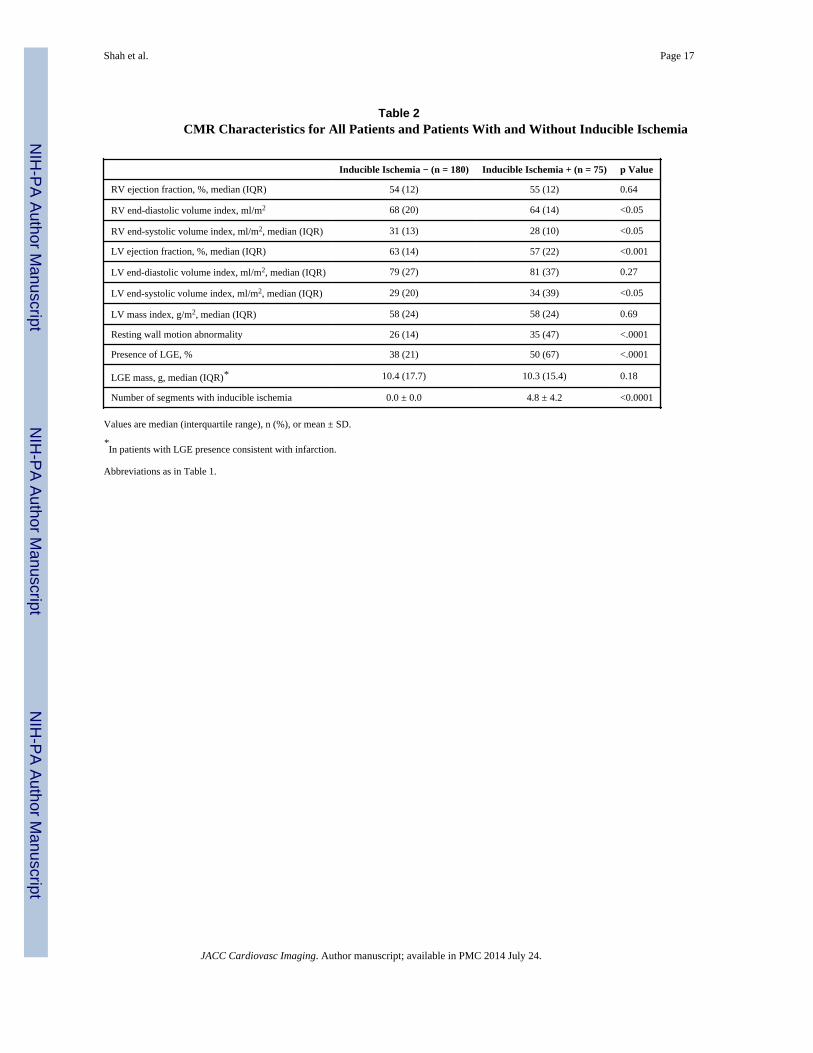
Table 2CMR Characteristics for All Patients and Patients With and Without Inducible Ischemia
Inducible Ischemia − (n = 180) Inducible Ischemia + (n = 75) p Value
RV ejection fraction, %, median (IQR) 54 (12) 55 (12) 0.64
RV end-diastolic volume index, ml/m2 68 (20) 64 (14) <0.05
RV end-systolic volume index, ml/m2, median (IQR) 31 (13) 28 (10) <0.05
LV ejection fraction, %, median (IQR) 63 (14) 57 (22) <0.001
LV end-diastolic volume index, ml/m2, median (IQR) 79 (27) 81 (37) 0.27
LV end-systolic volume index, ml/m2, median (IQR) 29 (20) 34 (39) <0.05
LV mass index, g/m2, median (IQR) 58 (24) 58 (24) 0.69
Resting wall motion abnormality 26 (14) 35 (47) <.0001
Presence of LGE, % 38 (21) 50 (67) <.0001
LGE mass, g, median (IQR)* 10.4 (17.7) 10.3 (15.4) 0.18
Number of segments with inducible ischemia 0.0 ± 0.0 4.8 ± 4.2 <0.0001
Values are median (interquartile range), n (%), or mean ± SD.
*In patients with LGE presence consistent with infarction.
Abbreviations as in Table 1.
JACC Cardiovasc Imaging. Author manuscript; available in PMC 2014 July 24.
NIH
-PA
Author M
anuscriptN
IH-P
A A
uthor Manuscript
NIH
-PA
Author M
anuscript
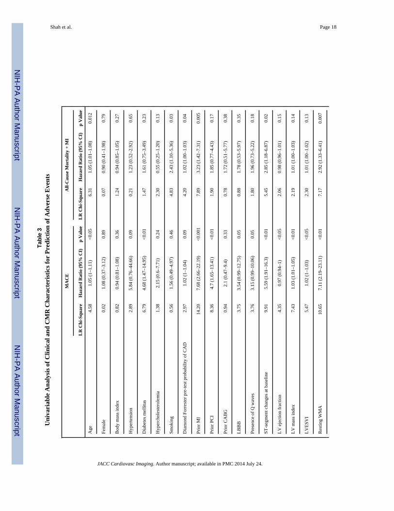
Shah et al. Page 18
Tab
le 3
Uni
vari
able
Ana
lysi
s of
Clin
ical
and
CM
R C
hara
cter
isti
cs f
or P
redi
ctio
n of
Adv
erse
Eve
nts
MA
CE
All-
Cau
se M
orta
lity
+ M
I
LR
Chi
-Squ
are
Haz
ard
Rat
io (
95%
CI)
p V
alue
LR
Chi
-Squ
are
Haz
ard
Rat
io (
95%
CI)
p V
alue
Age
4.58
1.05
(1–
1.11
)<
0.05
6.31
1.05
(1.
01–1
.08)
0.01
2
Fem
ale
0.02
1.08
(0.
37–3
.12)
0.89
0.07
0.90
(0.
41–1
.98)
0.79
Bod
y m
ass
inde
x0.
820.
94 (
0.81
–1.0
8)0.
361.
240.
94 (
0.85
–1.0
5)0.
27
Hyp
erte
nsio
n2.
895.
84 (
0.76
–44.
66)
0.09
0.21
1.23
(0.
52–2
.92)
0.65
Dia
bete
s m
ellit
us6.
794.
68 (
1.47
–14.
95)
<0.
011.
471.
61 (
0.75
–3.4
9)0.
23
Hyp
erch
oles
tero
lem
ia1.
382.
15 (
0.6–
7.71
)0.
242.
300.
55 (
0.25
–1.2
0)0.
13
Smok
ing
0.56
1.56
(0.
49–4
.97)
0.46
4.83
2.43
(1.
10–5
.36)
0.03
Dia
mon
d Fo
rres
ter
pre-
test
pro
babi
lity
of C
AD
2.97
1.02
(1–
1.04
)0.
094.
201.
02 (
1.00
–1.0
3)0.
04
Prio
r M
I14
.20
7.68
(2.
66–2
2.19
)<
0.00
17.
893.
23 (
1.42
–7.3
1)0.
005
Prio
r PC
I8.
364.
7 (1
.65–
13.4
1)<
0.01
1.90
1.85
(0.
77–4
.43)
0.17
Prio
r C
AB
G0.
942.
1 (0
.47–
9.4)
0.33
0.78
1.72
(0.
51–5
.77)
0.38
LB
BB
3.75
3.54
(0.
99–1
2.75
)0.
050.
881.
78 (
0.53
–5.9
7)0.
35
Pres
ence
of
Q w
aves
3.76
3.15
(0.
99–1
0.06
)0.
051.
801.
96 (
0.73
–5.2
2)0.
18
ST-s
egm
ent c
hang
es a
t bas
elin
e9.
915.
59 (
1.91
–16.
3)<
0.01
5.45
2.85
(1.
18–6
.87)
0.02
LV
eje
ctio
n fr
actio
n4.
350.
97 (
0.94
–1)
<0.
052.
060.
98 (
0.96
–1.0
1)0.
15
LV
mas
s in
dex
7.43
1.03
(1.
01–1
.05)
<0.
012.
191.
01 (
1.00
–1.0
3)0.
14
LV
ESV
I5.
471.
02 (
1–1.
03)
<0.
052.
301.
01 (
1.00
–1.0
2)0.
13
Res
ting
WM
A10
.65
7.11
(2.
19–2
3.11
)<
0.01
7.17
2.92
(1.
33–6
.41)
0.00
7
JACC Cardiovasc Imaging. Author manuscript; available in PMC 2014 July 24.

NIH
-PA
Author M
anuscriptN
IH-P
A A
uthor Manuscript
NIH
-PA
Author M
anuscript
Shah et al. Page 19
MA
CE
All-
Cau
se M
orta
lity
+ M
I
LR
Chi
-Squ
are
Haz
ard
Rat
io (
95%
CI)
p V
alue
LR
Chi
-Squ
are
Haz
ard
Rat
io (
95%
CI)
p V
alue
Any
str
ess
perf
usio
n de
fect
9.84
25.9
3 (3
.39–
198.
24)
<0.
0110
.29
3.76
(1.
67–8
.43)
0.00
1
Rev
ascu
lari
zatio
n w
ithin
60
days
of
CM
R4.
933.
46 (
1.16
–10.
34)
<0.
050.
671.
50 (
0.57
–3.9
9)0.
41
LG
E4.
616.
56 (
1.18
–36.
5)<
0.05
0.87
1.61
(0.
59–4
.38)
0.35
RV
EF
3.54
0.95
(0.
9–1)
0.06
1.49
0.97
(0.
93–1
.02)
0.22
Pres
ence
of
indu
cibl
e is
chem
ia12
.28
9.81
(2.
74–3
5.19
)<
0.00
110
.665
3.66
(1.
68–7
.97)
0.00
1
CA
D =
cor
onar
y ar
tery
dis
ease
; CI
= c
onfi
denc
e in
terv
al; L
BB
B =
left
bun
dle
bran
ch b
lock
; LR
chi
-squ
are
= c
hi-s
quar
e lik
elih
ood
ratio
; LV
ESV
I =
left
ven
tric
ular
end
-sys
tolic
vol
ume
inde
x; M
AC
E =
maj
or a
dver
se c
ardi
ac e
vent
s; M
I =
myo
card
ial i
nfar
ctio
n; R
VE
F =
rig
ht v
entr
icul
ar e
ject
ion
frac
tion;
WM
A =
wal
l mot
ion
abno
rmal
ity; o
ther
abb
revi
atio
ns a
s in
Tab
le 1
.
JACC Cardiovasc Imaging. Author manuscript; available in PMC 2014 July 24.