Vascularización cerebral (parte iii)
32
Effects of VEGF administration and inhibition in the visual cortex of developing rats Current research martes 15 de noviembre de 2011
-
Upload
enrike-g-argandona -
Category
Health & Medicine
-
view
365 -
download
2
Transcript of Vascularización cerebral (parte iii)

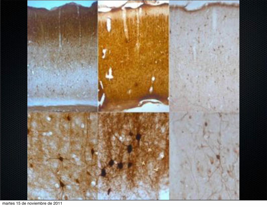
Effects of VEGF
administration and inhibition
in the visual cortex of
developing rats
Current research
martes 15 de noviembre de 2011

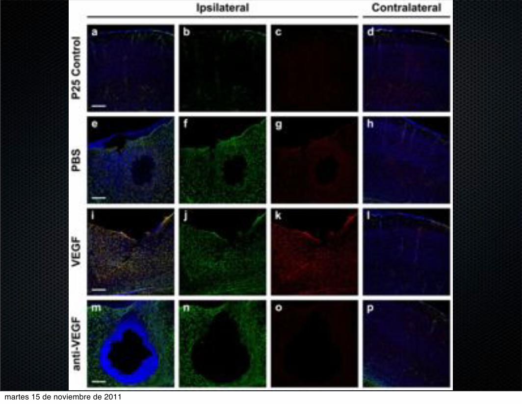
VEGF infusion
18 dpn Long Evans rats
Alzet minipumps for 1 week at a 1 µl /h rate.VEGF. 25 ng/ml.
anti-VEGF. 25 µg/ml.
PBS.
Untreated rats.
martes 15 de noviembre de 2011

martes 15 de noviembre de 2011

EBA
martes 15 de noviembre de 2011
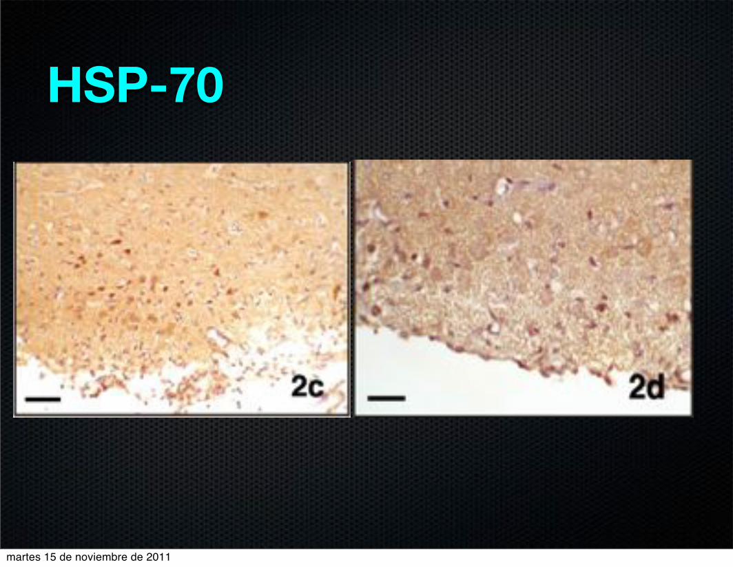
HSP-70
martes 15 de noviembre de 2011

GFAP
martes 15 de noviembre de 2011

martes 15 de noviembre de 2011

martes 15 de noviembre de 2011
martes 15 de noviembre de 2011

martes 15 de noviembre de 2011

martes 15 de noviembre de 2011

martes 15 de noviembre de 2011

0
20
40
60
80
100
120
Adq 1 Adq 2 Adq 3 Adq 4 Adq 5 Cue
Late
ncy
to ta
rget
(s)
Learning
Control SC SC Control SC EE PBS SC SC VEGF SC SC VEGF SC EE
0
20
40
60
80
100
120
Adq 1 Adq 2 Adq 3 Adq 4 Adq 5 Cue
Late
ncy
to ta
rget
Learning
Control SC SC Control SC EE
0
20
40
60
80
100
Adq 1 Adq 2 Adq 3 Adq 4 Adq 5 Cue
Late
ncy
to ta
rget
(s)
Learning
Control SC EE VEGF SC EE
0
20
40
60
80
100
Adq 1 Adq 2 Adq 3 Adq 4 Adq 5 Cue
Late
ncy
to ta
rget
(s)
Learning
VEGF SC SC VEGF SC EE
Morris water maze
martes 15 de noviembre de 2011

martes 15 de noviembre de 2011

martes 15 de noviembre de 2011

martes 15 de noviembre de 2011

martes 15 de noviembre de 2011

martes 15 de noviembre de 2011
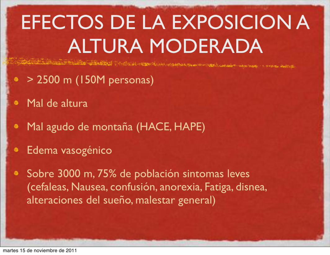
EFECTOS DE LA EXPOSICION A ALTURA MODERADA
> 2500 m (150M personas)
Mal de altura
Mal agudo de montaña (HACE, HAPE)
Edema vasogénico
Sobre 3000 m, 75% de población sintomas leves (cefaleas, Nausea, confusión, anorexia, Fatiga, disnea, alteraciones del sueño, malestar general)
martes 15 de noviembre de 2011

MECANISMOS DE PROTECCIÓN A ALTURA MODERADA
Genéticos (HIF). A. Bigham. PLoS 2010
Altura moderada protege frente patologias cardiovasculares. (12%/1000m) D. Faeh. Circulation 2009
martes 15 de noviembre de 2011

MATERIAL Y METODOS
Ratas Wistar criadas a 400 m (P47) y transportadas a 2800m en 35 minutos.
Ratas criadas en condiciones standard
Ratas con acceso a rueda de ejercicio
Ratas control a 400 m.
Se sacrifican a 48 horas.
martes 15 de noviembre de 2011

MATERIAL AND METHODS
Cuantificación densidad vascular mediante Histoquimia de Butiril Colinesterasa
Immunohistoquimia de NeuN, Calbindina, Parvalbumina y Calretinina.
martes 15 de noviembre de 2011

Vascular density
martes 15 de noviembre de 2011

Vascular density
martes 15 de noviembre de 2011

NeuN
martes 15 de noviembre de 2011
martes 15 de noviembre de 2011

martes 15 de noviembre de 2011

martes 15 de noviembre de 2011

martes 15 de noviembre de 2011

martes 15 de noviembre de 2011

[Cell Adhesion & Migration 3:2, 199-204; April/May/June 2009]; ©2009 Landes Bioscience
Neurovascular development in the central nervous system has a rich history and compelling significance. The developing central nervous system (CNS) does not produce vascular progenitor cells, and so ingression of blood vessels is required for continued CNS development and function. Classic studies provide elegant descriptions of formation of the vascular plexus that surrounds the embryonic brain and spinal cord, and the subsequent ingression of blood vessels into the neural tissue. Recent work has focused on the molecular pathways responsible for neurovascular cross-talk and development of the blood-brain barrier. Here we review neurovas-cular development in the central nervous system, with emphasis on the spinal cord. We discuss the historical work, the current status of our knowledge and unanswered questions. The importance of neurovascular development to diseases of the cerebral vasculature and the neural stem cell niche are discussed.
Introduction
Neurovascular development is the parallel emergence and patterning of the nervous system and the vascular system during embryogenesis and early life. This symbiosis is particularly important in the central nervous system (CNS) because there are no resident vascular precursor cells, so the vessels that invade the developing brain and spinal cord are absolutely essential for CNS growth and maturation. There is a rich history of descriptive studies of neuro-vascular development in the CNS and compelling medical relevance. Neurovascular cross-talk that initiates early in life sets the stage for a continuing relationship, exemplified by formation of the blood-brain barrier that protects the fragile CNS tissue from metabolic and cellular changes. Neurovascular communication is also the basis for fMRI (functional magnetic resonance imaging), a technique that reveals changes in local blood flow and oxygenation that correlate with immediate and localized neural activity in the brain, thus allowing us to non-invasively watch people “think.”1 Perturbations
of neurovascular function are also the hallmarks of diseases such as cerebral cavernous malformations and vascular dementia. Yet little is known regarding the molecular controls and mechanisms that are important in neurovascular development, and how these two complex organs communicate with each other and integrate informa-tion. This is beginning to change, as developmental biologists apply modern tools and models to questions of neurovascular develop-ment and function. This review is not comprehensive; it will focus on neurovascular development and patterning in the CNS, with emphasis on the spinal cord, since the differentiation and patterning events of both the neural and vascular compartments are accessible to examination and manipulation in the developing spinal cord. We present a brief overview of historical work, and then discuss our current knowledge and future goals.
Historical Overview
There are elegant early descriptions of blood vessel formation and invasion of the CNS. The vascularization of the fetal brain was described by several investigators, primarily through analysis of embryos injected with India ink to visualize patent vessels.2,3 These early investigators realized that blood vessels invaded from the surrounding peri-neural vascular plexus (PNVP) at specific times, and that vessels formed stereotypical patterns once they entered the brain. The patterns were thought to eventually reflect functional domains, but how initial blood vessel patterns were set up within the developing brain was not known. In 1946, Feeney and Watterson elegantly documented the formation of the PNVP around the developing spinal cord in chick embryos, and the stereotypical vessel ingression patterns exhibited over time.2 They concluded, amazingly, that the precise vessel ingression patterns they documented “…would suggest that the points where penetration of capillaries into the cord first occurs are determined by conditions within the cord…”. They did not know what conditions would promote vessel ingression, but they prophetically pointed out that experimental manipulations would be required for further informa-tion. A later comprehensive electron microscopic study of spinal cord vascularization in the developing mouse provided important infor-mation regarding cellular interactions between endothelial cells and neural cells, and described potential contributions of both cell types to the blood-brain barrier.4 These studies and others set the stage for more recent experiments examining the cellular and molecular basis for co-ordinated neurovascular development.
*Correspondence to: Victoria L. Bautch; Professor of Biology; Department of Biology, CB#3280; University of North Carolina at Chapel Hill; Chapel Hill, NC 27599 USA; Tel.: 919.966.6797; Fax: 919.962.8472; Email: [email protected]
Submitted: 01/27/09; Accepted: 03/13/09
Previously published online as a Cell Adhesion & Migration E-publication: http://www.landesbioscience.com/journals/celladhesion/article/8397
Special Focus: Angiogenesis in the Central Nervous System
Neurovascular developmentThe beginning of a beautiful friendship
Victoria L. Bautch1,2,* and Jennifer M. James1
1Department of Biology; 2Carolina Cardiovascular Biology Center; The University of North Carolina at Chapel Hill; Chapel Hill, NC USA
Key words: neural development, vascular development, neural tube, spinal cord, central nervous system, peri-neural vascular plexus, vessel sprouting, angiogenesis, neural stem cell, vascular niche
www.landesbioscience.com Cell Adhesion & Migration 199
martes 15 de noviembre de 2011

www.slideshare.net/nfpguare
www.ehu.es/ehusfera/lance
Contacto
martes 15 de noviembre de 2011