VASCULAR PURPURAS Arial Font. 2 Hemostasis Haemostasis refers to spontaneous arrest of bleeding...
35
VASCULAR PURPURAS Arial Font
-
Upload
alexia-henry -
Category
Documents
-
view
218 -
download
0
Transcript of VASCULAR PURPURAS Arial Font. 2 Hemostasis Haemostasis refers to spontaneous arrest of bleeding...
VASCULAR PURPURAS
Arial Font
2
Hemostasis
• Haemostasis refers to spontaneous arrest of bleeding caused by injury of small blood vessels.
• Small vessels are continuously exposed to trauma during normal activities can not bleed due to effects of haemostasis.
3
Hemostasis - Functions
• Maintains blood in a fluid, clot free state in normal vessel.
• Induce rapid and localized haemostatic plug at the site of injury.
4
Hemostasis - Mechanism
Haemostasis depends on three components• Vascular wall• Platelets • Coagulation cascade
5
Hemorrhage and its Forms
• Hemorrhage refers to extravasation of blood due to rupture of blood vessels. Haemorrhage may be external or may be enclosed within a tissue.
• Hematoma means accumulation of haemorrhagic blood in a tissue.
• Petechae is minute 1 to 2 mm haemorrhage into skin, mucous membrane or serous surface.
6
Hemorrhage and its Forms• Purpura is haemorrhage larger than 3 mm into
skin and mucous membrane.• Echymosis is subcutaneous haematoma larger
than 2 cm.• Large accumulation of blood in body cavities
like pleural cavity, peritoneal cavity, pericardial cavity, synovial cavity are called – haemothorax, – haemoperitoneum, – haemopericardium – haemarthrosis.
7
Clinical Significance of Hemorrhage
• Clinical effects of haemorrhage depend on – volume and rate of blood loss - rapid removal of up to
20% of blood volume or slow losses of even larger amount may have little impact in healthy adult. Greater losses may result in haemorrhagic shock.
– site of haemorrhage is also important. Bleeding that would be insignificant in subcutaneous tissue may cause death if located in the brain because the skull is unyielding and bleeding can result increased intracranial pressure and herniated.
– chronic or recurrent external blood loss may lead to iron deficiency anaemia.
8
Hemorrhagic Disorders
Hemorrhagic disorders are a group of disorders of many different causes
characterized by an abnormal tendency of bleeding due to failure of hemostasis.
9
Clinical Character of Hemorrhagic Disorders
• Spontaneous bleeding in the skin, mucous membrane or internal tissue.
• Extensive or prolonged bleeding following trauma.
• Bleeding from more than one site.

10
Causes of Hemorrhagic Disorders
• Due to vascular defects• Due to platelet defects• Due to coagulation defects.• Combination of all
11
Bleeding due to vascular defects
Common characters:• More common but less severe• Usually in the form of petechae or purpura• Increased bleeding time and frequently
positive tourniquet test.• Normal platelet count or function.• Normal CT, APTT, PT and TT
12
Vascular Disorders - Classification
Acquired:• Simple easy bruising (devil’s pinches)• Senile purpura• The symptomatic vascular purpura (non-thromcytopenic purpura)
– Infection– Drugs– Uraemia– Cushing’s syndrome and adrenocorticosteroid administration– Scurvy– Dysproteinaemia– Henoch-Schoenlein purpura
• Miscellaneous– Orthostatic purpura– Mechanical purpura– Fat embolism– Auto-erythrocyte sensitization– Systemic disorders – collagen diseases, polyarteritis nodusa,
amyloidosis, allergy
Congenital:• Hereditary haemorrhagic telangiectasis (Osler-Rendu-weber disease)• Ehlars-danlos disease
Henoch-Schönlein Purpura (HSP)
HSP - Background
HSP is a small-vessel vasculitis characterized by purpura, arthritis, abdominal pain, and hematuria.
It is also known as anaphylactoid pupura.
It is the most common cause of non-thrombocytopenic purpura in children
HSP - Epidemiology
• The etiology of HSP is unknown, but it typically follows as upper respiratory tract infection.
• It used to account for about 1% of the admissions to hospital but changes in the medical practice have reduced the frequency to about 0.6%.
• It is more frequent in children more than adult, and mostly occurs between the ages of 2-8 years.
HSP - Epidemiology
• Males are affected twice as frequently as females.
• And the overall incidence is estimated to be 9/100000 population
HSP - Etiology
The etiology of HSP is unknown.
About 50% of patients have a preceding upper respiratory illness (URI).
Multiple infectious agents as well as drugs, foods, and insect bites may trigger HSP.
HSP - Pathophysiology
This illness is considered by histopathology to be an IgA mediated vasculitis of small vessels.
And immunoflorescent techniques may show deposition of IgA and C3 in the small vessels of the skin and the renal glomeruli
HSP - Pathophysiology
HSP and IgA nephropathy are related disorders.
Both illnesses have elevated serum IgA levels and identical findings on renal biopsy.
however, IgA nephropathy almost exclusively involves young adults and predominantly affects the kidneys only.
HSP affects mostly children and involves the skin and connective tissues, gastrointestinal tract, joints, and scrotum as well as the kidneys.
HSP - Mortality/Morbidity
In general, HSP is a benign self-limited disorder.
Fewer than 5% of cases cause chronic symptoms.
Fewer than 1% of cases progress to end-stage renal failure. Sex: Male-to-female ratio is about 2:1.
Age: Seventy-five percent of patients affected are aged 2-8 years.
HSP - History
• The onset of the disease maybe acute with the appearance of several manifestations simultaneously, or insidious with sequential occurrence of symptoms over period of weeks to months.
• Low grade fever and fatigue occur in more than half of affected children
HSP – Cinical Manifestations
Rash (95-100%), especially involving the legs, may not be present on initial presentation
Beginning as pinkish maculopapules that initially blanches on pressure.
Then progress petechiae or purpura which are characterized clinically as palpable pupura that evolve from red to purple rusty brown before they eventually fade.
HSP – Cinical Manifestations
The lesions tend to occur in crops lasting from 3-10 days and may appear at intervals that vary from a few days to as long as 3-4 months.
In fewer than 10% of children, recurrence of the rash may not end until as late as a year and rarely several years after the initial episode
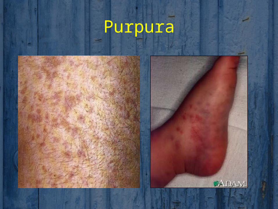
Purpura
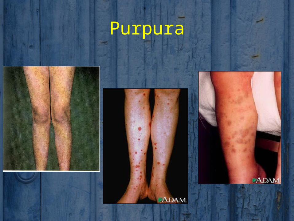
Purpura
HSP – Cinical Manifestations Subcutaneous edema (20-50%)
Damage to cutanuous vessels also may result in local angiodema, which may preceed the palpable purpura.
Edema occurs primarily in dependent areas, as below the waist, over the buttocks or on the back and posterior scalp of infants, or in areas of greater tissue destisibility such as eyelids, lips, scrotum, or dorsum of the hands or feet.
HSP – Cinical Manifestations
Joint pain (60-80%), especially involving knees and ankles
Arithritis is usually localized to the knees and ankles and appears to be concomitant with edema.
The effusion are serous, not hemorrhagic, in nature and resolve after few days without residual deformity or articular damage.
They may recur during a subsequent reactive phase of the disease
HSP – Cinical Manifestations Abdominal pain and vomiting (85%)
Edema and damage to the vasculature of the GIT may also lead to intermittent abdominal pain that is often colicky in nature.
More than half of patients have occult positive stools, diarrhea or hematemesis.
The recognition of peritoneal exudates, enlarged mesenteric lymph nodes, segmental edema, and hemorrhage into the bowel may prevent unnecessary laporotomy for acute abdomen.
Intussusception may occur, which is suggested by an empty right lower abdominal quadrant on physical examination or by cuurant jelly stools, which may be followed by complete obstruction or infarction with bowel perforation.
HSP – Cinical Manifestations• Several other organs may be involved during the acute phase of
the disease.
• Renal involvement occur in 20-50% of patients.
• Hepatosplenomegaly and lymphadenopathy may also occur.
• A rare but potentially serious outcome of the CNS involvement is the development of seizures, paresis, or coma.
• Other rare complications include cardiac and eye involvement, mononeuropathy, pancreatitis and pulmonary or intramuscular hemorrhage
HSP – Physical
Palpable purpura, particularly on the buttocks and legs Edema of the hands, feet, scalp, and ears
Arthritis, most commonly involving the knees and ankles
Abdominal tenderness
Gastrointestinal bleeding
Acute scrotal edema that may mimic testicular torsion
HSP - Lab Studies
Platelet count and coagulation studies: Platelet count is usually in the reference range but may be
elevated; the platelet count should not be low in HSP. A normal platelet count rules out idiopathic thrombocytopenic
purpura (ITP). A normal platelet count and normal coagulation studies (ie, PT,
aPTT, fibrin split products) rule out thrombotic thrombocytopenic purpura (TTP).
Lipase: A normal lipase makes acute pancreatitis very unlikely.
HSP - Lab Studies CBC and differential: WBC count may be in the reference
range or elevated. Eosinophilia is sometimes present.
Erythrocyte sedimentation rate: Sedimentation rate may be in the reference range or elevated.
BUN and creatinine levels: These may be elevated from renal involvement of HSP or from dehydration.
Urinalysis: Hematuria and/or proteinuria are present in 10-20% of patients.
• Definitive diagnosis of vasculitis, confirmed by biopsy of an involved cutanuous site, shows leuckocytoclastic angiits.
• Renal biopsy may show IgA mesengial deposition and occasionally IgM,C3, and fibrin.
HSP - Medications
• Nonsteroidal anti-inflammatory drugs (NSAIDs) may help joint pain and do not worsen the purpura. However, NSAIDs should be used cautiously in patients with renal insufficiency.
• Clinicians often use corticosteroids to treat abdominal pain, subcutaneous edema, and nephritis, but no prospective, placebo-controlled studies have demonstrated their effectiveness. Most patients recover quickly without treatment.
HSP - Medications
• Treatment in patients with severe HSP nephritis includes many different modalities, but again, no prospective controlled studies prove the efficacy of any of these treatments.
• Treatment regimens have included IV or oral steroids with or without any of the following: azathioprine, cyclophosphamide, cyclosporine, high-dose IV immunoglobulin G (IVIg), danazol, or fish oil.