Vascular permeability expression
5
Proc. Natl. Acad. Sci. USA Vol. 92, pp. 905-909, January 1995 Biochemistry Vascular endothelial growth factor/vascular permeability factor expression in a mouse model of retinal neovascularization (angiogenesis/hypoxia/Muller cells/retinopathy of prematurity) ERIC A. PIERCE*, ROBERT L. AVERYt, ELIOT D. FOLEY*, LLOYD P. AIELLOt, AND LOIS E. H. SMITH*§ *Department of Ophthalmology, Children's Hospital and Harvard Medical School, Boston, MA 02115; tNeuroscience Research Institute, University of California, Santa Barbara, CA 93103; and tBeetham Eye Institute and Research Division, Joslin Diabetes Center and Harvard Medical School, Boston, MA 02215 Communicated by Hector F. DeLuca, University of Wisconsin, Madison, WI, October 7, 1994 ABSTRACT Neovascular diseases of the retina are a major cause of blindness worldwide. Hypoxia is thought to be a common precursor to neovascularization in many retinal diseases, but the factors involved in the hypoxic neovascular response have not been fully identified. To investigate the role of vascular endothelial growth factor/vascular permeability factor (VEGF/VPF) in retinal neovascularization, the expres- sion of VEGF/VPF mRNA and protein were studied in a mouse model of proliferative retinopathy. RNA (Northern) blot analysis revealed that retinal VEGF/VPF mRNA expres- sion increased 3-fold between 6 and 12 hr of relative retinal hypoxia and remained elevated during the development of neovascularization. In situ hybridization localized VEGF/VPF mRNA to cells bodies in the inner nuclear layer of the retina. Immunohistochemical confocal microscopy dem- onstrated that VEGF/VPF protein levels increase with a time course similar to that of the mRNA. The cells in the inner nuclear layer of the retina that produce VEGF/VPF were identified morphologically as Muller cells. These data suggest that VEGF/VPF expression in the retina plays a central role in the development of retinal ischemia-induced ocular neo- vascularization. Retinal neovascularization is a major cause of blindness in the United States (1). Pathologic retinal angiogenesis is a final common pathway leading to vision loss in diseases such as retinopathy of prematurity (ROP), diabetic retinopathy, and age-related macular degeneration (2-4). Despite the preva- lence of these diseases, the biochemical events responsible for retinal neovascularization are not fully understood. It is hoped that a better understanding of the fundamental basis of retinal neovascularization will improve treatment and prevention of retinal vascular diseases. In these eye diseases, as in tumor growth and wound healing, hypoxia appears to be a common precursor to neovascular- ization (5, 6). In the 1940s and 1950s, Michaelson (7) and Ashton et al. (8) postulated that retinal neovascularization was caused by release of a "vasoformative factor" from the retina in response to hypoxia. Since these initial hypotheses, it has become widely accepted that retinal hypoxia results in the release of factors that influence new blood vessel growth (3). The angiogenic factors responsible for retinal neovasculariza- tion, however, have not been conclusively identified. Vascular endothelial growth factor/vascular permeability factor (VEGF/VPF) is an endothelial cell-specific mitogen that was identified and cloned by several groups of investiga- tors (9-12). The expression of VEGF/VPF is stimulated by hypoxia, and VEGF/VPF has been demonstrated to be re- quired for tumor-associated angiogenesis (13-15). These char- acteristics make VEGF/VPF an ideal candidate for a retinal vasoformative factor. It has recently been reported that VEGF/VPF levels are elevated in the vitreous of patients with retinal neovascularization (16). It has also been demonstrated that several types of cultured retinal cells secrete VEGF/VPF, and that hypoxia stimulates VEGF/VPF production in some cell types (17-20). VEGF/VPF levels also increase in a primate model of iris neovascularization (21). However, the location and time course of VEGF/VPF expression in association with retinal neovascularization has not been delineated nor has it been determined if VEGF/VPF has a direct role in the induction of retinal neovascularization. The goal of the present study was to examine the role of VEGF/VPF in retinal neovascularization by studying the time course and location of VEGF/VPF expression in a mouse model of proliferative retinopathy (22). In this model, mice are exposed to hyperoxia, resulting in obliteration of the posterior retinal vessels. The mice are then returned to room air, which is presumed to cause relative hypoxia of the now nonperfused retina, producing a quantifiable neovascular response in 100% of animals. Using this model, the expression of VEGF/VPF mRNA and protein were studied by RNA (Northern) blot analysis, in situ hybridization, and immunohistochemical con- focal microscopy. These data demonstrate the role of VEGF/ VPF in an animal model of retinal neovascularization, which has not previously been done to our knowledge. EXPERIMENTAL PROCEDURES Mouse Model. The study adhered to the "Association for Research in Vision and Ophthalmology Statement for the Use of Animals in Ophthalmic and Vision Research." To produce retinal neovascularization, litters of 7-day-old [postnatal day 7 (P7)] C57BL/6J mice with nursing mothers were exposed to 75 ± 2% oxygen for 5 days and then returned to room air at age P12 as described (22). Mice of the same age kept in room air were used as controls. Flat-mounted, fluorescein-conjugated dextran-perfused retinas were examined to assess the retinal vasculature (22). In Situ Hybridization. For in situ hybridization studies, mice at different time points during the induction of neovascular- ization were deeply anesthetized with Avertin and sacrificed by perfusion through the left ventricle with 4% paraformalde- hyde in phosphate-buffered saline. Eyes were enucleated, fixed in 4% paraformaldehyde at 4°C overnight, and embedded in paraffin. Serial 6-,um sections of the whole eyes were placed on microscope slides, and the slides were stored at 4°C. Several slides from each eye were also stained with either hematoxylin alone or periodic acid/Schiff reagent (PAS) and hematoxylin. Tissue sections were prehybridized and hybridized as de- scribed (23). An 35S-labeled RNA probe prepared from rat Abbreviations: PAS, periodic acid/Schiff reagent; RPE, retinal pig- ment epithelium; VEGF/VPF, vascular endothelial growth factor/ vascular permeability factor; P7, etc., postnatal day 7, etc. §To whom reprint requests should be addressed at: Department of Ophthalmology, Fegan 4, Children's Hospital, 300 Longwood Ave- nue, Boston, MA 02115. 905 The publication costs of this article were defrayed in part by page charge payment. This article must therefore be hereby marked "advertisement" in accordance with 18 U.S.C. §1734 solely to indicate this fact. Downloaded by guest on November 25, 2021
Transcript of Vascular permeability expression

Proc. Natl. Acad. Sci. USAVol. 92, pp. 905-909, January 1995Biochemistry
Vascular endothelial growth factor/vascular permeability factorexpression in a mouse model of retinal neovascularization
(angiogenesis/hypoxia/Muller cells/retinopathy of prematurity)
ERIC A. PIERCE*, ROBERT L. AVERYt, ELIOT D. FOLEY*, LLOYD P. AIELLOt, AND LOIS E. H. SMITH*§*Department of Ophthalmology, Children's Hospital and Harvard Medical School, Boston, MA 02115; tNeuroscience Research Institute, University of California,Santa Barbara, CA 93103; and tBeetham Eye Institute and Research Division, Joslin Diabetes Center and Harvard Medical School, Boston, MA 02215
Communicated by Hector F. DeLuca, University of Wisconsin, Madison, WI, October 7, 1994
ABSTRACT Neovascular diseases of the retina are amajor cause of blindness worldwide. Hypoxia is thought to bea common precursor to neovascularization in many retinaldiseases, but the factors involved in the hypoxic neovascularresponse have not been fully identified. To investigate the roleof vascular endothelial growth factor/vascular permeabilityfactor (VEGF/VPF) in retinal neovascularization, the expres-sion of VEGF/VPF mRNA and protein were studied in amouse model of proliferative retinopathy. RNA (Northern)blot analysis revealed that retinal VEGF/VPF mRNA expres-sion increased 3-fold between 6 and 12 hr of relative retinalhypoxia and remained elevated during the development ofneovascularization. In situ hybridization localizedVEGF/VPF mRNA to cells bodies in the inner nuclear layerofthe retina. Immunohistochemical confocal microscopy dem-onstrated that VEGF/VPF protein levels increase with a timecourse similar to that of the mRNA. The cells in the innernuclear layer of the retina that produce VEGF/VPF wereidentified morphologically as Muller cells. These data suggestthat VEGF/VPF expression in the retina plays a central rolein the development of retinal ischemia-induced ocular neo-vascularization.
Retinal neovascularization is a major cause of blindness in theUnited States (1). Pathologic retinal angiogenesis is a finalcommon pathway leading to vision loss in diseases such asretinopathy of prematurity (ROP), diabetic retinopathy, andage-related macular degeneration (2-4). Despite the preva-lence of these diseases, the biochemical events responsible forretinal neovascularization are not fully understood. It is hopedthat a better understanding of the fundamental basis of retinalneovascularization will improve treatment and prevention ofretinal vascular diseases.
In these eye diseases, as in tumor growth and wound healing,hypoxia appears to be a common precursor to neovascular-ization (5, 6). In the 1940s and 1950s, Michaelson (7) andAshton et al. (8) postulated that retinal neovascularization wascaused by release of a "vasoformative factor" from the retinain response to hypoxia. Since these initial hypotheses, it hasbecome widely accepted that retinal hypoxia results in therelease of factors that influence new blood vessel growth (3).The angiogenic factors responsible for retinal neovasculariza-tion, however, have not been conclusively identified.
Vascular endothelial growth factor/vascular permeabilityfactor (VEGF/VPF) is an endothelial cell-specific mitogenthat was identified and cloned by several groups of investiga-tors (9-12). The expression of VEGF/VPF is stimulated byhypoxia, and VEGF/VPF has been demonstrated to be re-quired for tumor-associated angiogenesis (13-15). These char-acteristics make VEGF/VPF an ideal candidate for a retinalvasoformative factor. It has recently been reported that
VEGF/VPF levels are elevated in the vitreous of patients withretinal neovascularization (16). It has also been demonstratedthat several types of cultured retinal cells secrete VEGF/VPF,and that hypoxia stimulates VEGF/VPF production in somecell types (17-20). VEGF/VPF levels also increase in a primatemodel of iris neovascularization (21). However, the location andtime course ofVEGF/VPF expression in association with retinalneovascularization has not been delineated nor has it beendetermined if VEGF/VPF has a direct role in the induction ofretinal neovascularization.The goal of the present study was to examine the role of
VEGF/VPF in retinal neovascularization by studying the timecourse and location of VEGF/VPF expression in a mousemodel of proliferative retinopathy (22). In this model, mice areexposed to hyperoxia, resulting in obliteration of the posteriorretinal vessels. The mice are then returned to room air, whichis presumed to cause relative hypoxia of the now nonperfusedretina, producing a quantifiable neovascular response in 100%of animals. Using this model, the expression of VEGF/VPFmRNA and protein were studied by RNA (Northern) blotanalysis, in situ hybridization, and immunohistochemical con-focal microscopy. These data demonstrate the role of VEGF/VPF in an animal model of retinal neovascularization, whichhas not previously been done to our knowledge.
EXPERIMENTAL PROCEDURESMouse Model. The study adhered to the "Association for
Research in Vision and Ophthalmology Statement for the Useof Animals in Ophthalmic and Vision Research." To produceretinal neovascularization, litters of 7-day-old [postnatal day 7(P7)] C57BL/6J mice with nursing mothers were exposed to 75± 2% oxygen for 5 days and then returned to room air at ageP12 as described (22). Mice of the same age kept in room airwere used as controls. Flat-mounted, fluorescein-conjugateddextran-perfused retinas were examined to assess the retinalvasculature (22).
In Situ Hybridization. For in situ hybridization studies, miceat different time points during the induction of neovascular-ization were deeply anesthetized with Avertin and sacrificed byperfusion through the left ventricle with 4% paraformalde-hyde in phosphate-buffered saline. Eyes were enucleated, fixedin 4% paraformaldehyde at 4°C overnight, and embedded inparaffin. Serial 6-,um sections of the whole eyes were placed onmicroscope slides, and the slides were stored at 4°C. Severalslides from each eye were also stained with either hematoxylinalone or periodic acid/Schiff reagent (PAS) and hematoxylin.
Tissue sections were prehybridized and hybridized as de-scribed (23). An 35S-labeled RNA probe prepared from rat
Abbreviations: PAS, periodic acid/Schiff reagent; RPE, retinal pig-ment epithelium; VEGF/VPF, vascular endothelial growth factor/vascular permeability factor; P7, etc., postnatal day 7, etc.§To whom reprint requests should be addressed at: Department ofOphthalmology, Fegan 4, Children's Hospital, 300 Longwood Ave-nue, Boston, MA 02115.
905
The publication costs of this article were defrayed in part by page chargepayment. This article must therefore be hereby marked "advertisement" inaccordance with 18 U.S.C. §1734 solely to indicate this fact.
Dow
nloa
ded
by g
uest
on
Nov
embe
r 25
, 202
1
Proc. Nati Acad ScL USA 92 (1995)
VEGF/VPF cDNA (24) was used to detect VEGF/VPF in theprepared tissue sections. Single-stranded antisense and sense35S-labeled RNA probes were prepared by in vitro transcrip-tion with uridine 5'-[a-(35S)thio]triphosphate (New EnglandNuclear). Specific hybridization was detected by autoradiog-raphy with NTB-2 emulsion (Kodak). Hybridizations withlabeled sense RNA probe on adjacent serial sections served ascontrols for nonspecific binding. Slides were counterstainedwith hematoxylin and photographed with both light and dark-field microscopy.Northern Blot Analysis. For Northern blot analyses, retinas
from mice at different time points during the induction ofretinopathy were isolated and frozen immediately in liquidnitrogen. For each time point, 10 retinas from five mice werepooled, and total retinal RNAwas prepared (25). RNA (15 jig)from each time point was then electrophoresed on agarose/formaldehyde gels and transferred by capillary technique toBiotrans nylon membranes (ICN). Hybridization was per-formed with labeled DNA probes according to establishedmethods (26). DNA probes were prepared from the humanVEGF/VPF cDNA (11) by the random hexamer labelingmethod (27) and [a-32P]dATP (New England Nuclear).Washed membranes were exposed to autoradiography filmand analyzed on a PhosphorImager (Molecular Dynamics).Total RNA content was normalized at each time point byprobing all blots with 36B4 cDNA (28). ImageQuant software(Molecular Dynamics) was used to quantify the VEGF/VPFand 36B4 signals for each lane on the blots.
Confocal Microscopy. For confocal microscopy, eyes wereharvested and fixed in paraformaldehyde as described for insitu hybridization studies. Sections were then prepared by usinga modification of described techniques (29). Briefly, the
posterior segments of paraformaldehyde-fixed eyes were em-bedded in low-melting-point agarose. Thick serial sections(100 gm) were cut and incubated overnight at 4°C withpolyclonal anti-VEGF/VPF antibodies (30). Adjacent sectionsincubated with normal rabbit serum served as controls. Afterextensive washing, the sections were incubated with anti-rabbitIgG labeled with the fluorochrome CY3. After additionalwashing, the sections were mounted in n-propyl galate andglycerol and imaged with a Bio-Rad 500 confocal microscope.
RESULTSMouse Model of Proliferative Retinopathy. To investigate
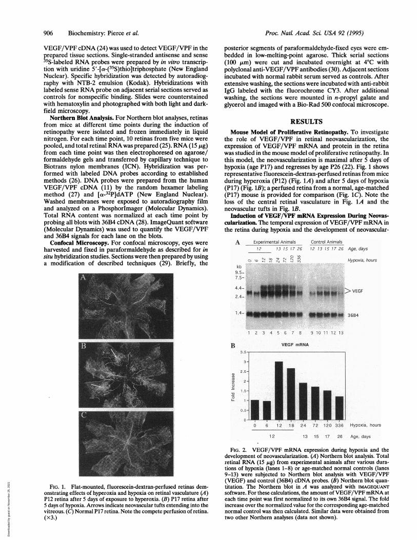
the role of VEGF/VPF in retinal neovascularization, theexpression of VEGF/VPF mRNA and protein in the retinawas studied in the mouse model of proliferative retinopathy. Inthis model, the neovascularization is maximal after 5 days ofhypoxia (age P17) and regresses by age P26 (22). Fig. 1 showsrepresentative fluorescein-dextran-perfused retinas from miceduring hyperoxia (P12) (Fig. 1A) and after 5 days of hypoxia(P17) (Fig. 1B); a perfused retina from a normal, age-matched(P17) mouse is provided for comparison (Fig. 1C). Note theloss of the central retinal vasculature in Fig. 1A and theneovascular tufts in Fig. 1B.
Induction of VEGF/VPF mRNA Expression During Neovas-cularization. The temporal expression ofVEGF/VPF mRNA inthe retina during hypoxia and the development of neovascular-
A Experimental Animals72 73 75 17 26
kb9. 5-7.5-:4.4-
2.4-
Control Animals72 73 75 77 26 Age, days
Hypoxia, hours
MkjAO > VEGF
-_yw~3 6 B41.4-
1 2 3 4 5 6 7 8 9 10 11 12 13
VEGF mRNA
3
2.5-
CD 2-
-01.5-
0.5-
0 6 1 2 1 8 24 72 120 336 Hypoxia, hours
13 15 17 26 Age, days
FIG. 1. Flat-mounted, fluorescein-dextran-perfused retinas dem-onstrating effects of hyperoxia and hypoxia on retinal vasculature (A)P12 retina after 5 days of exposure to hyperoxia. (B) P17 retina after5 days of hypoxia. Arrows indicate neovascular tufts extending into thevitreous. (C) Normal P17 retina. Note the compete perfusion of retina.(x3.)
FIG. 2. VEGF/VPF mRNA expression during hypoxia and thedevelopment of neovascularization. (A) Northern blot analysis. Totalretinal RNA (15 ,g) from experimental animals after various dura-tions of hypoxia (lanes 1-8) or age-matched normal controls (lanes9-13) were subjected to Northern blot analysis with VEGF/VPF(VEGF) and control (36B4) cDNA probes. (B) Northern blot quan-titation. The Northern blot in A was analyzed with IMAGEQUANTsoftware. For these calculations, the amount ofVEGF/VPFmRNA ateach time point was first normalized to its own 36B4 signal. The foldincrease over the normalized value for the corresponding age-matchednormal control was then calculated. Similar data were obtained fromtwo other Northern analyses (data not shown).
B
1 2
906 Biochemistry: Pierce et aL
Dow
nloa
ded
by g
uest
on
Nov
embe
r 25
, 202
1

Proc. Natl. Acad Sci. USA 92 (1995) 907
FIG. 3. In situ hybridization ofVEGF/VPF mRNA expression. Sections from the eyes of animals exposed to 24 hr of hypoxia (A-C) and normalage-matched (P13) controls (D-F) were analyzed by in situ hybridization and photographed with light (A and D) and dark-field (B, C, E, and F)illumination. (A) Hybridization with antisense probe after 24 hr of hypoxia. The ganglion cell layer (G), inner nuclear layer (I), and the retinalpigment epithelium (RPE) (designated R) are indicated. (B) Dark-field image ofA. (C) Control hybridization of adjacent serial section with thesense probe, dark-field image. (D) Hybridization with antisense probe, age-matched (P13) normal control. (E) Dark-field image of D. (F) Controlhybridization of adjacent serial section with the sense probe, dark-field image. (x 12.5.)
ization was evaluated by Northern blot analysis. Fig. 2A shows adramatic increase in VEGF/VPFmRNA levels between 6 and 12hr after return of experimental P12 mice to room air-that is,after 6 to 12 hr of hypoxia (Fig. 2A, lanes 2 and 3). TheVEGF/VPF mRNA levels remained elevated for several daysand then decreased toward baseline with regression of theretinopathy (P26; Fig. 2A, lane 8). Control RNA from age-matched animals raised in room air demonstrated a compara-tively constant, low level of VEGF/VPF mRNA (Fig. 2A, lanes
9-13). The sizes of the VEGF/VPF transcripts detected in thesestudies are approximately 3.7 kb and 4.5 kb.
Fig. 2B shows the fold increase in VEGF/VPF mRNA foreach hypoxic time point compared with age-matched controlsafter normalization to the 36B4 signal in each lane. Phosphor-Imager quantitation of the VEGF/VPF mRNA signal on theNorthern blot shown in Fig. 2A showed a maximal 3-foldincrease of VEGF/VPF mRNA at 12 hr of hypoxia comparedwith normal, age-matched controls.
FIG. 4. In situ hybridization analysis of VEGF/VPF mRNA expression during the development of neovascularization. Sections from the eyesof animals at the ages indicated were analyzed by in situ hybridization and photographed with light (A, C, E, G, and I) and dark-field (B, D, F,and H) illumination. All hybridizations shown were performed with antisense probe. The ganglion cell layer (G), inner nuclear layer (I), and RPE(R) are indicated in A. (A and B) Age P12, after 5 days of hyperoxia. (C and D) Age P13, after 24 hr of hypoxia. (E and F) Age P15, after 72 hrof hypoxia. (G and H) Age P17, after 120 hr of hypoxia. (I) Serial section adjacent to G and H, stained with PAS and hematoxylin to demonstrateneovascular tufts (arrows). (X30.)
Biochemistry: Pierce et al.
Dow
nloa
ded
by g
uest
on
Nov
embe
r 25
, 202
1

Proc. Natl. Acad Sci. USA 92 (1995)
To determine the location of VEGF/VPF mRNA expres-sion during the development of neovascularization, in situhybridization was performed. Fig. 3 shows the results of atypical in situ hybridization performed after 24 hr of hypoxiaand in age-matched (P13) normal controls. Hybridization ofthe hypoxic retinas with antisense probe showed an intensesignal for VEGF/VPF mRNA over the inner nuclear layer ofthe retina and a small amount of labeling over the ganglion celllayer (Fig. 3A and B). In contrast, age-matched nonhypoxiccontrol retinas showed very little VEGF/VPF mRNA signal(Fig. 3 D and E). Hybridization with sense probe showed lowbackground in both hypoxic and control retinas (Fig. 3 C andF, respectively). Note that the RPE has a bright signal ondark-field microscopy due to the presence ofpigment granules,and thus probe hybridization over this area cannot be accu-rately evaluated.
Fig. 4 shows the pattern of VEGF/VPF mRNA expressionduring the time course of hypoxia and the development ofretinal neovascularization. At age P12, just prior to removal ofthe animals from oxygen, some specific hybridization was seenin the inner nuclear and ganglion cell layers (Fig. 4A and B).After 24 hr of relative hypoxia, at age P13, the VEGF/VPFmRNA signal was dramatically increased (Fig. 4 C and D).After 72 and 120 hr of hypoxia (P15 and P17, respectively), theVEGF/VPF mRNA signal in the inner nuclear layer was stillstrong, and more mRNA in the ganglion cell layer was detected(Fig. 4E-H). By age P26, little VEGF/VPF mRNAwas detected(data not shown). Retinas from age-matched control animals ateach time point showed a pattern of hybridization similar to thatof the P13 controls shown in Fig. 3 D-F. Sense hybridizationcontrols were used in all experiments and demonstrated uni-formly low background, as seen in Fig. 3 C and F.At time points early in hypoxia, such as after 24 hr of hypoxia
(P13), the entire inner nuclear layer showed VEGF/VPFhybridization (Fig. 3A and B; Fig. 4 C and D). The neovasculartufts that develop in this model are concentrated in the middleretina at the junction of the perfused and nonperfused retina(Fig. 1) (22). Fig. 4 shows the pattern of hybridization at P17(Fig. 4H) compared to an adjacent serial section stained withPAS and hematoxylin to demonstrate the neovascular tufts(Fig. 41). The hybridization signal in the inner nuclear layer isconcentrated in the non-perfused retina just posterior to theneovascular tufts growing though the inner limiting membraneinto the vitreous (arrows).
Stimulation ofVEGF/VPF Protein Expression and CellularLocalization. To investigate VEGF/VPF protein expression,immunohistochemistry was performed with confocal micro-
FIG. 5. Confocal immunohistochemistry of VEGF/VPF. Sectionsfrom the eyes of animals at the ages indicated were analyzed byimmunohistochemistry with anti-VEGF/VPF antibodies (A, B, andD)or nonimmune control (C) serum and viewed by confocal microscopy.(A) P17, after 120 hr of hypoxia, stained with anti-VEGF/VPFantibodies. Note Muller cell processes spanning retina (arrow). (B)Higher power view ofA showing Muller cell foot plates in inner retina.(C) Staining of adjacent serial section with nonimmune serum. (D)Normal P17 control, stained with anti-VEGF/VPF antibodies. (A,X21; B-D, X42.)
scopy. Fig. S demonstrates that VEGF/VPF protein is pro-duced by cells in the inner nuclear layer of the retina, consis-tent with the results of the in situ hybridization studiesdescribed above. The additional resolution of confocal micros-copy makes it possible to determine that the VEGF/VPFprotein is located in cells morphologically consistent with theMuller cells of the retina. This is evident from the staining ofcell bodies in the inner nuclear layer, with spindle-shapedprocesses that span the entire width of the retina (Fig. SA andB) (31). At age P17, after 5 days of hypoxia, more VEGF/VPFis present in experimental (Fig. S A and B) than control (Fig.SD) retinas. The VEGF/VPF staining is specific, as indicatedby the low background observed with nonimmune serum (Fig.SC). Further controls with secondary antibody alone revealedno signal above baseline (data not shown). At mouse ages P12and P27, very little VEGF/VPF protein was detected (data notshown). Staining of the photoreceptor matrices was not ob-served consistently with different antibody preparations.
DISCUSSIONOur data demonstrate that VEGF/VPF is expressed in the retinaprior to the development of neovascularization in a mouse modelof proliferative retinopathy. The VEGF/VPF mRNA level in-creases dramaticallywithin 6-12 hr of relative retinal hypoxia andremains elevated until neovascularization develops. The VEGF/VPF mRNA level then declines as the neovascularization re-gresses to nearly control levels by P26, when the neovasculariza-tion has regressed. The levels of VEGF/VPF protein correlatewith the mRNA levels. This time course and the localization datadiscussed below are consistent with a central role forVEGF/VPFin the development of the neovascularization, and thus representsthe first reported demonstration of the association betweenVEGF/VPF expression and retinal neovascularization in ananimal model.Two VEGF/VPF transcripts with sizes of approximately 3.7
and 4.5 kb were detected in these studies. These sizes areconsistent with the size of mouse VEGF/VPF messages de-tected by other investigators (32, 33). Alternative splicing ofVEGF/VPF mRNA is known to occur and generates fourmolecular species of VEGF/VPF (34, 35). Additional studieswill be required to determine if the two transcripts detectedcode for different species of VEGF/VPF and if they aredifferentially regulated.
In situ hybridization studies show that VEGF/VPF mRNAis produced by cells in the inner nuclear layer of the retina. Thisis consistent with a prior report that VEGF/VPF mRNA wasproduced in the inner nuclear layer of the retina in a monkeymodel of iris neovascularization (21). Of further interest, ourdata demonstrate that as neovascularization develops, theVEGF/VPF message is concentrated just posterior to theneovascular tufts, at the junction of the perfused and nonper-fused portions of the retina. This demonstrates an especiallystrong correlation between VEGF/VPF expression and retinalneovascularization.The specific cell type in the inner nuclear layer responsible
for VEGF/VPF production has not been previously identified.Our immunohistochemical data demonstrate that VEGF/VPFis produced by cells morphologically consistent with Mullercells. This is evident from the unique morphology of Mullercells, with cell bodies in the inner nuclear layer and radialprocesses that span the entire retina. The other retinal cellswhose cell bodies are present in the inner nuclear layer are allneurons with morphologies distinctly different from the im-munostaining pattern observed (31). Muller cells are theprincipal glial cells of the neural retina. The finding thatVEGF/VPF is produced by Muller cells is consistent with theirknown role as regulators of the extracellular environment ofthe retina (31). Our data does not eliminate the possibility thatother retinal cells also produce VEGF/VPF.
908 Biochemistry: Pierce et al.
Dow
nloa
ded
by g
uest
on
Nov
embe
r 25
, 202
1

Proc. Natl. Acad. Sci. USA 92 (1995) 909
In the mouse model used for these studies, exposure ofneonatal mice to hyperoxia causes obliteration of the centralretinal vessels. Return to room air is then hypothesized tocause relative retinal hypoxia, which leads to the observedneovascularization (22). This approach to the creation ofproliferative retinopathy was first used by Ashton et al. (8) andhas been used by others more recently (36). While it ishypothesized that the avascular retina is hypoxic in thesemodels, there is no direct evidence that the central, avascularretina is hypoxic following return to room air in the mousemodel. There is, however, direct evidence from other animalmodels of retinal neovascularization that the avascular retinais hypoxic (37, 38). Fluorescein-dextran perfusion of miceexposed to hyperoxia demonstrates complete loss of the retinalcapillary bed in the central retina (22). It is a reasonablehypothesis, therefore, that the nonperfused retina is hypoxic inthe mouse model.
Given the assumption that the avascular retina is hypoxic in themouse model, the findings presented here are consistent withthose of other investigators, who have shown that VEGF/VPF isregulated by hypoxia. This has been demonstrated in cell culturesystems (18), in clinical tumor specimens (13, 14), and in otheranimal model systems (21). The mechanism by which hypoxiastimulates VEGF/VPF expression has not been determined.The data presented here provide evidence of a correlation
of VEGF/VPF expression with retinal neovascularizationsuggesting that VEGF/VPF plays a major role in retinalneovascularization. Further studies, however, will be requiredto determine if VEGF/VPF is directly responsible for retinalneovascularization. Several studies have demonstrated thatblocking of VEGF/VPF activity with antibodies can preventretinal endothelial cell growth in vitro and angiogenesis-dependent tumor growth in vivo (15, 16). The mouse model ofproliferative retinopathy is well-suited to such blocking stud-ies, as it is 100% reproducible (22). If VEGF/VPF is found tobe required for retinal neovascularization, then agents thatblock VEGF/VPF action or expression might be clinicallyuseful to prevent or limit neovascular disease.
In summary, the data presented here demonstrate theassociation between VEGF/VPF expression and retinal neo-vascularization in an animal model. In addition, Muiller cellshave been found to be the predominant producers of VEGF/VPF in the in vivo retina. These data, therefore, help clarify thepathogenesis of ischemic retinal vascular disease and elucidatea vasoformative factor hypothesized to exist over 40 years ago(7, 8). An improved understanding of the mechanism by whichVEGF/VPF illicits the ischemia-induced neovascular re-sponse may eventually lead to improved future therapeuticregimens for these blinding diseases.
We thank Dr. Gabriel Corfas for technical advice about in situhybridization and Richard Sullivan for assistance with tissue prepa-ration and photography. We are grateful to Dr. Napoleon Ferrara forhis gift ofanti-VEGF/VPF antibodies and human VEGF/VPF cDNA,and to Dr. Brygida Berse and Dr. Donald Senger for their gift of ratVEGF/VPF cDNA. This work was supported in part by grants fromthe National Eye Institute (Kll EY00343 to E.A.P. and EY08670 toL.E.H.S.), by a grant from the Knights Templar Eye Foundation toE.A.P., and by a grant from the Vitreoretinal Research Institute toR.L.A.
1. Anonymous (1993) Vision Research, A National Plan: 1994-1998,A Report of the National Advisory Eye Council (Natl. Inst.Health, Bethesda, MD), DHHS Publ. No. (NIH) 93-3186.
2. Flynn, J. T. (1990) in Treatment ofRetinopathy ofPrematurity, eds.Eichenbaum, J. W., Mamelok, A. E., Mittl, R. N. & Orellana, J.(Year Book Med., Chicago), pp. 81-117.
3. Patz, A. (1982) Am. J. Ophthalmol. 94, 715-743.4. Bressler, N. M., Bressler, S. B. & Gragoudas, E. S. (1993) in
Principles and Practice of Ophthalmology, eds. Albert, D. M. &Jakobiec, F. A. (Saunders, Philadelphia), pp. 834-852.
5. Knighton, D. R., Hunt, T. K., Scheuenstuhl, H., Halliday, B. J.,Werb, Z. & Banda, M. J. (1983) Science 221, 1283-1285.
6. Folkman, J. & Klagsbrun, M. (1987) Science 235, 442-446.7. Michaelson, I. C. (1948) Trans. Ophthalmol. Soc. U.K 68, 137-
180.8. Ashton, N., Ward, B. & Serpell, G. (1954) Br. J. Ophthalmol. 38,
397-432.9. Senger, D. R., Galli, S. J., Dvorak, A. M., Perruzzi, C. A., Har-
vey, V. S. & Dvorak, H. F. (1983) Science 219, 983-985.10. Ferrara, N. & Henzel, W. J. (1989) Biochem. Biophys. Res.
Commun. 161, 851-858.11. Leung, D. W., Cachianes, G., Kuang, W. J., Goeddel, D. V. &
Ferrara, N. (1989) Science 246, 1306-1309.12. Keck, P. J., Hauser, S. D., Krivi, G., Sanzo, K., Warren, T., Feder,
J. & Connolly, D. T. (1989) Science 246, 1309-1312.13. Plate, K H., Breier, G., Weich, H. A. & Risau, W. (1992) Nature
(London) 359, 845-848.14. Shweiki, D., Itin, A., Soffer, D. & Keshet, E. (1992) Nature
(London) 359, 843-845.15. Kim, K. J., Li, B., Winer, J., Armanini, M., Gillett, N., Phillips,
H. S. & Ferrara, N. (1993) Nature (London) 362, 841-844.16. Aiello, L. P., Avery, R. L., Arrigg, P. G., Keyt, B., Aiello, L. M.,
Jampel, H. D., Shah, S. T., Pasquale, L. R., Thieme, H., Iwamoto,M. A., Park, J. E., Nguyen, H. V., Ferrara, N. & King, G. L.(1994) N. Engl. J. Med. 331, 1480-1487.
17. Adamis, A. P., Shima, D. T., Yeo, K. T., Yeo, T. K., Brown, L. F.,Berse, B. & D'Amore, P. A. F. (1993) Biochem. Biophys. Res.Commun. 193, 631-638.
18. Aiello, L. P., Ferrara, N. & King, G. L. (1993) Invest. Ophthalmol.Vis. Sci. 35, 1868 (abstr.).
19. Simorre-Pinatel, V., Guerrin, M., Chollet, P., Penary, M., Cla-mens, S., Malecaze, F. & Plouet, J. (1994) Invest. Ophthalmol. Vis.Sci. 35, 3393-3400.
20. Thieme, H., Aiello, L. P., Ferrara, N. & King, G. L. (1994)Diabetes, in press.
21. Miller, J. W., Adamis, A. P., Shima, D. T., D'Amore, P. A.,Moulton, R. S., O'Reilly, M. S., Folkman, J., Dvorak, H. F.,Brown, L. F., Berse, B., Yeo, T. & Yeo, K. (1994) Am. J. Pathol.145, 574-584.
22. Smith, L. E. H., Wesolowski, E., McLellan, A., Kostyk, S. K.,D'Amato, R., Sullivan, R. & D'Amore, P. A. (1994) Invest.Ophthalmol. Vis. Sci. 35, 101-111.
23. Sasson, D. A. & Rosenthal, N. (1993) Methods Enzymol. 225,384-404.
24. Brown, L. F., Yeo, K. T., Berse, B., Yeo, T. K., Senger, D. R.,Dvorak, H. F. & van deWater, L. (1992) J. Exp. Med. 176,1375-1379.
25. Chomczynski, P. & Sacchi, N. (1987) Anal. Biochem. 162, 156-159.
26. Sambrook, J., Fritsch, E. F. & Maniatis, T. (1989) MolecularCloning: A Laboratory Manual (Cold Spring Harbor Lab. Press,Plainview, NY).
27. Feinberg, A. P. & Vogelstein, B. (1983) Anal. Biochem. 132,6-13.
28. Laborda, J. (1991) Nucleic Acids Res. 19, 3998.29. Matsumoto, B. & Hale, I. (1993) Methods Neurosci. 15, 54-71.30. Houck, K. A., Leung, D. W., Rowland, A. M., Winer, J. &
Ferrara, N. (1992) J. Biol. Chem. 267, 26031-26037.31. Neuman, E. A. (1993) in Principles and Practice of Ophthalmol-
ogy, eds. Albert, D. M. & Jakobiec, F. A. (Saunders, Philadel-phia), pp. 398-419.
32. Claffey, K. P., Wilkison, W. 0. & Spiegelman, B. M. (1992) J.Biol. Chem. 267, 16317-16322.
33. Breier, G., Albrecht, U., Sterrer, S. & Risau, W. (1992) Devel-opment (Cambridge, UK) 114, 521-532.
34. Houck, K. A., Ferrara, N., Winer, J., Cachianes, G., Li, B. &Leung, D. W. (1991) Mol. Endocrinol. 5, 1806-1814.
35. Tischer, E., Mitchell, R., Hartman, T., Silva, M., Gospodarowicz,D., Fiddes, J. C. & Abraham, J. A. (1991) J. Biol. Chem. 266,11947-11954.
36. Penn, J. S., Tolman, B. L. & Lowery, L. A. (1993) Invest. Oph-thalmol. Vis. Sci. 34, 576-585.
37. Pournaras, C. J., Tsacopoulos, M., Strommer, K., Gilodi, N. &Leuenberger, P. M. (1990) Ophthalmology 97, 1321-1328.
38. Pournaras, C. J., Tsacopoulos, M., Strommer, K., Gilodi, N. &Leuenberger, P. M. (1990) Ophthalmology 97, 1329-1333.
Biochemistry: Pierce et al.
Dow
nloa
ded
by g
uest
on
Nov
embe
r 25
, 202
1