Varicella-Zoster Virus Modulates NF- B Recruitment on ... · Varicella-zoster virus (VZV) is a...
13
JOURNAL OF VIROLOGY, Dec. 2007, p. 13092–13104 Vol. 81, No. 23 0022-538X/07/$08.000 doi:10.1128/JVI.01378-07 Copyright © 2007, American Society for Microbiology. All Rights Reserved. Varicella-Zoster Virus Modulates NF-B Recruitment on Selected Cellular Promoters Nadia El Mjiyad, Se ´bastien Bontems, Geoffrey Gloire, Julie Horion, Patricia Vandevenne, Emmanuel Dejardin, Jacques Piette, and Catherine Sadzot-Delvaux* GIGA-Research, Virology and Immunology Unit, GIGA B34, University of Lie `ge, B-4000 Lie `ge, Belgium Received 25 June 2007/Accepted 31 August 2007 Intercellular adhesion molecule 1 (ICAM-1) expression is down-regulated in the center of cutaneous vari- cella lesions despite the expression of proinflammatory cytokines such as gamma interferon and tumor necrosis factor alpha (TNF-). To study the molecular basis of this down-regulation, the ICAM-1 induction of TNF- was analyzed in varicella-zoster virus (VZV)-infected melanoma cells (MeWo), leading to the following observations: (i) VZV inhibits the stimulation of icam-1 mRNA synthesis; (ii) despite VZV-induced nuclear translocation of p65, p52, and c-Rel, p50 does not translocate in response to TNF-; (iii) the nuclear p65 present in VZV-infected cells is no longer associated with p50 and is unable to bind the proximal NF-B site of the icam-1 promoter, despite an increased acetylation and accessibility of the promoter in response to TNF-; and (iv) VZV induces the nuclear accumulation of the NF-B inhibitor p100. VZV also inhibits icam-1 stimulation of TNF- by strongly reducing NF-B nuclear translocation in MRC5 fibroblasts. Taken together, these data show that VZV interferes with several aspects of the immune response by inhibiting NF-B binding and the expression of target genes. Targeting NF-B activation, which plays a central role in innate and adaptive immune responses, leads to obvious advantages for the virus, particularly in melanocytes, which are a site of viral replication in the skin. Varicella-zoster virus (VZV) is a human alphaherpesvirus responsible for two diseases. It causes varicella (chicken pox), establishes latency in sensory ganglia, and may reactivate to cause herpes zoster (shingles) in the host (58). VZV enters the host via the respiratory mucosal epithelium, from which it is transported to the skin via a cell-associated viremia (3, 36, 38, 41). It was proposed that melanocytes are the main site of viral replication in the skin prior to the appearance of the typical vesicular lesions (23). Finally, the virus reaches the sensory ganglia, where it may remain latent for years (12, 13, 20, 50). The innate immune response to VZV seems to be charac- terized by the production of granulysin by NK cells and of alpha interferon (IFN-), reducing viral replication (1, 24). In vitro, inflammatory cytokines were also shown to be produced, in a Toll-like receptor 2-dependent manner, by monocytes/ macrophages (57). In addition, peripheral blood mononuclear cells from patients with acute varicella infection produce larger amounts of IFN-, tumor necrosis factor alpha (TNF-), and interleukin-12 (IL-12) in vitro than do those from naı ¨ve pa- tients, and these cytokines were also shown to be capable of inhibiting viral replication at an early time of infection (29, 54). Furthermore, these cytokines have been detected in the blood- stream of VZV-infected patients, suggesting that they have a crucial role in the control of VZV infection (for reviews, see references 1 and 54). Like many other viruses, VZV evades the immune response in different ways (2, 4, 43). A study on skin biopsies reported that VZV down-regulates intercellular ad- hesion molecule 1 (ICAM-1; CD54) in infected cells located in the center of cutaneous vesicular lesions, despite the presence of IFN- and TNF- (45), suggesting that the modulation of ICAM-1 expression in response to proinflammatory cytokines could have a major influence on viral replication in skin. ICAM-1, an 80- to 114-kDa inducible surface glycoprotein, belongs to the immunoglobulin superfamily and is involved in a wide range of immune responses (52). ICAM-1 binds to LFA-1 and Mac-1, two integrins expressed by leukocytes. This interaction promotes cell adhesion and leukocyte extravasation (17, 52). It has also been shown that ICAM-1 expression on nonendothelial cells also plays a key role in the immune re- sponse, especially for the activation of CD8 T cells (39, 40). The icam-1 promoter contains two putative TATA boxes, two sites for NF-B, AP-1, AP-2, and the GC receptor element, and one IFN- activated site (GAS) (14, 28, 56). ICAM-1 expression can be induced by proinflammatory cytokines such as IL-1 and TNF-, which stimulate its expression mainly via NF-B activation (49). NF-B consists of two subunits of either homo- or het- erodimers of RelA (p65), RelB, c-Rel, NF-B1 (p105 pro- cessed to p50), and NF-B2 (p100 processed to p52) (25). Most NF-B complexes are sequestered in the cytoplasm and prevented from activating transcription by proteins referred to as inhibitors of NF-B or IB proteins (IB,-, and -ε, p100, and p105). After stimulation, the IB proteins are phosphory- lated by the IB kinases (IKKs), ubiquitinated, and degraded by the proteasome, allowing nuclear translocation of freed NF-B (35). Two main pathways leading to NF-B activation have been described. The classical pathway, activated by proin- flammatory cytokines such as TNF- and IL-1, leads to IKK complex activation, IB degradation, and mostly p50-p65 het- erodimer nuclear translocation. On the other hand, the so- * Corresponding author. Mailing address: GIGA-Research, Virology and Immunology Unit, GIGA B34, University of Lie `ge, B-4000 Lie `ge, Belgium. Phone: 32-4-3663673. Fax: 32-4-3664534. E-mail: csadzot@ulg .ac.be. Published ahead of print on 12 September 2007. 13092
-
Upload
duongthuan -
Category
Documents
-
view
220 -
download
0
Transcript of Varicella-Zoster Virus Modulates NF- B Recruitment on ... · Varicella-zoster virus (VZV) is a...

JOURNAL OF VIROLOGY, Dec. 2007, p. 13092–13104 Vol. 81, No. 230022-538X/07/$08.00!0 doi:10.1128/JVI.01378-07Copyright © 2007, American Society for Microbiology. All Rights Reserved.
Varicella-Zoster Virus Modulates NF-"B Recruitment on SelectedCellular Promoters!
Nadia El Mjiyad, Sebastien Bontems, Geoffrey Gloire, Julie Horion, Patricia Vandevenne,Emmanuel Dejardin, Jacques Piette, and Catherine Sadzot-Delvaux*
GIGA-Research, Virology and Immunology Unit, GIGA B34, University of Liege, B-4000 Liege, Belgium
Received 25 June 2007/Accepted 31 August 2007
Intercellular adhesion molecule 1 (ICAM-1) expression is down-regulated in the center of cutaneous vari-cella lesions despite the expression of proinflammatory cytokines such as gamma interferon and tumornecrosis factor alpha (TNF-!). To study the molecular basis of this down-regulation, the ICAM-1 induction ofTNF-! was analyzed in varicella-zoster virus (VZV)-infected melanoma cells (MeWo), leading to the followingobservations: (i) VZV inhibits the stimulation of icam-1 mRNA synthesis; (ii) despite VZV-induced nucleartranslocation of p65, p52, and c-Rel, p50 does not translocate in response to TNF-!; (iii) the nuclear p65present in VZV-infected cells is no longer associated with p50 and is unable to bind the proximal NF-"B siteof the icam-1 promoter, despite an increased acetylation and accessibility of the promoter in response toTNF-!; and (iv) VZV induces the nuclear accumulation of the NF-"B inhibitor p100. VZV also inhibits icam-1stimulation of TNF-! by strongly reducing NF-"B nuclear translocation in MRC5 fibroblasts. Taken together,these data show that VZV interferes with several aspects of the immune response by inhibiting NF-"B bindingand the expression of target genes. Targeting NF-"B activation, which plays a central role in innate andadaptive immune responses, leads to obvious advantages for the virus, particularly in melanocytes, which area site of viral replication in the skin.
Varicella-zoster virus (VZV) is a human alphaherpesvirusresponsible for two diseases. It causes varicella (chicken pox),establishes latency in sensory ganglia, and may reactivate tocause herpes zoster (shingles) in the host (58). VZV enters thehost via the respiratory mucosal epithelium, from which it istransported to the skin via a cell-associated viremia (3, 36, 38,41). It was proposed that melanocytes are the main site of viralreplication in the skin prior to the appearance of the typicalvesicular lesions (23). Finally, the virus reaches the sensoryganglia, where it may remain latent for years (12, 13, 20, 50).
The innate immune response to VZV seems to be charac-terized by the production of granulysin by NK cells and ofalpha interferon (IFN-#), reducing viral replication (1, 24). Invitro, inflammatory cytokines were also shown to be produced,in a Toll-like receptor 2-dependent manner, by monocytes/macrophages (57). In addition, peripheral blood mononuclearcells from patients with acute varicella infection produce largeramounts of IFN-$, tumor necrosis factor alpha (TNF-#), andinterleukin-12 (IL-12) in vitro than do those from naıve pa-tients, and these cytokines were also shown to be capable ofinhibiting viral replication at an early time of infection (29, 54).Furthermore, these cytokines have been detected in the blood-stream of VZV-infected patients, suggesting that they have acrucial role in the control of VZV infection (for reviews, seereferences 1 and 54). Like many other viruses, VZV evades theimmune response in different ways (2, 4, 43). A study on skinbiopsies reported that VZV down-regulates intercellular ad-
hesion molecule 1 (ICAM-1; CD54) in infected cells located inthe center of cutaneous vesicular lesions, despite the presenceof IFN-$ and TNF-# (45), suggesting that the modulation ofICAM-1 expression in response to proinflammatory cytokinescould have a major influence on viral replication in skin.
ICAM-1, an 80- to 114-kDa inducible surface glycoprotein,belongs to the immunoglobulin superfamily and is involved ina wide range of immune responses (52). ICAM-1 binds toLFA-1 and Mac-1, two integrins expressed by leukocytes. Thisinteraction promotes cell adhesion and leukocyte extravasation(17, 52). It has also been shown that ICAM-1 expression onnonendothelial cells also plays a key role in the immune re-sponse, especially for the activation of CD8! T cells (39, 40).The icam-1 promoter contains two putative TATA boxes, twosites for NF-"B, AP-1, AP-2, and the GC receptor element,and one IFN-$ activated site (GAS) (14, 28, 56). ICAM-1expression can be induced by proinflammatory cytokines suchas IL-1% and TNF-#, which stimulate its expression mainly viaNF-"B activation (49).
NF-"B consists of two subunits of either homo- or het-erodimers of RelA (p65), RelB, c-Rel, NF-"B1 (p105 pro-cessed to p50), and NF-"B2 (p100 processed to p52) (25).Most NF-"B complexes are sequestered in the cytoplasm andprevented from activating transcription by proteins referred toas inhibitors of NF-"B or I"B proteins (I"B#, -%, and -ε, p100,and p105). After stimulation, the I"B proteins are phosphory-lated by the I"B kinases (IKKs), ubiquitinated, and degradedby the proteasome, allowing nuclear translocation of freedNF-"B (35). Two main pathways leading to NF-"B activationhave been described. The classical pathway, activated by proin-flammatory cytokines such as TNF-# and IL-1%, leads to IKKcomplex activation, I"B# degradation, and mostly p50-p65 het-erodimer nuclear translocation. On the other hand, the so-
* Corresponding author. Mailing address: GIGA-Research, Virologyand Immunology Unit, GIGA B34, University of Liege, B-4000 Liege,Belgium. Phone: 32-4-3663673. Fax: 32-4-3664534. E-mail: [email protected].
! Published ahead of print on 12 September 2007.
13092

called alternative pathway, activated by LT%, BAFF, CD40,and some viruses, leads to p100 processing into p52, whichgenerally translocates with RelB (8, 15, 25, 30, 35). NF-"Bpossesses the ability to promote the expression of numerousproteins involved in innate and adaptive immunity and thusregulates the immune response following various stimuli, in-cluding infections. Several viruses, herpesviruses in particular,have developed strategies to interfere with NF-"B activation inorder to evade the immune response or to activate it for theirown interest (5, 6, 42). A microarray analysis of VZV-infectedfibroblasts revealed that several NF-"B-dependent genes weredown-regulated during VZV infection (31), and it was recentlyshown that VZV inhibits NF-"B translocation in infected fi-broblasts (30), but not much is known about the molecularbasis of this NF-"B inhibition in VZV-infected cells.
Since a down-regulation of ICAM-1 was observed in VZV-induced skin lesions despite the presence of TNF-#, we studiedthe inhibition of ICAM-1 stimulation by TNF-# in VZV-infectedhuman melanoma cells (MeWo) and human fibroblasts (MRC5).We demonstrate in this paper that this inhibition occurs at thetranscriptional level, since the icam-1 mRNA was not synthesizedin VZV-infected cells. We also show by electrophoretic mobilityshift assay (EMSA) that the nuclear NF-"B subunits present inVZV-infected cells are unable to bind the NF-"B site on theicam-1 promoter following TNF-# treatment. We also highlightthe observation that despite a steady-state level of p65, this sub-unit was drastically less associated with p50 in VZV-infectedMeWo cells. Using coimmunoprecipitation experiments, we dem-onstrate that both p65 and p50 are more associated with p100 inVZV-infected MeWo cells. Moreover, using chromatin immuno-precipitation (ChIP) assays of MeWo cells, we highlight that therecruitment of the NF-"B subunits p50, p65, and p52 to theicam-1 promoter upon TNF-# treatment is strongly inhibited byinfection. This inhibition occurs while both protein and mRNAlevels of I"B# are already very low. The inhibition of the NF-"Bpathway is specific, since the STAT-1 activation pathway wasintact in VZV-infected MeWo cells. We concluded that VZVspecifically inhibits the NF-"B activation pathway by destabilizingp65-p50 heterodimers and increasing their cytoplasmic associa-tion with p100.
MATERIALS AND METHODS
Cell culture and viruses. The human melanoma cell line MeWo and thefibroblast cell line MRC5 were cultured in Eagle’s minimal essential medium(Biowhitaker, Petit-Rechain, Belgium) supplemented with 10% fetal bovine se-rum, 1% glutamine, and 1% nonessential amino acids (GIBCO, Merelbeke,Belgium). We used the VZV strain rOKA-gfp, a recombinant strain expressingthe gfp gene under the control of the cytomegalovirus immediate-early promoter,to infect MeWo cells and the VZV strain rOKA, which does not express greenfluorescent protein (a kind gift from Marvin Sommer) (59), to infect MRC5 cells.The cells were infected by coculture with infected cells at a ratio of 1 to 10.
Protein extraction, EMSAs, and supershift experiments. Nuclear and cyto-plasmic extraction and gel shift experiments were performed as previously de-scribed (55), using either a probe carrying the proximal NF-"B sequence of thehuman icam-1 promoter or the "B sequence of the human immunodeficiencyvirus (HIV) long terminal repeat (LTR) promoter. The supershift experimentswere performed by incubating 5 &g of nuclear proteins with 2 &g of antibody for20 min prior to reaction with a DNA probe (for p65, sc109x; for p50, sc1191x; forcRel, sc6955x; for RelB, sc226x; and for p52, sc7386x [Tebu Bio, CA]).
Western blotting. Twenty micrograms of cytoplasmic or nuclear extract wasincubated with sodium dodecyl sulfate (SDS) loading buffer (10 mM Tris-HCl,pH 6.8, 1% SDS, 25% glycerol, 0.1 mM %-mercaptoethanol, and 0.03% bromo-phenol blue), boiled for 3 min, and subjected to SDS-polyacrylamide gel elec-
trophoresis. After transfer to a polyvinylidene fluoride membrane (Roche Ap-plied Sciences, Basel, Switzerland) and blocking with PBS-Tween containing 5%dry milk, the membrane was incubated at room temperature under agitation withdifferent antibodies, including antibodies to ICAM-1, p105/p50, and p65 (TebuBio, CA); actin (Sigma, Bornem, Belgium); RelB, c-Rel, p100/p52, and Nijmegenbreakage syndrome (NBS) (Upstate Cell Signaling, Charlottesville, VA); andI"B# (kindly provided by R. Hay, St. Andrews, Scotland).
Coimmunoprecipitation assays. Cells were lysed in 300 &l of immunoprecipi-tation buffer (25 mM HEPES, 150 mM NaCl, 0.5% Triton, 10% glycerol, 1 mMdithiothreitol, 1 mM Na3VO4, 25 mM %-glycerophosphate, 1 mM NaF, Com-plete, 1 mM nitrophenyl phosphate). One milligram of total cell extract wasincubated overnight in 1 ml of immunoprecipitation buffer with 2 &g of antibody(for p65, sc-109 [Tebu Bio, CA]; for p50, 06-886 [Upstate Cell Signaling, Char-lottesville, VA]; for p52/p100, 06-413 [Upstate Cell Signaling, Charlottesville,VA]; and for hemagglutinin, 1867423 [Roche Applied Sciences, Basel, Switzer-land]). The cell lysate and the antibody were then incubated for 2 h with proteinG-agarose beads (SPA-806; Stressgene). The beads were washed three times inthe immunoprecipitation buffer. The beads were then incubated with 30 &l of 2%SDS for 10 min at 37°C and centrifuged at 14,000 rpm for 1 min. The elutedproteins were incubated with the SDS loading buffer, boiled for 3 min, andsubjected to SDS-polyacrylamide gel electrophoresis. Western blot analysis wasused to detect the coimmunoprecipitated proteins.
The same protocol was used for coimmunoprecipitation in nuclear extracts,except that 500 &g of nuclear extract was incubated in 1 ml of immunoprecipi-tation buffer.
mRNA expression studies. Total RNAs were extracted and cDNAs preparedas previously described (55). Two microliters of cDNA was subjected to quan-titative PCR using the SYBR green mix method (Applied Biosystems, War-rington, United Kingdom). The following primers were used: for icam-1, FW(5'-GCAGACAGTGACCATCTACAGCTT-3') and RV (5'-CTTCTGAGACCTCTGGCTTCGT-3'); for il-8, FW (5'-GAAGGAACCATCTCACTGTGTGTAA-3') and RV (5'-ATCAGGAAGGCTGCCAAGAG-3'); for i"b#, FW (5'-CCAACCAGCCAGAAATTGCT-3') and RV (5'-TCTCGGAGCTCAGGATCACA-3'); and for 18S, FW (5'-AACTTTCGATGGTAGTCGCCG-3') and RV(5'-CCTTGGATGTGGTAGCCGTTT-3').
ChIP assays. ChIP assays were carried out according to the Upstate CellSignaling protocol. Protein A-agarose beads were saturated with herring spermDNA (Sigma-Aldrich, Bornem, Belgium) at 1 &g DNA/20 &l protein A-agarose.All ChIP assays were performed at least three times. Quantitative PCR targetingthe promoter region of each gene was performed on the immunoprecipitatedDNA. The following primers were designed using the software Primer Express:for icam-1, FW (5'-CCCGATTGCTTTAGCTTGGAA-3') and RV (5'-CCGGAACAAATGCTGCAGTTAT-3'); and for il-8, FW (5'-GCCATCAGTTGCAAATCGTG-3') and RV (5'-AGTGCTCCGGTGGCTTTT-3'). As a control ofbinding specificity, we amplified a noncoding region downstream of the albumingene (as previously described [37]). For specific binding to the beads, treated cellextracts were incubated with 2 &g of irrelevant antibody (anti-Flag). The follow-ing antibodies were used: antibodies to p65, p50, histone H3 acetylated at K9,histone H3 acetylated at S10, p52 (Upstate Cell Signaling, Charlottesville, VA),and histone H3 (Abcam, Cambridge, United Kingdom).
Western blot quantification. Quantification was achieved by quantifying thedensities of the bands obtained by Western blotting, using the program QuantityOne (Bio-Rad Laboratories Inc.).
RESULTS
ICAM-1 is not induced by TNF-! in VZV-infected cells. SinceICAM-1 was shown to be down-regulated in the center of VZVcutaneous lesions while proinflammatory cytokines were present(45), the basis of this down-regulation in VZV-infected cells wasinvestigated. A human melanoma cell line (MeWo) infected withthe VZV strain rOka labeled with green fluorescent protein,allowing follow-up of the infection, was used. At 24 and 36 hpostinfection, 50 and 90% of the MeWo cells, respectively, wereinfected. Cells were then treated for 24 h with TNF-# (500 U/ml),a proinflammatory cytokine known to induce ICAM-1 expression,mostly via the NF-"B classical activation pathway (49). ICAM-1expression was detected by Western blot analysis of total celllysates. TNF-# induced ICAM-1 expression in mock-infected
VOL. 81, 2007 VZV INTERFERES WITH NF-"B 13093

13094 EL MJIYAD ET AL. J. VIROL.
cells, but the induction was strongly reduced in VZV-infectedcells as early as 24 h postinfection and totally inhibited when cellswere treated at 36 h postinfection (Fig. 1A). Similar results wereobserved using IL-1%, another proinflammatory cytokine alsoknown to stimulate ICAM-1 expression via NF-"B activation (49)(data not shown). It can be concluded that VZV-infected cells donot respond to proinflammatory cytokine-induced ICAM-1 ex-pression, mimicking the situation described for skin lesions (45).
icam-1 gene transcription is down-regulated in VZV-in-fected cells treated with TNF-!. Quantitative reverse transcrip-tion-PCR (RT-PCR) was used to determine whether icam-1mRNA synthesis was induced in VZV-infected MeWo cells inresponse to TNF-# (18S RNA was used as an internal control).As shown in Fig. 1B, VZV reduced the basal level of icam-1gene expression (0 h). As expected, TNF-# (500 U/ml) up-regulated icam-1 gene transcription up to sixfold as early as 3 hafter treatment of mock-infected cells, while icam-1 gene tran-scription was not induced in VZV-infected cells. To ensurethat these observations were not due to a destabilization of themRNA, the stability of icam-1 mRNA was measured by treat-ing cells with a transcription inhibitor, actinomycin D (5 &g/ml), and the transcript half-lives in mock- and VZV-infectedcells were compared. The icam-1 mRNA half-lives in bothmock- and VZV-infected cells were quite similar, at around 30min (data not shown). In conclusion, VZV does not alter themRNA stability but inhibits ICAM-1 expression in response toTNF-# by inhibiting its transcription.
Since the basal level of expression of the icam-1 gene wasalready lowered by the VZV infection, we could not excludethe possibility that there was a general shutoff of the cellularpromoters. We then investigated whether the il-8 gene, anNF-"B-dependent gene that was not down-regulated upon in-fection, could respond to TNF-# in VZV-infected cells. il-8gene transcription following TNF-# treatment was induced upto 180-fold after 1 hour of TNF-# treatment in mock-infectedcells (Fig. 1C). Interestingly, VZV infection alone was able toup-regulate il-8 gene transcription up to 7-fold, and this up-regulation was further increased, up to 34-fold, in VZV-in-fected cells after TNF-# treatment (500 U/ml). Yet even if theil-8 promoter still responded to TNF-# treatment, the induc-tion was very weak compared to that in mock-infected cells,and it rapidly reached a plateau, meaning that there was still alimiting factor induced by VZV infection. Altogether, thesedata show that VZV infection is able to interfere with or evento inhibit the transcription of NF-"B-dependent genes in re-sponse to TNF-#.
In vitro, NF-"B DNA-binding ability is specifically inhibitedin VZV-infected cells. The icam-1 promoter contains two pu-tative TATA boxes, two sites for NF-"B, AP-1, AP-2, and theGC receptor element, and one site for GAS (14, 28, 56). Whilethe distal NF-"B site seems to be dispensable, the proximal
one has a major role in the response to proinflammatory cy-tokines, such as TNF-#, IL-1%, and IFN-$ (49). TNF-# andIL-1% activate NF-"B via the classical pathway, inducing thedegradation of I"B# and thus releasing p50-p65 heterodimers(25). The binding of this dimer to the proximal NF-"B sitewithin the icam-1 promoter is essential and sufficient to triggertranscription activation in response to TNF-# (56).
Nuclear NF-"B binding to the icam-1 proximal NF-"B sitewas investigated in mock- and VZV-infected cells by mobilityshift assay. As shown in Fig. 2A (upper panel), TNF-# inducedNF-"B binding to the icam-1 probe as early as 30 min in
FIG. 1. ICAM-1 is not induced by TNF-# treatment in VZV-infected cells. (A) MeWo cells infected with VZV for 24 and 36 h were treatedwith TNF-# (500 U/ml) for 24 h. Total cell lysates were harvested, and the level of ICAM-1 was analyzed by Western blotting. After 24 h, 50%of the MeWo cells were infected, and after 36 h, 80 to 90% of the cells were infected. The actin expression level was used as a loading control.(B) MeWo cells were mock or VZV infected for 36 h and treated for increasing times with TNF-# (500 U/ml). icam-1 mRNA expression wasanalyzed by quantitative RT-PCR and normalized using the 18S RNA level. (C) MeWo cells were either mock or VZV infected for 36 h beforebeing treated with TNF-# (500 U/ml) for increasing times. Total RNA extracts were isolated and analyzed by real-time RT-PCR, using primersfor the il-8 mRNA. RT-PCR was normalized using the 18S RNA expression level.
FIG. 2. NF-"B binding ability is inhibited by VZV infection.(A) MeWo cells were either mock or VZV infected for 36 h prior totreatment with TNF-# (200 U/ml) for increasing times. Nuclear ex-tracts were harvested. The nuclear extracts were used to study NF-"Bactivation by EMSA, using a radiolabeled probe carrying the sequenceof the icam-1 promoter’s proximal NF-"B site (top) or the HIV LTRpromoter NF-"B sequence (bottom). (B) The NF-"B subunits in-volved in binding were characterized by supershift experiments. MeWocells not infected by VZV were treated with TNF-#, and nuclearextraction was performed. The nuclear extracts were incubated withantibodies directed against every NF-"B subunit before starting theEMSA. n.s., nonspecific.
VOL. 81, 2007 VZV INTERFERES WITH NF-"B 13095

mock-infected MeWo cells, whereas this binding was com-pletely inhibited in VZV-infected cells. It should also be notedthat the basal level of NF-"B binding on the icam-1 probe wasalso reduced by the infection. The binding of NF-"B to anotherprobe, carrying the NF-"B binding sequence of the HIV LTRpromoter, was also tested in order to see whether the inhibi-tion was specific to the icam-1 promoter. While VZV totallyinhibited the NF-"B binding on the icam-1 probe, it less se-verely inhibited binding to the HIV probe (Fig. 2A, lowerpanel). Finally, a supershift experiment using antibodies di-rected against the different NF-"B subunits was carried out. Asshown in Fig. 2B, p65 and p50 were the main subunits bindingthe icam-1 and HIV probes in response to TNF-#.
Mock- and VZV-infected cells were then treated with IFN-$to check STAT-1 activation in order to ensure that NF-"Binhibition was specific. As shown in Fig. 3, not only wasSTAT-1 still phosphorylated on both Y701 and S727 (Fig. 3A),but it translocated to the nuclei of VZV-infected cells in re-sponse to IFN-$ (Fig. 3B) and was still able to bind a probecarrying the GAS sequence of the icam-1 gene (Fig. 3C). In-terestingly, binding to the GAS probe was increased uponVZV infection. These experiments clearly highlighted theVZV-specific inhibition of NF-"B activation while the cellswere still able to mount a normal STAT-1 activation.
I"B! expression is reduced in VZV-infected cells at latetimes of infection. I"B# is the cytoplasmic inhibitor of NF-"B.Western blot analysis revealed that in mock-infected cells,
TNF-# induced a transient degradation of I"B#, while inVZV-infected MeWo cells, the basal level of I"B# protein wasalready very low and slightly decreased after 1 h of TNF-#treatment (Fig. 4A). The i"b# transcription level analyzed byquantitative RT-PCR revealed that mRNA synthesis was nolonger induced by TNF-# in VZV-infected cells (Fig. 4B).Moreover, the basal level of expression of this gene was re-duced by VZV infection. Altogether, these data suggest thatVZV infection inhibits I"B# neosynthesis.
VZV infection impairs p50 but not p65 nuclear transloca-tion. In order to determine whether the VZV-induced inhibi-tion of NF-"B binding in the presence of TNF-# was due to acytoplasmic retention of some or all NF-"B components, theintracellular localization of the different NF-"B subunits wasanalyzed by Western blotting of nuclear and cytoplasmic ex-tracts. As shown in Fig. 5A (panel 1), p65 translocated into thenuclei of mock-infected cells upon TNF-# treatment. VZV byitself led to p65 translocation, as indicated by the high basallevel of p65 in the nuclei of untreated infected cells (0 min).However, the amount of nuclear p65 was not increased anyfurther by TNF-#. p50 analysis revealed its correct nucleartranslocation in mock-infected cells upon TNF-# treatment,while its basal level was not increased in infected cells and wasmaintained low upon TNF-# induction (Fig. 5A, panel 2).From these observations, it appears that VZV infection pro-motes the nuclear translocation of p65 independently of p50. Itis interesting that while the level of I"B# was already low inVZV-infected cells, p50-containing dimers still failed to trans-locate into the nucleus.
FIG. 3. The STAT-1 activation pathway is not inhibited at 36 hpostinfection. (A) Mock- and VZV-infected cells were treated withIFN-$ for increasing times. STAT-1 was then immunoprecipitatedfrom total cell lysates. The phosphorylation on Y701 and S727 wasanalyzed by Western blotting. A Western blot against STAT-1 wasperformed as a loading control. (B) Nuclear extracts were harvestedfrom mock- and VZV-infected cells treated with IFN-$ for 30 and 60min. Western blot analysis was carried out using an antibody directedagainst STAT-1. (C) MeWo cells were either mock or VZV infectedfor 36 h prior to treatment with IFN-$ (150 U/ml) for increasing times.Nuclear extracts were harvested and analyzed for STAT-1 activation byEMSA, using a radiolabeled probe carrying the sequence of the icam-1promoter GAS site. n.s., nonspecific.
FIG. 4. I"B# level is lowered at late times of infection. (A) MeWocells were either mock or VZV infected for 36 h prior to treatmentwith TNF-# (200 U/ml) for increasing times. The cytoplasmic extractswere harvested and used for Western blot analysis of I"B# proteinexpression. A Western blot of the HSP60 protein was used as a loadingcontrol. (B) MeWo cells were either mock or VZV infected for 36 hbefore treatment for increasing times with TNF-# (500 U/ml). TotalRNA extracts were isolated and analyzed by real-time RT-PCR, usingprimers for i"b# mRNA. RT-PCR was normalized using the 18S RNAexpression level.
13096 EL MJIYAD ET AL. J. VIROL.

The analysis of the other NF-"B subunits revealed that c-Relnuclear translocation was observed in mock-infected cellstreated with TNF-#, while VZV infection led to a basal level ofc-Rel which was significantly higher than that in mock-infectedcells and did not increase in response to TNF-# (Fig. 5A, panel3). Therefore, c-Rel behaves rather similarly to p65. Neverthe-less, this study was not pursued any further since it appearedthat c-Rel does not bind the icam-1 promoter upon TNF-#treatment (Fig. 2B).
Other NF-"B subunits, such as p100/p52 and RelB, werealso analyzed. Interestingly, nuclear levels of both p100 andp52 were strongly increased in VZV-infected cells but were notmodified by TNF-# treatment (Fig. 5B, panel 1). No cleardifferences in the nuclear level of RelB between mock- andVZV-infected cells, either treated or not with TNF-#, weredetected (Fig. 5B, panel 2). p105, an exclusively cytoplasmicprotein, and NBS, an exclusively nuclear protein, were used asquality controls (Fig. 5B, panels 3 and 4). An anti-actin West-ern blot analysis was also done to control gel loading (Fig. 5B,panel 5).
VZV destabilizes p50, reducing the occurrence of p50-asso-ciated p65. Since p65-p50 heterodimers are critical for TNF-#-induced icam-1 transcription (51), the extent to which theNF-"B subunits interact with each other was investigated byimmunoprecipitation of mock- and VZV-infected cells. Asshown in Fig. 6A (lanes 1 and 2), p65 was significantly lessassociated with p50 in VZV-infected MeWo cells than in un-infected cells. While its association with p52 was not influencedby infection, p65 seemed slightly more associated with theinhibitor p100 in VZV-infected cells. Concerning p50-contain-ing complexes (Fig. 6A, lanes 3 and 4), it seemed that the p50remaining in the VZV-infected cells was still associated withp65 and p52, while p50 seemed slightly more associated withp100. Finally, p100 and p52 were immunoprecipitated (Fig.6A, lanes 5 and 6). For equal amounts of these subunits,equivalent amounts of p65 and p50 were coimmunoprecipi-tated in mock- and VZV-infected cells.
Finally, p65 was immunoprecipitated from nuclear extractsof TNF-#-treated mock- and VZV-infected MeWo cells (Fig.6B). It turned out that the nuclear p65 in VZV-infected cells
FIG. 5. Nuclear translocation of NF-"B subunits is disrupted by VZV infection of MeWo cells. MeWo cells were either mock or VZV infectedfor 36 h prior to treatment with TNF-# (200 U/ml) for increasing times. The nuclear and cytoplasmic levels of various NF-"B subunits, activatedmostly via the classical NF-"B activation pathway (p65, p50, and c-Rel) (A) or the alternative pathway (p100, p52, and RelB) (B), were analyzedby Western blotting. Nuclear and cytoplasmic contaminations were evaluated using anti-p105 and anti-NBS antibodies, respectively. Actin was usedas a loading control.
VOL. 81, 2007 VZV INTERFERES WITH NF-"B 13097

was no longer associated with a detectable amount of p50.With this dimer being the most important for icam-1 genestimulation by TNF-# treatment (14, 49, 51), it could be envi-sioned that its destabilization by the VZV infection stronglycompromised icam-1 transcription induced by TNF-#. In ad-dition, it should be mentioned that an equivalent amount ofp52 was coimmunoprecipitated with p65, while p100 could notbe coimmunoprecipitated with p65 from the same nuclear ex-tracts (data not shown).
VZV-infected fibroblasts also show an inhibition of NF-"Bnuclear translocation. Recently, it was shown that VZV in-duced I"B# stabilization in fibroblasts at the beginning of theinfection, with a cytoplasmic retention of both p65 and p50(30). In order to compare our results to these data, experi-ments were repeated with MRC5 fibroblasts infected with theVZV strain rOka. VZV-infected MRC5 cells did not respondto TNF-# (Fig. 7A). Indeed, ICAM-1 synthesis was inhibited asearly as 24 h postinfection. Band shift assays were also carriedout on nuclear extracts from mock- and VZV-infected cellsand clearly showed that NF-"B binding upon TNF-# treatmentwas as inhibited by infection in MRC5 cells as it was in MeWocells, even though the basal level of binding was not affected by
the infection (Fig. 7B). Finally, cell fractionation was carriedout, and p65, p50, p52, and p100 nuclear translocation wasanalyzed. As shown in Fig. 7C, p65 nuclear translocation in-creased as much in VZV-infected MRC5 cells as it did inMeWo cells. Nevertheless, the nuclear p65 level was slightlylower than that in the nuclei of TNF-#-treated mock-infectedcells. We also observed a slight increase of p50 which was notobserved in MeWo cells. p100 and p52 also increased in VZV-infected MRC5 nuclei. It is interesting that TNF-# induced thenuclear translocation of p52 in mock-infected MRC5 cells butnot in VZV-infected cells. Thus, it turned out that despitesome differences between the two cell lines considered, thevirus inhibited NF-"B induction of icam-1 in both MeWo andMRC5 cells.
VZV perturbs TNF-!-induced NF-"B binding to a specificpromoter in vivo. To go one step further into the mechanismby which VZV impairs ICAM-1 inducibility by TNF-#, ChIPassays were carried out with MeWo cells to compare the invivo recruitment of p50, p65, and p52 to the icam-1 and il-8promoters. Cells infected for 36 h were treated for 60 minwith TNF-# (200 U/ml). After ChIP assays using specificantibodies against p50, p52, and p65, quantitative PCR am-
FIG. 6. VZV destabilizes p65-p50 heterodimers and increases their p100 association. (A) Total cell extracts from mock- and VZV-infectedMeWo cells were incubated overnight with 2 &g of antibodies directed against p65, p50, and p100/p52. An antibody directed against hemagglutinin(HA) was used to control the specificity of the immunoprecipitation (IP). A Western blot (Wb) analysis was used to identify the complexes presentin the cells. The results were quantified using Quantity One software from Bio-Rad. The mean value is presented under each band. The resultswere quantified by considering the mock-infected cell value as 1. (B) Nuclear extracts were made from mock- and VZV-infected MeWo cellstreated with TNF-# for 1 h. p65 was then immunoprecipitated from these nuclear extracts, and Western blot analysis was used to identify thep65-associated subunits.
13098 EL MJIYAD ET AL. J. VIROL.

plification of a 100-bp region surrounding the proximalNF-"B site of the icam-1 promoter was carried out. Immu-noprecipitation using an irrelevant antibody (anti-Flag) aswell as a PCR with primers amplifying a noncoding regiondownstream of the albumin gene was used as a specificitycontrol (data not shown). As shown in Fig. 8A, TNF-#treatment induced p65 recruitment to the proximal NF-"Bsite of the icam-1 promoter in mock-infected cells, while itwas not recruited in VZV-infected cells. The basal level ofp65 at the promoter was not significantly increased by in-fection, despite the fact that, as shown above, the nuclearp65 level was increased in VZV-infected cells (Fig. 5A,panel 1). The level of p50 present at the promoter did notsignificantly increase after TNF-# treatment. The basal levelof p50 on the icam-1 promoter was significantly lower inVZV-infected cells than in mock-infected cells. Finally, nei-
ther TNF-# treatment nor VZV infection significantly mod-ulated p52 recruitment to the icam-1 promoter (Fig. 8A,bottom panel).
Unlike the case for icam-1, there was still a slight recruit-ment of p65 to the il-8 promoter in VZV-infected cells afterTNF-# treatment (Fig. 8B, top panel). In VZV-infected cells,p50 was not recruited to the il-8 promoter upon TNF-# treat-ment, and its level was even lower than that in mock-infectedcells (Fig. 8B, middle panel). p52 recruitment also showed thatit was removed from the il-8 promoter following TNF-# treat-ment or VZV infection (Fig. 8B, bottom panel). We couldtherefore postulate that despite the VZV-induced increasednuclear levels of several NF-"B subunits (p65, c-Rel, and p52),infection abrogates p50/p65 recruitment to the icam-1 pro-moter, thereby explaining the loss of response of this gene toTNF-#.
FIG. 7. VZV-infected fibroblasts also show inhibition of NF-"B nuclear translocation. (A) MRC5 cells infected with the VZV strain rOka for24 and 36 h were treated with TNF-# (500 U/ml) for 24 h. Total cell lysates were harvested, and the level of ICAM-1 was analyzed by Westernblotting. Actin was used as a loading control. (B) MRC5 cells were either mock or VZV infected for 36 h prior to treatment with TNF-# (200 U/ml)for increasing times. The nuclear extracts were incubated with a radiolabeled probe carrying the sequence of the icam-1 promoter proximal NF-"Bsite. (C) MRC5 cells were either mock or VZV infected for 36 h prior to treatment with TNF-# (200 U/ml) for increasing times. The nuclear andcytoplasmic levels of various NF-"B subunits (p65, p50, and p100/p52) were analyzed by Western blotting. The nuclear translocation of the differentNF-"B subunits was quantified using Quantity One software from Bio-Rad, and the mean value is shown under each band. The results werequantified by considering the quantity of subunits present in mock-infected untreated cells as 1. The I"B# protein level was also evaluated in thiscell line. Nuclear contamination was assessed with anti-p105, and actin was used as a loading control.
VOL. 81, 2007 VZV INTERFERES WITH NF-"B 13099

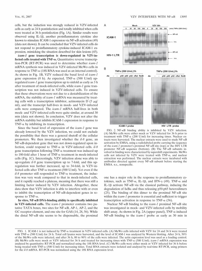
VZV does not inhibit icam-1 and il-8 promoter region acet-ylation. Histone acetylation and histone deacetylase (HDAC)removal are often correlated with increased chromatin acces-sibility at NF-"B-responsive promoters (27). Promoter avail-ability was evaluated by analyzing histone H3 acetylation on K9and the presence of HDAC3 on the promoters of the icam-1and il-8 genes in MeWo cells. As shown by ChIP analysis inFig. 9, there was an increase in the acetylated histone H3 levelat the icam-1 and il-8 promoters after TNF-# treatment inmock-infected cells. VZV alone induced an increased acetyla-tion of these promoters, which was further enhanced by TNF-#(Fig. 9A). The experiment was normalized by performing aChIP assay using an anti-histone H3 antibody. By ChIP assay,the HDAC3 level was shown to be decreased at the icam-1 andil-8 promoters after both TNF-# treatment and VZV infection,which correlates with the increased acetylation (Fig. 9B).
From these data, it can be concluded that VZV inhibited
NF-"B recruitment to the icam-1 promoter region and, to alesser extent, to the il-8 promoter after TNF-# treatment. Theinhibition was observed both in vitro and in vivo, althoughVZV did not inhibit promoter acetylation, which appeared tobe enhanced.
DISCUSSION
In this work, it was demonstrated for the first time that VZVis able to inhibit the expression of ICAM-1 in response toproinflammatory cytokines in infected melanoma cells and fi-broblasts. ICAM-1 has a crucial role in different aspects of theimmune response. Expressed on endothelial cells, ICAM-1allows the migration of leukocytes to inflammation sites, whileits expression on antigen-presenting cells gives costimulatorysignals and plays a crucial role in CD8! T-cell activation (40).ICAM-1 is poorly expressed in skin cells, but its expression
FIG. 8. VZV perturbs NF-"B binding induced by TNF-# on the targeted promoters in vivo. Mock- and VZV-infected MeWo cells were treatedfor 1 h with TNF-# (200 U/ml), and ChIP assays using p65, p50, or p52 antibody were carried out on total cell lysates. Real-time PCR amplificationof a 100-bp fragment from the icam-1 (A) or il-8 (B) promoter encompassing the proximal NF-"B site was carried out. The results were normalizedby performing PCR on the DNA input. *, statistically different (P ( 0.05); **, not statistically different (P ) 0.05).
13100 EL MJIYAD ET AL. J. VIROL.
level can be up-regulated by proinflammatory cytokines such asIFN-$ or TNF-#, as is the case in the periphery of VZVcutaneous lesions, where the viral load is low. On the otherhand, in the center of the lesions, where cells support a highviral load, ICAM-1 is not up-regulated in response to proin-flammatory cytokines (45). These cutaneous sites bear a highlevel of viral replication (7). Diminishing immune surveillanceat these sites might allow the virus to replicate more efficiently.
The most interesting feature beyond the down-regulation ofICAM-1 expression is that this inhibition actually specificallytargets NF-"B, a transcription factor having a central role inthe activation pathways of several genes. Indeed, the NF-"Bactivation pathway was inhibited, while STAT-1 was normallyactivated at 36 h postinfection (Fig. 3).
While I"B# protein has been demonstrated to be stabilizedat short times of infection in VZV-infected fibroblasts (30) andHEK293 cells (data not shown), we observed a decrease inI"B# protein level in both MeWo and MRC5 cells at late timesof infection. I"B# protein has a short turnover time (between30 and 180 min) (26), and since VZV decreased the basal levelof i"b# mRNA, this probably led to the decreased I"B# pro-tein level observed in both melanoma cells and fibroblasts atlate times of infection. Furthermore, VZV inhibited its TNF-#-induced resynthesis. The NF-"B subunits (p65, c-Rel, andp52) present in the nuclei of VZV-infected cells were shown tobe unable to bind the studied promoters. Our results for fibro-blasts show that the nuclear translocation of p50 and, to alesser extent, p65 is strongly decreased by VZV infection. Nev-ertheless, there is still a p65 nuclear translocation induced byinfection, but it never reaches the level observed in mock-infected TNF-#-treated cells. Furthermore, despite the I"B#
decrease at late times of infection, p50 did not translocate intothe nucleus upon TNF-# treatment, and this was observed intwo different cell lines, i.e., MeWo and MRC5. The coimmu-noprecipitation assays performed with MeWo cells tended toshow that VZV destabilizes p50 and thus reduces the occur-rence of p65-p50 heterodimers, leading to an accumulation ofp65 in the nuclei of infected cells independently of p50. It hasbeen shown by ChIP assay that the icam-1 promoter, in oppo-sition to the il-8 promoter, has less affinity for NF-"B dimerscontaining p50-independent p65 (51), explaining partly whythe il-8 promoter seemed less affected by the inhibitory effectof the infection, with the promoters with the most affinity forp50-independent complexes being the least inhibited by VZV.
Another observation that has to be highlighted is that VZV-induced nuclear NF-"B subunits did not bind a probe carryingthe icam-1 promoter NF-"B sequence in vitro, while they stillslightly bound the NF-"B sequence present in the HIV LTRpromoter. This could be correlated with a higher affinity of theVZV-induced nuclear NF-"B subunits for the LTR sequencethan for the icam-1 sequence. Furthermore, supershift assayswere performed to identify the subunits binding to each of theprobes used. Despite a slight presence of p52, it was shown thatthese two probes bound the same subunits in response toTNF-#, i.e., p50 and p65.
VZV also induced a strong nuclear accumulation of p52 inMeWo cells and, to a lesser extent, in MRC5 cells. This phe-nomenon has been described for other viruses, such as Ep-stein-Barr virus and human T-cell leukemia virus (9, 19, 44,46), but never for an alphaherpesvirus. We have also reportedfor the first time an alphaherpesvirus-induced nuclear translo-cation of p100 in both melanoma cells and fibroblasts. Not
FIG. 9. VZV does not shut off the icam-1 and il-8 promoter regions. ChIP assay analysis was carried out on mock- and VZV-infected MeWocells, using an antibody specific for the K9-acetylated form of histone H3, normalized using an anti-H3 ChIP assay (A), or an antibody specific forHDAC3 (B). The immunoprecipitated DNA was subjected to PCR analysis using primers amplifying the promoter region of either icam-1 or il-8.*, statistically different (P ( 0.05); **, not statistically different (P ) 0.05).
VOL. 81, 2007 VZV INTERFERES WITH NF-"B 13101

much is known about the mechanisms leading to the nuclearlocalization of p100, but its processing in the nucleus is IKK#dependent (48). The nuclear accumulation of p100 could verywell play a role in the inhibition of NF-"B binding to cellularpromoters. Indeed, p100 is one of the multiple NF-"B inhibi-tors containing ankyrin motifs, along with the I"Bs and p105(11, 16, 34). Furthermore, an increase in p100-associated p65in VZV-infected MeWo total cell lysates was observed, andp100 also seemed to be more associated with p50 in total celllysates, which could partly explain its cytoplasmic retention.Nevertheless, the coimmunoprecipitation of p100 with p65 innuclear extracts of both mock- and VZV-infected cells was notfeasible (data not shown). The mechanisms underlying theVZV-induced nuclear accumulation of p100, along with itspossible consequences, are currently under investigation.
It was shown that VZV activates NF-"B in macrophages andHEK293 cells in a Toll-like receptor 2-dependent manner,leading to proinflammatory cytokine expression (57). In thiswork, the induction of TNF-# (data not shown), IL-6 (data notshown), and IL-8 mRNA expression in VZV-infected MeWocells was shown. Nevertheless, IL-8 induction by the viruscould not solely be explained by p65 recruitment to its pro-moter. Indeed, ChIP assay analysis showed that VZV did notincrease p65 recruitment to this promoter.
Herpes simplex virus type 1, another alphaherpesvirusclosely related to VZV, activates NF-"B via the IKK complexpathway and needs this activation to replicate properly (21,47). It was recently shown that the NF-"B complex selectivelyactivates the expression of viral genes, such as the ICP0 gene,and inhibits cellular ones. This inhibition is due to modifica-tions of the chromatin structure in infected cells (6). Weshowed that VZV-induced NF-"B inhibition was not due tochromatin modifications at the promoter region, since histoneH3 was still acetylated and HDAC3-containing complexeswere removed. Nevertheless, while the shutoff does not seemto be due to acetylation inhibition or mRNA destabilization,we demonstrated in another study that the VZV immediate-early protein IE63 was able to interact with the transcriptioninitiation complex and inhibit several cellular genes (10, 18).Furthermore, it has been shown for other NF-"B-dependentgenes that to fully respond to NF-"B, the promoter must bederepressed by removal of a repressor complex containing p50homodimers from the promoter region (27). Therefore, wepostulate that the lower level of p50 at the promoters in VZV-infected cells could mean that this complex was removed by theinfection, whereas no transcriptionally active heterodimerswere recruited. Moreover, we also observed an inhibition ofNF-"B recruitment in vitro by mobility shift assay, suggestingthat it is probably a direct modification of the NF-"B dimerthat is responsible for the inhibition. Besides, in the case ofherpes simplex virus type 1, NF-"B was needed for virus rep-lication, especially for ICP0 expression. The occurrence offunctional NF-"B binding sites in the VZV genome has notbeen studied much, but it was recently shown that the inter-genic region between orf63 and orf62 contains a nonfunctionalone (32). It would be worth studying the replication and viralgene expression of VZV in cells unable to activate both NF-"Bpathways. Nuclear NF-"B could either have a direct effect onviral promoter stimulation or an indirect effect by activatingcellular factors that promote or slow down viral replication.
Indeed, NF-"B is also involved in cell cycle progression, par-ticularly by stimulating cyclin D1 expression (for a review, seereference 33). Also, CDK1 has been shown to greatly influencethe localization and repressor activity of the VZV protein IE63(22), and the use of the CDK1 inhibitor roscovitine signifi-cantly slows down VZV replication in MeWo cells (53).
Taken together, these results show that VZV is able toinhibit NF-"B nuclear translocation by destabilizing p50. Be-cause p65-p50 heterodimers are crucial for TNF-# induction ofthe icam-1 gene, this could explain why this gene is no longerinduced despite correct acetylation of the promoter (Fig. 10).Therefore, it seems that VZV modulates several aspects of theimmune response by modulating NF-"B nuclear shuttling andits recruitment to cellular promoters. NF-"B plays a centralrole in innate and adaptive immune responses. Its activationduring early stages of viral infection leads to the expression ofseveral immune response genes, such as those for proinflam-matory cytokines (IFN-%, TNF-#, IL-6, and IL-8), chemokines(RANTES), and adhesion molecules (ICAM-1 and VCAM-1).NF-"B also strongly induces major histocompatibility complexclass I and CD80/86 expression on antigen-presenting cells,thus increasing T-cell activation.
Regulating the actions of this transcription factor will lead toevident advantages for the control of infection progression,especially in melanocytes, which bear high viral replicationloads in the skin.
ACKNOWLEDGMENTS
This work was supported by a grant from the National Fund forScientific Research (FNRS, Brussels, Belgium).
N.E. is a Ph.D. student and teacher’s assistant at the University ofLiege, E.D. is a research associate at the FNRS, C.S.-D. is seniorresearch associate at the University of Liege, and J.P. is a researchdirector at the FNRS.
FIG. 10. VZV inhibits icam-1 transcription upon TNF-# treatmentby targeting p50-p65 heterodimers. VZV inhibits NF-"B recruitmentto the icam-1 promoter region and, to a lesser extent, to the il-8promoter after TNF-# treatment. Inhibition was observed both in vitroand in vivo, although VZV did not inhibit promoter acetylation but, onthe contrary, enhanced it. VZV is able to inhibit NF-"B nuclear trans-location by destabilizing p50. p65-p50 heterodimers are crucial forTNF-# induction of the icam-1 gene, explaining why this gene is nolonger induced.
13102 EL MJIYAD ET AL. J. VIROL.
REFERENCES
1. Abendroth, A., and A. Arvin. 2000. Host response to primary infection, p.142–156. In A. Arvin and A. Gershon (ed.), Varicella-zoster virus: virologyand clinical management. Cambridge University Press, Cambridge, UnitedKingdom.
2. Abendroth, A., I. Lin, B. Slobedman, H. Ploegh, and A. M. Arvin. 2001.Varicella-zoster virus retains major histocompatibility complex class I pro-teins in the Golgi compartment of infected cells. J. Virol. 75:4878–4888.
3. Abendroth, A., G. Morrow, A. L. Cunningham, and B. Slobedman. 2001.Varicella-zoster virus infection of human dendritic cells and transmission toT cells: implications for virus dissemination in the host. J. Virol. 75:6183–6192.
4. Abendroth, A., B. Slobedman, E. Lee, E. Mellins, M. Wallace, and A. M.Arvin. 2000. Modulation of major histocompatibility class II protein expres-sion by varicella-zoster virus. J. Virol. 74:1900–1907.
5. Amici, C., G. Belardo, A. Rossi, and M. G. Santoro. 2001. Activation of Ikappa B kinase by herpes simplex virus type 1. A novel target for anti-herpetic therapy. J. Biol. Chem. 276:28759–28766.
6. Amici, C., A. Rossi, A. Costanzo, S. Ciafre, B. Marinari, M. Balsamo, M.Levrero, and M. G. Santoro. 2006. Herpes simplex virus disrupts NF-kappaBregulation by blocking its recruitment on the IkappaBalpha promoter anddirecting the factor on viral genes. J. Biol. Chem. 281:7110–7117.
7. Arvin, A. M. 1996. Varicella-zoster virus. Clin. Microbiol. Rev. 9:361–381.8. Beinke, S., and S. C. Ley. 2004. Functions of NF-kappaB1 and NF-kappaB2
in immune cell biology. Biochem. J. 382:393–409.9. Beraud, C., S. C. Sun, P. Ganchi, D. W. Ballard, and W. C. Greene. 1994.
Human T-cell leukemia virus type I Tax associates with and is negativelyregulated by the NF-kappa B2 p100 gene product: implications for virallatency. Mol. Cell. Biol. 14:1374–1382.
10. Bontems, S., E. Di Valentin, L. Baudoux, B. Rentier, C. Sadzot-Delvaux, andJ. Piette. 2002. Phosphorylation of varicella-zoster virus IE63 protein bycasein kinases influences its cellular localization and gene regulation activity.J. Biol. Chem. 277:21050–21060.
11. Brown, K., S. Park, T. Kanno, G. Franzoso, and U. Siebenlist. 1993. Mutualregulation of the transcriptional activator NF-kappa B and its inhibitor, Ikappa B-alpha. Proc. Natl. Acad. Sci. USA 90:2532–2536.
12. Chen, J. J., Z. Zhu, A. A. Gershon, and M. D. Gershon. 2004. Mannose6-phosphate receptor dependence of varicella zoster virus infection in vitroand in the epidermis during varicella and zoster. Cell 119:915–926.
13. Croen, K. D., J. M. Ostrove, L. J. Dragovic, and S. E. Straus. 1988. Patternsof gene expression and sites of latency in human nerve ganglia are differentfor varicella-zoster and herpes simplex viruses. Proc. Natl. Acad. Sci. USA85:9773–9777.
14. Degitz, K., L. J. Li, and S. W. Caughman. 1991. Cloning and characterizationof the 5'-transcriptional regulatory region of the human intercellular adhe-sion molecule 1 gene. J. Biol. Chem. 266:14024–14030.
15. Dejardin, E. 2006. The alternative NF-kappaB pathway from biochemistry tobiology: pitfalls and promises for future drug development. Biochem. Phar-macol. 72:1161–1179.
16. Dejardin, E., G. Bonizzi, A. Bellahcene, V. Castronovo, M. P. Merville, andV. Bours. 1995. Highly-expressed p100/p52 (NFKB2) sequesters other NF-kappa B-related proteins in the cytoplasm of human breast cancer cells.Oncogene 11:1835–1841.
17. Diamond, M. S., D. E. Staunton, S. D. Marlin, and T. A. Springer. 1991.Binding of the integrin Mac-1 (CD11b/CD18) to the third immunoglobulin-like domain of ICAM-1 (CD54) and its regulation by glycosylation. Cell65:961–971.
18. Di Valentin, E., S. Bontems, L. Habran, O. Jolois, N. Markine-Goriaynoff, A.Vanderplasschen, C. Sadzot-Delvaux, and J. Piette. 2005. Varicella-zostervirus IE63 protein represses the basal transcription machinery by disorga-nizing the pre-initiation complex. Biol. Chem. 386:255–267.
19. Eliopoulos, A. G., J. H. Caamano, J. Flavell, G. M. Reynolds, P. G. Murray,J. L. Poyet, and L. S. Young. 2003. Epstein-Barr virus-encoded latent infec-tion membrane protein 1 regulates the processing of p100 NF-kappaB2 top52 via an IKKgamma/NEMO-independent signalling pathway. Oncogene22:7557–7569.
20. Gilden, D. H., Y. Rozenman, R. Murray, M. Devlin, and A. Vafai. 1987.Detection of varicella-zoster virus nucleic acid in neurons of normal humanthoracic ganglia. Ann. Neurol. 22:377–380.
21. Gregory, D., D. Hargett, D. Holmes, E. Money, and S. L. Bachenheimer.2004. Efficient replication by herpes simplex virus type 1 involves activationof the I"B kinase-I"B-p65 pathway. J. Virol. 78:13582–13590.
22. Habran, L., S. Bontems, E. Di Valentin, C. Sadzot-Delvaux, and J. Piette.2005. Varicella-zoster virus IE63 protein phosphorylation by roscovitine-sensitive cyclin-dependent kinases modulates its cellular localization andactivity. J. Biol. Chem. 280:29135–29143.
23. Harson, R., and C. Grose. 1995. Egress of varicella-zoster virus from themelanoma cell: a tropism for the melanocyte. J. Virol. 69:4994–5010.
24. Hata, A., L. Zerboni, M. Sommer, A. A. Kaspar, C. Clayberger, A. M.Krensky, and A. M. Arvin. 2001. Granulysin blocks replication of varicella-
zoster virus and triggers apoptosis of infected cells. Viral Immunol. 14:125–133.
25. Hayden, M. S., and S. Ghosh. 2004. Signaling to NF-kappaB. Genes Dev.18:2195–2224.
26. Herrero, J. A., P. Mathew, and C. V. Paya. 1995. LMP-1 activates NF-kappaB by targeting the inhibitory molecule I kappa B alpha. J. Virol. 69:2168–2174.
27. Hoberg, J. E., F. Yeung, and M. W. Mayo. 2004. SMRT derepression by theIkappaB kinase alpha: a prerequisite to NF-kappaB transcription and sur-vival. Mol. Cell 16:245–255.
28. Hou, J., V. Baichwal, and Z. Cao. 1994. Regulatory elements and transcrip-tion factors controlling basal and cytokine-induced expression of the geneencoding intercellular adhesion molecule 1. Proc. Natl. Acad. Sci. USA91:11641–11645.
29. Ito, M., T. Nakano, T. Kamiya, K. Kitamura, T. Ihara, H. Kamiya, and M.Sakurai. 1991. Effects of tumor necrosis factor alpha on replication of vari-cella-zoster virus. Antivir. Res. 15:183–192.
30. Jones, J. O., and A. M. Arvin. 2006. Inhibition of the NF-"B pathway byvaricella-zoster virus in vitro and in human epidermal cells in vivo. J. Virol.80:5113–5124.
31. Jones, J. O., and A. M. Arvin. 2005. Viral and cellular gene transcription infibroblasts infected with small plaque mutants of varicella-zoster virus. An-tivir. Res. 68:56–65.
32. Jones, J. O., M. Sommer, S. Stamatis, and A. M. Arvin. 2006. Mutationalanalysis of the varicella-zoster virus ORF62/63 intergenic region. J. Virol.80:3116–3121.
33. Joyce, D., C. Albanese, J. Steer, M. Fu, B. Bouzahzah, and R. G. Pestell.2001. NF-kappaB and cell-cycle regulation: the cyclin connection. CytokineGrowth Factor Rev. 12:73–90.
34. Kanno, T., G. Franzoso, and U. Siebenlist. 1994. Human T-cell leukemiavirus type I Tax-protein-mediated activation of NF-kappa B from p100(NF-kappa B2)-inhibited cytoplasmic reservoirs. Proc. Natl. Acad. Sci. USA91:12634–12638.
35. Karin, M., and Y. Ben-Neriah. 2000. Phosphorylation meets ubiquitination:the control of NF-"B activity. Annu. Rev. Immunol. 18:621–663.
36. Koropchak, C. M., S. M. Solem, P. S. Diaz, and A. M. Arvin. 1989. Investi-gation of varicella-zoster virus infection of lymphocytes by in situ hybridiza-tion. J. Virol. 63:2392–2395.
37. Kouskouti, A., and I. Talianidis. 2005. Histone modifications defining activegenes persist after transcriptional and mitotic inactivation. EMBO J. 24:347–357.
38. Ku, C. C., L. Zerboni, H. Ito, B. S. Graham, M. Wallace, and A. M. Arvin.2004. Varicella-zoster virus transfer to skin by T cells and modulation of viralreplication by epidermal cell interferon-alpha. J. Exp. Med. 200:917–925.
39. Lebedeva, T., N. Anikeeva, S. A. Kalams, B. D. Walker, I. Gaidarov, J. H.Keen, and Y. Sykulev. 2004. Major histocompatibility complex class I-inter-cellular adhesion molecule-1 association on the surface of target cells: im-plications for antigen presentation to cytotoxic T lymphocytes. Immunology113:460–471.
40. Lebedeva, T., M. L. Dustin, and Y. Sykulev. 2005. ICAM-1 co-stimulatestarget cells to facilitate antigen presentation. Curr. Opin. Immunol. 17:251–258.
41. Moffat, J. F., M. D. Stein, H. Kaneshima, and A. M. Arvin. 1995. Tropism ofvaricella-zoster virus for human CD4! and CD8! T lymphocytes and epi-dermal cells in SCID-hu mice. J. Virol. 69:5236–5242.
42. Montag, C., J. Wagner, I. Gruska, and C. Hagemeier. 2006. Human cyto-megalovirus blocks tumor necrosis factor alpha- and interleukin-1%-medi-ated NF-"B signaling. J. Virol. 80:11686–11698.
43. Morrow, G., B. Slobedman, A. L. Cunningham, and A. Abendroth. 2003.Varicella-zoster virus productively infects mature dendritic cells and alterstheir immune function. J. Virol. 77:4950–4959.
44. Munoz, E., and A. Israel. 1995. Activation of NF-kappa B by the Tax proteinof HTLV-1. Immunobiology 193:128–136.
45. Nikkels, A. F., C. Sadzot-Delvaux, and G. E. Pierard. 2004. Absence ofintercellular adhesion molecule 1 expression in varicella zoster virus-infectedkeratinocytes during herpes zoster: another immune evasion strategy? Am. J.Dermatopathol. 26:27–32.
46. Paine, E., R. I. Scheinman, A. S. Baldwin, Jr., and N. Raab-Traub. 1995.Expression of LMP1 in epithelial cells leads to the activation of a selectsubset of NF-kappa B/Rel family proteins. J. Virol. 69:4572–4576.
47. Patel, A., J. Hanson, T. I. McLean, J. Olgiate, M. Hilton, W. E. Miller, andS. L. Bachenheimer. 1998. Herpes simplex type 1 induction of persistentNF-kappa B nuclear translocation increases the efficiency of virus replica-tion. Virology 247:212–222.
48. Qing, G., and G. Xiao. 2005. Essential role of IkappaB kinase alpha in theconstitutive processing of NF-kappaB2 p100. J. Biol. Chem. 280:9765–9768.
49. Rothlein, R., M. Czajkowski, M. M. O’Neill, S. D. Marlin, E. Mainolfi, andV. J. Merluzzi. 1988. Induction of intercellular adhesion molecule 1 onprimary and continuous cell lines by pro-inflammatory cytokines. Regulationby pharmacologic agents and neutralizing antibodies. J. Immunol. 141:1665–1669.
50. Sadzot-Delvaux, C., S. Debrus, A. Nikkels, J. Piette, and B. Rentier. 1995.
VOL. 81, 2007 VZV INTERFERES WITH NF-"B 13103

Varicella-zoster virus latency in the adult rat is a useful model for humanlatent infection. Neurology 45:S18–S20.
51. Sasaki, C. Y., T. J. Barberi, P. Ghosh, and D. L. Longo. 2005. Phosphory-lation of RelA/p65 on serine 536 defines an I"B#-independent NF-"B path-way. J. Biol. Chem. 280:34538–34547.
52. Staunton, D. E., S. D. Marlin, C. Stratowa, M. L. Dustin, and T. A. Springer.1988. Primary structure of ICAM-1 demonstrates interaction betweenmembers of the immunoglobulin and integrin supergene families. Cell52:925–933.
53. Taylor, S. L., P. R. Kinchington, A. Brooks, and J. F. Moffat. 2004. Rosco-vitine, a cyclin-dependent kinase inhibitor, prevents replication of varicella-zoster virus. J. Virol. 78:2853–2862.
54. Torigo, S., T. Ihara, and H. Kamiya. 2000. IL-12, IFN-gamma, and TNF-alpha released from mononuclear cells inhibit the spread of varicella-zostervirus at an early stage of varicella. Microbiol. Immunol. 44:1027–1031.
55. Volanti, C., J. Y. Matroule, and J. Piette. 2002. Involvement of oxidative
stress in NF-kappaB activation in endothelial cells treated by photodynamictherapy. Photochem. Photobiol. 75:36–45.
56. Voraberger, G., R. Schafer, and C. Stratowa. 1991. Cloning of the humangene for intercellular adhesion molecule 1 and analysis of its 5'-regulatoryregion. Induction by cytokines and phorbol ester. J. Immunol. 147:2777–2786.
57. Wang, J. P., E. A. Kurt-Jones, O. S. Shin, M. D. Manchak, M. J. Levin, andR. W. Finberg. 2005. Varicella-zoster virus activates inflammatory cytokinesin human monocytes and macrophages via Toll-like receptor 2. J. Virol.79:12658–12666.
58. Weller, T. H., and H. M. Witton. 1958. The etiologic agents of varicella andherpes zoster; serologic studies with the viruses as propagated in vitro. J.Exp. Med. 108:869–890.
59. Zerboni, L., M. Sommer, C. F. Ware, and A. M. Arvin. 2000. Varicella-zostervirus infection of a human CD4-positive T-cell line. Virology 270:278–285.
13104 EL MJIYAD ET AL. J. VIROL.