Varicella Zoster Virus Infection in Pregnancy -...
22
8 Varicella Zoster Virus Infection in Pregnancy Irena Narkeviciute 1 and Jolanta Bernatoniene 2 1 Vilnius University Clinic of Children’s Diseases, 2 Bristol Royal Hospital for Children, 1 Lithuania 2 UK 1. Introduction Varicella-zoster virus (VZV) is one of the eight herpesviruses that infect humans. The virus causes two diseases: the primary infection, varicella (chickenpox), and secondary or clinical manifestation of latent infection, herpes zoster (shingles). Though VZV infection rarely occurs during pregnancy, the disease is likely to be associated with significant complications for both mother and fetus: pregnant women are several times more likely to develop fatal varicella than non-pregnant patients, the fetus is at high risk of congenital varicella syndrome (CVS), and the neonate is at high risk of a severe or fatal form of varicella. 1.1 Varicella Ninety percent of varicella cases occur in children between the age of 1 and 14 years. Varicella is a highly contagious disease and affects nearly all susceptible children with an attack rate approaching 90% following a household exposure to the illness. Transmission occurs mainly via direct contact and through respiratory droplets that contain the virus, making the disease highly contagious even before the first onset of rash. Varicella is generally regarded as a mild, self-limited viral illness with occasional complications in healthy children, and it is usually characterized by fever, malaise, and vesicular rash on the trunk, face, scalp, extremities, and oropharynx. The incubation period is usually 14-16 days, but can range from 10 to 21 days. The incubation period may be prolonged to 28 days if varicella zoster immunoglobulin (VZIG) has been administered. Transplacental transmission from mother to fetus occurs during maternal viraemia, and the incubation period for neonatal infection is 11 days (range 9-15 days) from the onset of maternal disease. The period of infectivity begins 1-2 days before the appearance of rash until the lesions crust over, usually 4 to 5 days after the onset of the rash (Centers for Disease Control and Prevention [CDC], 1996; Riley, 1998). Initially, the lesions appear as macules which tend to progress to vesicules which may erupt every 1-2 days. Once ruptured, these vesicules crust over. The lesions may be at various stages in the same area of skin and the rash is characteristically pruritic. The illness normally evolves over about 5 days. Seroepidemiological studies performed between 1973 and 2000 in different industrial countries revealed that up to 26% of women of reproductive age do not possess VZV- specific IgG class antibodies (Sauerbrei & Wutzler, 2005). Currently, most pregnant women living in the US and Europe are not susceptible to the development of varicella, because www.intechopen.com
Transcript of Varicella Zoster Virus Infection in Pregnancy -...
8
Varicella Zoster Virus Infection in Pregnancy
Irena Narkeviciute1 and Jolanta Bernatoniene2 1Vilnius University Clinic of Children’s Diseases,
2Bristol Royal Hospital for Children, 1Lithuania
2UK
1. Introduction
Varicella-zoster virus (VZV) is one of the eight herpesviruses that infect humans. The virus causes two diseases: the primary infection, varicella (chickenpox), and secondary or clinical manifestation of latent infection, herpes zoster (shingles). Though VZV infection rarely occurs during pregnancy, the disease is likely to be associated with significant complications for both mother and fetus: pregnant women are several times more likely to develop fatal varicella than non-pregnant patients, the fetus is at high risk of congenital varicella syndrome (CVS), and the neonate is at high risk of a severe or fatal form of varicella.
1.1 Varicella Ninety percent of varicella cases occur in children between the age of 1 and 14 years. Varicella is a highly contagious disease and affects nearly all susceptible children with an attack rate approaching 90% following a household exposure to the illness. Transmission occurs mainly via direct contact and through respiratory droplets that contain the virus, making the disease highly contagious even before the first onset of rash. Varicella is generally regarded as a mild, self-limited viral illness with occasional complications in healthy children, and it is usually characterized by fever, malaise, and vesicular rash on the trunk, face, scalp, extremities, and oropharynx. The incubation period is usually 14-16 days, but can range from 10 to 21 days. The incubation period may be prolonged to 28 days if varicella zoster immunoglobulin (VZIG) has been administered. Transplacental transmission from mother to fetus occurs during maternal viraemia, and the incubation period for neonatal infection is 11 days (range 9-15 days) from the onset of maternal disease. The period of infectivity begins 1-2 days before the appearance of rash until the lesions crust over, usually 4 to 5 days after the onset of the rash (Centers for Disease Control and Prevention [CDC], 1996; Riley, 1998). Initially, the lesions appear as macules which tend to progress to vesicules which may erupt every 1-2 days. Once ruptured, these vesicules crust over. The lesions may be at various stages in the same area of skin and the rash is characteristically pruritic. The illness normally evolves over about 5 days. Seroepidemiological studies performed between 1973 and 2000 in different industrial countries revealed that up to 26% of women of reproductive age do not possess VZV-specific IgG class antibodies (Sauerbrei & Wutzler, 2005). Currently, most pregnant women living in the US and Europe are not susceptible to the development of varicella, because
www.intechopen.com
Herpesviridae – A Look into This Unique Family of Viruses
174
more than 90% of women of childbearing age are protected by virus-specific IgG class antibodies. The average incidence of varicella in pregnant women has been calculated as 2-3 per 1,000 pregnancies (Enders & Miller, 2000). The epidemiology of varicella differs between countries with temperate climates and those with tropical climates. VZV is acquired at less frequency and at older ages in tropical and subtropical geographical zones. The rate of susceptibility to VZV is 16% (CDC, 1996). The latest seroepidemiological study carried out in 7980 pregnant women from various regions of the word demonstrated a prevalence of VZV antibodies of 93.1% in women born in Western European countries and 80.3% in women born in Central and Eastern Europe, Asia and Africa (Knowles et al., 2004). Although the clinical course of varicella in children is usually mild, it may be severe or fatal
in pregnant women. Varicella is estimated to be 25 times more likely to be severe in adults
than in children. There is thought to be an even greater risk associated with pregnancy,
particularly during the third trimester. Presumably, this is caused by maternal
immunosuppression, which is most intense during this period (Gershon, 2001).
1.2 Herpes zoster Herpes zoster (HZ) is caused by reactivation of latent VZV infection in sensory nerve root
ganglia, which resulted during the attack of primary varicella infection. It usually occurs in
about 15% of people over their lifetime, most commonly in elderly and
immunocompromised populations. People with herpes zoster are capable of transmitting
VZV to varicella susceptible individuals, although the patients with zoster infection are
thought to be less infectious than the patients suffering with the primary varicella. Herpes
zoster or shingles is characterized by a painful, unilateral vesicular rash that is usually
restricted to unilateral dermatomal distribution and can occur on the trunk or face, although
the rash may involve an extremity. The lesions, full of infectious virus, tend to coalesce. The
individual is considered contagious to varicella susceptible until the skin lesions have dried.
Most lesions may be present for days to weeks in immunocompromised patients. Pain is
often a major complaint rather than itching. Zoster seems to increase in severity with
increasing age; young people rarely have a severe form of the illness. Zoster is essentially
not a fatal illness in otherwise healthy individuals. There is little information on the risk of
developing zoster during pregnancy. Fortunately, herpes zoster during pregnancy and
during the perinatal period is not associated with any known birth defect and problems for
newborns, unless the women are immunocompromised (Enders et al., 1994; Miller et al.,
1989; Sauerbrei & Wutzler, 2000).
2. Varicella in pregnancy
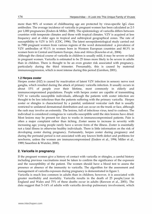
If the pregnant women give a history of contact with varicella or shingles, a careful history
including previous vaccinations must be taken to confirm the significance of the exposure
and the susceptibility of the patient. The women should have a blood test to assess the
presence or absence of the immunity to varicella. The algorithm for the assessment and
management of varicella exposure during pregnancy is demonstrated in figure 1.
Varicella is much less common in adults than in children; however, it is associated with greater morbidity and mortality. Varicella results in the death of 25 people/year in England and Wales and 75% of these deaths occur in adults (Rawson et al., 2001). The data suggest that 5–14% of adults with varicella develop pulmonary involvement, which
www.intechopen.com
Varicella Zoster Virus Infection in Pregnancy
175
may vary in severity from a subclinical form, only detected on X-ray or lung function tests, to a severe and potentially fatal illness (Nathwani et al., 1998). The disease usually develops within 3-5 days of the rash and is associated with cough, dyspnea, fever, and tachypnea. Additionally, cyanosis, pleuritic pain in the chest, and hemoptysis can occur and secondary bacterial infections are frequent. The chest X-ray findings include a diffuse or nodular infiltrative pattern often seen in peribronchial distribution involving both lungs. The X-ray findings may be more severe than the clinical appearance (Daley et al., 2008; Haake et al., 1990).
* varicella zoster immune globulin
Fig. 1. Management of exposure to varicella during pregnancy (Gardella & Brown, 2007, as
cited in Heuchan & Isaacs, 2001)
www.intechopen.com
Herpesviridae – A Look into This Unique Family of Viruses
176
Pregnant women appear no more likely than other adults to develop pneumonia (Royal College of Obstetricians and Gynaecologists [RCOG], 2001) which can occur in up to 10% of pregnant women suffering with varicella (Harger et al., 2002); however, varicella pneumonia may be more severe in pregnant compared to non-pregnant women. The severity of this complication seems to increase in later gestation (Tan & Koren, 2005). Smoking and the occurrence of at least 100 skin lesions are well known risk factors for the development of VZV pneumonia (Harger et al., 2002). The mortality rate in untreated pregnant women is as high as 40% (Haake et al., 1990), therefore varicella pneumonia in pregnancy is considered a medical emergency. However, more recent studies suggest that the mortality has decreased to 10-11% for both non-pregnant and pregnant patients, most likely due to the effective antiviral therapy and better respiratory management (Chandra et al., 1998). Encephalitis causing acute cerebellar ataxia can occur up to 21 days after the onset of the varicella rash (Sissons, 2003). Fortunately, more severe forms of encephalitis are uncommon (0.1–0.2% of cases of varicella), but they have a mortality of around 5–20% (Sissons, 2003). Possible haemorrhagic complications of chickenpox include acute thrombocytopenia, or purpura fulminans associated with arterial thrombosis and haemorrhagic gangrene (Sissons, 2003). Although encephalitis and haemorrhagic complications are rare, their development may severely complicate pregnancy. Multiorgan involvement including hepatitis, myocarditis, and pericarditis is associated with a high mortality. Fetal loss may result from maternal sepsis, fever and hypoxia (Daley et al., 2008). There is no evidence that uncomplicated chickenpox in the mother significantly increases the likelihood of spontaneous abortion during the first 20 weeks of pregnancy (3.0% in one study) or intrauterine death after the 20th week (0.7%) (Enders et al., 1994; Hinshaw & Fayyad, 2000; Office for National UK Statistics, 2005).
3. Management of the pregnant woman who develops varicella or herpes zoster
Most pregnant women who develop varicella illness do not require hospitalization; they can be reassured and sent home for daily or more frequent reviews if clinically indicated (Morgan-Capner et al., 2002). Indications for referral to hospital include the development of respiratory or/and neurological symptoms, a wide-spreading haemorrhagic rash or bleeding, a dense rash with or without mucosal lesions, the appearance of new lesions after 6 days, and a history of significant immunosuppression. Also, the women close to term should be considered for hospitalization because of the risk of developing haemorrhagic complications or varicella of the newborn (RCOG, 2001). Furthermore, if the woman smokes cigarettes, has chronic lung disease, is taking corticosteroids, or is in the latter half of pregnancy, a hospital assessment should be considered, even in the absence of complications (Heuchan & Isaacs, 2001; RCOG, 2001). If the pregnant woman develops the first signs of chickenpox illness, an appropriate treatment should be decided in consultation with a multidisciplinary team: obstetrician or fetal medicine specialist, virologist, and neonatologist. Depending on the severity of the maternal condition, a respiratory physician, intensive care specialist, and infectious disease specialist may also be involved. Timing and mode of delivery must be individualized (Morgan-Capner et al., 2002; RCOG, 2001). As the only safe therapeutic agent, acyclovir is indicated for pregnant women with varicella. The studies suggest that acyclovir administered in a dosage of 800mg five times a day for 7
www.intechopen.com
Varicella Zoster Virus Infection in Pregnancy
177
days reduces the duration of fever and symptomatology of varicella in immunocompetent patients if commenced within 24 hours of developing the rash when compared with placebo (Wallace et al. 1992). This randomized controlled trial did not have sufficient power to comment on the impact of early oral acyclovir on the serious complications of varicella. Data suggest that there is no increase in the risk of fetal malformation with acyclovir in pregnancy, although the theoretical risk of teratogenesis persists in the first trimester (Ratanajamit et al., 2003; Stone et al., 2004). Intravenous acyclovir (10mg/kg three times daily for at least 5 days (7-10)) is indicated for pregnant women with severe illness, complications and/or risk factors (Kempf et al., 2007; Morgan-Capner et al., 2002). VZIG has no therapeutic benefit once varicella has developed (Nathwani et al., 1998). The woman may also need symptomatic relief with paracetamol and/or ibuprofen,
although ibuprofen should be avoided after 30 weeks of gestation because it may cause
premature closure of the ductus arteriosus (Prodigy, 2005). Systemic antihistamines should
also be avoided during the first trimester and breast-feeding (Prodigy, 2005; Drug and
Therapeutics Bulletin [DTB], 2002).
Delivery during the viraemic period may be extremely hazardous. There are significant
maternal risks associated with bleeding, thrombocytopenia, disseminated intravascular
coagulopathy, and hepatitis. Also, there is a high risk of varicella of the newborn with
significant morbidity and mortality (Meyers, 1974; Miller et al., 1989). Therefore, where
relevant and practical, the delivery should be delayed until 5 days after the onset of maternal
illness to allow for passive transfer of antibodies that could protect the baby from the infection
(RCOG, 2001). Supportive treatment and intravenous acyclovir is therefore desirable, allowing
resolution of the rash, immune recovery, and transfer of protective antibodies from the mother
to the fetus. Delivery may be required in women to facilitate assisted ventilation in cases
where varicella pneumonia is complicated by respiratory failure (RCOG, 2001).
There is no evidence available to us to inform decisions about the optimum method of
anaesthesia for women requiring delivery by caesarean section. General anaesthesia may
exacerbate varicella pneumonia. There is a theoretical risk of transmitting the VZV from
skin lesions to the central nervous system via spinal anaesthesia. This results in advice that
epidural anaesthesia may be safer than spinal anaesthesia, because the dura is not
penetrated. A site free of cutaneous lesions should be chosen for needle placement (Brown
et al., 2003).
Women hospitalized with varicella should be nursed in isolation from babies or potentially
susceptible pregnant women or non-immune staff (RCOG, 2001).
Treatment of herpes zoster in imunocompetent pregnant women should be symptomatic;
topical or systemic antiviral therapy is not recommended (Kempf et al., 2007).
4. Consequences of varicella in pregnancy
Varicella acquired during pregnancy may have serious consequences for fetus and neonate.
Maternal varicella associated with viraemia can transmit the virus to the fetus by either
transplacental route or by ascending infection from the lesions in the birth canal. The
possible outcomes of VZV infection during pregnancy depends on the time of the disease
and includes spontaneous abortion, fetal malformation, premature delivery, fetal growth
restriction, or postnatal infection. In infancy, herpes zoster may be the first clinical
manifestation of VZV infection after the primary infection in utero (Table 1).
www.intechopen.com
Herpesviridae – A Look into This Unique Family of Viruses
178
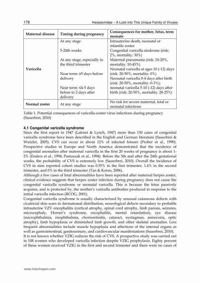
Maternal disease Timing during pregnancy Consequences for mother, fetus, term neonate
Varicella
At any stage 5-24th weeks At any stage, especially in the third trimester Near term: ≥5 days before delivery Near term: ≤4-5 days before to 2 days after delivery
Intrauterine death, neonatal or infantile zoster Congenital varicella sindrome (risk: 2%, mortality: 30%) Maternal pneumonia (risk: 10-20%, mortality: 10-45%) Neonatal varicella at ages 10 (-12) days (risk: 20-50%, mortality: 0%) Neonatal varicella 0-4 days after birth (risk: 20-50%, mortality: 0-3%); neonatal varicella 5-10 (-12) days after birth (risk: 20-50%, mortality: 20-25%)
Normal zoster At any stage No risk for severe maternal, fetal or neonatal infections
Table 1. Potential consequences of varicella-zoster virus infections during pregnancy (Sauerbrei, 2010)
4.1 Congenital varicella syndrome Since the first report in 1947 (Laforet & Lynch, 1947) more than 130 cases of congenital varicella syndrome have been described in the English and German literature (Sauerbrei &
Wutzler, 2005). CVS can occur in about 12% of infected fetuses (Prober et al., 1990).
Prospective studies in Europe and North America demonstrated that the incidence of congenital anomalies after maternal varicella in the first 20 weeks of pregnancy is about 1-
2% (Enders et al., 1994; Pastuszak et al., 1994). Before the 5th and after the 24th gestational weeks, the probability of CVS is extremely low (Sauerbrei, 2010). Overall the incidence of
CVS in nine reported cohort studies was 0.55% in the first trimester, 1.4% in the second trimester, and 0% in the third trimester (Tan & Koren, 2006).
Although a few cases of fetal abnormalities have been reported after maternal herpes zoster, clinical evidence suggests that herpes zoster infection during pregnancy does not cause the
congenital varicella syndrome or neonatal varicella. This is because the fetus passively acquires, and is protected by, the mother’s varicella antibodies produced in response to the
initial varicella infection (RCOG, 2001). Congenital varicella syndrome is usually characterized by unusual cutaneous defects with cicatricial skin scars in dermatomal distribution, neurological defects secondary to probable intrauterine VZV encephalitis (cortical atrophy, spinal cord atrophy, limb paresis, seizures, microcephaly, Horner’s syndrome, encephalitis, mental retardation), eye disease (microphthalmia, enophthalmia, chorioretinitis, cataract, nystagmus, anisocoria, optic atrophy), limb hypoplasia or diminished limb growth, and other skeletal anomalies. Less frequent abnormalities include muscle hypoplasia and affections of the internal organs as well as gastrointestinal, genitourinary, and cardiovascular manifestations (Sauerbrei, 2010). It is not known whether VZIG reduces the risk of CVS. A prospective study was carried out in 108 women who developed varicella infection despite VZIG prophylaxis. Eighty percent of these women received VZIG in the first and second trimester and there were no cases of
www.intechopen.com
Varicella Zoster Virus Infection in Pregnancy
179
CVS or infants with IgM antibodies at birth reported (Enders & Miller, 2000). However, no conclusion can be drawn from this, given the rarity of CVS. Criteria for diagnosis of CVS listed in Table 2.
- Appearance of maternal varicella during pregnancy - Neonate or fetus with
congenital skin lesions in dermatomal distribution and/or
neurological defects,
eye diseases,
limb hypoplasia - Proof of intrauterine VZV infection by
detection of viral DNA using polymerase chain reaction and/or
presence of specific IgM/persistence of IgG beyond 7 months of age
appearance of zoster during early infancy
Table 2. Criteria used for diagnosis of congenital varicella syndrome (Sauerbrei, 2010)
Prenatal diagnosis of CVS is possible using detailed fetal ultrasonography and/or fetal
magnetic resonance imaging and can be useful to look for limb anomalies or other
morphological abnormalities caused by intrauterine varicella. VZV DNA can be detected by
PCR in fetal blood, amniotic fluid, or placental villi. VZV IgM in fetal blood can be detected.
No cases of CVS occurred when the amniotic fluid obtained during amniocentesis was
negative for VZV DNA. If the amniotic fluid is PCR positive for VZV and the ultrasound is
normal at 17–21 weeks, the risk of CVS is still low. If the repeat ultrasound is normal at 23–24
weeks the risk of CVS is remote. However, the risk of CVS increases significantly if the
ultrasound scan reveals features compatible with CVS and the DNA is found in the amniotic
fluid. (Mouly et al., 1997). A negative PCR for VZV in amniotic fluid, and a normal ultrasound
scan results from 23 weeks onwards, suggest a low risk of intrauterine infection (Table 3).
Weeks of gestation PCR High level ultrasound
Risk for CVS with severe malformation
Initial 17-21
(+) Amniocentesis
Normal
Uncertain
Repeat 23-24 22-24
18-22/>23
(+) Amniocentesis (+) Amniocentesis
(±) cord blood (-)Amniocentesis
Normal
Abnormal
Normal
Unlikely
High
Low
Table 3. Prenatal diagnosis using ultrasound and PCR for risk of CVS (Enders & Miller, 2000)
The prognosis of infants born with CVS is poor, with death in infancy resulting from intractable gastroesophageal reflux, severe recurrent aspiration pneumonia, and respiratory failure due to dysfunction of the autonomic nervous system (Smith & Arvin, 2009). Nearly 30% of neonates born with CVS died during the first months of life. A follow-up report in the literature showed that in spite of initially poor prognosis, a good long-term outcome can occur in patients with CVS (Schulze & Dietzsch, 2000).
www.intechopen.com
Herpesviridae – A Look into This Unique Family of Viruses
180
4.2 Neonatal varicella If maternal varicella occurs 1–4 weeks before delivery, up to 50% of babies may be infected.
Approximately 23% of these develop clinical varicella despite high titers of passively acquired maternal antibodies (Miller et al., 1989). When the mother’s rash appeared 7–3
days before delivery, progressively fewer infants had antibodies. Infection may occur by transplacental viraemia, ascending infection from the birth canal, or
through direct contact with infectious lesions during and after delivery. Varicella occurring in the first 12 days of life is described as intrauterine-acquired neonatal varicella infection. If
the infection is diagnosed after the 12th day of the neonatal period, VZV is most likely acquired postnatally. (Enders & Miller, 2000). The severity of intrauterine-acquired neonatal
varicella is closely related to the time of onset of the maternal infection, as transplacentally transmitted antibodies may reduce the severity of symptoms in the neonate (Table 1).
Generalized neonatal varicella can be fatal if mothers develop varicella rash between 4-5 days before and 2 days after delivery since these neonates are not protected from severe
disease by the maternal antibodies. A fatal outcome is more likely if the neonatal disease occurs between 5 and 10 days after delivery. Infants born to mothers with varicella within
this high risk period are usually initially well-appearing. Varicella presents with the classical skin lesions, but can disseminate with pneumonia, hepatitis, encephalitis, and severe
coagulopathy resulting from liver failure and thrombocytopenia (Prober & Arvin, 1987). Before VZV immunoglobulin was available, the risk of death among neonates born to
mothers with the onset of rash up to four days before delivery was 31% (Meyers, 1974). The rate decreased to 7% when the use of VZIG was introduced and neonatal intensive care
improved (Miller et al., 1989). Neonatal varicella within the first 4 days after birth has usually been found to be mild (Sauerbrei & Wutzler, 2001).
When the history of maternal varicella is in the few weeks preceding delivery, the infants
may be asymptomatic or may have cutaneous lesions at birth or developing shortly
thereafter, but are at low risk of dissemination of varicella disease or complications (Smith &
Arvin, 2008).
Premature neonates younger than 28 weeks gestation must be considered to have an
increased risk for severe neonatal varicella during the first 6 weeks after birth (Advisory
Committee on Immunization Practices [ACIP], 1996; Deutsche Gesellschaft für Pädiatrische
Infektologie [DGPI], 2003). They are unlikely to have protecting maternal antibodies due to
the reduced gestation period and the lack the transplacental transfer of maternal IgG to
VZV.
The diagnosis of neonatal varicella is usually based on typical clinical picture. The clinical
findings may be confirmed by serological methods, detection of VZV DNA in skin swabs or
biopsies, or liquor and tissue samples using PCR (Sauerbrei, 2010). When varicella pneumonia
is suspected, broncho-alveolar lavage may be obtained for the VZV DNA detection.
We report three clinical cases of neonatal varicella: two transmitted in utero and one
acquired postnatally.
4.2.1 Case report 1 A 6-day-old baby boy was hospitalized on the first day of his illness with a history of a maculovesicular skin rash noticed on his face and back. On admission, he had normal temperature and he weighed 3960 g. The next day, he was noticed to have haemorrhagic fluid in some of the vesicles, and subsequently he developed pustules with infiltration as
www.intechopen.com
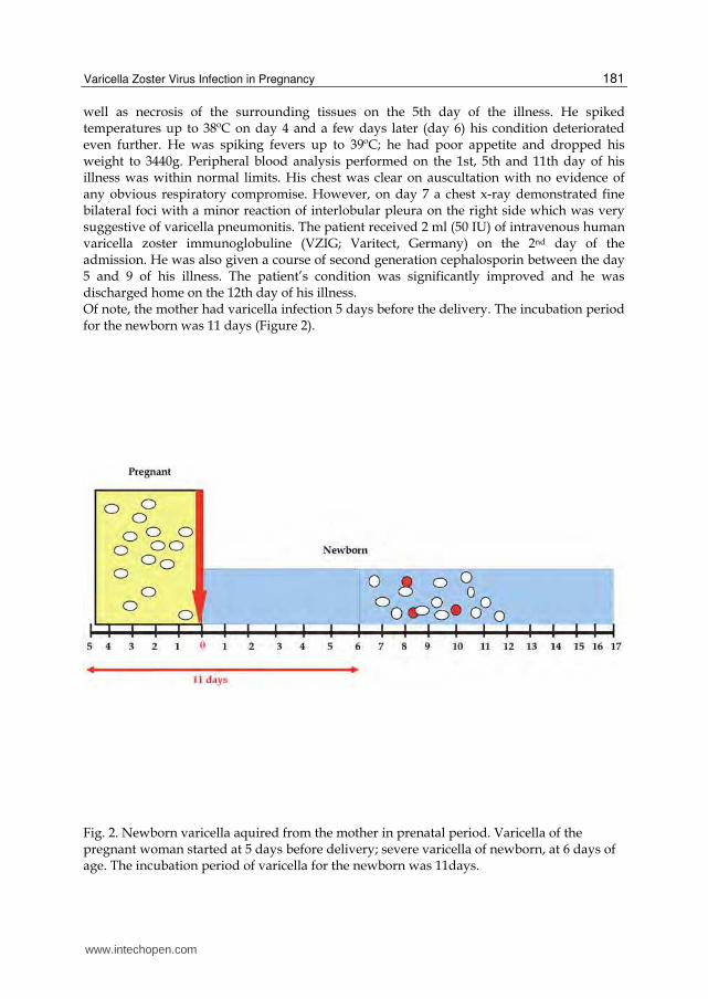
Varicella Zoster Virus Infection in Pregnancy
181
well as necrosis of the surrounding tissues on the 5th day of the illness. He spiked temperatures up to 38ºC on day 4 and a few days later (day 6) his condition deteriorated even further. He was spiking fevers up to 39ºC; he had poor appetite and dropped his weight to 3440g. Peripheral blood analysis performed on the 1st, 5th and 11th day of his illness was within normal limits. His chest was clear on auscultation with no evidence of any obvious respiratory compromise. However, on day 7 a chest x-ray demonstrated fine bilateral foci with a minor reaction of interlobular pleura on the right side which was very suggestive of varicella pneumonitis. The patient received 2 ml (50 IU) of intravenous human varicella zoster immunoglobuline (VZIG; Varitect, Germany) on the 2nd day of the admission. He was also given a course of second generation cephalosporin between the day 5 and 9 of his illness. The patient’s condition was significantly improved and he was discharged home on the 12th day of his illness. Of note, the mother had varicella infection 5 days before the delivery. The incubation period for the newborn was 11 days (Figure 2).
Fig. 2. Newborn varicella aquired from the mother in prenatal period. Varicella of the pregnant woman started at 5 days before delivery; severe varicella of newborn, at 6 days of age. The incubation period of varicella for the newborn was 11days.
www.intechopen.com
Herpesviridae – A Look into This Unique Family of Viruses
182
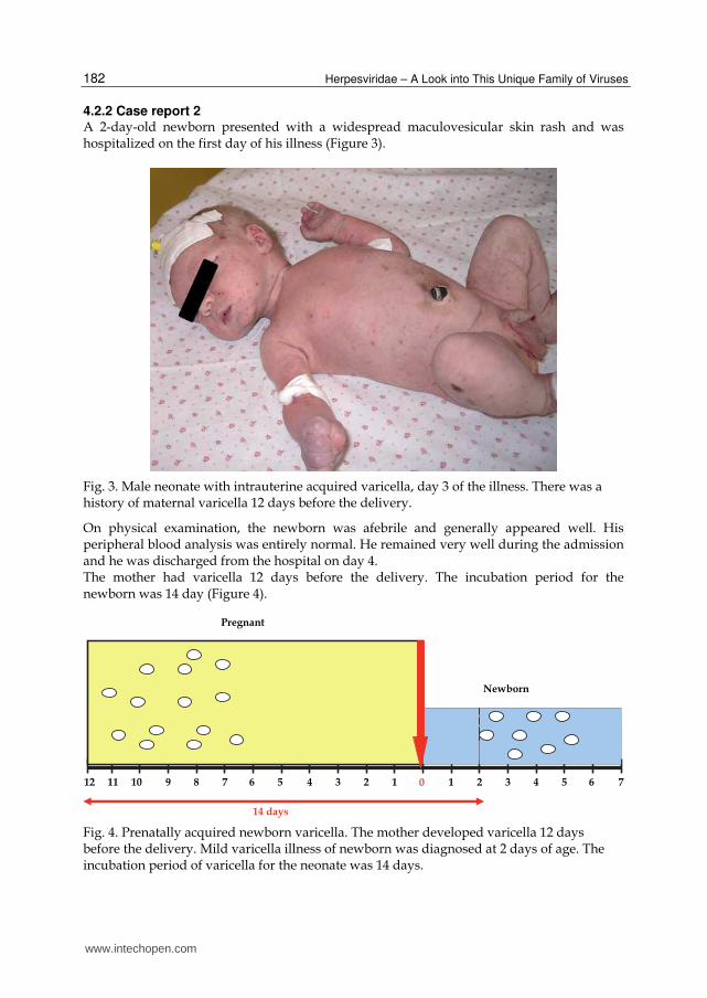
4.2.2 Case report 2 A 2-day-old newborn presented with a widespread maculovesicular skin rash and was hospitalized on the first day of his illness (Figure 3).
Fig. 3. Male neonate with intrauterine acquired varicella, day 3 of the illness. There was a history of maternal varicella 12 days before the delivery.
On physical examination, the newborn was afebrile and generally appeared well. His peripheral blood analysis was entirely normal. He remained very well during the admission and he was discharged from the hospital on day 4. The mother had varicella 12 days before the delivery. The incubation period for the newborn was 14 day (Figure 4).
Fig. 4. Prenatally acquired newborn varicella. The mother developed varicella 12 days before the delivery. Mild varicella illness of newborn was diagnosed at 2 days of age. The incubation period of varicella for the neonate was 14 days.
www.intechopen.com
Varicella Zoster Virus Infection in Pregnancy
183
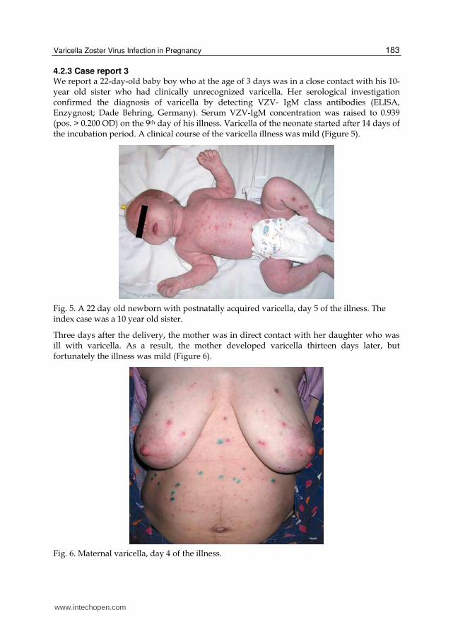
4.2.3 Case report 3 We report a 22-day-old baby boy who at the age of 3 days was in a close contact with his 10-year old sister who had clinically unrecognized varicella. Her serological investigation confirmed the diagnosis of varicella by detecting VZV- IgM class antibodies (ELISA, Enzygnost; Dade Behring, Germany). Serum VZV-IgM concentration was raised to 0.939 (pos. > 0.200 OD) on the 9th day of his illness. Varicella of the neonate started after 14 days of the incubation period. A clinical course of the varicella illness was mild (Figure 5).
Fig. 5. A 22 day old newborn with postnatally acquired varicella, day 5 of the illness. The index case was a 10 year old sister.
Three days after the delivery, the mother was in direct contact with her daughter who was ill with varicella. As a result, the mother developed varicella thirteen days later, but fortunately the illness was mild (Figure 6).
Fig. 6. Maternal varicella, day 4 of the illness.
www.intechopen.com
Herpesviridae – A Look into This Unique Family of Viruses
184
4.3 Herpes zoster in young infants Nearly 20% of infants with intrauterine-acquired VZV primary infection develop herpes zoster in the first or second year of life, usually with uncomplicated course (Sauerbrei & Wutzler, 2003). The observed risk of zoster when maternal infection occurs before 24 weeks was 0.8%, compared to a 1.7% observed risk if maternal varicella was after 25 weeks (Enders et al., 1994). The risk of postnatal herpes zoster infection in infancy was 3.8% in cases when maternal amniotic fluid had been positive for VZV DNA by PCR (Mouly et al., 1997). The symptoms are the same as in adults, although the skin lesions are less prominent and the symptoms of acute neuritis are mild or absent. Unlike adults, children do not suffer with post-herpetic neuralgia (Helgason et al., 2000). Serious complications have been reported in only one case of a four-month-old infant whose mother had had varicella at 17 weeks’ gestation. The infant was born full term without complications; however, aged three months, he developed herpes zoster associated with generalized seizures. The clinical picture and VZV DNA detected in his cerebrospinal fluid by PCR were consistent with central nervous system infection (Sauerbrei et al., 2003). Herpes zoster in infants who acquired VZV in utero has been self-limited. If the zoster is extensive and painful, treatment with acyclovir (initially by intravenous, followed by oral) for seven to 10 days is recommended. We report two patients with typical herpes zoster, whose mothers had varicella during the second-third trimester of their pregnancy.
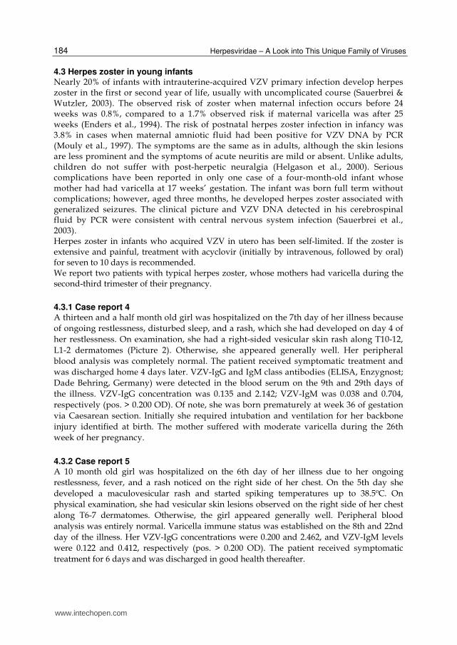
4.3.1 Case report 4 A thirteen and a half month old girl was hospitalized on the 7th day of her illness because
of ongoing restlessness, disturbed sleep, and a rash, which she had developed on day 4 of
her restlessness. On examination, she had a right-sided vesicular skin rash along T10-12,
L1-2 dermatomes (Picture 2). Otherwise, she appeared generally well. Her peripheral
blood analysis was completely normal. The patient received symptomatic treatment and
was discharged home 4 days later. VZV-IgG and IgM class antibodies (ELISA, Enzygnost;
Dade Behring, Germany) were detected in the blood serum on the 9th and 29th days of
the illness. VZV-IgG concentration was 0.135 and 2.142; VZV-IgM was 0.038 and 0.704,
respectively (pos. > 0.200 OD). Of note, she was born prematurely at week 36 of gestation
via Caesarean section. Initially she required intubation and ventilation for her backbone
injury identified at birth. The mother suffered with moderate varicella during the 26th
week of her pregnancy.
4.3.2 Case report 5 A 10 month old girl was hospitalized on the 6th day of her illness due to her ongoing
restlessness, fever, and a rash noticed on the right side of her chest. On the 5th day she
developed a maculovesicular rash and started spiking temperatures up to 38.5ºC. On
physical examination, she had vesicular skin lesions observed on the right side of her chest
along T6-7 dermatomes. Otherwise, the girl appeared generally well. Peripheral blood
analysis was entirely normal. Varicella immune status was established on the 8th and 22nd
day of the illness. Her VZV-IgG concentrations were 0.200 and 2.462, and VZV-IgM levels
were 0.122 and 0.412, respectively (pos. > 0.200 OD). The patient received symptomatic
treatment for 6 days and was discharged in good health thereafter.
www.intechopen.com
Varicella Zoster Virus Infection in Pregnancy
185
Fig. 7. The patient with herpes zoster following maternal varicella acquired during pregnancy. Day 7 of the illness. a) A typical anterior vesicular skin rash along the T10-12 and L1-2 dermatomes on the right side. b) A typical posterior vesicular skin rash along the T10-12 and L1-2 dermatomes on the right side. c) Post inflammatory skin hyperpigmentation following zoster, day 29 of the illness.
Early manifestation of herpes zoster may be explained by the immature cell-mediated immune response in young children and/or the decrease of maternally derived antibodies (Sauerbrei & Wutzler, 2000). The hypothesis was confirmed by our two clinical cases. Neither of them had detectable VZV-IgG antibodies at the beginning of the illness. High titers of serum VZV-IgG and IgM antibodies were discovered only 3–4 weeks later.
5. Management of neonatal varicella
Varicella zoster immunoglobulin, administered to the infants whose mothers developed varicella (but not herpes zoster) at around term, may attenuate infection and significantly reduce the risk of life-threatening neonatal varicella (Enders, 1985; Miller et al., 1989). VZIG should be given at birth to neonates who meet any of the following criteria (Sauerbrei, 2010):
their mothers develop rash between 7 days before and 7 days after birth;
they were born within the last 7 days, the mother is seronegative and they have had significant non-maternal post-natal exposure (e.g. from a sibling);
they have been exposed to varicella and are at risk because of potentially inadequate transfer of maternal antibodies. This includes neonates born before 28 weeks gestation; or weighing less than 1,000g; or who have had repeated blood sampling with replacement by packed red cell infusion; or those requiring intensive or prolonged special care nursing. VZIG can be issued without antibody testing but, where possible, such infants should be tested.
Other infants whose mothers have a positive history of varicella and/or a positive VZV
antibody result will usually have maternal antibody and do not require VZIG. Severe
neonatal varicella can occur despite VZIG administration (Reynolds et al., 1999). Neonatal
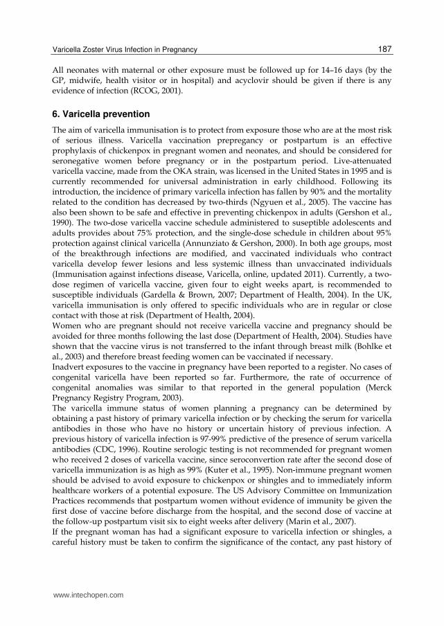
varicella should be treated promptly with acyclovir (Table 4, Figure 8).
Postnatally, the mother and neonate should be isolated from other mothers and babies on
the ward, but not from each other. Breast-feeding of neonates exposed to maternal varicella
should be encouraged. If the mother has varicella lesions close to the nipple, milk should be
expressed until the lesions have crusted (Morgan-Capner et al., 2002). Where other members
of the family have varicella at home and the mother is seronegative, discharge should be
delayed until the baby is at least 7 days old (Morgan-Capner et al., 2002).
www.intechopen.com
Herpesviridae – A Look into This Unique Family of Viruses
186
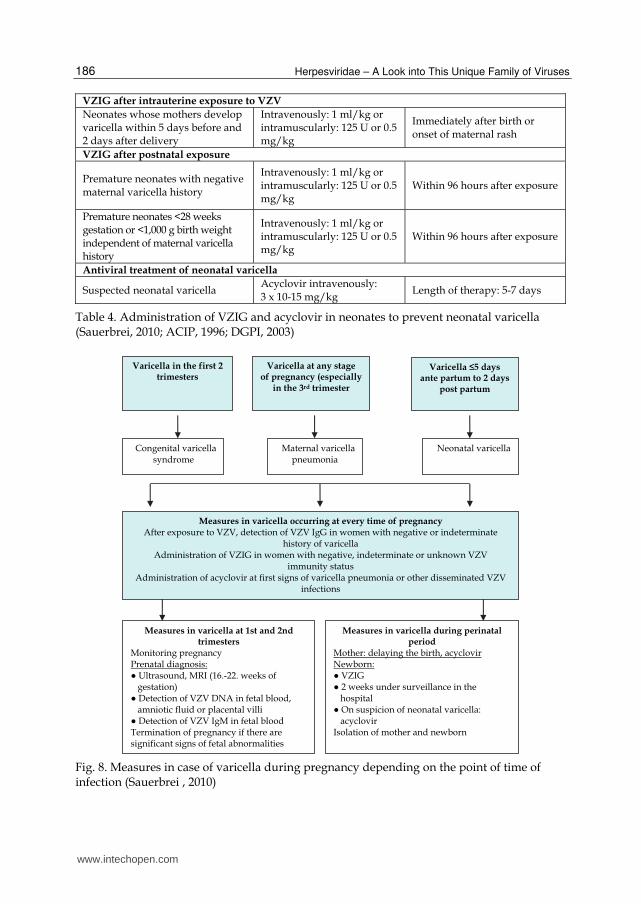
VZIG after intrauterine exposure to VZV
Neonates whose mothers develop varicella within 5 days before and 2 days after delivery
Intravenously: 1 ml/kg or intramuscularly: 125 U or 0.5 mg/kg
Immediately after birth or onset of maternal rash
VZIG after postnatal exposure
Premature neonates with negative maternal varicella history
Intravenously: 1 ml/kg or intramuscularly: 125 U or 0.5 mg/kg
Within 96 hours after exposure
Premature neonates <28 weeks gestation or <1,000 g birth weight independent of maternal varicella history
Intravenously: 1 ml/kg or intramuscularly: 125 U or 0.5 mg/kg
Within 96 hours after exposure
Antiviral treatment of neonatal varicella
Suspected neonatal varicella Acyclovir intravenously: 3 x 10-15 mg/kg
Length of therapy: 5-7 days
Table 4. Administration of VZIG and acyclovir in neonates to prevent neonatal varicella
(Sauerbrei, 2010; ACIP, 1996; DGPI, 2003)
Fig. 8. Measures in case of varicella during pregnancy depending on the point of time of infection (Sauerbrei , 2010)
www.intechopen.com
Varicella Zoster Virus Infection in Pregnancy
187
All neonates with maternal or other exposure must be followed up for 14–16 days (by the GP, midwife, health visitor or in hospital) and acyclovir should be given if there is any evidence of infection (RCOG, 2001).
6. Varicella prevention
The aim of varicella immunisation is to protect from exposure those who are at the most risk of serious illness. Varicella vaccination prepregancy or postpartum is an effective prophylaxis of chickenpox in pregnant women and neonates, and should be considered for seronegative women before pregnancy or in the postpartum period. Live-attenuated varicella vaccine, made from the OKA strain, was licensed in the United States in 1995 and is currently recommended for universal administration in early childhood. Following its introduction, the incidence of primary varicella infection has fallen by 90% and the mortality related to the condition has decreased by two-thirds (Ngyuen et al., 2005). The vaccine has also been shown to be safe and effective in preventing chickenpox in adults (Gershon et al., 1990). The two-dose varicella vaccine schedule administered to suseptible adolescents and adults provides about 75% protection, and the single-dose schedule in children about 95% protection against clinical varicella (Annunziato & Gershon, 2000). In both age groups, most of the breakthrough infections are modified, and vaccinated individuals who contract varicella develop fewer lesions and less systemic illness than unvaccinated individuals (Immunisation against infections disease, Varicella, online, updated 2011). Currently, a two-dose regimen of varicella vaccine, given four to eight weeks apart, is recommended to susceptible individuals (Gardella & Brown, 2007; Department of Health, 2004). In the UK, varicella immunisation is only offered to specific individuals who are in regular or close contact with those at risk (Department of Health, 2004). Women who are pregnant should not receive varicella vaccine and pregnancy should be avoided for three months following the last dose (Department of Health, 2004). Studies have shown that the vaccine virus is not transferred to the infant through breast milk (Bohlke et al., 2003) and therefore breast feeding women can be vaccinated if necessary. Inadvert exposures to the vaccine in pregnancy have been reported to a register. No cases of congenital varicella have been reported so far. Furthermore, the rate of occurrence of congenital anomalies was similar to that reported in the general population (Merck Pregnancy Registry Program, 2003). The varicella immune status of women planning a pregnancy can be determined by obtaining a past history of primary varicella infection or by checking the serum for varicella antibodies in those who have no history or uncertain history of previous infection. A previous history of varicella infection is 97-99% predictive of the presence of serum varicella antibodies (CDC, 1996). Routine serologic testing is not recommended for pregnant women who received 2 doses of varicella vaccine, since seroconvertion rate after the second dose of varicella immunization is as high as 99% (Kuter et al., 1995). Non-immune pregnant women should be advised to avoid exposure to chickenpox or shingles and to immediately inform healthcare workers of a potential exposure. The US Advisory Committee on Immunization Practices recommends that postpartum women without evidence of immunity be given the first dose of vaccine before discharge from the hospital, and the second dose of vaccine at the follow-up postpartum visit six to eight weeks after delivery (Marin et al., 2007). If the pregnant woman has had a significant exposure to varicella infection or shingles, a careful history must be taken to confirm the significance of the contact, any past history of
www.intechopen.com
Herpesviridae – A Look into This Unique Family of Viruses
188
chickenpox or shingles, vaccination, and the susceptibility of the patient. Significant contact is defined as contact in the same room for 15 minutes or more, face-to-face contact and contact in the setting of a large open ward. The UK Advisory Group on Chickenpox considers any close contact during the period of infectiousness to be significant (Nathwani et al., 1998). If the woman’s immunity to chickenpox is unknown and if there is any doubt about previous infection, or if there is no previous history of chickenpox or shingles, serum should be tested for VZV IgG. At least 80–90% of women tested will have VZ IgG and can be reassured (McGregor et al., 1987). If the woman has a past history of chickenpox or shingles or two doses of a varicella containing vaccine, and is not immunosuppressed, protection can be assumed and reassurance given. If the pregnant woman is VZV negative, has indeterminate or unknown serologic status, she
should be given VZIG as soon as possible (Figure 1), preferably within 72-96 h after the
exposure (Smith & Arvin, 2009). VZIG is effective when given up to 10 days after contact
(RCOG, 2001). VZIG recommended intramuscularly at a concentration of 125 U/10 kg of
body weight, up to a maximum of 625 U (ACIP, 1996) or 0.5 ml/kg of body weight (DGPI,
2003). A dosage of 1 ml/kg of body weight can be administered intravenously as alternative
(DGPI, 2003). The primary reason for VZIG prophylaxis in pregnant women is to reduce
severity of maternal disease and reduce the risk of fetal infection for women contracting
varicella in the first 20 weeks of pregnancy. The risk of fatal varicella is estimated to be
about five times higher in pregnant than non-pregnant adults, with fatal cases concentrated
late in the second or early in the third trimester (Enders & Miller, 2000). One study showed a
significant reduction in the risk of congenital VZV infection in women who developed
varicella after VZIG prophylaxis compared with women who developed varicella without
VZIG prophylaxis; however, the study was too small to assess whether the risk of CVS was
reduced (Enders et al., 1994). A case of CVS has been reported in the infant of a woman
exposed at the eleventh week of gestation and who developed clinical varicella despite post-
exposure prophylaxis with VZIG (Pastuszak et al., 1994). About 50% of susceptible pregnant
women given VZIG after a household exposure to chickenpox will develop clinical varicella,
although the disease may be attenuated; the clinical attack rates are similar whether VZIG is
given within 72 hours or four to ten days after contact (Enders & Miller, 2000; Miller et al.,
1993). A further quarter will be infected sub-clinically (Miller et al., 1993). Severe maternal
varicella may still occur despite VZIG prophylaxis. Prompt treatment with acyclovir is
indicated in such cases. Women who have had exposure to chickenpox or shingles
(regardless of whether or not they have received VZIG) should be asked to notify their
doctor or midwife early if a rash develops (Nathwani et al., 1998).
Neither immunoglobulin nor acyclovir treatment have been shown to prevent vertical
transmission or CVS (McKendrick et al., 2007).
If there is another exposure to chickenpox or shingles three weeks or more after the first use
of VZIG, the need for VZIG needs to be reassessed. If more than six weeks have elapsed
since first issue, antibody testing should be performed using a new (recent) sample ((Health
Protection Agency [HPA], 2011).
As VZIG does not always prevent chickenpox, the woman should be managed as being
possibly infectious 8–28 days after VZIG, and should be asked to contact her family doctor if
she develops a rash. Up to 50% may develop a modified form of disease. Maternal
pneumonia associated with chickenpox infection has been reported despite the timely VZIG
administration (HPA, 2001).
www.intechopen.com
Varicella Zoster Virus Infection in Pregnancy
189
7. References
Advisory Committee on Immunization Practices (ACIP). (1996). Prevention of varicella. Morbidity and Mortality Weekly Report, Vol.45, pp. 1-36
Annunziato, P.W. & Gershon, A.A. (2000). Primary vaccination against varicella, In: Varicella-zoster virus, A.M. Arvin & A.A. Gershon (Eds), pp. ??? Cambridge, Cambridge University Press
Bohlke, K.; Galil, K.; Jackson, L.A.; Schmid, D.S.; Starkovich, P.; Loparev, V.N. & Seward, J.F. (2003). Postpartum varicella vaccination: is the vaccine virus excreted in breast milk? Obstetrics & Gynecology. Vol.102 (5 Pt 1), pp. 970-977
Brown, N.W.; Parsons, A.P. & Kam, P.C. (2003). Anaesthetic considerations in a parturient with varicella presenting for Caesarean section. Anaesthesia, Vol.58, No.11, pp. 1092-1095
Centers for Disease Control and Prevention (CDC). (1996). Prevention of varicella: Recommendations of the Advisory Committee on Immunization (ACIP). Morbidity and Mortality Weekly Report, Vol.45 (RR-11), pp. 1-25
Chandra, P.C.; Patel, H., Schiavello, H.J. & Briggs, S.L. (1998). Successful pregnancy outcomeafter complicated varicella pneumonia. Obstetrics & Gynecology, Vol.92, (4 Pt 2), pp. 680-682
Chickenpox, pregnancy and the newborn. (2005). Drug and Therapeutics Bulletin, Vol.43, No.9, pp. 69-72 [Downloaded from dtb.bmj.com on May 16, 2011 – Published by group.bmj.com]
Daley, A.J.; Thorpe, S. & Garland, S.M. (2008). Varicella and the pregnant woman:prevention and management. Australian and New Zealand J of Obstetrics and Gynaecology, Vol.48, No.1 (Feb), pp. 26-33 Department of Health. (2004). Varicella [online]. Available: http://www.dh.gov.uk/assetRoot/04/07/31/40/04073140.pdf [Accessed 18 August 2005]
Deutsche Gesellschaft für Pädiatrische Infektiologie. (2003). Handbuch 2003: Infektionen im Kindesalter, pp. 732-739, Futuramed, München
Enders, G. & Miller, E. (2000). Varicella and herpes zoster in pregnancy and the newborn. In: Varicella-zoster virus. Virology and Clinical Management, A.M. Arvin & A.A. Gershon (Eds.), pp. 317-347, University Press, Cambridge
Enders, G.; Miller, E.; Cradock-Watson, J.; Bolley, I. & Ridehalgh, M. (1994). Consequences of varicella and herpes zoster in pregnancy: prospective study of 1739 cases. Lancet, Vol.343, No.8912 (Jun 18), pp. 1548-1551
Enders, G. (1985). Management of varicella-zoster contact and infection in pregnancy using a standartized varicella-zoster ELISA test. Postgraduate Medical Journal, Vol.61, (Suppl 4), pp. 23-30
Gardella, C. & Brown, Z.A. Managing varicella zoster infection in pregnancy. (2007). Cleveland Clinic Journal of Medicine, Vol.74, No.4 (Apr), pp. 290-296
Gershon, A.A. & Steinberg, S.P. (1990). Live attenuated varicella vaccine: protection in healthy adults compared with leukemic children. National Institute of Allergy and Infectious Diseases Varicella Vaccine Collaborative Study Group. Journal of Infectious Diseases, Vol.161, No.4, pp. 661-666
Gershon, A.A. (2001). Chickenpox, measles, and mumps. In: Infections of the Fetus and Newborn Infant, 5th ed., J. Remington, J. Klein, (Eds.), pp. 683-732, Saunders, Philadelphia
www.intechopen.com
Herpesviridae – A Look into This Unique Family of Viruses
190
Haake, D.A.; Zakowski, P.C.; Haake, D.L. & Bryson, X.J. (1990). Early treatment with acyclovir for varicella pneumonia in otherwise healthy adults: retrospective controlled study and review. Reviews of Infectious Diseases, Vol.12, No.5 (Sep-Oct), pp. 788-798
Harger, J.H.; Ernest, J.M.; Thurnau, G.R.; Moawad, A.; Momirova, V.; Landon, M.B.; Paul, R.; Miodovnik, M.; Dombrowski, M.; Sibai, B. & Van Dorsten P. (2002). Risk factors and outcome of varicella-zoster virus pneumonia in pregnant women. Journal of Infectious Diseases, Vol. 185, No.4, (Jan 17), pp. 422-427 Health Protection Agency, UK (2011). Guidance on viral rash in pregnancy [online]. Available: http://www.hpa.org.uk/web/HPAweb&HPAwebStandard/HPAweb_C/1195733745858
Helgason, S; Petursson, G.; Gudmundsson, S. & Sigurdsson, J.A. (2000). Prevalence of postherpetic neuralgija after a first episode of herpes zoster: prospective study with long term follow up. British Medical Journal, Vol.321, No.7264 (Sep 30), pp. 794-796
Petursson, G. & Sigurdsson, J.A. (2000). Prevalence of postherpetic neuralgia after a first episode of herpes zoster: prospective study with long term follow up. British Medical Journal, Vol.321, No.7264, pp. 794-796
Heuchan, A.M. & Isaacs, D. (2001). The management of varicella-zoster virus exposure and infection in pregnancy and the newborn period. The Medical Journal of Australia, Vol.174, No.6, pp. 288-292
Hinshaw, K. & Fayyad, A. (2000). The management of early pregnancy loss – (25) Oct 2000 [online]. Available: http://www.rcog.org.uk/printindex.asp?PageID=515 [Accessed 18 August 2005]
Department of Health. (2006). Varicella, In: Immunisation against infectious disease – The Green Book, D. Salisbury, M. Ramsy, K. Noakes, (Eds), pp.421-442, TSO, UK (updated 2011). [online]. Available: http://www.dh.gov.uk/prod_consum_dh/groups/dh_digitalassets/@dh/@en/documents/digitalasset/dh_128074.pdf.
Kempf, W.; Meylan, P.; Gerber, S.; Aebi, C.; Agosti, R.; Büchner, S.; Coradi, B.; Garweg, J.;Hirsch, H.H.; Kind, C.; Lauper, U.; Lautenschlager, S.; Russer, P.; Ruef, C.;Wunderli, W. & Nadal, D. (2007). Swiss recommendations for the management of
varicella zoster virus infections. Swiss Medical Weekly, Vol.137, No.17-18 (May 5), pp. 239-251 Knowles, S.J.; Grindy, K.; Cahill, I. & Cafferkey M.T. (2004). Susceptibility to infection rash
illness in pregnant women from diverse geographical regions. Communicable Disease and Public Health, Vol.7, No.4, pp. 344-348
Kuter, B.J.; Ngai, A.; Patterson, C.M.; Staehle, B.O.; Cho, I.; Matthews, H.; Provost, P.J. & White, C.J. (1995). Safety, tolerability, and immunogenicity of two regimens of Oka/Merck varicella vaccine (Varivax) in healthy adolescents and adults. Vaccine, Vol.13, No.11, pp. 967-972
Laforet, E.G. & Lynch, C.L. Jr. (1947). Multiple congenital defects following maternal varicella. New England Journal of Medicine, Vol.236, No.15 (April 10), pp. 534-537
McGregor, J.A.; Mark, S.; Crawford, G.P. & Levin, M.J. (1987). Varicella zoster antibody testing in the care of pregnant women exposed to varicella. American Journal of Obstetrics and Gynecology, Vol.157, No.2, pp.281–284
Marin, M.; Guris, D.; Chaves, S.S.; Schmid, S. & Seward, J.F. (2007). Prevention of varicella: recommendations of the Advisory Committee on Immunization Practices (ACIP).
www.intechopen.com
Varicella Zoster Virus Infection in Pregnancy
191
Morbidity and Mortality Weekly Report. Recommendations and Reports, Vol.56 (RR-4), pp. 1-40
McKendrick, M.W.; Lau, J.; Alston, S. & Bremner, J. (2007). VZV infection in pregnancy: a retrospective review over 5 years in Sheffield and discussion on the potential utilization of varicella vaccine in prevention. Journal of Infection, Vol.55, No.1, pp.64-67
Merck Pregnancy Registry Program (2003). Merck/CDC pregnancy registry for Varivax, the eight annual report
Meyers, J.D. (1974). Congenital varicella in term infants: risk reconsidered. Journal of Infectious Diseases, Vol.129, No.2 (Feb), pp. 215-217
Miller, E.; Cradock-Watson, J.E. & Ridehalgh, M.K. (1989). Outcome in newborn babies given anti-varicella zoster immunoglobulin after perinatal maternal infection with varicella zoster virus. Lancet, Vol.2, No.8659 (Aug 12), pp. 371-373
Miller, E.; Marshall, R. & Vurdien, J.E. (1993). Epidemiology, outcome and control of varicella-zoster infection. Reviews of Medical Microbiology, Vol.4, pp. 222-230
Morgan-Capner, P. & Crowcroft, N.S. (2002). Guidelines on the management of, and exposure to, rash illness in pregnancy (including consideration of relevant antibody screening programmes in pregnancy). Communicable Disease and Public Health, Vol.5, No.1 (Mar), pp. 59-71
Mouly, F.; Mirlesse, V.; Meritel, J.F.; Rozenberg, F.; Poissonier, M.H.; Lebon, P. & Daffos, F. (1997). Prenatal diagnosis of fetal varicella-zoster virus infection with polymerase chain reaction of amniotic fluids in 107 cases. American Journal of Obstetrics and Gynecology, Vol. 177, No.4 (Oct), pp. 894-898
Nathwani, D.; Maclean, A.; Conway, S. & Carrington, D. (1998). Varicella infections in pregnancy and the newborn. Journal of Infection, Vol.36, (Suppl 1), pp. 59-71
Nguyen, H.Q.; Jumaan, A.O. & Seward, J.F. (2005). Decline in mortality due to varicella after implementation of varicella vaccination in the United States. New England Journal of Medicine, Vol.352, No.5 (Feb 3), pp. 450-458
Office for National Statistics (2005). Deaths 2001: Childhood, infant and perinatal mortality: stillbirths, infant deaths and childhood deaths under 15 (rates) [online]. Available: http://www.statistics.gov.uk/STATBASE/xsdataset.asp?%20%20%20More=Y&vlnk=6667&All=Y&B2.x=24&B2.y=11 [Accessed 18 August 2005]
Oral antihistamines for allergic disorders. (2002). Drug and Therapeutics Bulletin, Vol.40, pp. 59-62
Pastuszak, A.L.; Levy, M.; Schick, B.; Zuber, C.; Feldkamp, M.; Gladstone, J.; Bar-Levy, F.; Jackson, E, Donnenfeld, A.; Meschino, W. & Koren, G. (1994). Outcome after maternal varicella infection in the first 20 weeks of pregnancy. New England Journal of Medicine, Vol.330, No.13 (Mar 31), pp. 901-905
Prober, C.G. & Arvin, A.M. (1987). Perinatal viral infections. European Journal of Clinical Microbiology, Vol.6, No.3 (Jun), pp. 245-261
Prober, C.G.; Gershon, A.A.; Grose, C.; McCracken, G.H. Jr.& Nelson, J.D. (1990). Consensus: varicella-zoster infections in pregnancy and the perinatal period. Pediatric Infectious Disease Journal, Vol.9, No.12 (Dec), pp. 865-869 Prodigy. (2004). Chickenpox [online]. Available from: www.prodigy.nhs.uk/guidance.asp?gt= Chickenpox [Accessed 18 August 2005]
Ratanjamit, C.; Vinther Skriver, M.; Jepsen, P.; Chongsuvivatwong, V.; Ober, J. & Sorensen, H.T. (2003). Adverse pregnancy outcome in women exposed to acyclovir during
www.intechopen.com
Herpesviridae – A Look into This Unique Family of Viruses
192
pregnancy: a population based observational study. Scandinavian Journal of Infectious Diseases, Vol.35, No.4, pp. 255-259
Rawson, H.; Crampin, A. & Noah, N. (2001). Deaths from chickenpox in England and Wales.1995-7: analysis of routine mortality data. Britsh Medical Journal, Vol.323, pp. 1091-1093
Reynolds, L.; Struik, S. & Nadel, S. Neonatal varicella: varicella zoster immunoglobulin (VZIG) does not prevent disease. (1999). Archives of Disease in Childhood. Fetal and Neonatal Ed, Vol.81, No.1 (Jul), F69-70
Riley, L. (1998). Varicella-zoster virus infection in pregnancy. Available from: UptoDate, 34 Washington St., Suite 100, Wellesley, MA 02481, on CD-ROM and by subscription at www.uptodate.com
Royal College of Obstetricians and Gynaecologists. (2001). Chickenpox in pregnancy [online]. Available: http://www.rcog.org.uk/resourses/Public/pdf/Chickenpox_No13.pdf
[Accessed 18 August 2005] Sauerbrei, A. & Wutzler, P. (2000). The congenital varicella sindrome. Journal of Perinatology,
Vol.20 (8 Pt 1), pp. 548-554 Sauerbrei, A. & Wutzler, P. (2003). Das fetale Varizellensyndrom. Monatsschrift fur
Kinderheilkunde, Vol.151, pp. 209-213 Sauerbrei, A,; Pawlak, J.; Luger, C. & Wutzler, P. (2003). Intracerebral varicella-zoster virus
reactivation in congenital varicella sindrome. Development Medicine and Child Neurology, Vol.45, No.12 (Dec), pp. 837-840
Sauerbrei, A. & Wutzler, P. (2005). Varicella-zoster virus infections during pregnancy: Epidemiology, clinical symptoms, diagnosis, preventon and therapy. Current Pediatric Reviews, Vol.1, pp. 205-216
Sauerbrei, A. & Wutzler, P. (2001). Neonatal varicella. Journal of Perinatology, Vol.21, No.8 (Dec), pp. 545-549
Sauerbrei, A. (2010). Review of varicella-zoster virus infections in pregnant women and neonates. Health, Vol.2, No.2, pp. 143-152
Schulze, A. & Dietzsch, H.J. (2000). The natural history of varicella embryopathy: A 25-year follow-up. Journal of Pediatrics, Vol.137, No.6 (Dec), pp. 871-874
Sissons, J.G. (2003). Herpesviruses (ecluding Epstein-Barr virus). In: Oxford Textbook of Medicine, D.A. Warrell et al. (Eds.), Fourth Editon, Vol.1, Sections 1-10, Oxford University press, Oxford
Smith, C.K. & Arvin, M.A. (2009). Varicella in the fetus and newborn. Seminars in Fetal & Neonatal Medicine, Vol.14, No.4 (Aug), pp. 209-217
Stone, K.M.; Reiff-Eldridge, R.; White, A.D.; Cordero, J.F.; Brown, Z.; Alexander, E.R. & Andrews, E.B. (2004). Pregnancy outcomes following systemic prenatal acyclovir exposure: conclusions from the International acyclovir pregnancy registry, 1984- 1999. Birth Defects Research.Part A, Clinical and Molecular Teratology, Vol.70, No.4 (Apr), pp. 201-207.
Tan, M.P. & Koren, G. (2006). Chickenpox in pregnancy: revisited. Reproductive Toxicology, Vol.21, No.4 (May), pp. 410-420
Wallace, M.R.; Bowler, W.A.; Murray, N.B.; Brodine, S.K. & Oldfield, E.C. (1992). Treatment of adult varicella with oral acyclovir. A randomized, placebo-controlled trial. Annals of Internal Medicine, Vol.117, No.5 (Sep 1), pp. 358-363
www.intechopen.com
Herpesviridae - A Look Into This Unique Family of VirusesEdited by Dr. George Dimitri Magel
ISBN 978-953-51-0186-4Hard cover, 320 pagesPublisher InTechPublished online 07, March, 2012Published in print edition March, 2012
InTech EuropeUniversity Campus STeP Ri Slavka Krautzeka 83/A 51000 Rijeka, Croatia Phone: +385 (51) 770 447 Fax: +385 (51) 686 166www.intechopen.com
InTech ChinaUnit 405, Office Block, Hotel Equatorial Shanghai No.65, Yan An Road (West), Shanghai, 200040, China
Phone: +86-21-62489820 Fax: +86-21-62489821
In order to fully understand the nature of viruses, it is important to look at them from both, their basic scienceand clinical, standpoints. Our goal with this book was to dissect Herpesviridae into its biological properties andclinical significance in order to provide a logical, as well as practical, approach to understanding and treatingthe various conditions caused by this unique family of viruses. In addition to their up-to-date and extensivetext, each chapter is laced with a variety of diagrams, tables, charts, and images, aimed at helping us achieveour goal. We hope that this book will serve as a reference tool for clinicians of various specialties worldwide.
How to referenceIn order to correctly reference this scholarly work, feel free to copy and paste the following:
Irena Narkeviciute and Jolanta Bernatoniene (2012). Varicella Zoster Virus Infection in Pregnancy,Herpesviridae - A Look Into This Unique Family of Viruses, Dr. George Dimitri Magel (Ed.), ISBN: 978-953-51-0186-4, InTech, Available from: http://www.intechopen.com/books/herpesviridae-a-look-into-this-unique-family-of-viruses/varicella-zoster-virus-infection-in-pregnancy

© 2012 The Author(s). Licensee IntechOpen. This is an open access articledistributed under the terms of the Creative Commons Attribution 3.0License, which permits unrestricted use, distribution, and reproduction inany medium, provided the original work is properly cited.