Vanadate inhibits endoplasmic reticulum stress responses...Thus, IRE1 and PERK activation is...
22
Short communication Vanadate inhibits endoplasmic reticulum stress responses Toru Hosoi 1 *, Atsushi Saito 2 *, Ayaka Kume 1 , Yasunobu Okuma 3 , Yasuyuki Nomura 2,4 , and Koichiro Ozawa 1 1 Department of Pharmacotherapy, Graduate School of Biomedical Sciences, Hiroshima University, Kasumi 1-2-3, Minami-ku, Hiroshima 734-8551, Japan Telephone/FAX: 81-82-257-5332 2 Department of Pharmacology, Graduate School of Pharmaceutical Sciences, Hokkaido University, Sapporo 060-0812, Japan 3 Department of Pharmacology, Faculty of Pharmaceutical Sciences, Chiba Institute of Science, Choshi, Chiba 288-0025, Japan 4 Yokohama College of Pharmacy, Yokohama, Kanagawa 245-0066, Japan Address correspondence to: Koichiro Ozawa, Department of Pharmacotherapy, Graduate School of Biomedical Sciences, Hiroshima University, Kasumi 1-2-3, Minami-ku, Hiroshima 734-8551, Japan e-mail: [email protected] * These authors contributed equally to the present work Acknowledgements: This research was supported by Grants-in-Aid for Scientific Research from the Ministry of Education, Science, Sports and Culture of Japan. 1
Transcript of Vanadate inhibits endoplasmic reticulum stress responses...Thus, IRE1 and PERK activation is...

Short communication
Vanadate inhibits endoplasmic reticulum stress responses
Toru Hosoi1*, Atsushi Saito2*, Ayaka Kume1, Yasunobu Okuma3, Yasuyuki Nomura2,4 ,
and Koichiro Ozawa1
1Department of Pharmacotherapy, Graduate School of Biomedical Sciences, Hiroshima University, Kasumi 1-2-3, Minami-ku, Hiroshima 734-8551, Japan Telephone/FAX: 81-82-257-5332 2Department of Pharmacology, Graduate School of Pharmaceutical Sciences, Hokkaido University, Sapporo 060-0812, Japan 3Department of Pharmacology, Faculty of Pharmaceutical Sciences, Chiba Institute of Science, Choshi, Chiba 288-0025, Japan 4Yokohama College of Pharmacy, Yokohama, Kanagawa 245-0066, Japan Address correspondence to: Koichiro Ozawa, Department of Pharmacotherapy, Graduate School of Biomedical Sciences, Hiroshima University, Kasumi 1-2-3, Minami-ku, Hiroshima 734-8551, Japan e-mail: [email protected] * These authors contributed equally to the present work
Acknowledgements: This research was supported by Grants-in-Aid for Scientific
Research from the Ministry of Education, Science, Sports and Culture of Japan.
1

ABSTRACT
The disruption of endoplasmic reticulum function leads to an accumulation of
unfolded proteins, which results in endoplasmic reticulum stress. In the present study,
we investigated the effect of vanadate on such stress. Endoplasmic reticulum stress
increased glucose-regulated protein 78 (GRP78) and CCAAT/enhancer-binding protein
homologous protein (CHOP) expressions in glial cell cultures. We found that vanadate
inhibited the endoplasmic reticulum stress-induced increase in GRP78 and CHOP
expressions at both mRNA and protein levels. Thus, these results suggest that
vanadate modulates endoplasmic reticulum stress responses and that novel
vanadate-responsive protein(s) might be involved in these processes.
Key Words: endoplasmic reticulum stress; vanadate; glucose-regulated protein 78
(GRP78); CCAAT/enhancer-binding protein homologous protein (CHOP)
2

1. Introduction
Endoplasmic reticulum stress has been implicated in the pathogenesis of diseases
such as neurodegenerative diseases, diabetes, and virus infection (Katayama et al.,
1999; Imai et al., 2001; Kaufman et al., 2002). When unfolded proteins accumulate in
the endoplasmic reticulum, cells activate the endoplasmic reticulum stress response
pathways for cellular protection, such as translational attenuation, the induction of
endoplasmic reticulum chaperones (i.e., induction of glucose-regulated protein 78
(GRP78)), and degradation of unfolded proteins (Mori et al., 2000). On the other hand,
when endoplasmic reticulum functions are severely impaired, the apoptotic pathway is
activated. This apoptosis is mediated by factors such as caspases (Nakagawa et
al.,2000; Hitomi et al., 2004) or C/EBP homologues protein (CHOP) (Zinszner et al.,
1998; Harding et al., 2000). We previously reported that phosphatidylinositide-3-OH
kinase (PI3K) and heat shock protein 90 (HSP90) inhibitors induced CHOP expression
through 4-(2-aminoethyl)-benzenesulfonyl fluoride (AEBSF)-sensitive serine protease
(Hyoda et al., 2006; Hosoi et al., 2007a; Hosoi et al., 2007b). However, the
intracellular mechanisms of these endoplasmic reticulum-regulated integrated stress
responses are not well-understood.
Vanadate is a potent inhibitor of protein-tyrosine phosphatases (Swarup et al.,
1982a; Swarup et al., 1982b), Na+/K+ ATPase (Cantley et al., 1977), and alkaline
phosphatases (Lopez et al., 1976; Seargeant et al., 1979). In contrast, vanadate has
been reported to activate adenylate cyclase (Schwabe et al., 1979). Moreover, it has
been reported to alter a number of cellular functions (Shechter et al., 1980; Seglen et al.,
1981; Carpenter et al., 1981). However, the pharmacological actions of vanadate in
mammalian cells have not been well-clarified. In the present study, we investigated
the possible involvement of vanadate in the endoplasmic reticulum stress response.
3

We found that vanadate inhibited endoplasmic reticulum stress-induced GRP78 and
CHOP expressions at both mRNA and protein levels. These unique observations
suggest that novel vanadate-responsive protein(s) might be involved in endoplasmic
reticulum stress, and the possible physiological significance of this finding was
discussed.
4

2. Materials and Methods
2.1. Materials and reagents
Tunicamycin and thapsigargin were obtained from Wako Pure Chemical Ltd. (Japan).
Sodium orthovanadate was purchased from Nacalai Tesque (Japan).
2.2. Cell culture
The mouse DBT astrocytoma cell line was maintained in Dulbecco’s modified
Eagle’s medium (DMEM) supplemented with 10% (v/v) heat-inactivated fetal calf
serum and antibiotics (100 units/ml penicillin G, 100 μg/ml streptomycin, and 0.25
μg/ml amphotericin B; Nacalai Tesque, JAPAN) at 37°C in humidified 5% CO2, 95%
air.
2.3. Preparation of primary cultured glial cells
Glial cells were prepared from the whole brains of neonatal C57BL/6 mice, as
described previously (Hosoi et al., 2000). The cells were allowed to grow to
confluency (10 days) in DMEM medium with 10% FCS and antibiotics (100 units/ml
penicillin G, 100 μg/ml streptomycin, and 0.25 μg/ml amphotericin B; Nacalai Tesque,
JAPAN). All cultured cells were kept at 37°C in 5% CO2/95% air. Subsequently,
mixed glial cells were shaken at 120 rpm for 18 h, cultured again for 5 to 7 days in 35
mm dishes, and then used in the following experiments. At this point, the astrocyte
cultures were routinely >95% positive for glial fibrillary acidic protein.
2.4. Reverse transcriptase-polymerase chain reaction (RT-PCR) analysis
Total RNA was isolated using TRI Reagent (Sigma-Aldrich, St. Louis, MO, USA).
RT-PCR was performed as described previously (Hosoi et al., 2007a). Specifically,
5
cDNA was synthesized from total RNA by reverse transcription using 100 U of
Superscript Reverse Transcriptase (Invitrogen) and Oligo (dt)12-18 primer (Invitrogen) in
a 20 μl reaction mixture containing Superscript buffer (Invitrogen), 1 mM dNTP mix,
10 mM dithiothreitol (DTT), and 40 U of RNase inhibitor. Total RNA and Oligo
(dt)12-18 primer were incubated at 70°C for 10 min prior to the reverse transcription.
After incubation for 1 h at 42°C, the RT reaction was terminated by denaturing the
Reverse Transcriptase enzyme for 15 min at 70°C. For PCR amplification, 1.2 μl of
cDNA was added to 12 μl of a reaction mix containing 0.2 μM of each primer, 0.2 μM
of dNTP mix, 0.6 U of Taq polymerase, and reaction buffer. PCR was performed in a
DNA Thermal Cycler (GeneAmp® PCR System 9700). The following primers were
used: GRP78 upstream, 5’-ctg ggt aca ttt gat ctg act gg-3’; GRP78 downstream, 5’-gca
tcc tgg tgg ctt tcc agc cat tc-3’; CHOP upstream, 5’-ccc tgc ctt tca cct tgg-3’; CHOP
downstream, 5’-ccg ctc gtt ctc ctg ctc-3’; glyceraldehyde-3-phosphate dehydrogenase
(GAPDH) upstream, 5’-aaa ccc atc acc atc ttc cag -3’; and GAPDH downstream, 5’-agg
ggc cat cca cag tct tct-3’. The PCR products (10 μl) were resolved by electrophoresis
in an 8% polyacrylamide gel in TBE buffer. The gels were stained with ethidium
bromide and then photographed under ultraviolet light. cDNA for GAPDH, GRP78,
and CHOP were amplified and these PCR reactions were run separately.
2.5. Western blotting analysis
Western blotting was performed as described previously (Hosoi et al., 2007c). Cells
were washed with ice-cold PBS and lysed in a buffer containing 10 mM HEPES-NaOH
(pH 7.5), 150 mM NaCl, 1 mM EGTA, 1 mM Na3VO4, 10 mM NaF, 10 μg/ml aprotinin,
10 μg/ml leupeptin, 1 mM phenylmethyl sulfonyl fluoride (PMSF), and 1% NP-40 for
20 min. The lysates were centrifuged at 15,000 rpm for 20 min at 4°C, and the
6

supernatants were collected. The samples were boiled with laemmli’s buffer (62.5 mM
Tris, 2% sodium dodecyl sulfate, 10% glycerol, 5% 2-mercaptoethanol, and 0.01%
bromophenol blue, pH 6.8) for 3 min, fractionated by sodium dodecyl
sulfate-polyacrylamide gel electrophoresis (SDS-PAGE), and transferred at 4°C to
nitrocellulose membranes. The membranes were incubated with anti-KDEL (for
GRP78) (Stressgen; 1:1,000), anti-CHOP (Santa Cruz; 1:500) and anti-phospho Tyr
(Upstate; 1:1,000) antibodies and then with anti-horseradish peroxidase-linked antibody.
Peroxidase was detected by chemiluminescence using an ECL system (Amersham).
The protein bands that cross-reacted with antibodies were detected on X-ray films.
2.6. Statistics
Results are expressed as means ± S.E.M. Statistical analysis was performed with
Student’s t-test or the paired t-test.
7

3. Results
3.1. Vanadate inhibited endoplasmic reticulum stress responses
Endoplasmic reticulum stress triggers the cellular unfolded protein responses, which
result in an increase in the unfolded protein response-regulated genes such as GRP78,
an endoplasmic reticulum-resident chaperone, or CHOP, an endoplasmic reticulum
stress-induced apoptotic transcription factor. Endoplasmic reticulum stress was
induced by tunicamycin, which inhibits protein glycosylation (Wang et al., 1998;
Iwawaki et al., 2001). To investigate whether endoplasmic reticulum stress was
induced in the present experimental conditions, we measured GRP78 and CHOP
expression. As assessed by RT-PCR, endoplasmic reticulum stress (Tm: tunicamycin,
0.1 μg/ml, 6 h) markedly increased GRP78 and CHOP expression in the DBT cell line
(Fig. 2A-D). Moreover, endoplasmic reticulum stress (Tm: tunicamycin, 0.1 μg/ml, 24
h) also increased GRP78 and CHOP expression at protein levels (Fig. 3). Vanadate is
known to be a potent inhibitor of tyrosine phosphatases. As shown in Figure 1, we
observed an increase in phosphorylated tyrosine levels in vanadate-treated cells. Thus,
we next investigated whether vanadate affects these endoplasmic reticulum stress
responses. Vanadate (100 μM, 7 h) alone did not affect the expression levels (mRNA
or protein levels) of GRP78 or CHOP (Figs. 2 and 3). However, vanadate (100 μM)
completely inhibited the endoplasmic reticulum stress-induced increase in GRP78 and
CHOP expression both at mRNA and protein levels in the DBT astrocytoma cell line
(Figs. 2CD and 3AB). Similar results were obtained using mouse primary cultured
glial cells. Vanadate inhibited endoplasmic reticulum stress (tunicamycin- or
thapsigargin-induced endoplasmic reticulum stress)-induced GRP78 or CHOP
expression in the primary cultured glial cells (Fig. 3C). The inhibitory effect of
vanadate on the endoplasmic reticulum stress-induced expression of GRP78 or CHOP
8
was dose dependent (10-1,000 μM, Fig. 2AB). Thus, these results suggest that
vanadate modulates endoplasmic reticulum stress responses, and that novel
vanadate-responsive protein(s) might be involved in these processes.
On the other hand, we observed that the long-term treatment (36 h~) of cells with Tm
alone increased cell death. Interestingly, we also found that vanadate alone increased
cell death and that this was inhibited by treatment with Tm (unpublished observation).
The mechanisms underlying these observations are not understood, and so future studies
are needed.
9

4. Discussion
Increasing evidence has suggested that endoplasmic reticulum stress is involved in
neurodegenerative diseases, diabetes, and viral infection (Katayama et al., 1999; Y. Imai
et al., 2001; Kaufman et al., 2002). However, detailed mechanisms of endoplasmic
reticulum stress responses are poorly understood. In the present study, we found that
vanadate inhibited endoplasmic reticulum stress responses (GRP78 and CHOP
induction), suggesting that vanadate-responsive factor(s) are involved in these
processes.
Endoplasmic reticulum stress-regulated signal transducing sensor kinase is
characterized as an endoplasmic reticulum-resident transmembrane kinase such as
inositol-requiring protein-1 (IRE1) or double-stranded-RNA-dependent protein
kinase-like endoplasmic reticulum kinase (PERK) (Tirasophon et al., 1998; Wang et al.,
1998; Harding et al., 1999). In response to endoplasmic reticulum stress, IRE1
activates cJUN NH2-terminal kinases (JNK) through TNFR-associated factor 2
(TRAF2), an adaptor protein that couples plasma membrane receptors to JNK activation
(Urano et al., 2000). PERK activation phosphorylates eukaryotic initiation factor 2α
(eIF2α) at Ser51, which results in the inhibition of translation initiation (Harding et al.,
2000a). Thus, IRE1 and PERK activation is regulated through its Ser/Thr protein
kinase activity. Indeed, okadaic acid, a protein Ser/Thr phosphatase inhibitor, has been
reported to enhance GRP78 expression in endoplasmic reticulum-stressed cells (Price et
al., 1992; Cao et al., 1995). On the other hand, vanadate is a potent inhibitor of protein
tyrosine, but not protein serine phosphatases (Swarup et al., 1982a; Swarup et al.,
1982b). We observed an increase in phosphorylated tyrosine levels in a
vanadate-treated DBT cell line. As vanadate inhibited endoplasmic reticulum
stress-induced GRP78 or CHOP expression, it is possible that protein tyrosine
10

phosphatases are involved in endoplasmic reticulum stress. Indeed, protein-tyrosine
phosphatase 1B (PTP-1B) has been reported to potentiate IRE1 signaling during
endoplasmic reticulum stress (Gu et al., 2004). Thus, it is possible that
protein-tyrosine phosphatases may positively regulate endoplasmic reticulum stress
responses. On the other hand, in agreement with previous reports (Price et al., 1992;
Cao et al., 1995), we observed that genistein, a tyrosine kinase inhibitor, inhibited the
endoplasmic reticulum stress-induced expression of GRP78 and CHOP (unpublished
observation). These results raise the possibility that the inhibitory effect of vanadate
on endoplasmic reticulum stress would be mediated independently through inhibiting
protein tyrosine phosphatases. Vanadate has other pharmacological actions in addition
to inhibiting protein tyrosine phosphatases. Thus, it is possible that the inhibitory
effect of vanadate on endoplasmic reticulum stress would be mediated through these
other vanadate-responsive factor(s). It is possible that the effect of vanadate on the
endoplasmic reticulum stress signal would be mediated through Na+/K+ ATPase and/or
alkaline phosphatases, as vanadate has been reported to inhibit these factors (Cantley et
al., 1977; Seargeant et al., 1979). Na+/K+ ATPase has been reported to be inhibited up
to 50% by 40 nM vanadate under optimal conditions (28 mM Mg2+) (Cantley et al.,
1977). In addition, vanadate was shown to be a potent competitive inhibitor (Ki less
than 1 μM) of purified alkaline phosphatase from the human liver, intestine, or kidney
(Seargeant et al., 1979). On the other hand, vanadate has been reported to activate
adenylate cyclase in membranes isolated from rat fat cells (Schwabe et al., 1979). The
stimulatory effect of vanadate was concentration (10-1,000 μM)-dependent, which led
to significant activation at 10 μM (Schwabe et al., 1979). Moreover, vanadate
dose-dependently (10-80 μM) activated both NF-κB and c-Jun N-terminal kinase (JNK)
in macrophages (Chen et al., 1999). Furthermore, vanadate has been shown to induce
11

cyclooxygenase (COX) isoenzymes, key enzymes in prostaglandin synthesis (Hirai et
al., 1997). In the present study, we found that vanadate dose-dependently (10-1,000
μM) inhibited endoplasmic reticulum stress responses. The present dose range of the
pharmacological effects of vanadate would correspond with these reported
concentrations. Thus, it is possible that these vanadate-sensitive proteins would be
involved in endoplasmic reticulum stress signal transduction. However, as the
experimental conditions of the previous reports are different from the present ones,
detailed analysis is needed in future experiments. At present, it is unknown whether
and what endoplasmic reticulum-residing proteins are responsible for vanadate-sensitive
endoplasmic reticulum stress signals, but the results provide novel information to aid in
the understanding of endoplasmic reticulum stress-related diseases.
12

Figure legends
Fig.1 Effect of vanadate on phosphorylated tyrosine levels in DBT cell line.
DBT cells were treated with vanadate (Vd: 100 μM) for 6 h, and Western blotting was
performed using phospho-tyrosine antibody. Vanadate markedly increased
phospho-Tyr levels in the DBT cell line.
13

14

Fig. 2 Effect of vanadate on endoplasmic reticulum stress-induced GRP78 and
CHOP mRNA expression.
(A) DBT cells were pre-treated with vanadate (Vd: 10-1000 μM) for 1 h and then with
tunicamycin (Tm: 0.1 μg/ml) for 6 h. RT-PCR analysis was performed using specific
primers for each mRNA. (B) Quantification of the RT-PCR analysis was performed by
densitometric analysis using image-analyzing software. (C) DBT cells were
pre-treated with vanadate (Vd: 100 μM) for 1 h and then with tunicamycin (Tm: 0.1
μg/ml) for 6 h. RT-PCR analysis was performed using specific primers for each
mRNA. Data are from an experiment representative of three independent experiments.
(D) The quantification of RT-PCR analysis was performed by densitometric analysis
using image-analyzing software. Values are presented as means ± S.E.M. (n=3 per
group). GRP78 and CHOP: *P < 0.05 (Student’s t-test: significant difference between
15
Tm and Tm+Vd)
16

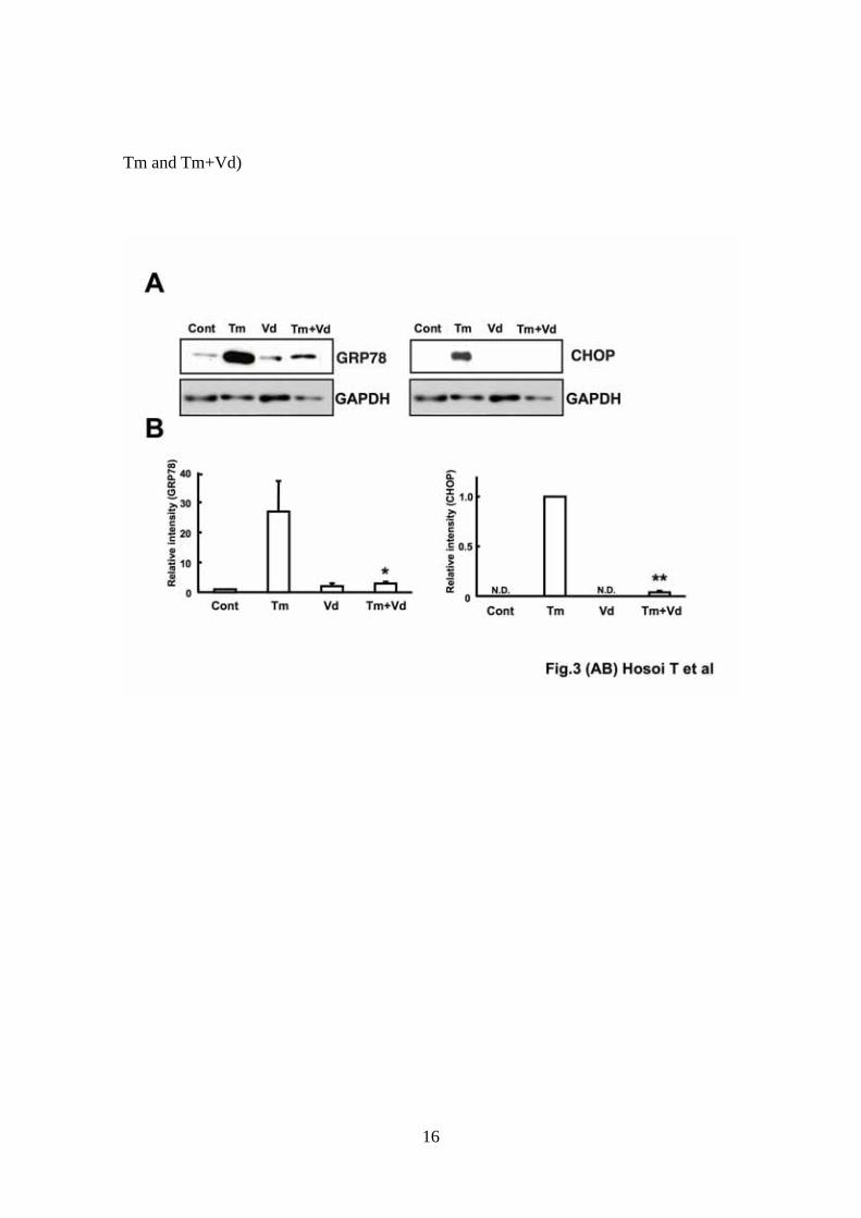
Fig. 3 Effect of vanadate on endoplasmic reticulum stress-induced GRP78 and
CHOP protein expression.
(A) DBT cells were pre-treated with vanadate (Vd: 100 μM) for 1 h and then with
tunicamycin (Tm: 0.1 μg/ml) for 24 h. Samples were separated by SDS gel
electrophoresis followed by immunoblot analysis using anti-KDEL (GRP78) or
anti-CHOP antibodies. CHOP expression was not detected (N.D.) in control (Cont)
and vanadate (Vd)-treated cells. Data are from an experiment representative of three
independent experiments. (B) The quantification of Western blot analysis was
performed by densitometric analysis using image-analyzing software. Values are
presented as means ± S.E.M. (n=3 per group). GRP78: *P < 0.05 (Student’s t-test:
significant difference between Tm and Tm+Vd), CHOP: **P < 0.01 (Paired t-test:
significant difference between Tm and Tm+Vd). (C) Mouse primary cultured glial
17

cells were treated with vanadate (Vd: 500 μM) for 1 h and then stimulated with
tunicamycin ( Tm: 0.1μg/ml) or thapsigargin (Tg: 0.1 μM) for 24 h. Samples were
separated by SDS gel electrophoresis followed by immunoblot analysis using
anti-KDEL (GRP78) or anti-CHOP antibodies.
18

References
Cantley, L.C. Jr, Josephson, L., Warner, R., Yanagisawa, M., Lechene, C., Guidotti, G.,
1977. Vanadate is a potent (Na,K)-ATPase inhibitor found in ATP derived from
muscle. J. Biol. Chem. 252, 7421-7423.
Cao, X., Zhou, Y., Lee, A.S., 1995. Requirement of tyrosine- and serine/threonine
kinases in the transcriptional activation of the mammalian grp78/BiP promoter by
thapsigargin. J Biol Chem. 270, 494-502.
Carpenter, G., Vanadate, epidermal growth factor and the stimulation of DNA synthesis.
1981. Biochem. Biophys. Res. Commun. 102, 1115-1121.
Chen, F., Demers, L.M., Vallyathan, V., Ding, M., Lu, Y., Castranova, V., Shi, X., 1999.
Vanadate induction of NF-κB involves IκB kinase β and SAPK/ERK kinase 1 in
macrophages. J Biol Chem. 274, 20307-20312.
Gu, F., Nguyên, D.T., Stuible, M., Dubé, N., Tremblay, M.L., Chevet, E., 2004.
Protein-tyrosine phosphatase 1B potentiates IRE1 signaling during endoplasmic
reticulum stress. J. Biol. Chem. 279, 49689-49693.
Harding, H.P., Zhang, Y., Ron, D., 1999. Protein translation and folding are coupled by
an endoplasmic-reticulum-resident kinase. Nature 397, 271-274.
Harding, H.P., Zhang, Y., Bertolotti, A., Zeng, H., Ron, D., 2000a. Perk is essential for
translational regulation and cell survival during the unfolded protein response. Mol.
Cell 5, 897-904.
Harding, H., Novoa, I., Zhang, Y., Zeng, H., Wek, R.C., Schapira, M., and Ron, D.
2000b. Regulated translation initiation controls stress-induced gene expression in
mammalian cells. Mol. Cell 6, 1099-1108.
Hirai, K., Takayama, H., Tomo, K., Okuma, M., 1997.
Protein-tyrosine-kinase-dependent expression of cyclo-oxygenase-1 and -2 mRNAs
19
in human endothelial cells. Biochem J. 322, 373-377.
Hitomi, J., Katayama, T., Eguchi, Y., Kudo, T., Taniguchi, M., Koyama, Y., Manabe, T.,
Yamagishi, S., Bando, Y., Imaizumi, K., Tsujimoto, Y., Tohyama, M., 2004.
Involvement of caspase-4 in endoplasmic reticulum stress-induced apoptosis and
Abeta-induced cell death. J. Cell Biol. 165, 347-356.
Hosoi, T., Okuma, Y., Nomura, Y., 2000. Expression of leptin receptors and induction of
IL-1β transcript in glial cells. Biochem Biophys Res Commun. 273, 312-315.
Hosoi, T., Hyoda, K., Okuma, Y., Nomura, Y., Ozawa, K., 2007a. Inhibitory effect of
4-(2-aminoethyl)-benzenesulfonyl fluoride, a serine protease inhibitor, on PI3K
inhibitor-induced CHOP expression. Eur. J. Pharmacol. 554, 8-11.
Hosoi, T., Hyoda, K., Okuma, Y., Nomura, Y., Ozawa, K., 2007b. Geldanamycin
induces CHOP expression through a 4-(2-aminoethyl)-benzenesulfonyl
fluoride-responsive serine protease. Cell Res. 17, 184-186.
Hosoi, T., Hyoda, K., Okuma, Y., Nomura, Y., Ozawa, K., 2007c. Akt up- and
down-regulation in response to endoplasmic reticulum stress. Brain Res. 1152,
27-31.
Hyoda, K., Hosoi, T., Horie, N., Okuma, Y., Ozawa, K., Nomura, Y., 2006. PI3K-Akt
inactivation induced CHOP expression in endoplasmic reticulum-stressed cells.
Biochem Biophys Res Commun. 340, 286-290.
Imai, Y., Soda, M., Inoue, H., Hattori, N., Mizuno, Y., Takahashi, R., 2001. An unfolded
putative transmembrane polypeptide, which can lead to endoplasmic reticulum
stress, is a substrate of Parkin. Cell 105, 891-902.
Iwawaki, T., Hosoda, A., Okuda, T., Kamigori, Y., Nomura-Furuwatari, C., Kimata Y.,
Tsuru, A., Kohno, K., 2001. Translational control by the ER transmembrane
kinase/ribonuclease IRE1 under ER stress. Nat. Cell Biol. 3, 158-164.
20

Katayama, T., Imaizumi, K., Sato, N., Miyoshi, K., Kudo, T., Hitomi, J., Morihara, T.,
Yoneda, T., Gomi, F., Mori, Y., Nakano, Y., Takeda, J., Tsuda, T., Itoyama, Y.,
Murayama, O., Takashima, A., St. George-Hyslop, P., Takeda, M., Tohyama, M.,
1999. Presenilin-1 mutations downregulate the signalling pathway of the
unfolded-protein response. Nat. Cell Biol. 1, 479-485.
Kaufman, R.J., 2002. Orchestrating the unfolded protein response in health and disease.
J. Clin. Invest. 110, 1389-98.
Nakagawa, T., Zhu, H., Morishima, N., Li, E., Xu, J., Yankner, B.A., Yuan, J., 2000.
Caspase-12 mediates endoplasmic-reticulum-specific apoptosis and cytotoxicity by
amyloid-β. Nature. 403, 98-103.
Lopez, V., Stevens, T., Lindquist, R.N., 1976. Vanadium ion inhibition of alkaline
phosphatase-catalyzed phosphate ester hydrolysis. Arch. Biochem. Biophys. 175,
31-38.
Mori, K., 2000. Tripartite management of unfolded proteins in the endoplasmic
reticulum. Cell 101, 451-454.
Price, BD., Mannheim-Rodman, L.A., Calderwood, S.K., 1992. Brefeldin A,
thapsigargin, and AIF4- stimulate the accumulation of GRP78 mRNA in a
cycloheximide dependent manner, whilst induction by hypoxia is independent of
protein synthesis. J. Cell Physiol. 152, 545-552.
Schwabe, U., Puchstein, C., Hannemann, H., Söchtig, E., 1979. Activation of adenylate
cyclase by vanadate. Nature 277, 143-145.
Seargeant, L.E., Stinson, R.A., 1979. Inhibition of human alkaline phosphatases by
vanadate. Biochem J. 181, 247-250.
Seglen, P.O., Gordon, P.B. 1981. Vanadate inhibits protein degradation in isolated rat
hepatocytes. J. Biol. Chem. 256, 7699-76701.
21

Shechter, Y., Karlish, S.J., 1980. Insulin-like stimulation of glucose oxidation in rat
adipocytes by vanadyl (IV) ions. Nature. 284, 556-558.
Swarup, G., Cohen, S., Garbers, D.L., 1982a Inhibition of membrane
phosphotyrosyl-protein phosphatase activity by vanadate. Biochem. Biophys. Res.
Commun. 107, 1104-1109.
Swarup, G., Speeg, K.V. Jr., Cohen, S., Garbers, D.L., 1982b. Phosphotyrosyl-protein
phosphatase of TCRC-2 cells. J. Biol. Chem. 257, 7298-7301.
Tirasophon, W., Welihinda, A.A., Kaufman, R.J., 1998. A stress response pathway from
the endoplasmic reticulum to the nucleus requires a novel bifunctional protein
kinase/endoribonuclease (Ire1p) in mammalian cells. Genes Dev. 12, 1812-1824.
Urano, F., Wang, X., Bertolotti, A., Zhang, Y., Chung, P., Harding, H.P., Ron, D., 2000.
Coupling of stress in the ER to activation of JNK protein kinases by transmembrane
protein kinase IRE1. Science. 287, 664-666.
Wang, X.Z., Harding, H.P., Zhang, Y., Jolicoeur, E.M., Kuroda, M., Ron, D., 1998.
Cloning of mammalian Ire1 reveals diversity in the ER stress responses. EMBO J.
17, 5708-5717.
Zinszner, H., Kuroda, M., Wang, X., Batchvarova, N., Lightfoot, R.T., Remotti, H.,
Stevens, J.L., Ron, D., 1998. CHOP is implicated in programmed cell death in
response to impaired function of the endoplasmic reticulum. Genes Dev. 12,
982-995.
22