Validation of a new grading system for endoscopic...
4
ORIGINAL RESEARCH Validation of a new grading system for endoscopic examination of adenoid hypertrophy Sanjay R. Parikh, MD, Mark Coronel, MD, James J. Lee, MD, and Seth M. Brown, MD, New York, New York OBJECTIVE: To propose and validate a new subjective grading system of adenoid size with flexible fiberoptic evaluation. STUDY DESIGN AND SETTING: Digital video clips of 24 flexible fiberoptic nasopharyngeal exams were presented to 24 exam- iners (otolaryngology resident and consultant physicians) at a tertiary care institution. Examiners were asked to use the proposed grading system to rate adenoid hypertrophy. Kappa statistical analysis was used to evaluate the degree of intergrader agreement or disagreement. RESULTS: Statistical analysis of intergrader agreement demon- strated an overall Kappa score of 0.71 suggesting a “substantial” strength of agreement. The Kappa strength of agreement was found to be 0.83 (almost perfect) among consultant physicians and 0.62 (substantial) among resident physicians. CONCLUSIONS: The proposed adenoid staging system is a reliable and consistent method of staging adenoid tissue size. SIGNIFICANCE: This new validated grading system may be a useful standard for reporting adenoid size in future clinical out- come studies. © 2006 American Academy of Otolaryngology–Head and Neck Surgery Foundation. All rights reserved. A denoid hypertrophy is a common cause of nasal ob- struction in the pediatric population. This can lead to such issues as obstructive sleep apnea, recurrent otitis me- dia, and Eustachian tube dysfunction. Evaluation of the adenoids in the child can be a difficult endeavor that his- torically has been performed via lateral skull radiographs. With the introduction of flexible fiberoptic endoscopes, a greater number of children are being evaluated without radiation. The fiberoptic endoscope can be passed along the floor of the nose to provide an excellent view of the ade- noids and its relation to adjacent structures. There have been very few studies published on the eval- uation of adenoid hypertrophy. One study 1 used lateral skull radiographs to categorize adenoid size as normal or large. Some research groups 2,3 have tried to use acoustic rhinometry to evaluate adenoid size. One other study 4 attempted to classify adenoid hypertrophy at the time of adenoidectomy through the use of concurrent intraoperative flexible fiber- optic examination. Only 1 study has attempted to grade adenoid hypertrophy endoscopically in the outpatient set- ting. Wang et al 5 examined 180 patients and graded ade- noids from 1 to 3 based on the distance between the vomer and the adenoid tissue during nasal endoscopic examina- tion. In that study, a significant relationship between the size of the adenoids and upper respiratory symptoms such as nasal obstruction and snoring were demonstrated, but vali- dation of their grading system was not performed. The objective of this study is to construct and validate a subjective grading system for endoscopic adenoid examina- tion that could be used as a possible standard for reporting adenoid size in future clinical outcome studies. MATERIALS AND METHODS A grading system for adenoid hypertrophy was created based on the anatomical relationships between the adenoid tissue and the following structures: vomer, soft palate, and torus tubaris (Table 1). The grading is based on the rela- tionship of the adenoids to adjacent structures when the patient is at rest (ie, when the soft palate is not elevated). A prospective protocol for validation of this grading system was proposed and then Institutional Review Board approval for the study was obtained. From the Department of Otorhinolaryngology–Head and Neck Surgery, Albert Einstein College of Medicine. Reprint requests: Sanjay R. Parikh, MD, Director of Pediatric Otolar- yngology, Children’s Hospital at Montefiore, 3400 Bainbridge Avenue, 3rd Floor, Bronx, NY 10467. E-mail address: sparikh@montefiore.org. Otolaryngology–Head and Neck Surgery (2006) 135, 684-687 0194-5998/$32.00 © 2006 American Academy of Otolaryngology–Head and Neck Surgery Foundation. All rights reserved. doi:10.1016/j.otohns.2006.05.003
-
Upload
nguyendang -
Category
Documents
-
view
218 -
download
3
Transcript of Validation of a new grading system for endoscopic...

Otolaryngology–Head and Neck Surgery (2006) 135, 684-687
ORIGINAL RESEARCH
Validation of a new grading system for endoscopic
examination of adenoid hypertrophy
Sanjay R. Parikh, MD, Mark Coronel, MD, James J. Lee, MD,
and Seth M. Brown, MD, New York, New YorkOBJECTIVE: To propose and validate a new subjective gradingsystem of adenoid size with flexible fiberoptic evaluation.STUDY DESIGN AND SETTING: Digital video clips of 24flexible fiberoptic nasopharyngeal exams were presented to 24 exam-iners (otolaryngology resident and consultant physicians) at a tertiarycare institution. Examiners were asked to use the proposed gradingsystem to rate adenoid hypertrophy. Kappa statistical analysis wasused to evaluate the degree of intergrader agreement or disagreement.RESULTS: Statistical analysis of intergrader agreement demon-strated an overall Kappa score of 0.71 suggesting a “substantial”strength of agreement. The Kappa strength of agreement wasfound to be 0.83 (almost perfect) among consultant physicians and0.62 (substantial) among resident physicians.CONCLUSIONS: The proposed adenoid staging system is areliable and consistent method of staging adenoid tissue size.SIGNIFICANCE: This new validated grading system may be auseful standard for reporting adenoid size in future clinical out-come studies.© 2006 American Academy of Otolaryngology–Head and NeckSurgery Foundation. All rights reserved.
Adenoid hypertrophy is a common cause of nasal ob-struction in the pediatric population. This can lead to
such issues as obstructive sleep apnea, recurrent otitis me-dia, and Eustachian tube dysfunction. Evaluation of theadenoids in the child can be a difficult endeavor that his-torically has been performed via lateral skull radiographs.With the introduction of flexible fiberoptic endoscopes, agreater number of children are being evaluated withoutradiation. The fiberoptic endoscope can be passed along thefloor of the nose to provide an excellent view of the ade-noids and its relation to adjacent structures.
There have been very few studies published on the eval-
From the Department of Otorhinolaryngology–Head and Neck Surgery,Albert Einstein College of Medicine.
Reprint requests: Sanjay R. Parikh, MD, Director of Pediatric Otolar-
0194-5998/$32.00 © 2006 American Academy of Otolaryngology–Head and Necdoi:10.1016/j.otohns.2006.05.003
uation of adenoid hypertrophy. One study1 used lateral skullradiographs to categorize adenoid size as normal or large.Some research groups2,3 have tried to use acoustic rhinometryto evaluate adenoid size. One other study4 attempted toclassify adenoid hypertrophy at the time of adenoidectomythrough the use of concurrent intraoperative flexible fiber-optic examination. Only 1 study has attempted to gradeadenoid hypertrophy endoscopically in the outpatient set-ting. Wang et al5 examined 180 patients and graded ade-noids from 1 to 3 based on the distance between the vomerand the adenoid tissue during nasal endoscopic examina-tion. In that study, a significant relationship between the sizeof the adenoids and upper respiratory symptoms such asnasal obstruction and snoring were demonstrated, but vali-dation of their grading system was not performed.
The objective of this study is to construct and validate asubjective grading system for endoscopic adenoid examina-tion that could be used as a possible standard for reportingadenoid size in future clinical outcome studies.
MATERIALS AND METHODS
A grading system for adenoid hypertrophy was createdbased on the anatomical relationships between the adenoidtissue and the following structures: vomer, soft palate, andtorus tubaris (Table 1). The grading is based on the rela-tionship of the adenoids to adjacent structures when thepatient is at rest (ie, when the soft palate is not elevated). Aprospective protocol for validation of this grading systemwas proposed and then Institutional Review Board approvalfor the study was obtained.
yngology, Children’s Hospital at Montefiore, 3400 Bainbridge Avenue, 3rdFloor, Bronx, NY 10467.
E-mail address: [email protected].
k Surgery Foundation. All rights reserved.
685Parikh et al Validation of a new grading system for endoscopic . . .
Patients were prospectively recruited and consented from2 outpatient hospital-based otolaryngology clinics and in-cluded any patients who required nasal endoscopy as part oftheir office visit. The majority of patients were recruited ina pediatric setting. No patient was recruited for the solepurpose of undergoing endoscopy.
After application of topical 0.5% tetracaine, a fiberopticexamination was carried out with either a 2.4 mm (PentaxFNL-7RP3) or a 3.5 mm (Vision Sciences ENT-2000) flex-ible endoscope. Video imaging was than captured with acamera (Storz telecam 2021230) and transferred to a laptopcomputer using a video bus (Belkin USB Videobus II).Digital video clips were then edited with software (MGIVideowave III) for presentation to examiners. Video clipswere excluded if they were deemed inadequate for visual-ization of adenoid tissue or adjacent anatomic structures.
Otolaryngology consultant physicians and resident phy-sicians were given a handout with the proposed adenoidgrading system, both in illustrated and written form. Eachexaminer was then asked to grade the adenoid tissue of 24fiberoptic nasal endoscopies by viewing approximately 15to 30 second digital video clips of each endoscopic exam-ination. Examiners were allowed to review each clip asoften as they required to provide a grade of adenoid hyper-trophy.
Statistical AnalysisKappa statistical analysis was used to evaluate the degree ofintergrader agreement or disagreement.6,7 The Kappa valuemeasures the amount of agreement that occurs beyond whatwould occur by chance alone. The range of the Kappa valueis between 1 (complete agreement) and 0 (complete dis-agreement) and is calculated by the following formula:
Kappa coefficient
�observed agreement � chance agreement
total possible agreement � chance agreement
The strength of agreement based on the Kappa value ranges
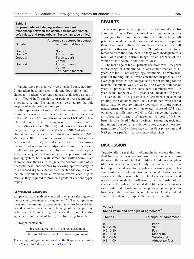
Table 1
Proposed adenoid staging system: anatomic
relationship between the adenoid tissue and vomer,
soft palate, and torus tubaris (Eustachian tube orifice)
GradeAnatomic structures in contact
with adenoid tissue
Grade 1 NoneGrade 2 Torus tubarisGrade 3 Torus tubaris
VomerGrade 4 Torus tubaris
VomerSoft palate (at rest)
from “poor” to “almost perfect” (Table 2).
RESULTS
Twenty-nine patients were prospectively recruited after In-stitutional Review Board approval in an outpatient otolar-yngology office based in a tertiary hospital setting. Allpatients were already undergoing nasal endoscopy as part oftheir office visit. Informed consent was obtained from allpatients for this study. Five of the 29 digital clips had to beremoved from the study because they were inadequate as aresult of bleeding, blurred image, or an absence of thevomer or soft palate in the field of view.
The mean age of the 24 patients reviewed was 10.9 yearswith a range of 4 months to 48 years and a median of 11years. Of the 24 otolaryngology examiners, 14 were resi-dents in training and 10 were consultants in practice. Theaverage postmedical school graduate year of training for theresident examiners was 3.6 years. The average number ofyears of practice for the consultant examiners was 10.2years with a range of 2 to 30 years and a median of 8 years.
Overall, 460 of a possible 576 agreements in adenoidgrading were obtained from the 24 examiners who scoredthe 24 nasal endoscopy digital video clips. With the Kappameasurement of agreement, an overall score of 0.71 wasobtained that falls into the 0.61 to 0.80 range and suggestsa “substantial” strength of agreement. A score of 0.81 orbetter is considered “almost perfect.” Separating residentsin training from consultants demonstrated a Kappa measure-ment score of 0.62 (substantial) for resident physicians and0.83 (almost perfect) for consultant physicians.
DISCUSSION
Traditionally, lateral skull radiographs have been the stan-dard for evaluation of adenoid size. There are several lim-itations to the use of lateral skull films. A radiographic plainfilm is only a 2-dimensional study that evaluates the rela-tionship of the adenoid to the palate in a single plane. Thiscan result in misinterpretation of adenoid obstruction incases where there is only bulky lateral adenoid growth andopen choanae medially. Furthermore, the relationship of theadenoid to the palate in a lateral skull film can be erroneousas a result of skull rotation or inappropriate palate positionfrom inspiration, expiration, or phonation. Finally, lateralskull films inherently expose the patient to radiation.
Table 2
Kappa value and strength of agreement7
Kappa Strength of agreement
0.01 Poor0.01-0.20 Slight0.21-0.40 Fair0.41-0.60 Moderate0.61-0.80 Substantial0.81-1.00 Almost perfect

686 Otolaryngology–Head and Neck Surgery, Vol 135, No 5, November 2006
Office nasal endoscopy offers several advantages overthe lateral skull radiograph in the evaluation of adenoidhypertrophy. There is no patient exposure to radiation and therelationship of the adenoid to important adjacent anatomicstructures can be evaluated dynamically in 3 dimensions. Al-though the image of an endoscope is 2-dimensional, the abilityto move the camera in and out of the nose allows the examinerto have a 3-dimensional sense of adenoid size. The relationshipof the adenoid to the adjacent torus tubaris, vomer, and softpalate can be easily evaluated in a dynamic fashion that allowsfor complete evaluation of the nasopharynx. Eustachian tubeand airway obstruction can be readily identified through nasalendoscopy in all 3 planes.
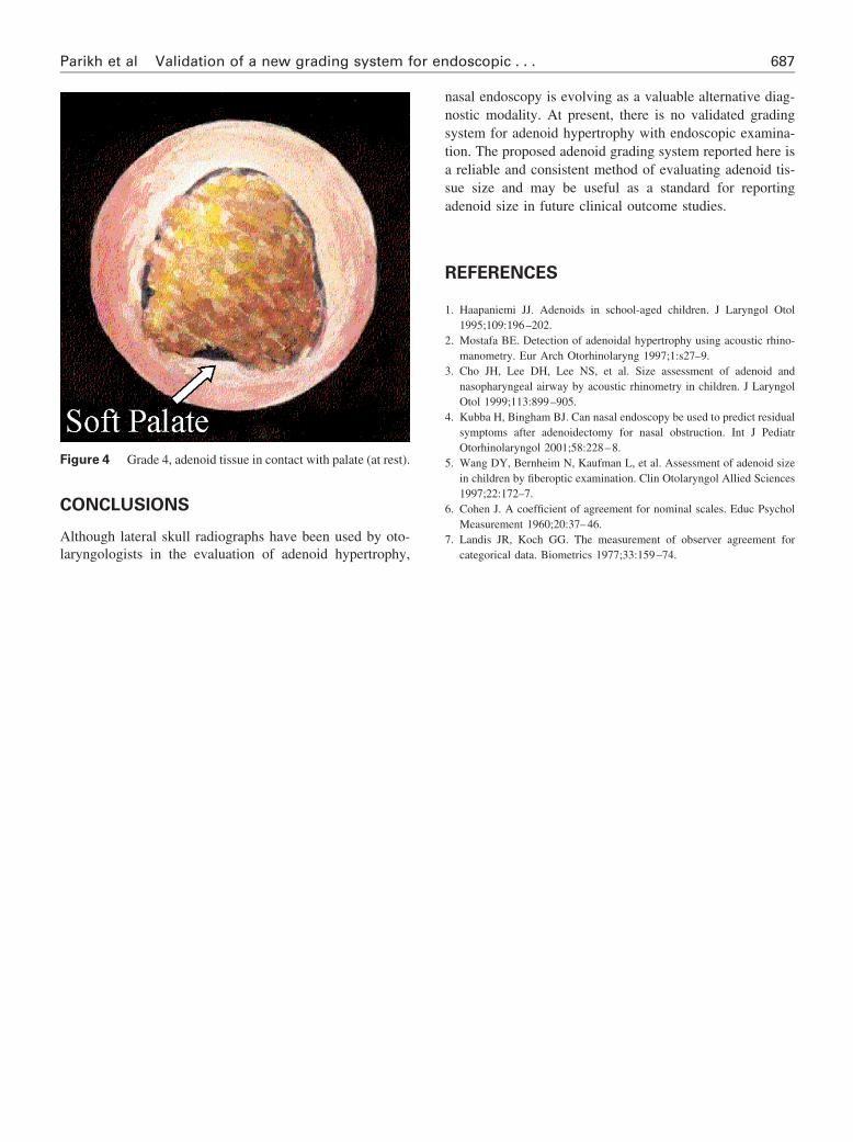
In this prospective study, a new grading system has beendevised based on the principle that the relationship of theadenoid tissue to adjacent structures is as important as theadenoid size itself. Impingement on the Eustachian tube oron the nasopharyngeal airway can both have negative se-quelae for the patient. The grading system presented hereincorporates the relationship of the adenoid to the torustubaris (Eustachian tube orifice), vomer (posterior nasalseptum), and soft palate (Table 1). Specifically grade 1adenoids are nonobstructive and do not contact any of thepreviously mentioned anatomic subsites (Fig 1). Subsequently,grade 2, 3, and 4 adenoids contact the torus tubaris, vomer, andsoft palate (at rest) respectively (Figs 2, 3, and 4).
Several factors may have had an impact on the final Kappascore and strength of this study. In particular, examiners maynot have had as clear a view of the adenoid tissue as theywould have if they were performing the endoscopy them-selves. The quality of digital video is inferior to direct visual-ization through a flexible nasal endoscope. Furthermore, the
Figure 1 Grade 1, adenoid tissue not in contact with adjacent
structures.learning curve for the grading system was not taken intoaccount. All endoscopic video clips examined were included inthe study, and examiners were not given practice video clips(the discrepancy between residents and consultants may po-tentially reflect how familiarity with the endoscopic view ofthe nasopharynx affects grading). Nevertheless, the Kappascore in this study suggests that the proposed endoscopic ad-enoid grading system has a substantial strength of agreementamong residents in training and an almost perfect strength ofagreement among consultants in practice.
Figure 3 Grade 3, adenoid tissue in contact with Vomer.
Figure 2 Grade 2, adenoid tissue in contact with torus tubaris.
687Parikh et al Validation of a new grading system for endoscopic . . .
CONCLUSIONS
Although lateral skull radiographs have been used by oto-
Figure 4 Grade 4, adenoid tissue in contact with palate (at rest).
laryngologists in the evaluation of adenoid hypertrophy,
nasal endoscopy is evolving as a valuable alternative diag-nostic modality. At present, there is no validated gradingsystem for adenoid hypertrophy with endoscopic examina-tion. The proposed adenoid grading system reported here isa reliable and consistent method of evaluating adenoid tis-sue size and may be useful as a standard for reportingadenoid size in future clinical outcome studies.
REFERENCES
1. Haapaniemi JJ. Adenoids in school-aged children. J Laryngol Otol1995;109:196–202.
2. Mostafa BE. Detection of adenoidal hypertrophy using acoustic rhino-manometry. Eur Arch Otorhinolaryng 1997;1:s27–9.
3. Cho JH, Lee DH, Lee NS, et al. Size assessment of adenoid andnasopharyngeal airway by acoustic rhinometry in children. J LaryngolOtol 1999;113:899–905.
4. Kubba H, Bingham BJ. Can nasal endoscopy be used to predict residualsymptoms after adenoidectomy for nasal obstruction. Int J PediatrOtorhinolaryngol 2001;58:228–8.
5. Wang DY, Bernheim N, Kaufman L, et al. Assessment of adenoid sizein children by fiberoptic examination. Clin Otolaryngol Allied Sciences1997;22:172–7.
6. Cohen J. A coefficient of agreement for nominal scales. Educ PsycholMeasurement 1960;20:37–46.
7. Landis JR, Koch GG. The measurement of observer agreement for
categorical data. Biometrics 1977;33:159–74.