UvA-DARE (Digital Academic Repository) Moving towards ... · 521408-L -bw-Roth Processed on:...
27
UvA-DARE is a service provided by the library of the University of Amsterdam (http://dare.uva.nl) UvA-DARE (Digital Academic Repository) Moving towards improved malaria control The position of molecular diagnostics and evaluating the efficacy of a novel treatment option Roth, J.M. Link to publication Citation for published version (APA): Roth, J. M. (2018). Moving towards improved malaria control: The position of molecular diagnostics and evaluating the efficacy of a novel treatment option. General rights It is not permitted to download or to forward/distribute the text or part of it without the consent of the author(s) and/or copyright holder(s), other than for strictly personal, individual use, unless the work is under an open content license (like Creative Commons). Disclaimer/Complaints regulations If you believe that digital publication of certain material infringes any of your rights or (privacy) interests, please let the Library know, stating your reasons. In case of a legitimate complaint, the Library will make the material inaccessible and/or remove it from the website. Please Ask the Library: https://uba.uva.nl/en/contact, or a letter to: Library of the University of Amsterdam, Secretariat, Singel 425, 1012 WP Amsterdam, The Netherlands. You will be contacted as soon as possible. Download date: 08 Aug 2019
Transcript of UvA-DARE (Digital Academic Repository) Moving towards ... · 521408-L -bw-Roth Processed on:...

UvA-DARE is a service provided by the library of the University of Amsterdam (http://dare.uva.nl)
UvA-DARE (Digital Academic Repository)
Moving towards improved malaria controlThe position of molecular diagnostics and evaluating the efficacy of a novel treatment optionRoth, J.M.
Link to publication
Citation for published version (APA):Roth, J. M. (2018). Moving towards improved malaria control: The position of molecular diagnostics andevaluating the efficacy of a novel treatment option.
General rightsIt is not permitted to download or to forward/distribute the text or part of it without the consent of the author(s) and/or copyright holder(s),other than for strictly personal, individual use, unless the work is under an open content license (like Creative Commons).
Disclaimer/Complaints regulationsIf you believe that digital publication of certain material infringes any of your rights or (privacy) interests, please let the Library know, statingyour reasons. In case of a legitimate complaint, the Library will make the material inaccessible and/or remove it from the website. Please Askthe Library: https://uba.uva.nl/en/contact, or a letter to: Library of the University of Amsterdam, Secretariat, Singel 425, 1012 WP Amsterdam,The Netherlands. You will be contacted as soon as possible.
Download date: 08 Aug 2019

521408-L-bw-Roth521408-L-bw-Roth521408-L-bw-Roth521408-L-bw-RothProcessed on: 13-9-2018Processed on: 13-9-2018Processed on: 13-9-2018Processed on: 13-9-2018 PDF page: 157PDF page: 157PDF page: 157PDF page: 157
Johanna M. Roth, Patrick Sawa, George Omweri, Victor Osoti, Nicodemus Makio, John Bradley, Teun Bousema, Henk D.F.H. Schallig, Pètra F. Mens
Malaria Journal 2018, 17:223
Chapter 5
Plasmodium falciparum gametocyte dynamics after pyronaridine-artesunate or artemether-lumefantrine treatment

521408-L-bw-Roth521408-L-bw-Roth521408-L-bw-Roth521408-L-bw-RothProcessed on: 13-9-2018Processed on: 13-9-2018Processed on: 13-9-2018Processed on: 13-9-2018 PDF page: 158PDF page: 158PDF page: 158PDF page: 158
158
Chapter 5
ABSTRACT
Background
Artemisinin-based combinations differ in their impact on gametocyte preva-
lence and density. This study assessed female and male gametocyte dynamics
after treating children with uncomplicated Plasmodium falciparum malaria
with either pyronaridine-artesunate (PA) or artemether-lumefantrine (AL).
Methods
Kenyan children with uncomplicated Plasmodium falciparum malaria were in-
cluded and randomly assigned to PA or AL treatment. Filter paper blood sam-
ples were collected as a source of RNA for quantitative reverse-transcription
PCR (qRT-PCR) and nucleic acid sequence based amplification (QT-NASBA)
to detect female gametocytes (targeting Pfs25 mRNA). Male gametocytes
were detected by qRT-PCR (targeting PfMGET mRNA). Duration of gameto-
cyte carriage, the female and male gametocyte response and the agreement
between qRT-PCR and QT-NASBA were determined.
Results
The mean duration of female gametocyte carriage was significantly longer for
PA (4.9 days) than for AL (3.8 days) as estimated by QT-NASBA (P=0.036), but
this difference was less clear when determined by Pfs25 qRT-PCR (4.5 days
for PA and 3.7 for AL, P=0.166). qRT-PCR based female gametocyte preva-
lence decreased from 100% (75/75) at baseline to 6.06% (4/66) at day 14 in
the AL group and from 97.7% (83/85) to 13.9% (11/79) in the PA group. Male
gametocyte prevalence decreased from 41.3% (31/75) at baseline to 19.7%
(13/66) at day 14 in the AL group and from 35.3% (30/85) to 22.8% (18/79)
in the PA group. There was good agreement between Pfs25 qRT-PCR and QT-
NASBA female gametocyte prevalence (0.85, 95% CI: 0.82 – 0.87).
Conclusions
This study indicates that female gametocyte clearance may be slightly faster
after AL compared to PA. Male gametocytes showed similar post-treatment
clearance between study arms. Future studies should further address poten-
tial differences between the post-treatment transmission potential after PA
compared to AL.

521408-L-bw-Roth521408-L-bw-Roth521408-L-bw-Roth521408-L-bw-RothProcessed on: 13-9-2018Processed on: 13-9-2018Processed on: 13-9-2018Processed on: 13-9-2018 PDF page: 159PDF page: 159PDF page: 159PDF page: 159
159
Gametocyte dynamics after ACT
BACKGROUND
Since artemisinin-based combination therapy (ACT) became widely adopt-
ed as first-line treatment for uncomplicated Plasmodium falciparum malaria,
it considerably contributed to the decline of the disease burden1–4. However,
resistance against commonly used artemisinin-based combinations is rising
in South-East Asia and the potential spread to African countries is a major
public health concern5,6. New drugs are under development that offer possible
alternatives to currently used artemisinin-based combinations. One of these
alternatives is the fixed-dose combination therapy pyronaridine-artesunate
(PA), which is found to be well tolerated and efficacious for the treatment of
uncomplicated P. falciparum malaria and the blood stage of Plasmodium vivax
malaria7–12. Mild and transient increases in transaminases are the main safety
concern11.
So far, the effect of PA on the transmission stages of P. falciparum (gameto-
cytes), has not been extensively studied in the clinical setting. In vitro data are
contradicting: a strong gametocytocidal effect of pyronaridine against stage II
to IV gametocytes has been found13, but was not confirmed elsewhere14. Delves
et al reported activity of pyronaridine against stage V gametocytes in vitro,
although only at concentrations close to cytotoxic levels, suggesting limited
clinical relevance15,16. With the increasing efforts to reduce malaria transmis-
sion, it becomes highly important to evaluate not only the potential of ACT
to cure the asexual stage of the parasite, but also their effect on gametocytes.
ACT is generally effective against asexual stages and immature gametocytes,
but its activity against mature gametocytes is limited14,17–19. However, differ-
ences between artemisinin-based combinations in the gametocyte response
after treatment exist. A recent meta-analysis showed that the appearance of
gametocytaemia in patients without gametocytes at baseline was lower after
artemether-lumefantrine (AL) and artesunate-mefloquine (AS-MQ) com-
pared to dihydroartemisinin-piperaquine (DP) and artesunate-amodiaquine
(AS-AQ)20. Among patients with gametocytes at baseline, clearance was fast-
er after AS-MQ and slower after DP, compared to AL. This meta-analysis by
the Worldwide Antimalarial Resistance Network (WWARN) hypothesized that
the non-artemisinin partner drug is a relevant determinant for differences in
the post-treatment gametocyte response.
To accurately evaluate the gametocyte response after ACT treatment, mo-
lecular tools are informative since post-treatment gametocyte densities are
5

521408-L-bw-Roth521408-L-bw-Roth521408-L-bw-Roth521408-L-bw-RothProcessed on: 13-9-2018Processed on: 13-9-2018Processed on: 13-9-2018Processed on: 13-9-2018 PDF page: 160PDF page: 160PDF page: 160PDF page: 160
160
Chapter 5
often below the detection threshold of microscopy21. Quantitative Nucleic
Acid Sequence Based Amplification (QT-NASBA) is a sensitive and reliable
technique for the detection of submicroscopic gametocytes, targeting the
female-specific Pfs2522,23. Recently, a sex-specific quantitative reverse tran-
scriptase PCR (qRT-PCR) has been developed and evaluated, differentiating
between female (Pfs25) and male (PfMGET) gametocytes24. This differentia-
tion may be important, because the minority male population (normally 3-5
females to 1 male) was shown in vitro to be more sensitive than females to a
range of anti-malarial drugs15. Thus, faster clearance of the male gametocyte
population during or after treatment might sterilize the infection, while the
female-dominated gametocyte density may not be reduced to the same ex-
tent25. The sex-specific qRT-PCR can be used to investigate both male and
female gametocyte dynamics in clinical trials.
In this study, the QT-NASBA and qRT-PCR based female specific gametocyte
response after PA-treatment was compared to that after AL. Furthermore,
qRT-PCR was used to evaluate and compare male and female gametocyte dy-
namics. Finally, the agreement between Pfs25 qRT-PCR and QT-NASBA for
the detection of female gametocytes was determined.

521408-L-bw-Roth521408-L-bw-Roth521408-L-bw-Roth521408-L-bw-RothProcessed on: 13-9-2018Processed on: 13-9-2018Processed on: 13-9-2018Processed on: 13-9-2018 PDF page: 161PDF page: 161PDF page: 161PDF page: 161
161
Gametocyte dynamics after ACT
METHODS
Study design
This observational study was part of a phase III randomized clinical trial in-
vestigating the efficacy and safety of PA compared to AL in Kenyan children
with uncomplicated P. falciparum malaria26. The study was conducted at St.
Jude’s Clinic, Mbita, Western Kenya, from October 2015 to June 2016 and from
January to August 2017. Ethical approval was obtained from the Ethical Review
Committee of the Kenya Medical Research Institute (KEMRI) (NON-SSC no.
479, registered at clinicaltrials.gov under NCT02411994). Children aged 6
months to 12 years seeking care at the clinic were eligible to participate if they
were living within 10 km range from the study clinic and had microscopically
confirmed P. falciparum mono-infection with a parasitaemia between 1000
and 200,000 µl-1. Exclusion criteria were signs and symptoms of complicated
malaria, non-P. falciparum or mixed Plasmodium infection, a history of he-
patic and/or renal impairment, a haemoglobin (Hb) concentration <6 g/dL,
severe malnutrition (defined as having a weight-for-age or height-for-age
z-score of <-3)27, having received anti-malarial therapy in the previous two
weeks, known hypersensitivity to artemisinins, previous participation in this
study, current participation in other anti-malarial drug intervention studies
or not being available for follow-up. Written informed consent from a par-
ent or guardian was required for study participation, assent was sought from
children able to understand the study.
Procedures
Study participants were randomized to receive a three-day course of either
artemether-lumefantrine (AL, Novartis, Basel, Switzerland) or pyronari-
dine-artesunate (PA, Shin Poong Pharmaceutical Company, Seoul, South
Korea). All study staff except the pharmacists responsible for drug admin-
istration were blinded to treatment allocation. Dosing was body-weight de-
pendent and drugs were administered according to manufacturer’s instruc-
tions (Additional file 1) with food (mandazi – a type of fried bread) or milk.
For PA, children <20 kg received granules dissolved in lemonade. Children
≥20 kg received the tablet formulation. In the AL group, all children received
tablets.
Participants returned to the study clinic on 1, 2, 3, 7, 14, 28 and 42 days after
start of treatment. Blood samples were taken by finger-prick at all time-points.
5

521408-L-bw-Roth521408-L-bw-Roth521408-L-bw-Roth521408-L-bw-RothProcessed on: 13-9-2018Processed on: 13-9-2018Processed on: 13-9-2018Processed on: 13-9-2018 PDF page: 162PDF page: 162PDF page: 162PDF page: 162
162
Chapter 5
Hb was determined on day 0, 3, 7 and 28 by HemoCue (Ängelholm, Sweden).
Giemsa-stained thick smears were used for determination and counting of
asexual parasites and gametocytes, according to WHO procedures28. Thick-
and-thin blood smears were prepared and read by local expert microscopists.
A slide was considered negative when 100 high-power fields were examined
at 1000x magnification and no parasites were observed. Parasitaemia was
determined from thick smears by counting the number of parasites against
200 leukocytes, with the assumption of 8000 leukocytes/µl blood. When the
number of parasites after counting 200 leukocytes was <100, counting con-
tinued up to 500 leukocytes.
Female specific Pfs25 QT-NASBA and sex-specific Pfs25 and PfMGET qRT-
PCR were used for gametocyte detection on day 0, 3, 7 and 14. To perform
these assays, 2x 50 µl finger-prick blood was collected on Whatman 903
protein saver cards (GE Healthcare, Chicago, USA), dried at room tempera-
ture for 24 hours, packed individually with silica and stored at -20°C until
shipment to the Netherlands. Nucleic acid extraction was done by Nuclisens
EasyMag (bioMérieux, Marcy-l’Étoile, France) and DNA/RNA was stored at
-70°C. QT-NASBA was performed as previously described22, with minor mod-
ifications. The reaction mixture (5 µl) and sample (2.5 µl) were incubated for
2 minutes at 65 °C and 2 minutes at 41 °C. Enzyme was added (2.5 µl) and the
reaction was allowed to run for 30 minutes at 41 °C. Quantification was done
using standard curves of 103 to 10-1 gametocytes/µl, which were produced
from in vitro cultures as reported22. qRT-PCRs were performed as previous-
ly described, using sex-specific standard curves (103 to 10-2 gametocytes/µl)
for quantification24. To produce these separate standard curves, male and fe-
male gametocytes were isolated by fluorescence activated cell sorting using
a transgenic parasite line expressing male and female specific fluorescence
markers29. Samples were declared negative for both QT-NASBA and qRT-PCR
if the estimated gametocytaemia was <0.02 gametocytes/µl (1 gametocyte/50
µl sample)30.
Outcomes
The primary outcome was the mean duration of female gametocyte carriage
in the PA arm compared to the AL arm, based on QT-NASBA. Secondary out-
comes were: the qRT-PCR based mean duration of female gametocyte car-
riage, the QT-NASBA and qRT-PCR based female gametocyte circulation
time, the QT-NASBA based area under the curve (AUC) of female gametocyte

521408-L-bw-Roth521408-L-bw-Roth521408-L-bw-Roth521408-L-bw-RothProcessed on: 13-9-2018Processed on: 13-9-2018Processed on: 13-9-2018Processed on: 13-9-2018 PDF page: 163PDF page: 163PDF page: 163PDF page: 163
163
Gametocyte dynamics after ACT
density over time (gametocytes/µl-1 days), and gametocyte prevalence and
density on day 3, 7 and 14 as determined by QT-NASBA and Pfs25/PfMGET
qRT-PCR. Finally, the agreement between QT-NASBA and Pfs25 qRT-PCR for
the detection but not quantification of female gametocytes was determined.
Statistical analysis
Stata software version 14.0 (Stata Corporation, Texas, USA) and SAS version
9.4 (SAS Institute Inc, NC, USA) were used for statistical analyses. A deter-
ministic compartmental model, as previously published, was fitted to deter-
mine the duration of female gametocyte carriage and gametocyte circulation
times31. The AUC was determined as described previously and log10-trans-
formed32. Linear regression was used to compare the log AUC in the PA group
to that in the AL group, adjusting for log10-transformed baseline gametocyte
density. The Wilcoxon rank-sum test was used for between-group compar-
isons of gametocyte density on day 0, 3, 7 and 14. A chi-square or Fisher’s
exact test was used to compare between-group gametocyte prevalences on
day 0, 3, 7 and 14. The agreement between Pfs25 qRT-PCR and QT-NASBA
was determined by calculating the weighted concordance correlation coef-
ficient (CCC) based on variance components, taking repeated measures into
account33,34.5

521408-L-bw-Roth521408-L-bw-Roth521408-L-bw-Roth521408-L-bw-RothProcessed on: 13-9-2018Processed on: 13-9-2018Processed on: 13-9-2018Processed on: 13-9-2018 PDF page: 164PDF page: 164PDF page: 164PDF page: 164
164
Chapter 5
RESULTS
Study population and baseline characteristics
A consecutive subset of 160 children from the main clinical trial participated
in the present study. Of these 160 participants, 85 received PA and 75 received
AL (Figure 1). Nine participants did not complete follow-up (day 14) in the AL
group: four withdrew consent, three moved away from the study area and two
missed their day 14 visit. In the PA group, six participants did not complete
follow-up: three missed their day 14 visit, one discontinued due to repeated
vomiting, one moved away from the study area and one had a treatment fail-
ure on day 7.
Figure 1. Participant flow.
- Missed day 14 visit: n=3 - Moved outside study area: n=1 - Treatment failure day 7: n=1
Completed day 14:
n= 79
- Consent withdrawn: n=2 - Moved outside study area: n=2 - Missed day 14 visit: n=2
Completed day 14:
n= 66
Screened:
n= 971
Enrolled:
n= 160
Assigned to AL:
n= 75
Assigned to PA:
n= 85
Excluded: n= 811 No Plasmodium infection: n= 570 Parasitaemia <1000 p/µl: n= 149 P. malariae coinfection: n= 63 Parasitaemia >200,000 p/µl: n= 16 Previous study participation: n=7 Living outside study area: n=3 Already on antimalarial medication: n=1 Only gametocytes detected: n=1 Complicated malaria: n=1
Completed day 7:
n= 83
Completed day 7:
n= 72
- Rescue treatment after repeated vomiting: n=1 - Missing day 7 sample: n=1*
- Consent withdrawn: n=2 - Moved outside study area: n=1
*A day 14 sample was available for this participant.
Schematic presentation of patient screening, inclusion and follow-up.

521408-L-bw-Roth521408-L-bw-Roth521408-L-bw-Roth521408-L-bw-RothProcessed on: 13-9-2018Processed on: 13-9-2018Processed on: 13-9-2018Processed on: 13-9-2018 PDF page: 165PDF page: 165PDF page: 165PDF page: 165
165
Gametocyte dynamics after ACT
Baseline characteristics were similar between intervention groups (Table
1). QT-NASBA and Pfs25 qRT-PCR based female gametocyte prevalence and
density at baseline were comparable and higher than PfMGET qRT-PCR male
estimates. As expected, microscopy based prevalence was lower compared to
the molecular methods and children with microscopy confirmed gametocytes
at baseline had significantly higher Pfs25 QT-NASBA gametocyte density
compared to those without microscopically detected gametocytes (P<0.001,
Wilcoxon rank-sum test).
Table 1. Baseline characteristics of study participants at enrollment
pyronaridine-artesunate artemether-lumefantrine
N 85 75
Malea 54.1 (46/85) 49.3 (37/75)
Age (years)b 7.0 (4.2-9.0) 6.0 (3.4-9.8)
Hb (g/dL)c 11.8 (11.4-12.2) 11.7 (11.2-12.2)
Temperature (°C)c 37.6 (37.4-37.9) 37.4 (37.1-37.7)
Fever (Temperature> 37.5 °C)a 55.3 (47/85) 48.0 (36/75)
Asexual parasite density (p/µl)b 28800 (12000-68640) 29280 (12000-67680)
Gametocyte prevalence – microscopya 2.35 (2/85) 5.33 (4/75)
Gametocyte prevalence – QT-NASBAa 95.3 (81/85) 94.7 (71/75)
Gametocyte density – QT-NASBA (p/µl)b 3.23 (0.68-18.1) 6.36 (1.19-22.2)
Gametocyte prevalence – Pfs25 qRT-PCRa 97.7 (83/85) 100 (75/75)
Gametocyte density – Pfs25 qRT-PCR (p/µl)b 2.88 (0.85-5.24) 1.94 (0.77-5.35)
Gametocyte prevalence – PfMGET qRT-PCRa 35.3 (30/85) 41.3 (31/75)
Gametocyte density – PfMGET qRT-PCR (p/µl)b 0.94 (0.11-14.1) 0.48 (0.21-10.1)Data are: a Percentage % (n/N), b Median and IQR or c Mean and 95% CI.
Gametocyte carriage after PA and AL
QT-NASBA based female gametocyte prevalence at day 3 was 37.0% (30/81)
in the PA group and 31.0% (22/71) in the AL group (P=0.433). At day 7, preva-
lence decreased to 21.7% (18/83) in the PA group and 16.7% (12/72) in the AL
group (P=0.430). Prevalence at day 14 was 15.2% (12/79) in the PA group and
7.58% (5/66) in the AL group (P=0.156). Female gametocyte prevalence esti-
mates were highly similar when assessed by Pfs25 qRT-PCR (Figure 2, Table
2).
5

521408-L-bw-Roth521408-L-bw-Roth521408-L-bw-Roth521408-L-bw-RothProcessed on: 13-9-2018Processed on: 13-9-2018Processed on: 13-9-2018Processed on: 13-9-2018 PDF page: 166PDF page: 166PDF page: 166PDF page: 166
166
Chapter 5
Figure 2. Female gametocytes by Pfs25 QT-NASBA and qRT-PCR.
A. Gametocyte prevalence determined by QT-NASBA. B. Gametocyte density determined by QT-NASBA. C. Gametocyte prevalence determined by qRT-PCR. D. Gametocyte density determined by qRT-PCR. 95% confidence intervals are presented for prevalences. Density is presented as median (IQR) for gameto-cyte-positive individuals only. Samples were considered negative if gametocyte levels were <0.02/µl. AL: artemether-lumefantrine, PA: pyronaridine-artesunate.
Table 2. Female and male gametocyte prevalence and density
pyronaridine-
artesunate
artemether-
lumefantrine
P-value*
Pfs25 QT-NASBA prevalence, % (no./No.)
Day 0 95.3 (81/85) 94.7 (71/75) 1.000
Day 3 37.0 (30/81) 31.0 (22/71) 0.433
Day 7 21.7 (18/83) 16.7 (12/72) 0.430
Day 14 15.2 (12/79) 7.58 (5/66) 0.156
Pfs25 QT-NASBA density, median (IQR)a
Day 0 3.23 (0.68-18.1) 6.36 (1.19-22.2) 0.229
Day 3 3.86 (0.33-16.4) 3.73 (0.33-6.91) 0.630
Day 7 2.82 (1.17-33.5) 0.74 (0.23-12.9) 0.150
Day 14 6.19 (2.10-27.2) 1.45 (0.65-1.69) 0.092

521408-L-bw-Roth521408-L-bw-Roth521408-L-bw-Roth521408-L-bw-RothProcessed on: 13-9-2018Processed on: 13-9-2018Processed on: 13-9-2018Processed on: 13-9-2018 PDF page: 167PDF page: 167PDF page: 167PDF page: 167
167
Gametocyte dynamics after ACT
pyronaridine-
artesunate
artemether-
lumefantrine
P-value*
Pfs25 qRT-PCR prevalence, % (no./No.)
Day 0 97.7 (83/85) 100 (75/75) 0.499
Day 3 30.9 (25/81) 33.8 (24/71) 0.699
Day 7 19.3 (16/83) 16.7 (12/72) 0.674
Day 14 13.9 (11/79) 6.06 (4/66) 0.122
Pfs25 qRT-PCR density, median (IQR)a
Day 0 2.88 (0.85-5.24) 1.94 (0.77-5.35) 0.568
Day 3 0.58 (0.30-1.66) 0.30 (0.14-0.62) 0.039
Day 7 0.78 (0.33-15.5) 0.28 (0.16-0.83) 0.126
Day 14 0.33 (0.14-6.42) 0.12 (0.08-0.90) 0.322
PfMGET qRT-PCR prevalence (%) (no./No.)
Day 0 35.3 (30/85) 41.3 (31/75) 0.433
Day 3 34.6 (28/81) 36.6 (26/71) 0.792
Day 7 31.3 (26/83) 30.6 (22/72) 0.918
Day 14 22.8 (18/79) 19.7 (13/66) 0.652
PfMGET qRT-PCR density, median (IQR)b
Day 0 0.94 (0.11-14.1) 0.48 (0.21-10.1) 0.920
Day 3 2.55 (0.21-11.0) 0.82 (0.17-5.07) 0.341
Day 7 3.23 (0.21-8.95) 1.22 (0.19-11.5) 0.551
Day 14 1.81 (0.30-11.2) 1.51 (0.66-6.91) 0.779
Total qRT-PCR prevalence, % (no./No.)
Day 0 97.7 (83/85) 100 (75/75) 0.499
Day 3 42.0 (34/81) 43.7 (31/71) 0.841
Day 7 32.5 (27/83) 31.9 (23/72) 0.920
Day 14 25.3 (20/79) 21.2 (14/66) 0.560
Total qRT-PCR density, median (IQR)c
Day 0 3.23 (0.86-7.54) 2.24 (0.91-6.13) 0.642
Day 3 1.20 (0.24-11.0) 1.06 (0.11-5.21) 0.248
Day 7 3.47 (0.26-15.5) 1.69 (0.27-12.3) 0.631
Day 14 1.66 (0.30-14.6) 1.34 (0.55-7.00) 0.662*P-values represent between group differences. Prevalence differences were tested with the chi-squared or Fisher’s Exact test. Differences in gametocyte density were tested with the Wilcoxon rank-sum test. Data included: afemale gametocyte positive individuals, bmale gametocyte positive individuals, cmale and/or female gametocyte positive individuals
5

521408-L-bw-Roth521408-L-bw-Roth521408-L-bw-Roth521408-L-bw-RothProcessed on: 13-9-2018Processed on: 13-9-2018Processed on: 13-9-2018Processed on: 13-9-2018 PDF page: 168PDF page: 168PDF page: 168PDF page: 168
168
Chapter 5
While prevalence estimates were comparable between QT-NASBA and qRT-
PCR, some differences in density measurements were observed. As can be
seen in Figure 2, the decrease in density over time is clearer by qRT-PCR as
compared to QT-NASBA, especially in the PA group. In the AL group, the QT-
NASBA based female gametocyte density decreased from a median of 6.36 ga-
metocytes/µl (IQR: 1.19 – 22.2) at baseline to 1.45 gametocytes/µl (IQR: 0.65
– 1.69) at day 14. However, in the PA group, the median female gametocyte
density estimated by QT-NASBA in gametocyte positive samples did not de-
crease over time and even appeared to increase slightly: 3.23 gametocytes/
µl (IQR: 0.68 – 18.1) at baseline versus 6.19 gametocytes/µl (IQR: 2.11 – 27.2)
at day 14. Given that the median gametocyte density is only determined over
gametocyte positive individuals, the median density on day 3, 7 and 14 is not
necessarily assessed over the same individuals used to determine the baseline
gametocyte density. To investigate whether this apparent increase over time
in the PA group was due to an absolute increase within individuals, the medi-
an QT-NASBA based gametocyte density on day 0 for individuals still positive
on day 14 was calculated and found to be 113.0 (IQR: 21.0 – 252). Thus, the
median female gametocyte density for participants in the PA group gameto-
cyte positive on day 14 decreased from baseline to day 14 and the apparent rise
in density as estimated by QT-NASBA could not be explained by an absolute
increase within individuals, but is rather a difference between the population
positives on day 0 and that on day 14.
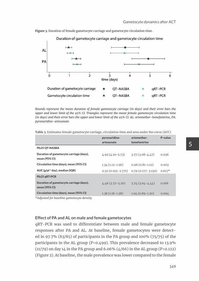
The mean duration of female gametocyte carriage as estimated by QT-NASBA
was significantly longer in the PA group (4.92 days, 95% CI: 4.10 – 5.73),
compared to the AL group (3.77 days, 95% CI: 3.08 – 4.47) (P= 0.036). By
qRT-PCR, the mean duration of female gametocyte carriage was also longer
in the PA group (4.46 days, 95% CI: 3.72 – 5.20), compared to the AL group
(3.74 days, 95% CI: 3.04 – 4.44), but this difference was not significant (P=
0.166). Similarly, the QT-NASBA based mean gametocyte circulation time
was longer for PA (1.34 days, 95% CI: 1.12 – 1.56) compared to AL (0.96 days,
95% CI: 0.81 – 1.12) (P=0.003). This was also the case for the qRT-PCR based
mean gametocyte circulation time (PA: 1.38 days, 95% CI: 1.18 – 1.58 and AL:
1.04 days, 95% CI: 0.89 – 1.20, P=0.004) (Figure 3 and Table 3). The AUC, on
the other hand, was not different between treatment arms (Table 3, P=0.617
after adjustment for baseline gametocyte density), possibly explained by the
slower gametocyte clearance in the PA group, but considerable contribution
of the higher baseline gametocyte density to the AUC in the AL group.
521408-L-bw-Roth521408-L-bw-Roth521408-L-bw-Roth521408-L-bw-RothProcessed on: 13-9-2018Processed on: 13-9-2018Processed on: 13-9-2018Processed on: 13-9-2018 PDF page: 169PDF page: 169PDF page: 169PDF page: 169
169
Gametocyte dynamics after ACT
Figure 3. Duration of female gametocyte carriage and gametocyte circulation time.
Rounds represent the mean duration of female gametocyte carriage (in days) and their error bars the upper and lower limit of the 95% CI. Triangles represent the mean female gametocyte circulation time (in days) and their error bars the upper and lower limit of the 95% CI. AL: artemether-lumefantrine, PA: pyronaridine-artesunate.
Table 3. Estimates female gametocyte carriage, circulation time and area under the curve (AUC)
pyronaridine-
artesunate
artemether-
lumefantrine
P-value
Pfs25 QT-NASBA
Duration of gametocyte carriage (days),
mean (95% CI)4.92 (4.10-5.73) 3.77 (3.08-4.47) 0.036
Circulation time (days), mean (95% CI) 1.34 (1.12-1.56) 0.96 (0.81-1.12) 0.003
AUC (g/µl-1 day), median (IQR) 0.52 (0.105-2.771) 0.79 (0.177-3.150) 0.617*
Pfs25 qRT-PCR
Duration of gametocyte carriage (days),
mean (95% CI)4.46 (3.72-5.20) 3.74 (3.04-4.44) 0.166
Circulation time (days), mean (95% CI) 1.38 (1.18-1.58) 1.04 (0.89-1.20) 0.004*Adjusted for baseline gametocyte density
Effect of PA and AL on male and female gametocytes
qRT-PCR was used to differentiate between male and female gametocyte
responses after PA and AL. At baseline, female gametocytes were detect-
ed in 97.7% (83/85) of participants in the PA group and 100% (75/75) of the
participants in the AL group (P=0.499). This prevalence decreased to 13.9%
(11/79) on day 14 in the PA group and 6.06% (4/66) in the AL group (P=0.122)
(Figure 2). At baseline, the male prevalence was lower compared to the female
5
521408-L-bw-Roth521408-L-bw-Roth521408-L-bw-Roth521408-L-bw-RothProcessed on: 13-9-2018Processed on: 13-9-2018Processed on: 13-9-2018Processed on: 13-9-2018 PDF page: 170PDF page: 170PDF page: 170PDF page: 170
170
Chapter 5
prevalence: 35.3% (30/85) in the PA group and 41.3% (31/75) in the AL group
(P=0.433). In both groups the decrease in male gametocyte prevalence was
less substantial than that of female prevalence (male gametocyte prevalence
22.8% (18/79) in the PA group and 19.7% (13/66) in the AL group on day 14
(P=0.652) (Figure 4).
The median female density decreased from 2.88 (IQR: 0.85 – 5.24) gameto-
cytes/µl at baseline to 0.58 (IQR: 0.30 – 1.66) on day 3 in the PA group. In the
AL group, female baseline density was 1.94 gametocytes/µl (IQR: 0.77 – 5.35),
which decreased to 0.30 (IQR: 0.14 – 0.62) on day 3. By day 14, median female
gametocyte density was 0.33 (IQR: 0.14 – 6.42) in the PA group and 0.12 (IQR:
0.08 – 0.90) in the AL group (P=0.322) (Table 2). Median male density, on the
other hand, did not decrease over time and even appears to increase slight-
ly. Similar to the analysis of QT-NASBA density in the PA group as described
above, the apparent increase of male gametocytes over time was investigated.
The median gametocyte density on day 0 for individuals still positive on day
14 was calculated and found to be 9.44 (IQR: 0.18 – 45.7) in the PA group and
10.1 (IQR: 0.81 – 36.5) in the AL group. Thus, the median gametocyte densi-
ty for participants gametocyte positive on day 14 decreased from baseline to
day 14 in both treatment groups (P=0.01 for both PA and AL groups, Wilcoxon
signed rank test). This illustrates also for male gametocytes that the apparent
rise in density could not be explained by an absolute increase within individ-
uals, but is rather a difference between the population positives on day 0 and
that on day 14.

521408-L-bw-Roth521408-L-bw-Roth521408-L-bw-Roth521408-L-bw-RothProcessed on: 13-9-2018Processed on: 13-9-2018Processed on: 13-9-2018Processed on: 13-9-2018 PDF page: 171PDF page: 171PDF page: 171PDF page: 171
171
Gametocyte dynamics after ACT
Figure 4. Male gametocytes by PfMGET qRT-PCR.
A. Gametocyte prevalence, including 95% confidence intervals. B. Gametocyte density, presented as medi-an (IQR) for gametocyte-positive individuals only. Samples were considered negative if gametocyte levels were <0.02/µl. AL: artemether-lumefantrine, PA: pyronaridine-artesunate.
Agreement between Pfs25 qRT-PCR and QT-NASBA
The agreement between binary female prevalence outcomes of Pfs25 qRT-PCR
and QT-NASBA was calculated using the CCC variance components method.
Only subjects with a complete dataset were included (140 participants and
560 samples). Out of 560, 12 samples were positive by QT-NASBA but not by
qRT-PCR. Similarly, 10 samples were positive by qRT-PCR but negative by
QT-NASBA. The two tests were in agreement for the remaining 538 samples
(209 positive and 329 negative). The CCC was 0.85 (95% CI: 0.82 – 0.87), in-
dicating good agreement between Pfs25 qRT-PCR and QT-NASBA for the de-
tection of female gametocytes.
5

521408-L-bw-Roth521408-L-bw-Roth521408-L-bw-Roth521408-L-bw-RothProcessed on: 13-9-2018Processed on: 13-9-2018Processed on: 13-9-2018Processed on: 13-9-2018 PDF page: 172PDF page: 172PDF page: 172PDF page: 172
172
Chapter 5
DISCUSSION
This is the first paper describing kinetics of submicroscopic gametocytes af-
ter PA treatment using molecular detection methods in comparison with the
most widely used first-line treatment for malaria in Africa, AL. The duration
of female gametocyte carriage and gametocyte circulation time appeared to
be slightly longer for PA compared to AL. There were no indications that PA or
AL preferentially cleared male gametocytes.
The failure of conventional anti-malarials, including ACT, to clear circu-
lating mature gametocytes may allow persisting malaria transmission in
the week(s) following treatment35. Gametocyte clearance time may thus be
a relevant indicator of the transmission-blocking potential of anti-malarial
drugs. Although it has been observed that some persisting gametocytes may
not be viable36, current evidence suggests that a comparison of anti-malar-
ial drugs on gametocytocidal properties would reach similar conclusions on
their relative transmission blocking effects18,35–37. The microscopy-based ga-
metocyte clearance time has previously been compared between PA and AL,
but no difference between the two drugs was found7,10. Since microscopy is
notoriously insensitive for gametocyte detection, molecular methods provide
more accurate estimates of post treatment gametocytaemia38. Importantly, it
has been shown that submicroscopic gametocytes may allow onward trans-
mission to mosquitoes39. In the present study, gametocytes were detected by
microscopy in only 3.75% (6/160) of study participants at baseline, compared
to 95.0% (152/160) by QT-NASBA. This contrast is even higher than in pre-
vious studies and emphasizes the underestimation of gametocyte prevalence
by microscopy38.
Different effects of ACT on the gametocyte response have previously been re-
ported and in a recent meta-analysis AL was shown to be better in preventing
the microscopic occurrence of gametocytes shortly after treatment compared
to DP or AS-AQ20. A point of caution when interpreting the results of this me-
ta-analysis, is the fact that sensitivities of parasites to the drugs fluctuat-
ed over the years and drug efficacy is setting dependent. However, there is
agreement in literature that, based on both microscopy as well as molecular
gametocyte detection, the duration of gametocyte carriage is significant-
ly shorter after AL treatment, compared to DP20,35. In the present study, the
duration of gametocyte carriage and gametocyte circulation time were sur-
prisingly short compared to other studies in the same area35,37. While the day

521408-L-bw-Roth521408-L-bw-Roth521408-L-bw-Roth521408-L-bw-RothProcessed on: 13-9-2018Processed on: 13-9-2018Processed on: 13-9-2018Processed on: 13-9-2018 PDF page: 173PDF page: 173PDF page: 173PDF page: 173
173
Gametocyte dynamics after ACT
3 QT-NASBA female gametocyte prevalence was 31.0% (22/71) for AL and
37.0% (30/81) for PA, others reported day 3 QT-NASBA prevalences >50%
after AL treatment among those positive at baseline35,37. Previous studies in
sub-Saharan Africa, using the same model to assess gametocyte clearance
and circulation time, found a duration of gametocyte carriage after AL of 12.4
days in a trial with similar inclusion criteria to the present study40 and of 19.7
days in a trial including patent gametocyte carriers41. These gametocyte car-
riage estimates are 3-5 fold longer compared to the present study. A possi-
ble explanation for this observation is the relatively low median gametocyte
density at baseline in the present study. Alternatively, the process of stor-
ing and extracting RNA from filter papers may have resulted in a suboptimal
yield and underestimated gametocyte prevalence during follow up. Despite
the shorter clearance estimates compared to other reports, there are no indi-
cations that this observation affected the comparison between PA and AL in
the present study.
Baseline prevalence of female gametocytes (estimated by qRT-PCR) was
98.8% (158/160), while male baseline prevalence was only 38.1% (61/160).
This is in contrast to the data presented by Stone et al., from the same study
site, where both female and male prevalence were 100% as estimated by the
same qRT-PCR. However, the study by Stone et al included only participants
with microscopically detectable gametocytes, while being gametocyte posi-
tive by microscopy was uncommon in the present study. This resulted in me-
dian baseline qRT-PCR-based gametocyte densities of 2.9/µl (PA) and 1.9/
µl (AL) for female gametocytes and 0.9/µl (PA) and 0.5/µl (AL) for males.
Working with such low densities, with presumably a female biased sex-ratio
at baseline, it is not unlikely that part of the samples with low density female
gametocytaemia at baseline had male densities below the detection thresh-
old, which may explain the difference in baseline prevalence between male
and female gametocytes.
No evidence of faster male compared to female gametocyte clearance was
found. In fact, the present data suggest that even though the proportion of
participants with male gametocytes at baseline was lower than that with fe-
male gametocytes, males may actually be cleared slower. Previous studies that
examined gametocyte sex ratio after DP or SP-AQ alone or with primaquine
observed that during the course of follow-up gametocyte sex ratios became
more female-biased while primaquine initially resulted in a male-biased sex
ratio24,36. In microscopy-based studies a female biased gametocyte response
5

521408-L-bw-Roth521408-L-bw-Roth521408-L-bw-Roth521408-L-bw-RothProcessed on: 13-9-2018Processed on: 13-9-2018Processed on: 13-9-2018Processed on: 13-9-2018 PDF page: 174PDF page: 174PDF page: 174PDF page: 174
174
Chapter 5
after various artemisinin-based combinations was commonly observed42,43.
In vitro results also indicate a more pronounced effect of most anti-malarial
drugs on male compared to female gametocytes. For example, the percent-
age inhibition of activation by artemether and artesunate was found to be ap-
proximately 39 and 10 times higher, respectively, for males than for females15.
The difference between the qRT-PCR used in the present study and the in vitro
system used by Delves et al. is that the latter evaluates the gametocytes’ abil-
ity to form gametes rather than the presence of mRNA. Whether the qRT-PCR
can detect mRNA from nonviable gametocytes is unknown40. Both the in vitro
and the mRNA results can be accurate if male gametocytes are more affect-
ed by the ACT than females, but remain present in the circulation during the
time of sampling as intact nonviable gametocytes24. Thus, despite the clear
added value of molecular techniques like QT-NASBA and qRT-PCR, function-
al assays that determine gametocyte fitness or infectivity remain crucial in
assessing transmission-blocking properties of anti-malarial drugs.
The apparent increase in male density (and female density in the PA arm
as estimated by QT-NASBA) could not be explained by an absolute increase
within individuals, but rather reflects a difference between the population
positives on day 0 and that on day 14. Stone et al. performed a similar analysis
and reported a small decrease of male density after DP treatment (from 3.8/µl
at baseline to 0.9/µl at day 7)24. Both studies had low baseline male gameto-
cyte density, but estimates were approximately 5 times higher in the study
by Stone et al. Baseline densities close to the detection limit could lead to an
increase in density by chance and this could possibly explain the difference in
density over time between the two studies. Additionally, a study by Dicko et
al. found a higher baseline density of male gametocytes and showed a more
distinct decrease over time compared to both the present study and Stone et
al.24,36.
A good level of agreement between QT-NASBA and Pfs25 qRT-PCR female ga-
metocyte prevalence was observed. This confirms data from a previous study
where both assays were shown to be suitable to detect and quantify submi-
croscopic levels of gametocytes, although the reproducibility of qRT-PCR
was found to be better than that of QT-NASBA30.
A limitation of the present study is that gametocyte infectiousness to mos-
quitoes could not be established. This was due to an infection of the estab-
lished mosquito colony with Microsporidia species, which has been shown
to inhibit the survival of Plasmodium in mosquitoes44. Since only mosquito

521408-L-bw-Roth521408-L-bw-Roth521408-L-bw-Roth521408-L-bw-RothProcessed on: 13-9-2018Processed on: 13-9-2018Processed on: 13-9-2018Processed on: 13-9-2018 PDF page: 175PDF page: 175PDF page: 175PDF page: 175
175
Gametocyte dynamics after ACT
feeding assays can provide evidence on the transmissibility of gametocytes,
an assessment of infectivity could not be done. Future studies should further
address potential differences between the post-treatment transmission po-
tential after PA compared to AL.
CONCLUSIONS
This study provides important data on the submicroscopic gametocyte re-
sponse after PA compared to AL treatment of uncomplicated P. falciparum
malaria. These data may contribute to estimates of impact differences be-
tween artemisinin-based combinations, for example based on a model that
demonstrated a higher reduction of clinical episodes using long-acting com-
binations in high transmission settings, while combinations with shorter
half-lifes but more pronounced gametocytocidal effects were shown to be
more suitable for low-transmission settings45.
5

521408-L-bw-Roth521408-L-bw-Roth521408-L-bw-Roth521408-L-bw-RothProcessed on: 13-9-2018Processed on: 13-9-2018Processed on: 13-9-2018Processed on: 13-9-2018 PDF page: 176PDF page: 176PDF page: 176PDF page: 176
176
Chapter 5
DECLARATIONS
Authors’ contributions
JR, PS, HS and PM designed the study. JR, PS, GO, VO and NM were involved in
data collection. JR, JB, TB, HS and PM contributed to the analysis and inter-
pretation of data. JR drafted the manuscript and all authors provided critical
comments. All authors read and approved the final version of the manuscript.
Acknowledgements
We thank the team of St. Jude’s Clinic, study participants and their parents/
guardians. We also thank Kjerstin Lanke (Radboud UMC) for sending qRT-
PCR trend lines and providing qRT-PCR instructions and Merlin van Loenen
(AMC) for assisting with the execution of qRT-PCRs. Finally, we would like to
thank Shin-Poong for providing pyronaridine-artesunate.
Competing interests
The authors declare that they have no competing interests. Shin Poong
Pharmaceutical Company (Seoul, South-Korea) provided pyronaridine-ar-
tesunate tablets and granules, but had no further role in study design, data
collection, data analysis and writing of the report.
Availability of data and materials
The datasets used and/or analysed during the current study are available on
request from the corresponding author.
Consent for publication
No details relating to individual participants are presented in this manuscript.
Ethics approval and consent to participate
Ethical approval was obtained from the Ethical Review Committee of the
Kenya Medical Research Institute (KEMRI) (NON-SSC no. 479, registered
at clinicaltrials.gov under NCT02411994). Written informed consent from a
parent or guardian was required for study participation, assent was sought
from children able to understand the study.

521408-L-bw-Roth521408-L-bw-Roth521408-L-bw-Roth521408-L-bw-RothProcessed on: 13-9-2018Processed on: 13-9-2018Processed on: 13-9-2018Processed on: 13-9-2018 PDF page: 177PDF page: 177PDF page: 177PDF page: 177
177
Gametocyte dynamics after ACT
Funding
This work was supported by the EU FP7-Health-2013.0-1 project “Translation
of the direct-on-blood PCR-NALFIA system into an innovative near point-
of-care diagnostic for malaria” (DIAGMAL) [grant number 601714]. Teun
Bousema is supported by a fellowship from the European Research Council
(ERC-2014-StG 639776).
5

521408-L-bw-Roth521408-L-bw-Roth521408-L-bw-Roth521408-L-bw-RothProcessed on: 13-9-2018Processed on: 13-9-2018Processed on: 13-9-2018Processed on: 13-9-2018 PDF page: 178PDF page: 178PDF page: 178PDF page: 178
178
Chapter 5
REFERENCES
1. Bhatt, S. et al. The effect of malaria control on Plasmodium falciparum in Africa be-tween 2000 and 2015. Nature 526, 207–211 (2015).
2. Maude, R. J. et al. The last man standing is the most resistant: Eliminating artemis-inin-resistant malaria in Cambodia. Malar. J. 8, 31 (2009).
3. Maude, R. J. et al. Optimising strategies for Plasmodium falciparum malaria elimi-nation in Cambodia: primaquine, mass drug administration and Artemisinin re-sistance. PLoS One 7, e37166 (2012).
4. Nosten, F. et al. Effects of artesunate-mefloquine combination on incidence of Plasmodium falciparum malaria and mefloquine resistance in western Thailand: a prospective study. Lancet 356, 297–302 (2000).
5. Ashley, E. A. et al. Spread of artemisinin resistance in Plasmodium falciparum ma-laria. NEJM 371, 411–423 (2014).
6. Fairhurst, R. M. & Dondorp, A. M. Artemisinin-resistant Plasmodium falciparum malaria. Microbiol. Spectr. 4, El10-0013-2016 (2016).
7. Tshefu, A. K. et al. Efficacy and safety of a fixed-dose oral combination of pyronar-idine-artesunate compared with artemether-lumefantrine in children and adults with uncomplicated Plasmodium falciparum malaria: a randomised non-inferiority trial. Lancet 375, 1457–67 (2010).
8. Poravuth, Y. et al. Pyronaridine-artesunate versus chloroquine in patients with acute Plasmodium vivax malaria: a randomized, double-blind, non-inferiority tri-al. PLoS One 6, e14501 (2011).
9. Rueangweerayut, R. et al. Pyronaridine–artesunate versus mefloquine plus arte-sunate for malaria. NEJM 366, 1298–1309 (2012).
10. Kayentao, K. et al. Pyronaridine-artesunate granules versus artemether-lumefan-trine crushed tablets in children with Plasmodium falciparum malaria: a random-ized controlled trial. Malar. J. 11, 364 (2012).
11. Duparc, S. et al. Safety and efficacy of pyronaridine-artesunate in uncomplicated acute malaria: an integrated analysis of individual patient data from six random-ized clinical trials. Malar. J. 12, 70 (2013).
12. Sagara, I. et al. Safety and efficacy of re-treatments with pyronaridine-artesunate in African patients with malaria: a substudy of the WANECAM randomised trial. Lancet Infect. Dis. 16, 189–198 (2016).
13. Chavalitshewinkoon-Petmitr, P., Pongvilairat, G., Auparakkitanon, S. & Wilairat, P. Gametocytocidal activity of pyronaridine and DNA topoisomerase II inhibitors against multidrug-resistant Plasmodium falciparum in vitro. Parasitol. Int. 48, 275–280 (2000).
14. Adjalley, S. H. et al. Quantitative assessment of Plasmodium falciparum sexual de-velopment reveals potent transmission-blocking activity by methylene blue. Proc. Natl. Acad. Sci. USA 108, E1214–E1223 (2011).

521408-L-bw-Roth521408-L-bw-Roth521408-L-bw-Roth521408-L-bw-RothProcessed on: 13-9-2018Processed on: 13-9-2018Processed on: 13-9-2018Processed on: 13-9-2018 PDF page: 179PDF page: 179PDF page: 179PDF page: 179
179
Gametocyte dynamics after ACT
15. Delves, M. J. et al. Male and female Plasmodium falciparum mature gametocytes show different responses to antimalarial drugs. Antimicrob. Agents Chemother. 57, 3268–3274 (2013).
16. Lelièvre, J. et al. Activity of clinically relevant antimalarial drugs on Plasmodium falciparum mature gametocytes in an ATP bioluminescence ‘transmission block-ing’ assay. PLoS One 7, e35019 (2012).
17. Kumar, N. & Zheng, H. Stage-specific gametocytocidal effect in vitro of the an-timalaria drug qinghaosu on Plasmodium falciparum. Parasitol. Res. 76, 214–218 (1990).
18. Targett, G. et al. Artesunate Reduces but Does Not Prevent Posttreatment Transmission of Plasmodium falciparum to Anopheles gambiae. J. Infect. Dis. 183, 1254–1259 (2001).
19. White, N. J. The role of anti-malarial drugs in eliminating malaria. Malar. J. 7 Suppl 1, S8 (2008).
20. WWARN Gametocyte Study Group. Gametocyte carriage in uncomplicated Plasmodium falciparum malaria following treatment with artemisinin combination therapy: a systematic review and meta-analysis of individual patient data. BMC Med. 14, 79 (2016).
21. Schneider, P. et al. (Sub)microscopic Plasmodium falciparum gametocytaemia in Kenyan children after treatment with sulphadoxine-pyrimethamine monothera-py or in combination with artesunate. Int. J. Parasitol. 36, 403–408 (2006).
22. Schneider, P. et al. Quantification of Plasmodium falciparum gametocytes in differ-ential stages of development by quantitative nucleic acid sequence-based amplifi-cation. Mol. Biochem. Parasitol. 137, 35–41 (2004).
23. Mens, P. F. et al. A randomized trial to monitor the efficacy and effectiveness by QT-NASBA of artemether-lumefantrine versus dihydroartemisinin-piperaquine for treatment and transmission control of uncomplicated Plasmodium falciparum malaria in western Kenya. Malar. J. 7, 237 (2008).
24. Stone, W. et al. A Molecular Assay to Quantify Male and Female Plasmodium fal-ciparum Gametocytes: Results From 2 Randomized Controlled Trials Using Primaquine for Gametocyte Clearance. J. Infect. Dis. 216, 457–467 (2017).
25. White, N. et al. Assessment of therapeutic responses to gametocytocidal drugs in Plasmodium falciparum malaria. Malar. J. 13, 483 (2014).
26. Roth, J. M. et al. Pyronaridine-artesunate and artemether-lumefantrine for the treatment of uncomplicated Plasmodium falciparum malaria in Kenyan children: a randomized controlled non‑inferiority trial. Malar. J. 17, 199 (2018).
27. WHO. WHO child growth standards and the identification of severe acute malnutrition in infants and children. (World Health Organization, 2009).
28. WHO. Basic malaria microscopy - part I: Learner’s guide. 2nd Edn. (World Health Organization, 2010).
29. Lasonder, E. et al. Integrated transcriptomic and proteomic analyses of P. falci-parum gametocytes: molecular insight into sex-specific processes and transla-tional repression. Nucleic Acids Res. 44, 6087–6101 (2016).
5

521408-L-bw-Roth521408-L-bw-Roth521408-L-bw-Roth521408-L-bw-RothProcessed on: 13-9-2018Processed on: 13-9-2018Processed on: 13-9-2018Processed on: 13-9-2018 PDF page: 180PDF page: 180PDF page: 180PDF page: 180
180
Chapter 5
30. Pett, H. et al. Comparison of molecular quantification of Plasmodium falciparum gametocytes by Pfs25 qRT-PCR and QT-NASBA in relation to mosquito infectivity. Malar. J. 15, 539 (2016).
31. Bousema, T. et al. Revisiting the circulation time of Plasmodium falciparum ga-metocytes: Molecular detection methods to estimate the duration of gametocyte carriage and the effect of gametocytocidal drugs. Malar. J. 9, 136 (2010).
32. Méndez, F., Muñoz, Á. & Plowe, C. Use of area under the curve to characterize trans-mission potential after antimalarial treatment. Am. J. Trop. Med. Hyg. 75, 640–644 (2006).
33. Carrasco, J. L., King, T. S. & Chinchilli, V. M. The concordance correlation coeffi-cient for repeated measures estimated by variance components. J. Biopharm. Stat. 19, 90–105 (2009).
34. Pan, Y. et al. Assessing agreement of repeated binary measurements with an appli-cation to the CDC’s anthrax vaccine clinical trial. Int. J. Biostat. 9, 19–32 (2013).
35. Sawa, P. et al. Malaria Transmission After Artemether-Lumefantrine and Dihydroartemisinin-Piperaquine: A Randomized Trial. J. Infect. Dis. 207, 1637–1645 (2013).
36. Dicko, A. et al. Efficacy and safety of primaquine and methylene blue for preven-tion of Plasmodium falciparum transmission in Mali: a phase 2, single-blind, ran-domised controlled trial. Lancet Infect. Dis. 18, 30044–6 (2018).
37. Bousema, J. T. et al. Moderate effect of artemisinin-based combination therapy on transmission of Plasmodium falciparum. J. Infect. Dis. 193, 1151–9 (2006).
38. Bousema, T. & Drakeley, C. Epidemiology and infectivity of Plasmodium falciparum and Plasmodium vivax gametocytes in relation to malaria control and elimination. Clin. Microbiol. Rev. 24, 377–410 (2011).
39. Schneider, P. et al. Submicroscopic Plasmodium falciparum gametocyte densities frequently result in mosquito infection. Am. J. Trop. Med. Hyg. 76, 470–474 (2007).
40. Eziefula, A. C. et al. Single dose primaquine for clearance of Plasmodium falciparum gametocytes in children with uncomplicated malaria in Uganda: a randomised, controlled, double-blind, dose-ranging trial. Lancet Infect. Dis. 14, 130–139 (2014).
41. Gonçalves, B. P. et al. Single low dose primaquine to reduce gametocyte carriage and Plasmodium falciparum transmission after artemether-lumefantrine in chil-dren with asymptomatic infection: A randomised, double-blind, placebo-con-trolled trial. BMC Med. 14, 40 (2016).
42. Sowunmi, A., Balogun, S. T., Gbotosho, G. O. & Happi, C. T. Plasmodium falciparum gametocyte sex ratios in symptomatic children treated with antimalarial drugs. Acta Trop. 109, 108–117 (2009).
43. Gbotosho, G. O. et al. Plasmodium falciparum gametocyte carriage, emergence, clearance and population sex ratios in anaemic and non-anaemic malarious chil-dren. Mem. Inst. Oswaldo Cruz 106, 562–569 (2011).
44. Koella, J. C., Lorenz, L. & Bargielowski, I. Microsporidians as Evolution-Proof Agents of Malaria Control? Adv. Parasitol. 68, 315–27 (2009).

521408-L-bw-Roth521408-L-bw-Roth521408-L-bw-Roth521408-L-bw-RothProcessed on: 13-9-2018Processed on: 13-9-2018Processed on: 13-9-2018Processed on: 13-9-2018 PDF page: 181PDF page: 181PDF page: 181PDF page: 181
181
Gametocyte dynamics after ACT
45. Okell, L. C. et al. Contrasting benefits of different artemisinin combination thera-pies as first-line malaria treatments using model-based cost-effectiveness anal-ysis. Nat. Commun. 5, 5606 (2014).
5

521408-L-bw-Roth521408-L-bw-Roth521408-L-bw-Roth521408-L-bw-RothProcessed on: 13-9-2018Processed on: 13-9-2018Processed on: 13-9-2018Processed on: 13-9-2018 PDF page: 182PDF page: 182PDF page: 182PDF page: 182
182
Chapter 5
ADDITIONAL FILES
Additional file 1: Weight-based dosing of treatments
Pyronaridine-Artesunate
Patient weight Number of sachets per day
≥5 to <8 kg 1 (total course: 3 sachets)
8 to <15 kg 2 (total course: 6 sachets)
15 to <20 kg 3 (total course: 9 sachets)
Patient weight Number of tablets per day
20 to <24 kg 1 (total course: 3 tablets)
24 to <45 kg 2 (total course: 6 tablets)
Artemether-Lumefantrine
Patient weight Number of tablets
≥5 to <15 kg 1 tablet as single initial dose, followed by 1 tablet after 8 hours, and then 1 tablet twice a day (morning and evening) for the following 2 days (total course: 6 tablets)
15 to <25 kg 2 tablets as single initial dose, followed by 2 tablets after 8 hours, and then 2 tablets twice a day (morning and evening) for the following 2 days (total course: 12 tablets)
25 to <35 kg 3 tablets as single initial dose, followed by 3 tablets after 8 hours, and then 3 tablets twice a day (morning and evening) for the following 2 days (total course: 18 tablets)
≥35 kg 4 tablets as single initial dose, followed by 4 tablets after 8 hours, and then 4 tablets twice a day (morning and evening) for the following 2 days (total course: 24 tablets)
Total duration of treatment for both pyronaridine-artesunate and artemether-lumefantrine: 3 days.