Formation of saliva and potassium transport in the perfused cat ...
Upload
truongphucCategory
view
217download
3
UvA-DARE is a service provided by the library of the University of Amsterdam (http://dare.uva.nl)
UvA-DARE (Digital Academic Repository)
Assessment and preservation of liver function in hepatic ischemia and reperfusion
Heijnen, B.H.M.
Link to publication
Citation for published version (APA):Heijnen, B. H. M. (2003). Assessment and preservation of liver function in hepatic ischemia and reperfusion
General rightsIt is not permitted to download or to forward/distribute the text or part of it without the consent of the author(s) and/or copyright holder(s),other than for strictly personal, individual use, unless the work is under an open content license (like Creative Commons).
Disclaimer/Complaints regulationsIf you believe that digital publication of certain material infringes any of your rights or (privacy) interests, please let the Library know, statingyour reasons. In case of a legitimate complaint, the Library will make the material inaccessible and/or remove it from the website. Please Askthe Library: http://uba.uva.nl/en/contact, or a letter to: Library of the University of Amsterdam, Secretariat, Singel 425, 1012 WP Amsterdam,The Netherlands. You will be contacted as soon as possible.
Download date: 22 Jun 2018

Chapterr 7
Decreasee in core liver temperature with 10°C by in situ
hypothermicc perfusion under total hepatic vascular exclusion
reducess liver ischemia and reperfusion injur y during partial
hepatectomyy in pigs.
Bobb H.M. Heijnen, Irene H. Straatsburg, Dirk J. Gouma, Thomas M. van Gulik
Dept.. of Surgery, Surgical Laboratory, Academic Medical Center, Amsterdam, The Netherlands
Surgery,Surgery, in press
111 1

Chapterr 7
Abstract t
Objective:: The aim of this study was to assess liver I/R injury under a mild decrease in core liver
temperaturee with 10°C by in situ hypothermic perfusion during ischemia.
Methods:: Liver ischemia was induced by total hepatic vascular exclusion with concomitant in
situsitu perfusion with hypothermic (4°C) Ringer-glucose (cold perfused group, core liver
temperaturee maintained at 28°C), with normothermic (38°C) Ringer-glucose (warm perfused
group)) or without in situ perfusion (control group).
Results:: In the cold perfused, warm perfused and control groups, 24 hrs survival was 5/5, 0/5 and
3/55 respectively. Hemodynamic parameters in the cold perfused group remained stable whereas
pigss in both other groups required circulatory support. Plasma AST and IL-6 levels were lower in
thee cold perfused group than in both other groups. Hepatocellular function was best preserved in
thee cold perfused group as indicated by complete recovery of bile production during reperfusion
andd no loss of indocyanine green (ICG) clearance capacity. In both other groups, bile production
andd ICG clearance capacity were significantly reduced. Hyaluronic acid uptake capacity of pigs in
thee cold perfused group or control group did not differ, indicating preserved sinusoidal endothelial
celll function. Histopathological injury-scores during reperfusion were significantly lower in the
coldd perfused group when compared to both other groups.
Conclusions:: A mild decrease in core liver temperature with 10°C by in situ hypothermic liver
perfusionn during ischemia significantly protects the liver from I/R injury. This protection owes to
coolingg of the liver rather than the wash out of blood during perfusion.
112 2

Inn situ hypothermic perfusion
Abbreviations s
ASTT aspartate aminotransferase
HAA hyaluronic acid
ICGG indocyanine green
I/RR ischemia/reperfusion
LHXX left hemihepatectomy
SECC sinusoidal endothelial cell
THVEE total hepatic vascular exclusion
113 3

Chapterr 7
Introductio n n
Duringg major liver resections, blood flow to the liver may be temporarily occluded to prevent
massivee blood loss. This maneuver, first described by Pringle in 1908, reduces intra-operative
bloodd loss and significantly improves post-operative outcome '"3. A second factor contributing to
intraoperativee blood loss during resection is back-bleeding from the hepatic veins. This problem
cann be tackled by occluding the supra- and infrahepatic caval vein in conjunction with vascular
infloww occlusion. This technique of total hepatic vascular exclusion (THVE) has been reported to
effectivelyy reduce blood loss during extensive resectional procedures of the liver 4'5. At the same
time,, this maneuver induces ischemia of the liver by vascular inflow occlusion of the portal vein
andd hepatic artery. Subsequent reperfusion is initiated when the clamps are released and the
circulationn to the liver is restored, giving rise to ischemia/reperfusion (I/R) injury 6.
Normothermicc ischemia up to one hour is well tolerated in normal human livers,
howeverr morbidity rates in patients with chronic hepatic disease are significantly higher 12,7.
Whereass in the majority of liver resections, parenchymal dissection can be completed within one
hour,, tumoral invasion in surrounding vascular structures may necessitate major liver resections
withh vascular reconstruction, leading to longer periods of normothermic ischemia 8'9. When liver
I/RR injury can be reduced, clamping times can be safely prolonged, allowing a more prudent,
unhurriedd transection of liver parenchyma and consequent decrease in the risk of technical error,
bilee leakage and intra- and postoperative hemorrhage.
Too attenuate ischemic injury, hypothermia is commonly used for the preservation of
liverr grafts and recently has also been applied during hepatic resections under THVE 10~12. The
influencee of ischemia on parenchymal cells and sinusoidal endothelial cells (SEC) under
hypothermicc conditions, are clearly distinct. Whereas parenchymal cells are protected from I/R
injury,, SEC injury is enhanced by hypothermic ischemia and subsequent reperfusion '\ Recent
studiess have pointed out that the optimal temperature for cooling of organs has not been defined
andd that mild hypothermia (26-34°C instead of 4°C) effectively protects the liver microcirculation 14 4
Thee aim of this study was to assess liver I/R injury under 120 min of THVE while
decreasingg the core temperature of the liver with 10°C by in situ hypothermic perfusion of the
liverr during left hemihepatectomy (LHX). Perfusion of the liver prevents stasis of blood in the
microcirculation,, which could be in part responsible for the attenuation of liver I/R injury.
Therefore,, to distinguish between the effects of cooling per se, and blood clearance of the liver on
114 4

Inn situ hypothermic perfusion
I/RR injury, a second group of pigs underwent LHX during THVE in combination with in situ,
normothermicc liver perfusion. A third group of pigs underwent LHX during 120 min of THVE
withoutt in situ perfusion as control series. The main outcome parameters were survival of the
animal,, hemodynamic changes, parenchymal and SEC injury, functional capacity of hepatocytes
andd inflammatory response during 24 hrs of reperfusion.
Material ss and methods
AnimalAnimal preparation
Thiss study was approved by the Animal Experiment Committee of the Academic Medical Center,
Universityy of Amsterdam, The Netherlands. Fifteen female pigs (36-46 kg; Vendrig, Amsterdam,
Thee Netherlands) were used. Al l pigs were allowed to acclimatize to the laboratory environment
forr 7 days with free access to water and standard laboratory food (Blok, Woerden, The
Netherlands).. Pigs were housed under standard environmental conditions with a 12-hour
light/darkk cycle. Pigs were fasted overnight with free access to water before use in experiments.
Anesthesia Anesthesia
Femalee pigs were premedicated with ketamine (10mg/kg; Nimatec , Eurovet, Bladel, The
Netherlands),, clonidine (5ug/kg) and atropine (0.1mg/kg). After inhalation of a mixture of 02 :
N200 ( 1 :1 1/min) and isoflurane (1-1.5%; Florene®, Abbott Laboratories Ltd., Queensborough,
Kent,, United Kingdom), pigs were intubated endotrachealy and ventilated with a mixture of 02
andd air. Anesthesia was maintained by intravenous administration (25 ml/h before LHX and 12
ml/hh after LHX) of a mixture of sufentanil citrate (20mg/l; Sufenta Forte®, Janssen-Cilag, Tilburg,
Thee Netherlands) and ketamine (20g/l). Muscle relaxation was accomplished by intravenous
administrationn (2 ml/h) of pancuronium bromide (2 mg/ml; Pavulon®, Organon Teknika B.V.,
Boxtel,, The Netherlands). Arterial blood pressure was maintained by fluid infusion (Hartman,
Ringer-glucose,, NPBI B.V., Emmer-Compascum, The Netherlands and eloHaes, Fresenius B.V.,
's-Hertogenbosch,, The Netherlands). If fluid infusion alone could not maintain mean arterial
bloodd pressure above 55 mmHg, phenylephrine (10 mg/ml; 2-25 ml/h i.v.) was administered.
Plasmaa glucose level was controlled by infusion of 20% glucose solution in saline.
SurgicalSurgical procedure
Al ll operative procedures were performed under sterile conditions. A cannula was inserted in the
earr vein for administration of all anesthetic drugs. A second cannula was inserted in the cephalic
115 5

Chapterr 7
veinn for fluid infusion. Furthermore, a cannula was inserted in the popliteal artery for monitoring
off blood pressure and heart rate and for the purpose of blood sampling and a Swann-Ganz catheter
wass inserted in the left jugular vein for measurement of pulmonary, wedge and central venous
pressures.. Urine production was monitored after surgical insertion of a catheter in the bladder. A
cell-saverr unit (Cellsaver̂ 3 plus, Haemonaticss, Massachusetts, USA) was used during the
procedure. .
Afterr midline laparotomy, all ligamentous connections to the liver were divided. After
isolationn of the hepatic pedicle, the common bile duct was cannulated to assess bile production
duringg the experiment and the common hepatic artery, portal and caval vein were dissected free.
Inn order to cut off any accessory blood supply to the liver, the right gastric and gastroduodenal
arteryy were divided. The left main branch(es) of the portal vein and hepatic artery were ligated
resultingg in discoloration of the left liver lobes, thereby defining the resection plane. Ischemia of
thee liver was induced by clamping the portal vein, common hepatic artery as well as the
suprahepaticc and infrahepatic caval vein for total hepatic vascular exclusion (THVE). A
Venflonn (16 GA) was inserted in the proximal stump of the severed, left hepatic artery for
retrogradee perfusion of cold or warm isotonic Ringer-glucose into the right liver. Because pigs do
nott resist splanchnic congestion, a polyethylene prosthesis with one side port was devised to
bypasss blood from the infrahepatic caval vein and the portal vein to the suprahepatic caval vein.
Afterr transverse incision in the infrahepatic caval vein, the prosthesis was inserted and guided in
craniall direction after which the distal end was fixed with a sling cranial to the hepatic veins.
Caudall to the incision in the infrahepatic caval vein, the prosthesis was fixed with a sling above
thee renal veins. A silicone tube (DLP* 16 Fr, Medtronic Cardiac Surgical Products, Grand Rapids,
MII 49504-6393 USA) was inserted in the portal vein and attached to the side port of the
prosthesiss as a portal-systemic shunt (Figure 1). The bypass procedure did not last for more than 5
minn on average to complete. During retrograde perfusion, the perfusate was drained from the
cavall vein via the previous incision. A left hemihepatectomy was performed using an
electrosurgicall knife. Hemostasis at the resection margin was achieved by suture closure of the
resectionn plane of the remnant liver. Reperfusion was initiated by removing the clamps from the
hepaticc artery and portal vein. Just prior to reperfusion, the sling above the hepatic veins was
released,, allowing blood from the liver to drain again into the caval vein and a second ligature
wass placed distal of the caval incision (preventing blood from the liver to enter the abdominal
cavityy during reperfusion). The portal-systemic shunt was removed and the abdomen was closed.
116 6

Inn situ hypothermic perfusion
Figuree 1. Situationn created during total hepatic vascular exclusion (THVE) in the pig. Both portal vein (PV) and hepaticc artery (HA) are occluded. A polyethylene prosthesis (P) is inserted in the inferior caval vein (ICV) andd slings are tightened at both ends, allowing blood from the intrahepatic vein to drain through the prosthesiss to the heart. A silicone tube is inserted in the PV and is attached to the side-port of the prosthesis ass a portal venous shunt. An infusion system is applied after division of left HA and retrograde perfusion is initiated,, resulting in discoloration (with or without cooling) of the right hepatic lobes. The perfusate is drainedd from the caval vein via the incision used for access of the prosthesis. THVE was continued for 2 hrs inn which time a left hemihepatectomy (LHX) was performed.
117 7

Chapterr 7
ExperimentalExperimental design
Fifteenn female pigs underwent 120 min of continuous liver ischemia under THVE, followed by 24
hrss of reperfusion. The first group received a left hemihepatectomy while the future remnant liver
wass perfused with cold (4°C) isotonic Ringer-glucose (cold-LHX group). The second group
underwentt a left hemihepatectomy while the future remnant liver was perfused with
normothermicc (38°C) isotonic Ringer-glucose (warm-LHX group), to compare with the effects of
inin situ cooling. The third group underwent a left hemihepatectomy without in situ fluid perfusion
(control-LHXX group, n=5). In all pigs, both rectal and esophageal temperature were monitored
continuouslyy as well as core liver temperature (Mon-a-therm Myocardial Thermistor YSI 400
seriess 30mm temperature probe connected to a Thermistor monitor model 4070, Mallinckrodt, St.
Louis,, USA).
AA pressurized standard clinical infusion system (Flexline, Medisize, Hillegom, The
Netherlands)) was used to perfuse the right liver lobes by retrograde infusion of Ringer-glucose
throughh the left hepatic artery. This infusion system allows for adjustment of flow rate.
Immediatelyy after induction of ischemia, maximum flow rate was used to rapidly drain the liver
fromm blood with or without cooling. In case of cooling, maximum flow rate was maintained until
thee desired core liver temperature of 28°C was reached. To maintain a liver temperature of 28°C
floww rate was adjusted accordingly. In case of normothermic ischemia, liver temperature was
maintainedd at 38°C (= body temperature).
AssessmentAssessment of hepatocellular injury
Hepatocellularr injury was assessed by measurement of aspartate aminotransferase (AST) levels in
plasma.. In pigs, alanine aminotransferase (ALT) and lactate dehydrogenese (LDH) are less
discriminativee and less sensitive parameters for hepatocellular injury, as was concluded from
previouss experiments in our laboratory 15. Plasma aspartate aminotransferase (AST) levels were
measuredd before ischemia and after 10 min, 6, 12 and 24 hrs of reperfusion.
AssessmentAssessment of hepatocellular function
Bilee production is an active secretory process of hepatocytes involving bile salt-dependent and
salt-independentt mechanisms. Both bile salt-dependent and bile salt-independent bile productions
aree considered to be reliable parameters for the assessment of hepatocellular function . Bile
production,, as parameter of excretory hepatocellular function, was monitored continuously and
wass expressed as ml/min. In order to make comparison between pre-ischemic and post-ischemic
118 8

Inn situ hypothermic perfusion
valuess possible, reported pre-ischemic levels reflect the bile production of the functional mass of
thee future remnant liver after resection.
Thee indocyanine green (ICG) clearance test, expressed as % of ICG cleared from the
circulationn 15 min after intravenous infusion, was used to assess hepatocellular function . ICG is
aa dark green synthetic dye which is exclusively cleared from the circulation by the liver and
secretedd into bile without being metabolized 18~21. After intravenous administration of ICG (0,5
mg/kgg bw, SIGMA®, Steinheim, Germany), plasma samples were collected every 5 min until 20
min.. ICG concentration in plasma was determined by spectrophotometric analysis (X=805nm).
Thee ICG clearance rate after 6 and 24 hrs of reperfusion was compared with baseline clearance
beforee ischemia.
AssessmentAssessment of SEC function
Thee SEC take up and metabolize more than 90% of circulating hyaluronic acid (HA) 22~24. The
percentagee of administered HA taken up by the SEC during 60 min was used as parameter for
SECC function. After intravenous infusion of 5 mg HA (Healon®, Pharmacia & Upjohn AB,
Uppsala,, Sweden), plasma samples were collected every 10 min during 60 min starting 1 min
afterr infusion. Plasma HA concentrations were measured using a radio-labeled binding assay
(Pharmaciaa & Upjohn AB, Uppsala, Sweden). We examined the ability of SEC to take up
exogenouss HA after 24 hours of reperfusion.
AssessmentAssessment of inflammatory response
Severall parameters can be used to assess a systemic inflammatory response. In this study, plasma
TNF-aa and IL-1 levels were measured in the pigs but proved undetectable in any sample at any
givenn time. Plasma IL-6 levels, however, could be accurately measured and are considered to be a
reliablee indicator of inflammatory response 25"27. IL-6 concentration was measured in plasma by
celll proliverative assay using the B9 cell line 28 and rHuIl-6 as a standard (CLB, Amsterdam, The
Netherlands)) before ischemia and after 10 min, 6, 12 and 24 hrs of reperfusion.
Histopathology Histopathology
Liverr biopsies were taken before ischemia, and after 10 min and 24 hrs of reperfusion,
respectively.. For light microscopy, biopsies were fixed in 4% buffered formaldehyde and were
routinelyy processed for paraffin embedding. Sections (4u) were cut and stained with haematoxylin
andd eosin. Semi-quantitative light microscopic evaluation was performed of all sections to assess
119 9
Chapterr 7
liverr parenchymal injury. Al l sections were examined for hepatocellular cytoplasmatic color
fading,, hepatocellular vacuolisation, hepatocellular nuclear condensation, hepatocellular nuclear
fragmentation,, hepatocellular nuclear fading and erythrocyte stasis. Each phenomenon was scored
accordingg to the percentage of cells showing this phenomenon per 10 microscopic fields (200x):
0=0%,, 1=0-10%, 2=10-50%, 3=50-100%.
StatisticalStatistical analysis
esultss are expressed as mean standard error of the mean (SEM). Statistical analysis (Student t-
testt and ANOVA for repeated measurements) was performed using GraphPad Prism version 3.02
forr Windows (GraphPad Software, San Diego California USA). A p-value <0.05 was considered
significant. .
Results s
Survival Survival
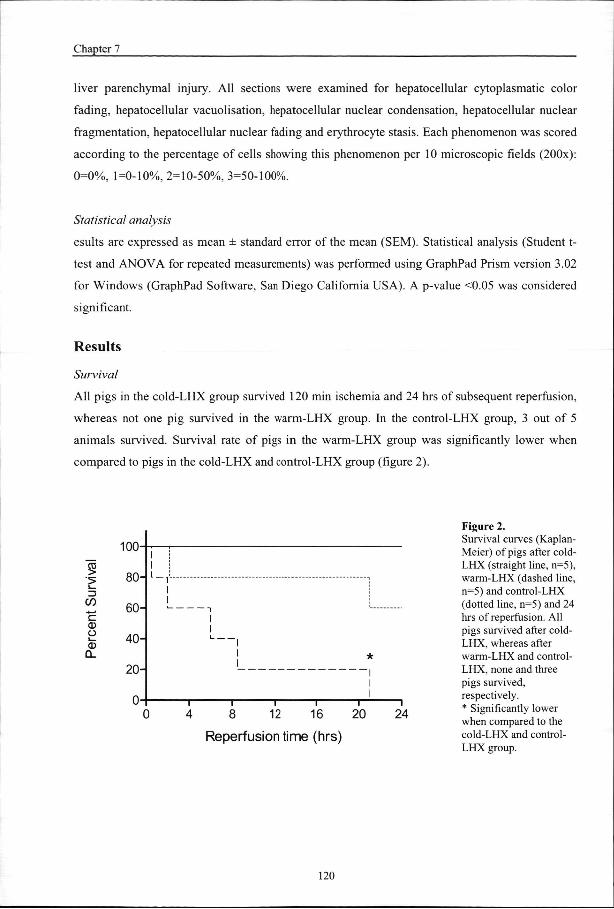
Al ll pigs in the cold-LHX group survived 120 min ischemia and 24 hrs of subsequent reperfusion,
whereass not one pig survived in the warm-LHX group. In the control-LHX group, 3 out of 5
animalss survived. Survival rate of pigs in the warm-LHX group was significantly lower when
comparedd to pigs in the cold-LHX and control-LHX group (figure 2).
3 3 <*> > "a. "a. CO CO o o CD D
100 0
80--
60--
40--
20--
0--
l l l l
0 0 1 1
4 4 I I
8 8 I I
12 2 i i
16 6
l l I I
20 0 i i
24 4
Reperfusionn time (hrs)
Figuree 2. Survivall curves (Kaplan-Meier)) of pigs after cold-LHXX (straight line, n=5), warm-LHXX (dashed line, n=5)) and control-LHX (dottedd line, n=5) and 24 hrss of reperfusion. All pigss survived after cold-LHX,, whereas after warm-LHXX and control-LHX,, none and three pigss survived, respectively. . ** Significantly lower whenn compared to the cold-LHXX and control-LHXX group.
120 0

Inn situ hypothermic perfusion
GeneralGeneral parameters
Inn pigs in the cold-LHX, warm-LHX and control-LHX groups, both resected liver weights
,, , 8 gr, resp.) and remnant , , 5 gr, resp.) liver
weightss ) were comparable.
Althoughh normal body temperature of pigs is considered 38°C, pigs in this study showed
higherr pre-ischemic body temperatures of 39°C due to anesthesia-induced stress, a commonly
observedd phenomenon in our laboratory. Body temperature of pigs in all groups was significantly
decreasedd with 1-2°C after 2 hrs ischemia when compared to pre-ischemic levels, showing no
significantt differences between pigs in all groups. During reperfusion body temperature remained
significantlyy below pre-ischemic temperature values in all groups (table 1).
Hemodynamicc parameters changed significantly during the experiment (table 1).
Hemodynamicc parameters of pigs in the cold-LHX group remained stable during the experiment,
withoutt the need for intravenous phenylephrine administration .
Experimentall groups
Bodyy temp (°C)
SAPP (mmHg)
DAPP (mmHg)
Heartt rate
(beats/min) )
Phenylephrine e
(mg/h) )
pre-I I
OhrR R
6hrsR R
244 hrs R
pre-I I
OhrR R
6hrsR R
244 hrs R
pre-I I
OhrR R
6hrsR R
244 hrs R
pre-I I
OhrR R
6hrsR R
244 hrs R
pre-I I
OhrR R
6hrsR R
244 hrs R
Cold-LHX X
39.11 3 A
37.11 1 A
38.00 2
38.11 0
110.66 6
99.88 7
95.66 2
105.88 1.2 Ü H 1 1
59.66 4.7 G
HJ J
59.00 4.3 '
699 4
944 0
777 °
811 0
0 0
0 0
0 P Q Q
0R R
Warm-LHX X
39.22 0.4 B
38.00 0.2 B
38.33 7
107.88 2.4 D t
67.00 12.9 D
87.00 2.3 E
K L L
40.88 5 K
37.33 5.7 JL
844 5
6 6
1111 °
0 0
0 0
2266 1 p
Control-LHX X
38.77 0.2 c
37.44 0.3 c
38.33 2
38.33 1
106.44 "
75.66 7.7 F
96.55 8
97.33 3
75.88 3.2 MN
43.22 5.2 M
46.33 3.5 N
46.00 8.9
799 6
1322 0
944 0
1188 30
0 0
3.33 3
900 19°
1066 1 R
121 1

Chapterr 7
Tablee 1 Meann rectal body temperature, hemodynamic parameters and use of phenylephrine before induction of ischemiaa and after 0, 6 and 24 hrs of reperfusion. Hemodynamic parameters remain most stable in pigs after cold-LHXX with no need for phenylephrine. Values are expressed as mean SEM. Temp=temperature,, SAP=systolic arterial pressure, DAP=diastolic arterial pressure Significantt differences exist between values tagged with equal fonts (p<0.05).
HepatocellularHepatocellular injury
Afterr control-LHX, AST levels were significantly elevated during reperfusion when compared to
pre-ischemicc levels and continued to increase during reperfusion, reaching maximum levels at 24
hrs.. AST levels after warm-LHX were higher when compared to pigs after control-LHX during
reperfusionn but this did not reach statistical significance (p=0.17). After cold-LHX, AST levels
weree significantly lower than in pigs after warm-LHX and control-LHX at all time points and
reachedd maximum levels after 6 hrs of reperfusion (figure 3).
Figuree 3 Plasmaa aspartate aminotransferase (AST)) assessed in pigs after cold-LHX (dashedd bar, n=5), warm-LHX (grey bar, n=5,, n=3 at 6 hrs of reperfusion) and control-LHXX (closed bar, n=5, n=4 at 6 andd 12 hrs of reperfusion, n=3 at 24 hrs off reperfusion) at pre-ischemia and after 100 min, 6, 12 and 24 hrs of reperfusion. Afterr cold-LHX, AST levels were significantlyy lower than after warm-LHXX and control-LHX. Bars represent meann SEM. ** Significantly different from pigs after cold-LHX. .
HepatocellularHepatocellular function
Bil ee production decreased to almost zero during ischemia in all pigs. During 24 hrs of reperfusion
bilee production in pigs after cold-LHX returned to pre-ischemic levels. In contrast, bile
productionn in pigs after warm-LHX and control-LHX did not recover and remained significantly
lowerr when compared to pigs after cold-LHX (figure 4).
Indocyaninee green (ICG) clearance in pigs in the cold-LHX group did not change during
thee time course of the experiments. However, after warm-LHX and control-LHX, ICG clearance
afterr 6 hrs of reperfusion was significantly decreased. Pigs in the control-LHX group did not show
anyy recovery of ICG clearance after 24 hrs of reperfusion and ICG clearance remained
significantlyy lower when compared to pigs after cold-LHX (figure 5).
3000--
="" 2000-3 3
3 3 1000--
flt flt
* *
—— a pre-ll 10m R 6h R 12hR 24h R
Samplingg time
122 2

Inn situ hypothermic perfusion
0.15 5
0.00 0 pre-ll Ischemia 24h R
Samplingg time
Figuree 4 Bilee production in pigs after cold-LHXX (dashed bar, n=5), warm-LHXX (grey bar, n=5, maximum n=4 duringg 21 hrs of reperfusion) and control-LHXX (closed bar, n=5, n=4 duringg reperfusion) assessed pre-ischemia,, during ischemia and duringg 24 hrs of reperfusion. In contrastt to pigs after warm-LHX andd control-LHX, bile production completelyy recovered during reperfusionn after cold-LHX. Bars representt mean SEM. ** Significantly different from pigs afterr warm-LHX and control-LHX.
24hR R
Samplingg time
Figuree 5 Indocyaninee green (ICG) clearance inn pigs after cold-LHX (dashed bar, n=5),, warm-LHX (grey bar, n=5, n=33 at 6 hrs of reperfusion) and control-LHXX (closed bar, n=5, n=4 att 6 hrs of reperfusion, n=3 at 24 hrss of reperfusion) at pre-ischemia andd after 6 and 24 hrs of reperfusion.. ICG clearance capacityy was maintained during reperfusionn after cold-LHX. After warm-LHXX and control-LHX, ICG clearancee was decreased during reperfusion,, showing rates compatiblee with severely impaired liverr function. Bars represent mean
. . ** Significantly different from pigs afterr cold-LHX.
123 3

Chapterr 7
SECSEC function
HAA uptake could only be measured in pigs after cold-LHX and control-LHX since all pigs after
warm-LHXX died within 24 hrs of reperfusion. The HA uptake profile after 24 hrs of reperfusion
wass not significantly different between both groups during the 60 min sampling period. After
cold-LHXX and control-LHX, 74% and 78%, resp. of exogenous HA was cleared from the
circulationn indicating near normal SEC function 29 after 24 hrs of reperfusion in both groups.
Figuree 6 Uptakee of exogenous hyaluronic acid (HA)) in a 60 min period following 2 hrss ischemia and 24 hrs of reperfusion afterr cold-LHX (dashed bar, n=5) and control-LHXX (closed bar, n=3). There weree no survivors in the warm-LHX groupp after 24 hrs of reperfusion. Resultss were expressed as the percentagee of exogenous administered HAA taken up by the sinusoidal endotheliall cells (SEC) during 60 min.. No decrease in SEC function wass observed in pigs after cold-LHX whenn compared to pigs after control-LHX.. Bars represent mean SEM.
Periodd (min)
InflammatoryInflammatory response
Inflammatoryy response was assessed by measurement of IL-6 in plasma. Plasma IL-6 was not
detectablee before induction of ischemia, indicating that none of the pigs suffered from infectious
diseasess prior to the experiment. After 10 min of reperfusion, pigs in all groups showed a marked
increasee in IL-6 levels, but levels in pigs after cold-LHX were significantly lower when compared
too both other groups. At 6 hrs of reperfusion IL-6 levels of pigs in the warm-LHX and control-
LHXX group reached maximum values whereas IL-6 levels of pigs in the cold-LHX group showed
aa decrease. After 12 hrs and 24 hrs of reperfusion IL-6 levels of pigs after cold-LHX were still
lowerr than in pigs after control-LHX but this did not reach statistical significance (p=0.09 and
p=0.088 resp., figure 7).
124 4

Inn situ hypothermic perfusion
Figuree 7 Plasmaa IL-6 levels measured in pigs after cold-LHXX (dashed bar, n=5), warm-LHX (greyy bar, n=5, n=3 at 6 hrs of reperfusion) andd control-LHX (closed bar, n=5, n=4 at 6 andd 12 hrs of reperfusion, n=3 at 24 hrs of reperfusion)) at pre-ischemia and after 10 min,, 6, 12 and 24 hrs of reperfusion. After cold-LHX,, IL-6 levels were significantly lowerr than after warm-LHX and control-LHXX until 6 hrs of reperfusion. Bars representt mean SEM. ** Significantly different from pigs after cold-LHX. .
Samplingg time
HistopathoHistopatho logy
Microscopicc evaluation of liver biopsies of all pigs showed a significant increase in
histopathologyy score after 10 min of reperfusion when compared to pre-ischemia. The score in
pigss after cold-LHX was significantly lower when compared to pigs after warm-LHX and control-
LHXX after 10 min of reperfusion. After 24 hrs of reperfusion, pigs in both the cold-LHX and the
control-LHXX group showed more pathologic changes when compared to 10 min of reperfusion
althoughh statistical significance was not reached (p=0.25 and p=0.05 resp.). The histopathological
scoress in biopsies collected at 24 hrs of reperfusion were significantly higher in pigs after control-
LHXX than after cold-LHX (figure 8). Histopathological scores in the warm-LHX group are
lackingg because there were no survivors in this group after 24 hrs of reperfusion.
Figuree 8 Histopathologyy scores (min=0, max=18)) of H&E stained liver sections off pigs after cold-LHX (dashed bar, n=5),, warm-LHX (grey bar, n=5) and control-LHXX (closed bar, n=5) at pre-ischemiaa and after 10 min as well as 24 hrss of reperfusion. After cold-LHX, histopathologyy scores were significantlyy lower than after warm-LHXX and control-LHX. Bars represent meann SEM. ** Significantly different from pigs after cold-LHX. .
Samplingg time
1500-, ,
11 1000-
to to
—— 500-
1 1
** *
ii il I II all *> I a i pre-ll 10m R 6h R 12h R 24h R
n r s J J - lOrvii D O/lhh D
125 5

Chapterr 7
Discussion n
Variouss vascular occlusion techniques have been used to prevent massive blood loss during
partiall hepatectomy. Portal triad clamping (Pringle maneuver) and THVE are two methods to
reducee hemorrhage during parenchymal dissection. Although hemorrhage can be more effectively
reducedd during partial hepatectomy under THVE, there is evidence that by applying this
technique,, liver tissue oxygenation is further hampered due to the lack of back-perfusion from the
cavall vein thereby enhancing I/R injury . This finding underscores the use of protective
measuress when applying THVE, such as in situ hypothermic perfusion of the future remnant liver.
Thee objective of this study was to examine whether in situ hypothermic perfusion is a
usefull tool to attenuate liver I/R injury and to investigate if this protective effect resulted from
clearancee of the liver from blood or to cooling of the liver per se. Cooling of the organ leads to a
decreasee in cell metabolism, as was demonstrated by a reduction in the velocity of various organic
reactionss by a factor 2 for every 10°C reduction in temperature 32. This results in a decrease in
hepaticc oxygen demand and uptake and thus provides an effective protection from hepatic oxygen
deprivationn thereby reducing reperfusion injury 3\ In literature, a major role is attributed to the
formationn of reactive oxygen species during reperfusion 34~36, which is attenuated by cooling of
thee organ. Other mechanistic factors, like maintenance of mitochondrial function by preventing
calciumm overload , suppression of nitric oxide formation or preservation of important
intracellularr concentrations of metabolites are also shown to play a role in temperature related I/R
injury.. This study showed that clearance of the liver from blood without concomitant cooling
severelyy aggravates liver I/R injury. The use of Ringer-glucose as perfusion fluid has several
implicationss for the observed outcome in this study. Although the Ringer-glucose solution was
isotonic,, the lack of proteins and the acidotic pH (4.65) probably enhanced endothelial cell
swellingg at normothermic temperatures, leading to microvascular disturbances and ultimately
enhancedd liver injury during reperfusion 39. This explanation is corroborated by the extensive
congestionn of the liver observed at the onset of reperfusion in pigs of the warm perfused group.
Inn the present study, a decrease in core liver temperature of 10°C was chosen to
investigatee if hepatocytes could be protected from I/R injury at this temperature without
hamperingg sinusoidal endothelial cell (SEC) function. Preserved hyaluronic acid (HA) uptake
capacityy after 24 hrs of reperfusion indicates that a drop of 10°C in liver core temperature did not
affectt SEC function. In fact, SEC still had a high reserve capacity for HA uptake after left
126 6

Inn situ hypothermic perfusion
hemihepatectomyy which is in agreement with previous experiments performed in our laboratory 29 9
Thiss pig liver I/R model constitutes a clinically relevant, large animal model, but at the
samee time has its limitations. Relatively long THVE times were used in this protocol for optimal
investigationn of the protective effect of in situ cooling on liver I/R injury. A THVE time of 2 hrs
resultss in sub-lethal liver failure, indicated by the survival of only 3 out of 5 pigs in the control-
LHXX group during 24 hrs of reperfusion. If circulatory support by infusion of phenylephrine was
nott provided in the control-LHX group, all pigs would have died during the 24 hrs of reperfusion
periodd and a statistical significant difference with the cold-LHX group would have been easily
reached.. Pigs in the cold-LHX group did not require phenylephrine infusion. Whereas plasma
ASTT levels in pigs in the cold-LHX group reached maximum levels after 6 hrs of reperfusion,
ASTT levels in pigs in the control-LHX group continued to increase during the 24 hrs of
reperfusionn period. This indicates that only pigs in the cold-LHX group showed recovery from the
sustainedd hepatic I/R injury.
Inn humans, an ICG clearance in livers of >90% is considered normal. In this study, ICG
clearancee in all pigs before induction of ischemia was 1 , which is
comparablee to the human situation. In literature, a correlation was observed between ICG
clearancee and functional liver mass 40. In this study however, no differences were observed
betweenn ICG clearance in pigs of the cold-LHX group before and after partial liver resection,
indicatingg an ample reserve capacity of the liver for ICG clearance. Pigs after warm-LHX and
control-LHX,, however, showed clearance rates of <70%, compatible with severely impaired liver
function. .
Usee of THVE can cause serious side effects like hemodynamic instability . Because
portosystemicc collateral circulation is poorly developed in pigs, portal venous drainage is
mandatoryy under THVE 31;42. In humans, however, collateral circulation of the portal venous
systemm is better developed and therefore, a portal venous shunt is usually not required. Sustained
hypotensionn by test clamping prior to resection ', indicates the need for a venovenous bypass
duringg THVE of which several systems have been described, both passive and active ' ' .
Usuallyy in these cases, systemic heparinisation is required to minimize the risk of intra-shunt
clotting,, thereby increasing the risk of uncontrolled hemorrhage. In the present experiments, a
customm made, stiff polycarbonate intraluminal prosthesis with one side-port was used to bypass
bloodd from the infrahepatic caval vein to the suprahepatic caval vein while at the same time
drainingg the portal venous system. Intra-shunt clotting was not observed and systemic
127 7

Chapterr 7
heparinisationn or local use of heparin was not required, which can be a major advantage during
thesee operative procedures.
Severall methods have been described for in situ cooling of the liver. Yang-IL et al. used
simplee in situ cooling by rapid infusion of chilled 450 ml Ringer-lactate before clamping of the
portall triad (Pringle maneuver). In their study, hypothermia of the liver lasted about one hour and
noo concomitant depression in body temperature was observed 44. These findings, however, could
nott be reproduced in our laboratory in which liver temperature in pigs could not be significantly
reducedd by rapid cold infusion without a concomitant decrease in body temperature (data not
shown).. Cooling of the liver can be achieved by infusion of different fluids via the hepatic artery
orr portal vein , or via both at the same time '°. Alternatively, topical surface cooling during
partiall hepatectomy can be used 45:46. It is not yet known which perfusion method is most
effective,, although our study shows that perfusion via the hepatic artery alone provides sufficient
coolingg of the liver.
Inn conclusion, a mild decrease in core liver temperature with 10°C by in situ
hypothermicc perfusion proved effective in attenuating liver I/R injury as demonstrated by
improvedd hepatocellular function, preservation of SEC function, less histopathological damage,
decreasedd systemic inflammatory response and increased survival in this pig model. Clearance of
thee liver from blood using Ringer-glucose without concomitant cooling severely aggravated I/R
injury. .
128 8

Inn situ hypothermic perfusion
References s 1.. Delva E, Camus,Y, Nordlinger,B, Hannoun.L, Parc,R, Deriaz,H, Lienhart,A, Huguet,C. Vascular
occlusionss for liver resections. Operative management and tolerance to hepatic ischemia: 142 cases.. Ann Surg 1989;209:211-218.
2.. Huguet C, Gavelli.A, Chieco,PA, Bona,S, Harb,J, Joseph,JM, Jobard,J, Gramaglia,M, Lasserre,M. Liverr ischemia for hepatic resection: where is the limit? Surgery 1992;111:251-259.
3.. Man K, Fan,ST, Ng,IO, Lo,CM, Liu,CL, Wong,J. Prospective evaluation of Pringle maneuver in hepatectomyy for liver tumors by a randomized study. Ann Surg 1997;226:704-711.
4.. Huguet C, Nordlinger,B, GalopinJJ, Bloch,P, Gallot,D. Normothermic hepatic vascular exclusion forr extensive hepatectomy. Surg Gynecol Obstet 1978;147:689-693.
5.. Bismuth H, Castaing,D, Garden,OJ. Major hepatic resection under total vascular exclusion. Ann Surgg 1989;210:13-19.
6.. Jaeschke H. Mechanisms of reperfusion injury after warm ischemia of the liver. J Hepatobiliary PancreatSurgg 1998;5:402-408.
7.. Huguet C, Gavelli,A, Bona,S. Hepatic resection with ischemia of the liver exceeding one hour. J Amm Coll Surg 1994;178:454-458.
8.. Neuhaus P, Jonas,S, Bechstein,WO, Lohmann,R, Radke,C, Kling,N, Wex,C, Lobeck,H, Hintze,R. Extendedd resections for hilar cholangiocarcinoma. Ann Surg 1999;230:808-818.
9.. Harmoun L, Balladur,P, Delva,E, Panis.Y, Camus,Y, HonigerJ, Levy,E, Parc,R. ["Ex situ-in vivo" surgeryy of the liver: a new technique in liver surgery. Principles and preliminary results]. Gastroenteroll Clin Biol 1991;15:758-761.
10.. Former JG, Shiu,MH, Kinne,DW, Kim,DK, Castro,EB, Watson,RC, Howland,WS, Beattie,EJ, Jr. Majorr hepatic resection using vascular isolation and hypothermic perfusion. Ann Surg 1974;180:644-652. .
11.. Hannoun L, Delriviere,L, gibbs,P, Borie,D, VaillantJC, Delva,E. Major extended hepatic resectionss in diseased livers using hypothermic protection: preliminary results from the first 12 patientss treated with this new technique. J Am Coll Surg 1996;183:597-605.
12.. Vaillant JC, Borie,DC, Hannoun,L. Hepatectomy with hypothermic perfusion of the liver. Hepatogastroenterologyy 1998;45:381 -388.
13.. Ikeda T, Yanaga,K, Kishikawa,K, Kakizoe,S, Shimada,M, Sugimachi,K. Ischemic injury in liver transplantation:: difference in injury sites between warm and cold ischemia in rats. Hepatology 1992;16:454-461. .
14.. Biberthaler P, Luchting,B, Massberg.S, Teupser,D, Langer,S, Leiderer,R, Messmer,K, Krombach,F.. THE INFLUENCE OF ORGAN TEMPERATURE ON HEPATIC ISCHEMIA-REPERFUSIONN INJURY: A Systematic Analysis 1. Transplantation 2001;72:1486-1490.
129 9

Chapterr 7
15.. van Wagensveld BA, van Gulik,TM, Gelderblom,HC, Scheepers, J J, Bosma,A, Endert,E, Gouma,DJ.. Prolonged continuous or intermittent vascular inflow occlusion during hemihepatectomyy in pigs. Arm Surg 1999;229:376-384.
16.. Bowers BA, Branum,GD, Rotolo,FS, Watters,CR, Meyers,WC. Bile flow-an index of ischemic injury.. J Surg Res 1987;42:565-569.
17.. El Desoky A, Seifalian,AM, Cope,M, Delpy.DT, Davidson,BR. Experimental study of liver dysfunctionn evaluated by direct indocyanine green clearance using near infrared spectroscopy. Br J Surgg 1999;86:1005-1011.
18.. Lau H, Man,K, Fan,ST, Yu,WC, Lo,CM, WongJ. Evaluation of preoperative hepatic function in patientss with hepatocellular carcinoma undergoing hepatectomy. Br J Surg 1997;84:1255-1259.
19.. Hemming AW, Scudamore,CH, Shackleton,CR, Pudek,M, Erb, SR. Indocyanine green clearance ass a predictor of successful hepatic resection in cirrhotic patients. Am J Surg 1992;163:515-518.
20.. Gottlieb ME, Stratton,HH, NewellJC, Shah,DM. Indocyanine green. Its use as an early indicator off hepatic dysfunction following injury in man. Arch Surg 1984;119:264-268.
21.. Tsubono T, Todo,S, Jabbour,N, Mizoe,A, Warty,V, Demetris,AJ, Starzl,TE. Indocyanine green eliminationn test in orthotopic liver recipients. Hepatology 1996;24:1165-1171.
22.. Tamaki S, Ueno,T, Torimura,T, Sata,M, Tanikawa,K. Evaluation of hyaluronic acid binding ability off hepatic sinusoidal endothelial cells in rats with liver cirrhosis. Gastroenterology 1996; 111:1049-1057. .
23.. Deaciuc IV, Bagby,GJ, Lang,CH, Spitzer,JJ. Hyaluronic acid uptake by the isolated, perfused rat liver:: an index of hepatic sinusoidal endothelial cell function. Hepatology 1993;17:266-272.
24.. Reinders ME, van Wagensveld,BA, van Gulik,TM, Frederiks,WM, Chamuleau,RAFM, Enderd,E, Klopper,PJ.. Hyaluronic acid uptake in the assessment of sinusoidal endothelial cell damage after coldd storage and normothermic reperfusion of rat livers. Transpl Int 1996;9:446-453.
25.. Namekata K, Takamori,S, Kojima,K, Beppu,T, Futagawa,S. Significant changes in the serum levelss of IL-6, h-HGF, and type IV collagen 7S during the perioperative period of a hepatectomy: relevancee to SIRS. Surg Today 2000;30:403-409.
26.. Fattori E, Cappelletti,M, Costa,P, Sellitto,C, Cantoni,L, Carelli,M, Faggioni,R, Fantuzzi,G, Ghezzi,P,, Poli,V. Defective inflammatory response in interleukin 6-deficient mice. J Exp Med 1994;180:1243-1250. .
27.. Ohzato H, Yoshizaki,K, Nishimoto,N, Ogata,A, Tagoh,H, Monden,M, Gotoh,M, KishimotoJ, Mori,T.. Interleukin-6 as a new indicator of inflammatory status: detection of serum levels of interleukin-66 and C-reactive protein after surgery. Surgery 1992;111:201-209.
28.. Helle M, Boeije,L, Aarden,LA. Functional discrimination between interleukin 6 and interleukin 1. Eurr J Immunol 1988;18:1535-1540.
130 0

Inn situ hypothermic perfusion
29.. van Wagensveld BA, van Gulik.TM, Gelderblom,HC, Scheepers,JJ, Bosma,A, Endert,E, Obertop,H,, Gouma,DJ. Continuous or intermittent vascular clamping during hemihepatectomy in pigs:: hyaluronic acid kinetics in the assessment of early microvascular liver damage. Eur J Surg 2000;166:255-261. .
30.. Tatsuma T, Kim,YI, Kai,T, Ishii,T, Akizuki,S, Takayama,F, Egashira,T, Kobayashi,M. Importance off hepatovenous back-perfusion for maintenance of liver viability during the Pringle manoeuvre. Brr J Surg 1995;82:1071-1075.
31.. Hiratsuka K, Kim,YI, Nakashima,K, Kawano,K, Yoshida,T, Kitano,S. Tissue oxygen pressure duringg prolonged ischemia of the liver. J Surg Res 2000;92:250-254.
32.. Belzer FO, Southard,JH. Principles of solid-organ preservation by cold storage. [Review] [27 refs]. Transplantationn 1988;45:673-676.
33.. Nagano K, Gelman,S, Bradley,EL, Jr., Parks,D. Hypothermia, hepatic oxygen supply-demand, and ischemia-- reperfusion injury in pigs. Am J Physiol 1990;258:G910-8.
34.. Zar HA, Tanigawa,K, Kim,YM, Lancaster,JR, Jr. Mild therapeutic hypothermia for postischemic vasoconstrictionn in the perfused rat liver. Anesthesiology 1999;90:1103-1 111.
35.. Karibe H, Chen,SF, Zarow,GJ, Gafni,J, Graham,SH, Chan,PH, Weinstein,PR. Mild intraischemic hypothermiaa suppresses consumption of endogenous antioxidants after temporary focal ischemia in rats.. Brain Res 1994;649:12-18.
36.. Globus MY, Busto,R, Lin,B, Schnippering,H, Ginsberg,MD. Detection of free radical activity duringg transient global ischemia and recirculation: effects of intraischemic brain temperature modulation.. J Neurochem 1995;65:1250-1256.
37.. Ferrari R, Raddino,R, Di Lisa,F, Ceconi,C, Curello,S, Albertini,A, Nayler,W. Effects of temperaturee on myocardial calcium homeostasis and mitochondrial function during ischemia and reperfusion.. J Thorac Cardiovasc Surg 1990;99:919-928.
38.. Kumura E, Yoshimine,T, Takaoka,M, Hayakawa,T, Shiga,T, Kosaka,H. Hypothermia suppresses nitricc oxide elevation during reperfusion after focal cerebral ischemia in rats. Neurosci Lett 1996;220:45-48. .
39.. Mazzoni MC, Borgstrom,P, Wamke,KC, Skalak,TC, Intaglietta,M, Arfors,KE. Mechanisms and implicationss of capillary endothelial swelling and luminal narrowing in low-flow ischemias. Int J Microcircc Clin Exp 1995;15:265-270.
40.. Bolognesi M, Merkel,C, Angeli,P, Caregaro,L, Gatta,A. [Liver metabolism of indocyanine green andd aminopyrine in liver cirrhosis: relations with functioning liver cell mass]. Minerva Med 1988;79:1035-1041. .
41.. Belghiti J, Noun,R, Zante,E, Ballet,T, Sauvanet,A. Portal triad clamping or hepatic vascular exclusionn for major liver resection. A controlled study. Ann Surg 1996;224:155-161.
42.. van Wagensveld BA, van Gulik,TM, Gabeler,EE, van der kleij,AJ, Obertop,H, Gouma,DJ. Intrahepaticc tissue p02 during continuous or intermittent vascular inflow occlusion in a pig liver resectionn model. Eur Surg Res 1998;30:13-25.
131 1

Chapterr 7
43.. Takeuchi K, Ohira,M, Yamashita,T, Sowa,M. An experimental study of hepatic resection using an inn situ hypothermic perfusion technique. Hepatogastroenterology 1997;44:1281-1294.
44.. Kim YI, Hiratsuka,K, Kitano,S, Joo,DH, Kamada,N, Sugimachi,K. Simple in situ hypothermia reducedd ischaemic injury to human liver during hepatectomy. Eur J Surg 1996;162:717-721.
45.. Yamanaka N, Okamoto,E. [Topical cooling as an adjunct for hepatectomy with inflow occlusion]. Nipponn Geka Gakkai Zasshi 1998;99:223-228.
46.. Imakita M, Yamanaka,N, Kuroda,N, Kitayama,Y, Okamoto,E. Does topical cooling alleviate ischemia/reperfusionn injury during inflow occlusion in hepatectomy? Results of an experimental andd clinical study [In Process Citation]. Surg Today 2000;30:795-804.
132 2