Utilization of autolytically active cell wall for...
116
UTILIZATION OF AUTOLYTICALLY ACTIVE CELL WALL FOR CHARACTERIZATION OF TOMATO FRUIT SOFTENING BY JAMES WAYNE RUSHING A DISSERTATION PRESENTED TO THE GRADUATE SCHOOL OF THE UNIVERSITY OF FLORIDA IN PARTIAL FULFILLMENT OF THE REQUIREMENTS FOR THE DEGREE OF DOCTOR OF PHILOSOPHY UNIVERSITY OF FLORIDA 1985
Transcript of Utilization of autolytically active cell wall for...

UTILIZATION OF AUTOLYTICALLY ACTIVE CELL WALLFOR CHARACTERIZATION OF TOMATO FRUIT SOFTENING
BY
JAMES WAYNE RUSHING
A DISSERTATION PRESENTED TO THE GRADUATE SCHOOLOF THE UNIVERSITY OF FLORIDA IN
PARTIAL FULFILLMENT OF THE REQUIREMENTSFOR THE DEGREE OF DOCTOR OF PHILOSOPHY
UNIVERSITY OF FLORIDA
1985

ACKNOWLEDGMENTS
I wish to thank my committee members and my family for their
assistance and support.
n

TABLE OF CONTENTS
Page
ACKNOWLEDGMENTS i i
LIST OF TABLES v
LIST OF FIGURES vi
ABSTRACT vii i
CHAPTERS
I INTRODUCTION 1
II LITERATURE REVIEW 3
Horticultural Significance of Tomato Fruit Softening 3
Approaches to the Study of Tomato Fruit Softening 4
Cell Wall Hydrolases Associated with Tomato FruitSoftening 11
Polygalacturonase 12
Pectinmethylesterase 13
Cellulase 14
S-Galactosidase 14
3-1,3-Glucanase 15
Isolation of Plant Cell Walls 15
Problems Inherent to Wall Isolation Procedures 15
Binding of Polygalacturonase to Cell Wall 20
Regulation of Polygalacturonase In Vitro. 23
Implications of Microbial Contamination in
Cell Wall Studies 28
III ENZYMICALLY ACTIVE CELL WALL AS A TOOL FORINVESTIGATION OF FRUIT SOFTENING 29
Materials and Methods 31
Plant Material 31
Cell Wall Preparation 31
Autolysis Experiments 32
Results and Discussion 33
iii

IV RELATIONSHIP BETWEEN HANDLING TECHNIQUES ANDSOLUBILIZATION OF CELL-WALL CONSTITUENTS DURINGAUTOLYSIS OF ISOLATED TOMATO CELL WALLS 42
Materials and Methods 44Plant Material 44Cell Wall Preparation 44Cell Wall Rehydration 45Sampling Techniques for Soluble Products 45Postautolysis Wall Recovery 46Soluble Product Analysis 46Wall Compositional Analysis 46Autolysis Experiments 47
Experiment 1 47Experiment 2 47Experiment 3 47Experiment 4 48Experiment 5 48
Results 48Experiment 1 48Experiment 2 49Experiment 3 51Experiment 4 55
Experiment 5 57
Discussion 63
V EFFECTS OF NaCl, dH, AND CALCIUM ON AUTOLYSIS OFISOLATED TOMATO CELL WALLS 71
Materials and MethodsPlant Material 72Cell Wall Preparation 72
Autolysi s Experiments 73
Experiment 1 74
Experiment 2 74Experiment 3 74Experiment 4 76
Resul ts „ 77
Experiment 1 77
Experiment 2 79
Experiment 3 81Experiment 4 83
Discussion 84
VI SUMMARY 90
ilBLIOGRAPHY 93
IIOGRAPHICAL SKETCH 104
IV

LIST OF TABLES
Table Page
3-1 Effect of pH on total amount of polyuronides releasedfrom cell wall of pink pericarp 39
4-1 Estimates of wall dry wts of samples recoveredpostautolysis by Miracloth filtration or centrifugation. . ..50
4-2 Yield of cell -walls extracted from 'Rutgers' tomato fruitat different stages of development 51
4-3 Dry wts and acid sugar content of cell-walls 55
4-4 Dry wts of individual components of the autolyticreaction 58
5-1 Protocol for cell -wall extraction in Experiment 3 75
5-2 Postautolysis dry wt of cell walls 79

LIST OF FIGURES
Figure Page
3-1 Release of polyuronide from cell wall of tomatopericarp at different stages of development 34
3-2 Characterization of the autolytic reaction in cell
wall from pericarp of pink tomato fruit 37
4-1 Effect of incubation time and postautolysis recoverymethod on estimate of solubilized acid sugars duringautolysis of freshly prepared cell walls from 'Rutgers'
pink pericarp 50
4-2 Effect of acetone-washing and drying on release ofpolyuronide from cell-walls isolated from 'Rutgers'pink or red pericarp 52
4-3 Comparison of methods of sampling for solubleautolysis products from cell walls isolated from'Rutgers' pink pericarp 54
4-4 Comparison of aliquot removal to centrifugation andsupernatant removal for sampling of solubilized acidsugars in cell walls from 'Rutgers' pink or
mature-green pericarp 56
4-5 Solubilization of uronic acids and proteins duringautolysis of cell walls extracted from 'Rutgers' pinkpericarp 58
4-6 Solubilization of neutral sugars and acidrneutralsugar ratio during autolysis 60
4-7 Neutral sugars released in the autolytic reaction 60
4-8 Bio-Gel P-10 chromatography of polyuronides releasedfrom autolytically active cell wall 61
4-9 Fractogel HW-50 chromatography of polyuronidesreleased from autolytically active cell wall 62
5-1 Effect of NaCl on release of polyuronide from cellwalls of pink or red tomato pericarp 78
VI

5-2 Autolytic activity in cell walls from pink pericarpincubated at pH 4.5 after pretreatment with 1 M NaClat different pH values 80
5-3 Effect of exposure to 1 M NaCl on solubilization ofacid sugars from cell walls of red tomato pericarp 82
5-4 Effects of pH, NaCl, and calcium on solubilizationof acid sugars from cell walls of red tomato pericarp 85
vn

Abstract of Dissertation Presented to the Graduate School
of the University of Florida in Partial Fulfillment of the
Requirements for the Degree of Doctor of Philosophy
UTILIZATION OF AUTOLYTICALLY ACTIVE CELL WALLFOR CHARACTERIZATION OF TOMATO FRUIT SOFTENING
BY
JAMES WAYNE RUSHING
December, 1985
Chairman: Donald J. HuberMajor Department: Horticultural Science
Cell walls from pericarp of normal tomato ( Lycopersicon
esculentum Mil 1 . cv 'Rutgers') fruit released acid sugars in vitro in
a reaction mediated by wall-bound polygalacturonase (PG;
EC 3.2.1.15). Release was negligible in walls from normal green and
the ripening mutant rin fruit, reflecting the absence of PG in these
tissues. Pectin solubilization was most extensive at pH 2.5 with a
less pronounced optimum at 5.5. Brief exposure of cell wall to low
(1.5) or high (7.5) pH resulted in reduction of autolytic activity,
which also was inhibited by high temperature, calcium, and treatments
employed to dissociate wall-bound protein.
The rate and extent of acid sugar solubilization were dependent
upon the techniques employed for handling the cell walls in vitro .
Enzymically active cell walls subjected to either intermittent or
continuous removal of the bathing medium released twice as much
vm

polyuronide as walls incubated in a static system. Protocol that
maximized acid sugar solubilization was employed to determine the
neutral sugar content and size distribution of pectic polymers
released during autolysis. Approximately 2% of the initial wall dry
weight was released as neutral sugars. The predominant products were
galactose (50%) and arabinose (30%). Minor quantities of rhamnose,
xylose, glucose, and mannose were also detected. The acid:neutral
sugar ratio of soluble autolysis products decreased from 10.3 to 2.9
as wall degradation proceeded. Pectin molecular size did not change
appreciably during autolytic reactions.
Polyuronide solubilization was optimally enhanced by NaCl in the
range of 200-300 mM. Pretreatment of enzymically active walls with
1 M NaCl in the pH range of 2.5-8.5 did not inhibit subsequent
autolysis. The tenacity of PG-binding was further illustrated by the
fact that 1 M NaCl treatments during wall extraction did not
eliminate release of acid sugars. Interactions between pH, NaCl, and
calcium were also investigated. The stimulation of wall-bound PG
activity by 150-300 mM NaCl was most apparent at pH 2.5. Calcium
(5 mM) did not inhibit autolysis at pH 2.5; however, at pH 4.5
calcium inhibition was apparent regardless of presence of NaCl.
Polyuronide solubilization was negligible at pH 6.5 or 8.5.
IX

CHAPTER 1
INTRODUCTION
Softening is one of the most dramatic events associated with
tomato fruit ripening. A degree of control over the rate of texture
change is desirable because consumer preferences for firmness vary
widely, e.g., soft, fully ripe tomatoes are desired for processing,
whereas firm fruit are preferred for shipping long distances or for
slicing in the fast food industry.
The cell wall is the most significant structural entity of the
plant cell (3artnicki-Garcia, 1984). Fruit softening involves
enzymic hydrolysis of specific cell wall polymers (Huber, 1983b).
Cells with partially degraded walls yield to external pressures and
slide against each other, i.e., they become soft. The enzyme most
often implicated in tomato fruit softening is polygalacturonase (PG;
EC 3.2.1.15) which is believed to be synthesized with the onset of
ripening (Tucker and Grierson, 1982). Its role is to catalyze
hydrolysis of pectic polymers of the middle lamella (Crookes and
Grierson, 1983). The most common approach to investigating the
action of PG and other cell wall hydrolases is to extract the
enzyme(s) of interest and assay for activity with commercially
prepared substrates (Wallner and Walker, 1975) or with a cell wall
preparation (Hobson, 1964; Pressey and Avants, 1973). Such studies
have provided much useful information. The interpretation of this
1

information in terms of in situ wall hydrolysis is limited by the
fact that techniques used to prepare cell walls or commercially
available carbohydrate polymers may remove factors that are crucial
to the hydrolytic activity of the enzyme. An alternative to the
above approach is to study cell wall autolysis, i.e., to extract
walls under conditions which preserve the bond between PG and its
substrate, then incubate the wall extract in vitro under conditions
that permit enzymic activity (autolysis). Products of this reaction
may constitute a more appropriate reflection of in situ wall
hydrolysis associated with fruit softening.
Several workers have used cell wall autolysis to study wall
metabolism associated with growth and development (Kivilaan et a!.,
1961; Lee et al., 1967; Huber and Nevins, 1979). This dissertation
addresses the use of enzymically active cell walls isolated from
tomato pericarp to investigate activity of wall-bound PG. Initial
experiments compared the autolysis system to other in vitro cell wall
studies of fruit softening. Variables such as salt stimulation or
inhibition, pH, temperature, and inactivation treatments were
investigated for their effect on autolysis. Several techniques for
cell wall preparation and handling were evaluated. The rate and
extent of acid sugar solubilization during in vitro autolysis were
significantly affected by handling techniques employed. A system
that maximized the release of autolysis products was chosen for
characterization of the neutral sugar component and size distribution
of solubilized pectic polymers. Further studies addressed the
effects of MaCl, pH, and calcium on autolysis of isolated wal Is.

CHAPTER 2
LITERATURE REVIEW
Horticultural Significance of Tomato Fruit Softening
Tomato ( Lycopersicon esculentum , Mill.) is the major fresh
market vegetable produced in the state of Florida, where skyrocketing
increases in production costs have emphasized the importance of
preserving product quality from field to consumer (Taylor and
Wilkowske, 1984). Climacteric fruits, such as tomato, may require
marketing within a relatively short period in order to avoid the
numerous pathological and physiological disorders that result in
product deterioration (Conway, 1984). Texture is an important
quality factor and change in texture (softening) often is measured in
postharvest studies because it is closely associated with other
features of ripening, such as color development (Hobson, 1964, 1965;
Khudairi, 1972), climacteric behavior (Gertman and Fuchs, 1974;
Tigchelaar et al., 1978; Yoshida et al., 1984) and catabolism of
membrane constituents (Borochov and Faiman-Weinberg, 1984). The
relationship between softening and ripening metabolism is so intimate
that several workers addressed the idea that softening enzymes
initiate ripening by solubilization of pectins. Wall -bound proteins
released during pectin solubilization presumably start a cascade of
metabolic reactions that collectively compose the phenomenon known as
ripening (Strand et al., 1976; Tigchelaar et al., 1978; Poovaiah and

Nukaya, 1979). This idea has been somewhat refuted by evidence that
the wall hydrolase primarily responsible for tomato fruit softening
is synthesized de novo with the onset of ripening and its synthesis
is preceded by the initiation of autocatalytic ethylene production
(Tucker and Grierson, 1982; Brady et al., 1982). Ethylene, the
"ripening hormone," is generally regarded as the initiator of
ripening (Tigchelaar et al., 1978); however, the concept of tissue
sensitivity to hormones has been proposed as a point of regulation of
developmental processes (Trewavas, 1982). At present, the
involvement of cell wall in initiation of ripening remains an
enigma. The molecular biological approach to the study of ripening
may provide the needed complement to physiological studies (Grierson
et al., 1931a; 1981b).
Approaches to Study of Tomato Fruit Softening
The following is a brief review of approaches that formed the
basis for this dissertation. Many of the investigations mentioned
here are discussed in more detail later.
The simplest approach to the study of softening is direct
measurement of fruit firmness. Although several techniques are
available for quantifying texture changes (Rushing and Huber, 1983),
electron microscopy (EM) provides a more direct way to observe the
dissolution of cell wall during softening. Simpson et al. (1976)
performed ul trastructural studies on ripening tomato fruit, utilizing
the mutant rin for comparison to normal tomato. In a more recent
investigation, Crookes and Grierson (1983) combined EM with
enzymology to confirm that polygalacturonase (PG) causes dissolution

of middle lamella and cell wall in thin sections of mature-green
pericarp in a manner indistinguishable from that caused by PG in situ
in ripening pericarp.
Early investigations of cell wall modifications during softening
involved unpurified extracts of wall hydrolases assayed in vitro with
acid sugar substrates of varying degrees of polymerization and
methylation (Jansen and MacDonnell, 1945; McCready et al., 1955;
Patel and Phaff, 1960a; 1960b). Tomato was the first fruit found to
contain PG, which had previously been isolated from fungi (McColloch
and Kertesz, 1949). The action pattern of tomato PG was the focus of
several early studies (McCready et al., 1955; Patel and Phaff,
1960a). Unpurified PG preparations degraded pectic acid extensively,
leading to the erroneous proposal that tomato contains both endo- and
exo-PG (Patel and Phaff, 1960a). The term "random cleavage" was
introduced to describe PG action (Patel and Phaff, 1960a), but it
seems unlikely the pattern of hydrolysis is left to chance in view of
the finding of Pressey and Avants (1971) that substrate size may
affect the rate of PG hydrolysis. Hobson (1964) introduced several
new concepts with his work on PG activity in normal and blotchy
tomato fruit. He reported that the appearance of PG during fruit
ripening coincides with color development and softening. The enzyme
is located primarily in pericarp and its extractabil i ty is enhanced
by addition of NaCl or EDTA to the homogenization medium. Unripened
(blotched) regions of pericarp did not develop PG nor did they soften
(Hobson, 1964), and later work demonstrated that firm tomato
varieties have less extractable PG than soft varieties (Hobson,

1965). The crude PG preparation had a bi-modal pH response which
Hobson (1964) speculated was evidence for isozymes. Others have
since employed purification schemes to characterize PG isozymes
(Pressey and Avants, 1973; Ali and Brady, 1982; Tucker et a!.,
1930). Investigations of these isozymes are discussed later in this
chapter.
The in vitro studies discussed above employed acid sugar
polymers as substrate for PG. A disadvantage of this approach is
that the structure of isolated polygalacturonan is drastically
different from the structure of pectin. The following discussion
addresses the use of isolated cell wall as substrate for tomato wall
hydrolases.
Wallner and Walker (1975) performed studies to ascertain which
hydrolases are present prior to and throughout tomato fruit
softening. They reported that PG is the only hydrolase found in
tomato that effectively degrades isolated cell walls. The authors
also detected 3-1,3-glucanase and s-galactosidase activity throughout
ripening; however, they acknowledged that these enzymes may not be
extracellular in origin, i.e., they may be artifacts of extraction.
In a later study, Wallner and Bloom (1977) determined the composition
of walls from green tomato pericarp before and after enzymic
digestion in vitro and compared the results to similar analysis of
walls from red pericarp that had undergone in situ hydrolysis during
softening. The authors found similarities in polysaccharide
degradation, e.g., polyuronide and neutral sugar solubilization, in
both systems and suggested that cell walls are more appropriate

substrates than refined polymers for studies of PG activity in vitro
if the objective is to approximate in situ conditions (Wallner and
Bloom, 1977). Wall composition analyses were soon followed by more
detailed characterization of the soluble products (Gross and Wallner,
1979). Crude enzyme hydrolysates of cell walls from green fruit were
compared to water soluble pectins released in vitro from walls of
ripening (pink) tomatoes. In addition, compositional changes (e.g.
polyuronide and neutral sugar solubilization) in walls of the
ripening mutant rin , which has no PG, were monitored over the period
of development in which ripening would normally occur. Polyuronide
solubilization from walls of rin fruit was not detected but was
extensive from normal cell walls. Neutral sugars, primarily
galactose and arabinose, were released from walls under all three
circumstances, suggesting that cell wall neutral sugar solubilization
in the latter stages of tomato fruit development is not dependent
upon the presence of PG (Gross and Wallner, 1979). However, PG-
mediated polyuronide solubilization without the loss of neutral
sugars has not been demonstrated (Huber, 1983a). One aspect of the
work of Gross and Wallner (1979) that is particularly pertinent to
this dissertation is the in vitro release of water-soluble-pectin
from cell walls of pink pericarp. The authors speculated that it was
an artifact of the wall extraction procedure; however, it seems more
likely to be a product of wall-bound PG activity (autolysis) since no
steps were taken to inactivate or remove bound hydrolases.
Themmen et al. (1982) proposed that the unpurified enzyme
preparations employed to investigate cell wall degradation may have

contained proteins other than PG that could enhance PG activity. The
authors extracted and purified a single PG isozyme (PG II) from ripe
tomatoes, then used it to hydrolyze walls from green fruit. They
found that acid sugar solubilization was similar to that reported in
previous work using unpurified enzyme (Hobson, 1964; Wallner and
Bloom, 1977). However, Themmen et al. (1982) could not conclude that
PG acts alone in wall solubilization because gel electrophoresis of a
protein extract from the cell wall preparation revealed the presence
of over 20 different polypeptides. Others have verified that a
diverse population of proteins of unknown origin are associated with
isolated cell walls (Kivilaan et al., 1961; Klis et al., 1974; Strand
et al., 1976; Hobson et al., 1983). The preceding review deals
specifically with investigations of cell wall hydrolases associated
with tomato fruit softening. Similar studies have been performed
with pears (Ahmed and Labavitch, 1980a; 1980b), apples (Knee, 1973;
Bartley, 1974), papaya (Paull and Chen, 1983; Lourenco and Catutani,
1984), avocado (McCready et al., 1955; Awad and Lewis, 1980),
strawberry (Huber, 1984a), and other fruit (Hinton and Pressey, 1980;
Huber, 1983b). Nearly all of these studies were performed with
extracted enzyme and employed commercially prepared carbohydrates or
isolated cell walls as substrate.
An additional approach to the investigation of cell wall
modifications during fruit softening is to extract the cell walls
under the mildest possible conditions in order to preserve the
enzyme-substrate complex. Incubation of the walls under conditions
that allow activity of the wall-bound hydrolases (autolysis) may

constitute a more appropriate reflection of in situ wall
hydrolysis. This concept was briefly introduced in the fruit
softening literature by Knee (1973), who reported autolytic
solubilization of neutral sugars from apple fruit cell walls in
vitro. However, the amount of neutral sugar released was extremely
low (< 0.1% wall dry wt) and polyuronide solubilization was only
slightly greater than neutral sugar loss (Knee, 1973). It was later
found that cell walls extracted from pink tomato pericarp released
polyuronide if incubated in distilled water; the solubilized product
was termed water-soluble-pectin and was reported to be an artifact of
wall extraction (Gross and Wallner, 1979). It seems more likely to
be a product of wall-bound PG activity. Gel filtration
chromatography (Bio-Gel P-10) demonstrated that these water-soluble-
pectins had mol wt greater than 20,000, whereas the acid sugars in a
PG-hydrolysate of cell walls from green pericarp varied in size from
mol wt greater than 20,000 down to oligosaccharides (Gross and
Wallner, 1979). No further studies that utilize enzymically active
walls have been reported in the fruit softening literature to date,
although several advantages seem apparent in taking this approach.
Perhaps the greatest advantage is that cell wall is presumably less
altered when extraction is carried out under mild conditions, e.g.,
cold aqueous extraction with no exposure to buffers or other salts.
It is especially desirable to preserve wall structure as much as
possible because unidentified components may regulate the activity of
wall hydrolases. Additionally, walls from ripening tissues may be

10
studied in vitro if autolysis is used to ascertain hydrolytic
activity.
Although cell wall autolysis studies are virtually absent from
the fruit softening literature, there are several reports of enzymic
activity associated with isolated walls from vegetative tissue.
Kivilaan et al. (1961) adopted, as a working hypothesis, the idea
that the enzymes involved in cell wall synthesis are located in the
walls. They performed assays using isolated cell walls as a source
of enzyme and determined that inorganic pyrophosphatase, ATP-ase,
uridine diphosphoglucose pyrophosphorylase, and invertase remain
associated with isolated walls. These enzymes also were present in
the cytoplasmic protein fraction but the specific activity was
significantly lower, suggesting that the amount of non-specific
enzyme binding relative to specific binding of protein to walls is
very low (Kivilaan et al., 1961).
Lee et al. (1967) employed the wall extraction procedures of
Kivilaan et al. (1961) to study the activity of wall-bound hydrolases
associated with extension growth in corn coleoptiles. A 10% decrease
in wall wt was attributed to autolysis of a non-cellulosic glucan
accompanied by release of traces of arabinose and xylose, leading the
authors to propose that both synthetic and hydrolytic enzymes are
components of the primary cell wall (Lee et al., 1967). Later
studies attributed the autolysis to endo- and exo-g-D-glucanases that
exhibited strong cooperati vity in hydrolyzing mixed-linkage glucans
demonstrated to be a part of the hemicellulose B fraction (Huber and
Nevins, 1979; 1981a). Galactose also was solubilized during

11
autolysis, suggesting that a wall-bound 3-galactosidase may have been
present. Autolytic activity increased with growth; however, this
could not be attributed to the presence of more enzyme because of the
simultaneous increase in the amount of substrate during growth (Huber
and Nevins, 1931a). The suggestion that the hydrolytic activity of
bound glucanases serves a wall-loosening role in extension growth is
supported by the fact that antibodies raised against an extract of
cell wall proteins inhibited growth of corn seedling sections as well
as autolysis of the isolated walls. The precise mechanism of
antibody inhibition in vivo is unresolved due to the assortment of
proteins in the antigen. However, the in vitro effect was indicative
of restricted enzyme mobility (Huber and Nevins, 1981b). One may
conclude from this brief perspective that the use of cell -wall
autolysis for softening studies was inevitable.
Cell Wall Hydrolases Associated with Tomato Fruit Softening
Considering the complexity of cell wall structure (Albersheim et
a!., 1984; 3artnicki-Garcia , 1984; Cooper et a!., 1984), it is not
surprising that so many cell wall hydrolases are found in softening
fruit. The high specificity of enzyme action dictates a
heterogeneous population of enzymes in order to effect wall
modifications (Lamport, 1973). The involvement of cell wall
hydrolases in fruit softening remains a timely topic and review
articles have appeared regularly (Rexova-Benkova, 1976; Pressey,
1977; Hobson, 1981; Knee and Bartley, 1981; Huber, 1983b).

12
Polygalacturonase (PG)
Several properties of PG (EC 3.2.1.15) were discussed in the
previous section of this review because it is the enzyme having the
greatest effect upon dissolution of middle lamella (Crookes and
Grierson, 1983) and several researchers working independently have
concluded that PG plays the most significant role in tomato fruit
softening (Hobson, 1964; Hallner and Walker, 1975; Rexova-Benkova and
Markovic, 1975). At least two isozymes, designated PG I and PG II,
are known to be present in tomato fruit. They have mol wts of
approximately 80,000 and 40,000 and thermostability limits of 78°C
and 57°C, respectively, for PG I and PG II (Pressey and Avants, 1972;
Tucker et al., 1930). More rigorous purification of PG II further
resolved two isozymes, designated PG 1 1 A and PG 113 having mol wts of
43,000 and 46,000, respectively (Ali and Brady, 1982). Antibodies
raised against PG 1 1 A reacted with all three isozymes, suggesting
similar polypeptides. An additional distinct molecular form of PG
was found in 'Longkeeper' tomato fruit (Pressey and Avants, 1932b).
A heat-stable, non-dialyzable factor that apparently converts PG II
to PG I was discovered in tomato but its role in softening metabolism
has not been ascertained (Tucker et al., 1981). Tomato PG exhibits
endo-activity (Hunter and Elkan, 1974) and its appearance in tomato
fruit correlates with polyuronide depolymerization (Huber, 1983a)
which also has been observed in ripening pear fruit
(Bartley, 1982). There is no conclusive evidence for the presence of
exo-PG in tomato but it is present in a number of other fruit types
(Huber, 1983b). Products solubilized from cell wall during

13
softening, presumably by PG action, include polyuronide (Hobson,
1965; Pressey and Avants, 1971), neutral sugars (Gross and Wallner,
1979; Huber, 1983a; Gross and Sams, 1984), protein (Hobson, 1983),
calcium (Rigney and Wills, 1981) and possibly other compounds.
Additional characteristics of tomato PG are discussed in a later
section on regulation of PG activity in vitro .
Pectinmethylesterase (PME)
Pectinmethylesterase (EC 3.1.1.11) has not been considered of
primary importance in tomato fruit softening. However, the
observation that PG does not hydrolyze highly methylated
polygalacturonan in vitro led to the proposal that PME action is
necessary prior to depolymerization by PG (Jansen and MacDonnell,
1945; Pressey and Avants, 1982b). Pectinmethylesterase is present
throughout development of tomato fruit and there is no conclusive
evidence that its activity may be limiting PG activity in situ during
softening. Pectinmethylesterase is highly specific for galacturonan
structure, attacking from the reducing end in a linear fashion to
leave discreet regions of demethylated polygalacturonic acid. It
does not proceed to complete deesteri f ication and the precise degree
of methylation at which its activity stops is unknown.
Polygalacturonan with a degree of polymerization _>10 is preferred
substrate for PME (Rexova-Benkova and Markovic, 1976). Numerous
isozymes of PME have been identified (Pressey and Avants, 1972;
Delincee, 1976) which have pH optima near neutrality and are
stimulated by calcium and sodium salts (Rexova-Benkova and Markovic,
1976).

14
Cellulase
Cellulase (EC 3.2.1.4) of the carboxymethylcellulase type is
found in tomato fruit throughout development (Hobson, 1968; Sobotka
and Stelzig, 1974). The level of activity in tomato fruit increases
prior to appearance of PG, suggesting that cellulase may facilitate
movement of PG to middle lamella (Babbit et al., 1973). Activity
declines at about the pink stage of ripeness (Sobotka and Stelzig,
1974). A prominent role for cellulase in tomato fruit softening has
not been established in spite of considerable research effort to do
so. However, it does appear to be involved in formation of locular
gel (Hall, 1964; Pharr and Dickinson, 1973; Huber, 1985). Avocado is
the only fruit in which the primary softening mechanism is reported
to be via cellulase activity (Awad and Lewis, 1980; Colinas-Leon and
Young, 1981).
S-Galactosidase
The precise action of 8-galactosidase (EC 3.2.1.23) is not well
defined in the fruit softening literature, where its prominence may
be due to the discovery that galactose is the neutral sugar
solubilized from cell wall in greatest quantity during softening
(Knee, 1973; Gross and Wallner, 1979). Pressey (1983) identified
three isozymes in tomato pericarp. Although the significance of 8-
galactosidase in tomato fruit softening is not well defined, it is
believed to be the enzyme responsible for softening in apple
(Bartley, 1974).

15
8-1,3-Glucanase
The enzyme 8-1,3-glucanase (EC 3.2.1.6) is present in tomato
fruit throughout development (Wa liner and Walker, 1975) but the level
of activity increases in the latter stages of lycopene biosynthesis
(Hinton and Pressey, 1980). Fruit had already softened considerably
prior to the increase in glucanase activity, suggesting that it may
be involved in the extensive wall degradation that occurs during
senescence (Hinton and Pressey, 1980).
The preceding paragraphs briefly described the enzymes
associated with cell wall degradation during tomato fruit
softening. While it is clear that all could make some contribution
to wall degradation, the enzyme PG is far more important at effecting
tomato cell wall solubilization than other enzymes. At present,
there is not a single study that correlates the activity of all of
these hydrolases with softening in comparable tissue samples. Paul 1
and Chen (1983) measured the activity of several wall hydrolases in
ripening papaya fruit but provided no firmness data to correlate with
enzyme activity.
Isolation of Plant Cell Walls
Problems Inherent to Wall Isolation Procedures
The plant cell wall may be regarded, for experimental purposes,
as one vast macromolecule. Isolation of the cell wall without
alteration is practically impossible; thus its disassembly is one of
the first problems encountered by the cell wall researcher. The
preferred method for cell wall disassembly is to employ degradative

16
enzymes, particularly carbohydrases, because of the specificity they
afford (Lamport, 1973). Unfortunately, such a battery of purified
enzymes is not available. A second, and more common, means of
dismantling cell wall is to apply force to the matrix until it simply
collapses. This may be accomplished by grinding with mortar and
pestle (Bates and Ray, 1981), homogenization (Wallner and Bloom,
1977; Ahmed and Labavitch, 1980a; Buescher and Hobson, 1982), ball-
milling (Lee et al., 1967; Selvendran, 1975), or utilization of a
pressure cell (Talmadge et al., 1973). A problem inherent to these
methods is how to retain an acceptable degree of structural integrity
in the isolated walls while applying adequate force to disrupt the
tissue (or cultured cells) so that no intact cells remain (Kivilaan
et al., 1961). Selvendran (1975) emphasized that overmilling tissue
produces powders that retain few of the characteristics of native
cell walls.
An additional concern regarding cellular disruption is the
medium in which the walls are to be isolated. If no liquid is added,
the cytosol becomes the bathing medium and the possibility for
bonding between cell wall and intracellular substances may be
enhanced. Employment of non-aqueous solvents for initial
homogenization particularly facilitates bonding of cytoplasmic
proteins to cell wall (Selvendran, 1975). The specificity of this
binding and the elution of proteins from cell wall are addressed
later as separate topics. A serious consequence of cytoplasmic
enzyme contamination is that the behavior of native wall-bound
enzymes may be obscured (Kivilaan et al., 1961; Selvendran, 1975).

17
An additional problem is that binding of cytoplasmic components to a
wall polymer may alter solubility of the carbohydrate (Selvendran,
1975). Solubility characteristics of wall constituents are important
to the elucidation of wall structure (Talmadge et al., 1973).
The bathing medium for cell wall disassembly may be employed in
subsequent washings to remove cytoplasmic contaminants and soluble
wall components. Physical separation of walls from other cellular
debris may be accomplished by either centrifugation (Bates and Ray,
1981), filtration (Buescher and Hobson, 1982) or both (Ahmed and
Labavitch, 1980a; Talmadge et al., 1983). Filtration through
materials such as Miracloth (Gross and Wallner, 1979; Buescher and
Hobson, 1982) or coarse sintered glass (Talmadge et al., 1973) seems
to offer the advantage over centrifugation of retaining only the wall
fragments larger than the filter mesh, thus providing a degree of
homogeneity to the wall preparation. The appropriate solvent for
washing a centrifuged pellet or filtered residue depends on what
needs to be solubilized and removed. Extracts that are exposed to
strictly non-aqueous media, e.g., alcohol or acetone-insoluble-
solids, should not be termed cell walls because they may contain
other macromolecules such as proteins, starch, nucleic acids, or
particulate cytoplasmic inclusions (Selvendran, 1975). Many of these
contaminants are removed by aqueous wash (Klis et al., 1974).
Cell walls are commonly extracted in buffers because of the
advantage of pH control (Talmadge et al., 1973; Gross, 1984).
Chelating buffers should be avoided because they contribute to pectin
solubilization (Jarvis, 1982; Buescher and Hobson, 1982). If minimal

13
wall modification is desired, the buffer pH should approximate that
of the native matrix. Unfortunately, the cell wall pH in situ is not
easily measured but is believed to be in the range of 4.0-5.0
(Lamport, 1978). Exposure to pH above neutrality may deesterify
galacturonic acid residues, while exposure to low pH may hydrolyze
methyl or acetyl groups (Ericson and Elbein, 1980). Manipulation of
pH for elution of wall-bound pectins is discussed later.
Removal of lipophilic substances from cell walls may be
accomplished by treatment with acetone or chloroform-methanol
(Selvendran, 1975). Some plasma membrane constituents, e.g.,
glycol ipids or glycoproteins, may be resistant to removal by these
solvents if they are covalently bound to the cell wall (Kivilaan et
al., 1961). Water-insoluble-minerals, particularly sodium and
calcium, are often of concern in cell wall studies (Pressey and
Avants, 1973; Jarvis, 1982; Demarty etal., 1984). These minerals
may be removed with chelators but not without solubilization of
pectins (Jarvis, 1982).
An additional consideration in selecting an isolation procedure
is whether or not it facilitates determination of wall yield. Direct
measurement of yield (weighing) is the method of choice but mandates
that the walls be dried. Air drying of walls is accelerated by
removal of water with ethanol or acetone (Talmadge et al., 1973).
Freeze drying is sometimes employed (Klis et al., 1974); however, the
possibility of low temperature inactivation of wall enzymes should be
considered (Pressey, 1983). A difficulty that may arise during
drying is the formation of aggregates that subsequently require

19
further mechanical disruption to facilitate uniform rehydration
(Buescher and Hobson, 1982).
Wall-isolation protocol is typically tailored to preserve the
wall component of greatest interest. For example, in studies that
required preservation of protein-cell wall interactions, 80% glycerol
was employed as the extraction medium. The glycerol was then removed
from walls by washing first with ethanol, then acetone (Kivilaan et
al., 1961; Lee et al., 1967). In a study directed to structural
analysis of the wall, Talmadge et al. (1973) required a homogenous
wall preparation and employed rather extensive protocol to obtain
same. They utilized cultured sycamore cells that had only primary
wall; these were ruptured in a pressure cell in neutral buffer,
centrifuged, and the pellet washed with buffer, 1^0, and chloroform-
methanol before filtering through coarse sintered glass. This
procedure apparently was adequate for sycamore wall isolation but the
structural analyses may have limited application to fruit cell wall,
which may contain up to 6 times as much pectin as found in sycamore
(Talmadge et al., 1973).
In an attempt to obtain a homogenous tomato pericarp cell wall
preparation, several solvents (e.g., HpO, ethanol, acetone, and
chloroform-methanol ) were utilized for repeated homogenization,
filtration, and washing of the walls (Buescher and Hobson, 1982).
In view of the variability found in wall isolation procedures,
it may be worthwhile to study several methods before choosing a
specific protocol.

20
Binding of Polygalacturonase to Cell Wall
Little is knov/n of the specific nature of PG-cell wall binding
and virtually no data are available to characterize the catalytic
site of PG. Speculation that PG binding sites in situ are associated
with specific neutral sugar cross-links (Gross and Wallner, 1979) is
not supported by the fact that PG will hydrolyze linear galacturonan
in vitro in the absence of neutral sugars other than those attached
to the PG protein itself (Ali and Brady, 1932). Although specific
information is limited, there are considerable data of a more general
nature on protein-cell wall binding.
Kivilaan et al. (1961) suggested that binding of non-wall
enzymes to cell wall during isolation is minimal based on
observations that specific activity of wall synthesizing enzymes was
much greater in cell wall than in the cytoplasmic protein fraction.
Also, oven-heated cell walls did not adsorb more enzyme (UDPG
phosphorylase) when exposed to purified enzyme and the isolation
procedure repeated. These results do not conclusively rule out
contamination by cytoplasmic enzymes because binding sites could have
been saturated during cellular disruption. It does, however, seem
intuitively correct that cell wall enzymes bind to wall with greater
specificity than non-wall enzymes. Bartley (1974) utilized walls
from apple fruit as a source of 6-galactosidase and found 100% more
activity associated with walls than was found in the soluble
fraction.
These observations bring up the question of percentage
saturation of protein-cell wall binding sites. Jansen et al. (1960)

21
reported that walls isolated from Avena coleoptiles could bind up to
200 times the amount of PME that would normally be found in the
unmodified matrix. There are no existing data on the frequency of PG
binding sites in tomato fruit cell wall so the percentage saturation
of these sites is not calculatable.
Polygalacturonase is a glycoprotein (Ali and Brady, 1982) so
there is the potential for covalent binding, however transient,
between PG and the cell wall (Lamport, 1980). Strand et al. (1976)
reported that a fungal endo-PG released detectable quantities of
protein from 2 M NaCl washed cell walls of three plant species.
Similar release by exo-PG was not observed. The authors suggested
that proteins are covalently bound to polygalacturonan and are
solubilized along with pectic polymers by the action of endo-PG. In
a study of protein solubilization from tomato fruit walls by fungal
PG, Hobson (1983) characterized protein-cell wall interactions as 1)
those broken by salts, 2) those released by PG, and 3) structural
glycoprotein resistant to salts and PG. Glycoproteins are sometimes
difficult to purify and characterize because of microheterogeneity,
i.e., subtle changes in the glycosyl moiety that may dramatically
alter properties of the protein, such as its solubility, thermal
stability, resistance to proteases, and extent of detergent binding
(Lamport, 1980).
Other evidence strongly suggests that PG-cell wall binding is
not exclusively covalent. Hobson (1964) reported that NaCl up to 1.7
M enhanced extraction of PG from homogenates of tomato pericarp. He
could not exclude the possibility that pectic polymers, which might

22
be covalently bound to PG, also were solubilized by NaCl. Whether or
not pectins are degraded by salts is not resolved. Knee (1973)
reported that salt did not enhance removal of polyuronide from apple
cell walls. In contrast, Gross (1984) reported that up to 20% of the
polyuronide in tomato cell walls was ionically associated.
Isozymes of PG apparently have differential NaCl solubilites;
PG II was extracted from tomato homogenate with 125 mM NaCl whereas
1 M NaCl was required to dissociate PG I from the pellet (Pressey and
Avants, 1932b). The assumption that salt soluble PG represents 100%
of the enzyme (Hunter and Elkan, 1974; Ali and Brady, 1982; Buescher
and Hobson, 1982; Pressey and Avants, 1982; Paul and Chen, 1983) is
not valid unless residual wall-bound PG is accounted for.
Wall -bound enzymes other than PG may be quite resistant to salt
treatments. Huber and Nevins (1979; 1981) demonstrated that 3 M LiCl
was required for dissociation of glucanases from autolytically active
corn coleoptile cell wall. Substrate was not removed by the salt
treatment. In addition, tomato cellulase extraction was not enhanced
by up to 2 M NaCl although PG yield was doubled by the presence of
salt (Babbit et al., 1973). The authors interpreted these results in
terms of differential binding characteristics of cellulase and PG to
the cell wall.
Binding of PG to cell wall apparently is perturbed by pH above
neutrality. Numerous workers have reported that PG extraction from
tomato fruit is enhanced by pH in the range of 7.0-10.0 (Babbit et
al., 1973; Pressey and Avants, 1973; Ali and Brady, 1982).
Polygalacturonase is a basic protein having an isoelectric point of
23
9.2-9.4 (Ali and Brady, 1932). Exposure to pH > 7.0 for prolonged
periods may cause an irreversible decrease in PG activity (Ali and
Brady, 1932).
Removal of PG or other proteins from cell wall is less of a
problem if retention of enzyme activity is not a consideration.
General protein extractants such as phenol racetic acidrwater (PAW) or
sodium dodecyl sulfate (SDS) effectively strip many proteins from
isolated walls (Selvendran, 1975). Non-ionic detergents such as
Triton X-100 or Tween-60 are generally less effective in protein
removal (Klis et al ., 1974).
Bound enzymes may be inactivated in situ by oven-heating
(Kivilaan et al., 1961), boiling 80% EtOH (Knee, 1973), or boiling
water (Pressey and Avants, 1982b). Boiling cell walls in aqueous
medium results in pectin degradation (Albersheim et al., 1960).
Regulation of Polygalacturonase In Vitro
No other factor affects apparent PG activity in vitro as
dramatically as the nature of the substrate employed to characterize
its activity. The reason for this is found in the diversity of D-
galacturonan structure, which is a function of the degrees of
polymerization and esterif ication, the ratio and distribution of
neutral sugars, and the presence of acetyl groups (lluber, 1983b).
These factors affect the extent of interchain association in the
presence of calcium, causing considerable difficulty in ascertaining
the structural configuration of substrate at the moment of hydrolysis
by PG. Substrates that have been utilized to characterize PG

24
activity in vitro vary in complexity from pure galacturonan, seldom
found in nature (Aspinall, 1980), to isolated pectic polymers or cell
wall preparations (Wallner and Bloom, 1977). The following
discussion provides an overview of factors that affect PG activity in
vitro with consideration given to the substrates employed.
A logical approach to investigating the regulation of an enzyme
is to search for factors that influence its activity. Unfortunately,
naturally occurring, specific inhibitors of tomato PG are not well
characterized. Benzyl isothiocyanate, found in papaya (Tang, 1971;
1974) and dextran sulfate both inhibited PG activity in vitro against
a simple galacturonan substrate (Ali and Brady, 1982). Calcium
inhibits in vitro PG activity to varying degrees (Ali and Brady,
1982; Buescher and Hobson, 1982). Its predominance in middle
lamella, plus the observation that PME is activated by calcium (Ali
and Brady, 1932) suggest that this divalent cation may have a
prominent role in the regulation of softening.
Review articles on the role of calcium in the cell wall (Demarty
et al., 1934) and its importance in ripening and senescence
(Poovaiah, 1979; Ferguson etal., 1984) are currently available.
Plant tissues generally contain 1-3% (dry wt) calcium, 60% of which
is associated with cell wall, 7% is membrane-bound, and 33% is
soluble (Demarty et al., 1984). Cell wall calcium is found in the
pectin-rich middle lamella where it serves as a cross-link between
adjacent polymers of rhamnogalacturonan (Aspinall, 1980; Demarty et
al., 1984). This binding is strongly cooperative and is affected by
pH; protons and calcium ions have approximately equal affinity for

25
carboxyl groups (Gidley etal., 1980). Thus the cell wall,
particularly the middle lamella, behaves as an ion-exchange system
for cationic molecular species. This probably accounts for the slow
apoplastic movement of calcium in plant tissues (Demarty et al.,
1984). The extent of calcium binding in situ is a function of the
degree of esterification and rhamnose substitution of galacturonan as
well as the presence of neutral sugar side chains (Huber, 1983b).
There is some evidence that calcium is solubilized from middle
lamella in situ during ripening, presumably by the action of PG
(Rigney and Wills, 1981). The fate of this solubilized calcium is
unknown. If transported across the plasmalemma, it could have many
effects on intracellular metabolism (Wills and Rigney, 1979;
Williamson, 1931; Marme, 1982).
The magnitude of calcium inhibition of in vitro PG activity is
quite variable and may be reversed by addition of chelators (Ali and
3rady, 1932; Buescher and Hobson, 1982). The variability may be
attributed to differences in the assay conditions employed by
different authors since there is no single, widely accepted technique
for determining PG activity. Assay conditions may vary in terms of
pH, ionic strength of bathing medium, choice of reaction buffer,
nature of the substrate, and presence of PG isozymes. The effects of
salts and pH have been studied in some detail and are discussed next
with regard to regulation of PG in vitro .
Sodium chloride within the 200-300 mM range significantly
stimulated in vitro activity of tomato PG (Pressey and Avants, 1973)
as well as PME (Pressey and Avants, 1972; Lourenco and Catutani,

26
1984). The stimulation of PG was most pronounced at low pH when
substrate was relatively large (Pressey and Avants, 1971). Under
these circumstances hydrogen bonding may cause aggregation of pectic
polymers and limit the access of PG to its site(s) of hydrolysis
(Pressey and Avants, 1971; Gidley et al., 1980). Concentrations of
NaCl higher than 300 mM caused a reduction in PG activity (Pressey
and Avants, 1973). Chloride salts other than NaCl generally do not
stimulate PG to the same extent, suggesting that the requirement for
Na may be rather specific (Pressey and Avants, 1973).
The pH optima for tomato PG isozymes in vitro is generally in
the range of 4.0-5.0 if simple polygalacturonan is employed as
substrate (Patel and Phaff, 1960a; Hobson, 1964; Pressey and Avants,
1973). The degree of polymerization of polygalacturonan influences
the pH optimum, which shifts to the acid side if substrate is
relatively small (Pressey and Avants, 1971). There is clearly an
interaction between pH, calcium, and NaCl in regulation of PG
activity in vitro . Any interpretation of these observations in terms
of in si tu PG regulation must take into consideration the effects of
pH, calcium, and NaCl on properties of cell wall. Therefore, it
seems pertinent to discuss the physical state of polysaccharides as a
feature of regulation of PG in vitro .
Degradation of cell wall by PG in vitro is measured by the
solubilization of wall fragments. Solubilized products within each
fragment must diffuse into the bathing medium before they are
detected, by which time more subtle, and perhaps more meaningful,
events may have occurred. The rate of diffusion of solubilized

27
polygalacturonans is a function of several factors that affect their
conformation and behavior in solution. High performance gel
exclusion chromatography was used to ascertain the radii of gyration
(Rq), i.e., rodlike vs. random coil behavior, of soluble pectins.
The R„ was dependent upon the polymer size, its degree of methylation
(Brant, 1980; Fishman et al., 1984), and the presence of calcium,
which increased the Rq
by cross-linking pectins to form larger
aggregates (Kawabata et al., 1981). There is no conclusive evidence
that softening in si tu requires the removal of polymers from their
structural site.
Other workers have approached cell wall rheology in terms of
soligel transitions (Gidley et al., 1980; Morris et al., 1982; Powell
et al., 1932; Oakenfull and Scott, 1984). These in vitro studies
with simple pectic polymers are extremely difficult to interpret in
terms of PG regulation in situ , where physiological restrictions are
imposed upon the variables in question. An important consideration
in cell wall sol: gel studies is the presence of wall -bound PME which,
in the presence of calcium, caused gelatinization of pectins in vitro
(Yamoaka, 1982; Yamoaka etal., 1983). The hypothesis that this
gelatinization might be due to direct protein-pectin interaction
rather than by calcium bridges (Bates and Ray, 1931) has been
discredited to some extent (Pressey, 1934). Non-specific pectin-
protein binding in processed tomato products is of interest because
it affects viscosity of the product (Takada and Nelson, 1983).
The preceding discussion of the regulation of PG activity in
vitro hopefully has drawn attention to the number of variables

28
involved. There is no reason to assume the process is less
complicated in situ .
Implications of Microbial Contamination in Cell Wall Studies
Cell wall is not merely a passive physical barrier to
microorganisms, but rather a dynamic entity having some capacity to
interact with its surroundings, particularly when invaded by
pathogens (Lamport, 1978). Pathogens show remarkable adaptability to
new hosts by secreting wall -degrading enzymes that facilitate the
spread of infection (Kolattukudy, 1984; Forster and Rasched, 1985).
The products of pathogen destruction are termed elicitors if the
plant cell responds by producing metabolites that aid in the
suppression of further infection. Pectic fragments may act as
elicitors (West et a!., 1984; Neff and Binns, 1985). Pathogen-
induced wall degradation is similar to that which occurs during fruit
softening; therefore, it is imperative that consideration be given to
the problems of microbes and their enzymes. Tomatoes intended for
use in cell wall studies should be cultivated under the most sanitary
conditions possible and injuries during harvesting and handling
should be minimized. Surface sterilization of fruit prior to
dissection further reduces the possibility of contamination. An
additional precaution for in vitro studies is to employ an
antibiotic; several have been evaluated for their interference of
carbohydrate assays (Lindner and Shomer, 1984).

CHAPTER 3
ENZYMICALLY ACTIVE CELL WALL AS A TOOLFOR INVESTIGATION OF FRUIT SOFTENING
Fruit softening is generally attributed to degradation of the
cell wall matrix by a series of enzymic, and perhaps some non-
enzymic, mechanisms. Hydrolytic enzymes that have been investigated
include polygalacturonases (Hobson, 1964), pectinmethylesterases
(Jansen and MacOonnell, 1945), cellulase (Awad and Lewis, 1980), b~
1,3-glucanase (Hinton and Pressey, 1980), g-galactosidase (Pressey,
1933) and others that are discussed in a recent review article
(Huber, 1983b).
The traditional approach to studying the wall metabolism of
softening has been to extract the enzymes of interest and assay with
a commercially prepared substrate or with some type of cell wall
preparation (Hobson, 1964; Wallner and Bloom, 1977). Such work has
been informative but is subject to certain limitations. For example,
the use of buffers containing citrate in experiments with cell wall
preparations should be avoided bcause the chelation properties of
citrate may enhance both enzymic and nonenzymic pectin solubilization
(Buescher and Hobson, 1982; Carr and Ng, 1959), thus introducing
uncertainty into both the quantitative and qualitative properties of
the reaction in question. Secondly, the carbazole method (Oische,
1947) for determination of uronic acid degradation products, still
29

30
widely employed, is subject to interference by neutral sugars
(Blumenkrantz and Asboe-Hansen, 1973). A more specific method for
total uronic acid determination employs m-hydroxydiphenyl in
chromagen formation and is not affected by the presence of other
sugars (Blumenkrantz and Asboe-Hansen, 1973). Finally, the use of a
reducing group assay specific for hexuronic acids (Milner and Avigad,
1957) has been generally ignored.
Perhaps the major criticism of cell wall softening studies is
that extraction procedures tend to disturb the intimate relationship
between the polysaccharide matrix and its bound protein. This
problem may be circumvented by isolating cell walls under conditions
which minimize the disruption of protein-cell wall interactions, then
incubating that preparation under conditions favorable for continued
enzymic activity. Several workers have used in vitro autolysis as a
tool to characterize the wall metabolism of coleoptile tissue (Huber
and Nevins, 1979; Kivilaan et a!., 1961; Lee et al., 1967). Such an
in vitro system seems ideal for study of the enzymic interactions
that occur in cell walls of ripening fruits. Gross and Wallner
(1979) reported a solubilization of uronic acids from cell walls of
tomato incubated in distilled water. They described this product as
water soluble pectin and assumed it was an artifact of extraction,
but it seems more likely that they were observing an autolytic
solubilization of wall pectins. Knee (1973) reported autolytic
activity in acetone insoluble residue from apple fruit tissue but the
analyses employed were for neutral sugars rather than the acid sugars
that predominate in pectins. The objective of this study is to

31
characterize autolytic activity in enzymatically active cell wall
preparations from tomato fruit through the course of their
development.
Materials and Methods
Plant Material
Normal tomato ( Lycopersicon esculentum Mill, cv. 'Rutgers')
plants and the ripening mutant rin plants were grown at the
University of Florida IFAS Horticultural Unit near Gainesville.
Fruit were harvested at the following developmental stages:
immature-green, mature-green, breaker, turning, pink, red (ripe), and
overripe, based on a U.S.D.A. visual aid for color classification of
tomatoes (Anon., 1975). Fruit were surface sterilized with 100 ppm
NaOCl and sectioned, seed and placental tissue were removed and
pericarp tissue stored at -20°C in sealed polyethylene bags.
Cell Wall Preparation
Modifications of cell wall during fractionation may result from
mechanical stress, heat, pH extremes, chelators, and wall-bound
enzymes. Since our objective was to preserve the integrity of
polyuronides and their bound proteins, exposure to heat, chelating
buffers, and strong ionic environment, was avoided. Approximately
100 g frozen pericarp tissue was partially thawed, peeled, and
homogenized in 250 ml distilled water (4°C) or in acetate buffer (40
mM, pH 4.5) for 1 minute in an Osterizer blender set at maximum
speed. The homogenate was transferred to Miracloth and washed with 3
liters distilled water (4°C) with continuous stirring. Excess water

32
was squeezed through the Miracloth and the remaining wall material
used for autolysis experiments.
Autolysis Experiments
Approximately 100 mg (dry wt) of freshly prepared wall material
were placed in 15 ml acetate buffer (40 mM, pH 4.5 unless otherwise
stated) and incubated 22 hours in a shaking water bath at 22°C except
in experiments involving temperature as a variable. An antibiotic
(Thimerosal, 0.02%) was included to suppress possible microbial
interference. Sodium azide is often employed but was inappropriate
here because of its strong interference with the Blumenkrantz
assay. In other experiments, buffer contained 150 mM NaCl and 1, 10,
or 100 mM CaC^. Some wall preparations were pre-treated by various
methods designed to inhibit the autolytic reaction, including boiling
30% EtOH (20), boiling H2 (24), and phenol :acetic acid:water (2:1:1,
w/v/v). The latter technique was employed by Jarvis (1982) to
inactivate cell walls and is based on a report by Selvendren (1975)
that such treatment effectively removes protein from extracted cell
walls.
Aliquots of 0.5 ml were removed from the reaction mixture at the
start of incubation and at intervals throughout the trial. These
aliquots were filtered through Whatman G/C glass fiber discs and
analyzed for total uronic acids (Blumenkrantz and Asboe-Hansen, 1973)
and for hexuronic acid reducing groups (Milner and Avigad, 1967).
Some samples were analyzed for 8-elimina tive breakdown of pectins as
described by Albersheim (1960) and modified by Ayers et al. (1966).
At the end of the incubation period (usually 22 hrs) remaining walls

33
were transferred to Miracloth, washed with distilled water, then
placed in 30 ml 100% acetone. This mixture was then filtered through
tared glass fibre filters and dried to a constant weight for cell
wall dry weight determination. Acid sugars (yg equivalents) released
during autolysis were added to obtain an estimate of initial dry
weight. All experiments were repeated at least twice with two or
three replications in each trial.
Results and Discussion
Homogenization of tomato pericarp in distilled H2
yielded an
extract with a pH of 4.3 for green fruit and about 4.2 for ripe
fruit. There was no apparent advantage to extracting in acetate
buffer in this pH range so the initial grinding was in distilled
H2O. All carbohydrate assays at time were negative, indicating
that soluble polysaccharides had been removed from the wall
preparation.
There was a dramatic increase in the in vitro cell wall
autolytic activity as ripening proceeded (Fig. 3-1). There was
virtually no autolytic activity in walls extracted from immature-
green fruit, with insignificant increases through the mature-green,
breaker, and turning stages. In contrast, walls from pink fruit
released up to 35 yg galacturonic acid equi valents/mg wall dry wt/hr
for the first two hours, eventually solubilizing acid sugars
constituting from 15 to 20% of the total wall weight in the 22 hr
incubation period. Slight increases in initial rate were observed in
red and overripe tissue, which is consistent with activity values

34
TIME (HRS)
Fig. 3-1. Release of polyuronide from cell wall of tomato pericarpat different stages of development. Autolytic reactionswere run at pH 4.5 in presence of 150 mM NaCl and 0.02%Thimerosal at 22°C. A = immature-green; B = mature-green;C = breaker; D = turning; E = pink; F = red (ripe); G =
overripe (senescent). Bars indicate standard error and,when absent, fall under the symbol.

35
reported by Hobson (1964) for polygalacturonase (PG) extracts
prepared from fruit at these stages of ripeness. He reported a
dramatic rise in PG activity as fruit approached the orange-red
(pink) stage with a gradual increase through overripeness. However,
we observed a decrease in total uronic acid released from walls from
overripe tissue (Fig. 3-1), which may be due to extensive
solubilization of polygalacturonans in situ and their removal during
the wall isolation procedure. Cell wall extracted from rin tissue
did not autolyze (data not shown), an observation consistent with
reports that PG is not present in rin (Buescher and Tigchelaar,
1975). To more fully characterize the features of enzymatically
active cell wall, pink tissue was selected for most experiments
because its initial rate of cell wall autolysis was rapid and it
probably contained more unsolubilized polyuronide than ripe or
senescent tissue.
Polygalacturonase activity in vitro is optimally enhanced by 150
mM NaCl (Pressey and Avants, 1982). We observed approximately a 100%
increase in both the initial rate of autolysis and in total product
solubilized when 150 mM NaCl was added to the reaction mixture
(Figure 3-2A). Similar treatment had no such effect on walls from
mature-green fruit, which is consistent with reports that PG is
absent at this stage of tomato development (Hobson, 1964).
Temperature within the physiological range affected autolytic
behavior in a manner consistent with the proposal that the activity
is enzymic (Fig. 3-2B). One might anticipate that exposure to higher
temperature could serve as a means of inactivating the walls.

Fig. 3-2. Characterization of the autolytic reaction in cell wallfrom pericarp of pink tomato fruit. Reactions were run at22°C (except in 3-2B) and in the presence of 150 mM NaCl
and 0.02% Thimerosal. Bars indicate standard error and,when absent, fall under the symbol.
A. Effect of 150 mM NaCl. (•= pink + 150 mM NaCl, O =
pink without NaCl, = mature-green + 150 mM NaCl, =
mature-green without NaCl)B. Effect of temperature. (X= 4°C, O = 24°C, • = 34 °C)
C. Effect of boiling 80% EtOH and hot (90°C) H2
pre-
treatments. (•= cold H20, O = hot Ho0, = cold
EtOH, D = hot EtOH)D. Effect of phenol racetic acid^O pretreatment. (• =
water pre-treatment control, A= reducing groups forwater control, O = phenol racetic acid^O pre-treatment; A = reducing groups for pre-treatmentsamples)
E. Effect of CaCl2 . ( • = no CaC1 2 , O = 1 mM, » 10 mM,
- 100 mM)

37

33
However, hot (90°C) water results in solubilization of pectin, and
possibly depolymerization via 3-elimination (Albersheim et al., 1960)
and, therefore, is not an appropriate means of inactivating autolytic
activity. Knee (1973) proposed boiling in 80% EtOH as a means of
enzyme inactivation without pectin degradation. In the present
study, both boiling H2 and 80% EtOH inactivated the wall preparation
(Fig. 3-2C); however, the boiling EtOH treatment did not result in
release of detectable amounts of wall material. Another method of
wall inactivation (Selvendren, 1975) involves exposure to
phenol :acetic acid:H2
(2:1:1, w/v/v) that presumably removes wall
protein. Jarvis (1982) used this technique in experiments that
presumed the absence of any wall-bound enzyme activity. However, our
wall preparations were not fully inactivated by this method, which
reduced initial rates and total product solubilized by about 75%
(Fig. 3-2D). The amount of reducing groups relative to total uronic
acid, shown in Figure 3-2D and Table 3-1, is typical of results
obtained throughout all experiments, with reducing groups generally
amounting from one-half to two-thirds of the total acid sugars
solubilized. Gel filtration analysis of autolytic products confirmed
the absence of monomer (data not shown), which is consistent with the
concept that the primary mechanism for polyuronide solubilization in
tomato fruit involves only endo-PG activity (Pressey, 1977).
Polygalacturonase activity in vitro is dramatically inhibited by
10"' M calcium (Wills and Rigney, 1979). Figure 3-2E illustrates the
effect of CaCl„ on release of pectins from wall from pink tissue. A
-3minimum of 10 M CaCl
?was required to achieve 50% inhibition of

39
Table 3-1. Effect of pH on total polyuronides released from cellwall of pink tissue during a 22 hr incubation at 22°Cperiod in presence of 150 mM NaCl and 0.02% Thimerosal.
y g galacturonic acid equivalents/mg wall dry wt
total hexuronic acidpH uronic acid reducing groups
1.5 23 8
2.5 292 66
3.5 202 89
4.5 180 74
5.5 232 66
6.5 83 22
7.5 42 19
autolysis, a concentration several orders of magnitude higher than
levels required to inhibit the activity of purified enzyme
preparations assayed using commercial pectin preparations. We
considered the possibility that this discrepancy may be due to the
presence of membrane fragments in our wall preparation that have
calcium binding sites, but pretreatment with 100% acetone did not
significantly alter the inhibitory effect of calcium. However, cell
walls have many such binding sites (Jarvis, 1982) that may have
sequestered the excess calcium and the inhibition may be due to an
interaction with substrate rather than to direct effects on the

40
protein. Similar results were obtained when CaSO^ was substituted
for CaCl2 (data not shown). Tomato PG activity in vitro is also
inhibited by 0.001% dextran sulfate (Ali and Brady, 1982) but such
effects were not observed in our system. This may be explained by
the inability of the high molecular weight dextran to diffuse through
the wall matrix and bind to the enzyme.
Experiments to determine the pH optimum for autolytic activity
revealed a peak at 5.5 that may represent the optimum for enzyme-
mediated solubilization. Exposure to low (1.5) or high (7.5) pH
extremes resulted in substantial reduction of activity (Table 3-1).
Other workers have reported pH optima for PG activity in vitro
between 4.0 and 5.0 (Hobson, 1964; Pressey and Avants, 1982b; Themmen
et al., 1932), but we observed at least a 30% increase in polyuronide
solubilization when pH was lowered to 2.5. This may be non-enzymic
degradation since reducing groups do not appear in comparable
quantity. This observation seems reasonable when one considers the
involvement of calcium in cross-linking of galacturonic acid residues
between adjacent pectic polymers. Presuming that increasing [H ]
could result in displacement of calcium, one might predict an
increase in polyuronic solubilization by at least two mechanisms.
The first is non-enzymic, whereby Ca -stabilized regions of
polygalacturonan are spontaneously solubilized with removal of Ca ,
thus accounting for fewer reducing groups at pH 2.5. Secondly,
loosening of wall matrix via Ca removal could reduce the physical
resistance encountered by enzymes as they migrate to potential sites
of hydrolysis, thus increasing the efficiency of enzymic activity. A

41
range of activity was observed up to pH 5.5 and we could not rule out
the possibility of 6-eliminative degradation. However, all assays
for the presence of 3-eli mi nation products were negative. One
interpretation of the pH effect is that slight changes in cell wall
matrix pH may provide an elegant means of regulation of enzyme-
mediated fruit softening.
Several distinct advantages are obvious in using enzymatically
active cell walls for study of softening metabolism. First, it more
closely resembles the situation in situ and thus is a more realistic
system with which to study the enzymology of softening. Secondly, it
may prove to provide a system for observation of sequential processes
during softening, e.g. pectin degradation accompanied by loss of
neutral sugars and hemicellulose modifications that, until recently,
had not been studied in tomato (Huber, 1983a). In addition, it is
ideal for study of binding characteristics of protein to the
polysaccharide matrix. No single in vitro system reported to date
has this potential for characterizing patterns of wall degradation in
ripening fruit. Interesting relationships have been observed between
pH and ionic strength of the extraction media and subsequent
autolytic activity. There seems to be a complex arrangement of ionic
and covalent bonds that are disrupted by various means to ultimately
lead to softer fruit. Work is continuing on analysis of solubilized
carbohydrate and protein products in the autolysis system.

CHAPTER 4
RELATIONSHIP BETWEEN HANDLING TECHNIQUES AND SOLUBILIZATIONOF CELL-WALL CONSTITUENTS DURING AUTOLYSIS
OF ISOLATED TOMATO CELL WALLS
The employment of enzymically active cell walls to investigate
fruit softening was described in Chapter 3. These experiments
confirmed that polygalacturonase-mediated softening metabolism may be
elucidated by examination of autolytically active wall. In order to
maximize its usefulness, several aspects of the in vitro autolysis
system need refinement.
Previous experiments (Chapter 3) were performed using freshly
prepared, hydra ted cell -walls which precluded the possibility of
taking accurate preautolysis dry weight measurement. Efficiency of
postautolysis cell-wall recovery also could not be determined under
those circumstances. A technique for cell-wall preparation and
handling is needed that allows accurate dry weight determination
prior to autolysis and ensures recovery of insoluble wall material
postautolysis. Additionally, earlier experiments addressed only the
acid sugars released by autolysis. Soluble neutral sugars also
should be defined because they are covalently bound to
rhamnogalacturonan and thus are important structural features of
pectin (Albersheim, 1976; Aspinall, 1980). Aliquot removal as a
technique for sampling solubilized acid sugars (Chapter 3) has
limitations when employed for neutral sugar determination. First,
42

43
neutral sugar levels are too low for detection in a small aliquot of
reaction buffer. Secondly, sequential release of products may be
masked unless complete removal of soluble constituents is conducted
at each sampling. A means of sampling must be employed that
maximizes the amount of soluble product available for analysis and
reduces the possibilities of product overlap in time-course
experiments.
An additional parameter requiring further investigation is the
size distribution of pectic polymers released during autolysis.
Examination of this property may offer insight into the substrate
specificity of wall-bound polygalacturonase (PG). Gross and Wallner
(1979) provided some groundwork for these analyses in studies of
water and enzyme-soluble pectins released from tomato fruit cell
walls. Water soluble pectins (presumably autolysis products)
consisted exclusively of large molecular weight polymers, while
enzyme soluble pectins ranged considerably in size. The present
study addresses the possibility of a temporal relationship between
cell-wall autolysis and the size distribution of solubilized acid
sugar.
The primary objective of this study is to examine various
methods of cell-wall preparation and handling available for further
characterizing the autolytic system. The conditions that appear most
satisfactory in circumventing the problems discussed above will be
employed to determine the neutral sugar content and relative size
distribution of pectins released during autolysis of tomato cell
walls.

44
Materials and Methods
Plant Material
Tomato ( Lycopersicon esculentum Mill, cv 'Rutgers') plants were
grown at the University of Florida Horticultural Unit near
Gainesville. Fruit were harvested at mature-green, pink, and red
(ripe) developmental stages based on a United States Department of
Agriculture visual aid for color classifications of tomatoes (Anon.,
1975). Fruit were surface sterilized with 100 ppm NaOCl and
sectioned; seed and placental tissue were removed and pericarp tissue
stored at -20°C in sealed polyethylene bags.
Cell Wall Preparation
Approximately 100 g of frozen pericarp were partially thawed,
peeled, and homogenized in 400 ml of distilled H2
(4°C) for 1 min in
a Sears blender set at maximum speed. The homogenate was transferred
to Miracloth (Calbiochem) and washed with 3 liters of distilled 1^0
(4°C) with continuous stirring. Excess water was squeezed through
the Miracloth and the remaining wall material used immediately for
autolysis experiments.
In other experiments, walls were washed with 1 liter of acetone
(1°C) prior to removal from Miracloth. The acetone-washed material
(cell-wall) was suspended in 100 ml of acetone for 1 hr. This
suspension was filtered through Whatman GF/C filters and rinsed with
acetone (approximately 500 ml) until pigment was removed, then the
cell walls were transferred to a beaker and dried at 30°C with
occasional stirring to reduce formation of aggregates. Dry

45
cell walls were stored at 22°C in sealed containers and were used for
autolysis experiments within 2 weeks after preparation.
Cell -Wall Rehydration
Cell walls (100 mg dry wt) were placed in a 50 ml centrifuge
tube in an ice bath and rehydrated by dropwise addition of 15 ml cold
(1°C) reaction buffer (50 mM Na acetate, pH 4.5, 150 miM NaCl, 0.02%
Thimerosal). Samples were held at 1°C for 30 min with occasional
stirring, then centrifuged (10 min at 10,000 RPM) with a Beckman
Model J2-21 centrifuge equipped with JA-20 rotor. The supernatant
was filtered through Whatman GF/C filters to obtain a time-zero
sample. Cell walls were resuspended in 15.0 ml reaction buffer and
incubated at 34°C.
Sampling Techniques for Soluble Products
Aliquots (0.5 ml) of reaction buffer were removed at intervals
as previously described (Chapter 3) or, in other experiments,
reaction mixtures were centrifuged (10 min at 10,000 RPM) at each
interval and supernatant filtered through Whatman GF/C filters.
Walls were then resuspended in 15.0 ml reaction buffer and the
incubation resumed.
An additional technique was designed that allowed continuous
sampling of soluble products throughout each experiment. Cell walls
(100 mg dry wt) were rehydrated and packed into a water- jacketed
column (1.6 x 6.0 cm) maintained at 34°C and continuously eluted with
reaction buffer to remove solubilized products. Fractions (5
ml /fraction) were collected with an ISC0 Model 1350 fraction
col lector.

46
Postautolysis Wall Recovery
Insoluble walls were recovered postautolysis either by Miracloth
filtration as previously described (Chapter 3) or by centrifugation
(5 min at 10,000 RPM), supernatant removal, and resuspension of walls
in 30 ml acetone. This suspension was filtered through tared Whatman
GF/C filters, dried at 30°C, and weighed.
Soluble Product Analysis
Neutral sugars were analyzed as alditol acetate derivatives
(Jones and Albersheim, 1972) using a Hewlett-Packard Model 5710A gas
chromatograph equipped with flame ionization detectors and a Hewlett-
Packard Model 3390A integrator. Total acid sugars and acid reducing
sugars were assayed using the techniques of Blumenkrantz and
Asboe-Hansen (1973) and Milner and Avigad (1967) respectively.
Soluble protein was determined with a 3io-Rad (3io-Rad Laboratories,
Richmond, CA) assay after the method of Bradford (1976). Gel
filtration analysis of 80% ethanol insoluble galacturonan was
performed on a bed (1.5 x 4.5 cm) of either Fractogel HW-50 (EM
Science, Gibbstown, NJ) or Bio-Gel P-10 (Bio-Rad Laboratories,
Richmond, CA). Samples were eluted with Na-acetate buffer (50 mM, pH
5.0) containing 100 mM NaCI and 5 mM Na2-EDTA. Fractions of 2 ml
(Fractogel) or 1.5 ml (Bio-Gel) were collected using an LK3 (Model
2070) fraction collector.
Wall Compositional Analysis
Dry cell walls were analyzed for total pectin (Ahmed and
Labavitch, 1977), lipids (Harborne, 1973), nitrogen (Nelson and

47
Sommers, 1973) and calcium (Soil Science Dept. Analytical Laboratory,
Univ. of Florida).
Autolysis Experiments
The following experiments were conducted at 34°C and were
repeated at least once with two or three replications each time.
Experiment 1 . Two variables (1. length of incubation time, and
2. method of postautolysis cell-wall recovery) were examined for
their effect on the estimation of acid-sugars released during
autolysis. Freshly prepared, hydrated cell walls from pink pericarp
were incubated for either 5 hrs or 22 hrs and recovered postautolysis
either by centrifugation or Miracloth filtration as described earlier
in this section.
Experiment 2 . Two techniques of cell -wall preparation were
examined for their effect on autolysis of cell walls isolated from
pink or red pericarp. Freshly prepared, hydrated cell walls were
compared to acetone-washed and dried cell-wall material. Sampling
for soluble products was done by aliquot (0.5 ml) removal and
postautolysis cell wall recovery accomplished by centrifugation as
described.
Experiment 3 . This is the first of two experiments that address
sampling techniques for soluble autolysis products. Acetone-washed
and dried cell walls from pink pericarp were rehydrated and packed
into a column that was eluted with reaction buffer as described.
Solubilization of cell walls pretreated with boiling 30% EtOH to
inactivate bound enzymes also was examined on the column. For
comparison to earlier studies, additional samples were incubated in

48
centrifuge tubes with soluble product sampling by aliquot removal and
postautolysis cell-wall recovery by centrifugation.
Experiment 4 . This experiment compares two methods of sampling
for soluble autolysis products (1. aliquot removal, and 2.
centrifugation and supernatant removal) from cell walls incubated in
centrifuge tubes. Acetone-washed and dried cell walls isolated from
either pink or mature-green pericarp were used and in some
experiments cell walls were exposed to boiling 80% EtOH prior to
incubation.
Experiment 5 . A system of cell wall handling was chosen from
preceding experiments for use in further characterization of soluble
autolysis products. Cell walls extracted from pink pericarp were
acetone-washed, dried, and rehydrated in centrifuge tubes to observe
the autolysis reaction. Centrifugation and supernatant removal were
used for soluble product sampling and postautolysis cell wall
recovery. Analyses of soluble products included acid sugars, neutral
sugars, protein, and gel filtration chromatography of pectin
polymers.
Results
Experiment 1
Postautolysis dry wt of cell walls was considerably lower in
samples recovered by Miracloth filtration compared to dry wt of
pellets recovered in centrifuged samples. Dry wts of pellets
recovered after 5 hrs incubation were 2.5 times greater than residue
retained by Miracloth and this weight difference increased to 3.5

49
fold when incubation was extended to 22 hrs (Table 4-1). All samples
were approximately identical in wt when autolysis was initiated and
the substantial difference in weights of cell walls recovered
postautolysis reflects a difference in methods used to recover the
insoluble material
.
Postautolysis dry weight is used to calculate the preautolysis
cell-wall weight and thus is a key factor in estimating the release
of solubilized products as a percentage of cell-wall weight. When
dry weight is low the corresponding value for soluble product may be
high. This is most exemplified in cell walls incubated 22 hrs and
recovered on Miracloth, which have a postautolysis dry weight
recovery less than one-third of those recovered using centrifugation
(Table 4-1). The calculated value for solubilized acid sugar is 4
times greater for samples recovered on Miracloth compared to
centrifuged samples after 22 hrs incubation (Fig. 4-1). This
difference is less pronounced (2.5 fold) in samples incubated for 5
hrs. Quantities of acid sugar released were similar in all samples
at intervals throughout the experiment (data not shown) therefore the
large differences observed in estimates of soluble acid sugars as a
percentage of wall dry weight (Fig. 4-1) are directly related to the
reduced amount of wall material recovered on Miracloth compared to
centrifugation (Table 4-1).
Experiment 2
Cell walls prepared from pink pericarp were acetone-washed and
dried (see Methods) and, upon rehydration, released acid sugars at

50
Table 4-1. Estimates of wall dry wts (mg) of samples described in
Fig. 4-1 below.
Reaction time (hrs!
Postautolysis Recovery Method
Miracloth filtration Centrifugation
5
22
51.9
30.9
124.2
112.7
400
10
TIME (HRS)
Figure 4-1. Effect of incubation time and postautolysis recoverymethod on estimate of solubilized acid sugars duringautolysis of freshly prepared cell walls from 'Rutgers'
pink pericarp at 34°C (•.O = Miracloth filtration; A , A =
centrifugation). The experiment was repeated withsimilar results each time. These data are from a singleexperiment with duplicate samples and standard errorsare smaller than the symbols.

51
rates comparable to freshly prepared cell walls (Fig. 4-2). However,
walls from red tissue that were acetone-washed and dried lost
approximately 25% autolytic capacity compared to the fresh extract
(Fig. 4-2). Dry walls from both pink and red pericarp were stored up
to 2 weeks in sealed containers with no loss of autolytic activity
(Fig. 4-2).
Yield (dry wt) of cell walls prepared in this manner decreased
over 50% during ripening (Table 4-2) but pectin content of the
extracts decreased only slightly from 34% in mature-green to 32% in
overripe. Compositional analyses revealed that approximately 1% of
wall dry weight is calcium, 6-8% is protein, and there was no
detectable lipid.
Table 4-2. Yield of cell-walls extracted from 'Rutgers' pericarp atdifferent stages of development.
Stage of Development Wall yield (mg/g fresh wt.
)
Mature-green
Pink
Red (ripe)
Overripe (senescent)
1217.3
950.7
790.3
546.8
Experiment 3
A technical difficulty encountered in designing the column was
the selection of an appropriate supportive material to retain the
cell walls. Small pore materials such as fine sintered glass,
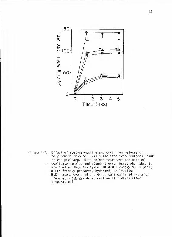
52
12 3 4 5
TIME (HRS)
Figure 4-2. Effect of acetone-washing and drying on release of
polyuronide from cell-walls isolated from 'Rutgers' pink
or red pericarp. Data points represent the mean of
duplicate samples and standard error bars, when absent,are smaller than the symbol (•,A,B = red;o,A,D = pink;
• ,0= freshly prepared, hydrated, cell-walls;,D = acetone-washed and dried cell-walls 24 hrs afterpreparation; A, A= dried cell-walls 2 weeks afterpreparation).

53
Whatman GF/C filters, and Spectramesh (Spectrum Medical Industries,
Los Angeles, CA; 10 micron pores) became plugged by eluted products
soon after the reaction was initiated. Results presented here were
obtained by using a double layer of Miracloth that was carefully
mounted so that it was not stretched and, presumably, its filtration
properties were not altered.
Profile for elution of acid sugars from predried, column-packed
walls from pink pericarp shows that most acid sugars were solubilized
within 2 hours (Fig. 4-3B). Soluble acid sugars and protein account
for 24% and 11%, respectively, of the preautolysis dry weight (100
mg). After 5 hours elution, 49.2 mg insoluble wall material was
recovered from the column (Table 4-3).
Cell walls pretreated with boiling 80% EtOH exhibited a profile
of acid sugar solubilization (Fig. 4-3C) similar to the profile
obtained for enzymically active walls (Fig. 4-33). The preautolysis
dry weight (100 mg) of pretreated walls decreased to 80.3 mg after 5
hrs elution (Table 4-3). Soluble acid sugars account for 12.4 mg of
the decrease during autolysis.
For comparison, wall samples were allowed to autolyze in a
static system (centrifuge tubes) with soluble product sampling by
aliquot (0.5 ml) removal. Approximately 10% of the preautolysis dry
weight (100 mg) was released from enzymically active walls as soluble
acid sugar during 7 hrs incubation (Fig. 4-3A) compared to 24% with
the column sampling system. Inactivated walls in the static system
released only 3% (dry wt) as acid sugar (Fig. 4-3A) compared to 12.4%
on the column. Rate of acid sugar solubilization declined to near

54
20 40FRACTION NO.
I 2 3TIME (HRS)
Figure 4-3. Comparison of methods of sampling for soluble autolysisproducts from cell walls isolated from 'Rutgers' pinkpericarp. Each sample consisted of 100 mg dry cellwalls and experiments were performed at 34 °C. Thesedata are from a single experiment that was repeated withsimilar results.A. Cell -walls incubated in static system (centrifuge
tubes) with sampling by aliquot (0.5 ml) removal.Data points are means of duplicate samples andstandard error is within each symbol (• =
enzymically active walls; o= pretreated withboiling 80% EtOH).
B. Profile of acid sugar released from enzymicallyactive walls placed on a column.
C. Profile of acid sugars released from cell walls thatwere pretreated with boiling 802 EtOH.

55
Table 4-3. Dry weights and acid sugar content of cell-wall samplesdescribed in Fig. 4-3.
Sample Dry weight (mg) Acid Sugar (% dry wt)
preautolysis 100 32.2
postautolysisA (•) 80.0 22.5
(O) 93.0 29.03 49.2 8.2C 80.3 19.8
zero after 5 hrs incubation in the static system compared to 2 hrs
elution time on the column (Fig. 4-3). Postautolysis dry weight of
samples incubated in static system were 80.0 and 93.0 mg,
respectively, for enzymically active and boiling 80% EtOH inactivated
samples (Table 4-3),
Experiment 4
An additional method of soluble product sampling involved
centrifugation of the reaction mixture and removal of supernatant at
each sampling throughout the experiment. This was compared to the
previously described technique of aliquot removal. Enzymically
active walls from pink tissue that were repeatedly subjected to
centrifugation, supernatant removal, and pellet resuspension in fresh
buffer exhibited a 50% increase in both rate of release and total
amount of acid sugars and acid reducing sugars released during
autolysis compared to those that were sampled by aliquot removal
(Fig. 4-4A.B). Samples that were pretreated with boiling 80% EtOH
were more drastically affected, releasing almost 3 times more product

56
2 3 4 5TIME (HRS)
Figure 4-4. Comparison of aliquot removal to centrifugation andsupernatant removal for sampling of solubilized acidsugars in cell walls from 'Rutgers' pink and mature-green pericarp incubated at 34°C. Each sample consistedof 100 mg dry cell walls. Results are from a singleexperiment that was repeated twice with similar resultseach time.A. Pink, aliquot removal; B. Pink-centrifugation; C.
Mature-green, aliquot removal; D. Mature-green,centrifugation (•,A= total acid sugars; O.A = acidsugar reducing groups; #,o= no inactivation pre-treatment; A»
A
= pretreated with boiling 80% EtOH).

57
if sampled by centrifugation (Fig. 4-4A.B). Walls from mature-green
tissue released acid sugars only if pretreated with boiling 80% EtOH
and centrifugation used for soluble product sampling (Fig. 4-4C.D).
Enzymically active samples that were centrifuged and resuspended in
the same buffer did not release more acid sugars than uncentrifuged
samples (data not shown).
Experiment 5
The procedures for cell-wall preparation and handling described
for data presented in Fig. 4-4B were used to further characterize
autolysis products. Solubilized products from enzymically active
walls were sampled at intervals throughout the time course.
Enzymically-inactive (pretreated with boiling 80% EtOH) walls were
sampled at time zero and at the end of the experiment.
Acid-sugar reducing groups were solubilized from enzymically
active walls (Fig. 4-5) in quantities equal to the earlier trial
(Fig. 4-43). However, the total soluble acid sugar was about 25%
lower the first time. Protein was released from the wall preparation
at a fairly constant rate (approx. 1 mg/hr) throughout the incubation
period (Fig. 4-5) and the total released accounted for almost 7% of
the preautolysis dry weight (Table 4-4).
Samples inactivated with boiling 80% EtOH lost 9.0% of their dry
weight during incubation, with the predominant products being acid
sugar and protein (Table 4-4). Total acid sugar content of the cell-
wall preparation was 34.5% before autolysis and decreased to

53
t-
>a:Q
200
2 3 4 5TIME (HRS)
Figure 4-5. Solubilization of uronic acids and proteins duringautolysis of cell walls extracted from 'Rutgers' pinkpericarp. Duplicate samples each contained 100 mg(preautolysis dry weight) cell walls and standard errorsare smaller than the symbols (• = total acid sugar;H= acid reducing sugars; A= protein; o,D,A =
pre treated with boiling 80% EtOH).
Table 4-4. Dry weights of individual components of the autolyticreaction described in Fig. 4-5 above.
Dry Weight (mg'
Enzymically active Pre-treated(Boiling 30% EtOH)
Cell -walls
preautolysis
postautolysis
Soluble products
acid sugar
protein
neutral sugar
100.0
74.6
21.0
6.8
2.4
100.0
91.0
3.2
2.9
0.3

59
13.5% and 31.3%, respectively, for enzymically active and inactivated
samples recovered postautolysis.
Neutral sugars solubilized during autolysis made up slightly
over 2% of the preautolysis wall dry wt (Table 4-4) and were released
at a rate of 5 ug/hr the first hour. This rate decreased to 2.5
yg/hr by the end of the incubation period (Fig. 4-6). The acid:
neutral sugar ratio was 10.8 after the first hour but had decreased
to 2.9 by the seventh hr of incubation (Fig. 4-6). Analysis of
individual neutral sugars revealed that galactose was the predominant
product, constituting approximately 50% of the total neutral sugar
released (Fig. 4-7). Arabinose accounted for 30% and minor
quantities of rhamnose, xylose, glucose, and mannose were also
detected (Fig. 4-7).
Gel filtration chromatography was used to analyze samples from
the reaction described in Fig. 4-5. Bio-Gel P-10 was chosen to
facilitate this analysis since Gross and Wallner (1979) found it
appropriate for fractionating enzyme (PG)-solubil ized-pectin (ESP)
from cell walls from mature-green tomatoes. Pectic polymers
solubilized during autolysis had a size distribution (Fig. 4-3) quite
similar to the profile obtained for ESP by Gross and Wallner
(1979). However, no apparent shift in polymer size during the time
course was observed with either Bio-Gel P-10 (Fig. 4-8) or Fractogel
HW-50 (Fig. 4-9). A portion of the supernatant was removed from
sample t, and incubated an additional six hours at 34°C, during which
no increase in acid reducing sugars was observed.

60
2 3 4 5TIME (HRS)
Figure 4-6. Solubilization of neutral sugars and acid:neutral sugarratio during autolytic reaction described in Fig. 4-5
( X= total neutral sugar; 0= acid neutral ratio).
Figure 4-7. Neutral sugars released in the autolytic reactiondescribed in Fig. 4-5 (Gal = Galactose; Ara = Arabinose;Rha = Rhamnose; Xyl = Xylose; Glc = Glucose; a
detectable trace of mannose was present).

61
BIO-GEL P-IO
0.4
0.3
0.2
0.1
0'
0.4
0.3
0.2
0.1
0-
0.4
0.3
0.2
0.1
0-
0.4
0.3
0.2
0.1
0- -i 1 1 1 1
—
10 20 30
FRACTION NO.
40 50
Figure 4-8. 3io-Gel P-10 chromatography of polyuronides released in
the autolytic reaction described in Fig. 4-5. (V =
exclusion limit; V,- = inclusion limit; subscriptsfollowing t = sampling time)

62
FRACTOGEL HW-50
20 30 40 50FRACTION NO.
Figure 4-9. Fractogel HW-50 chromatography of polyuronides releasedin the autolytic reaction described in Fig. 4-5. (V
e=
exclusion limit; V-j = inclusion limit; subscripts
following t = sampling time)

63
Discussion
The use of Miracloth for postautolysis recovery of cell walls
(Chapter 3) was based on the assumption that cell-wall material other
than that enzymically solubilized in autolytic reactions would be
quantitatively retained. Data presented in Table 4-1 do not support
that assumption. The autolysis reaction apparently generated
fragments that were not soluble in the supernatant of a centrifuged
sample yet were able to pass through Miracloth. Fragments lost
through Miracloth were unaccounted for and the resulting estimates of
dry weights were low (Table 4-1), leading to an overestimation of the
relative amount of soluble product (Fig. 4-1). There was an apparent
increase in the quantity of these fragments during the incubation
period between 5 hrs and 22 hrs since considerably lower amounts of
cell walls were recovered on Miracloth after extended incubation
(Table 4-1). The use of Miracloth for postautolysis cell-wall
recovery was discontinued because it does not appear to adequately
retain cell walls once a degree of degradation has occurred.
A significant source of error in estimating soluble products was
identified and, to some extent, resolved. However, the precise
preautolysis dry weight of the freshly prepared, hydrated cell walls
used in the previous experiment could not be ascertained. That
problem was circumvented by using acetone-dried walls prepared as
described in Materials and Methods.
Yields (dry wt) of acetone-washed cell walls from frozen
pericarp decreased approximately 50% as fruit ripened and senesced
(Table 4-1). Wallner and Bloom (1977) reported similar values and

64
suggested that the yield decrease is a consequence of in situ cell
wall hydrolysis associated with fruit softening. Acid sugar content
{% dry wt) of isolated walls decreased with fruit development in the
present study, from approximately 34% in mature-green to 32% in
overripe. Gross and Wallner (1979) reported a similar decrease (34%
to 30%).
The acetone washing and drying procedure had no apparent effect
on subsequent autolysis of walls from pink pericarp (Fig. 4-2);
however a decrease (25%) in activity was noted in dried walls
compared to freshly prepared, hydrated walls from ripe tissue (Fig.
4-2). A possible explanation for that difference is that softening
in the later stage of development is more closely associated with an
acetone soluble factor such as membrane lipid. Data presented here
do not address the involvement of factors other than those which are
wall-bound. All remaining experiments were performed with acetone-
washed and dried cell walls.
Several techniques for sampling soluble autolysis products were
evaluated in order to ascertain the most appropriate technique to
utilize for soluble neutral sugar analysis. Packing cell walls into
a column and eluting autolysis products with reaction buffer seemed
to offer the advantage of continuous removal of end-products that
might accumulate and inhibit autolysis. Almost double the amount of
acid sugar was released from column-packed walls compared to walls in
a static system (centrifuge tubes) with both enzymically active and
inactivated (boiling 80% EtOH) samples (Fig. 4-3). However, these
results must be interpreted with caution because Miracloth was used

65
to retain cell-walls in the column and an earlier study suggested it
may be inadequate for that purpose (Table 4-1). It was employed here
because conditions were such that the material was not subjected to
undue strain that might alter its filtration properties. Results of
the present study seem to confirm the earlier observation that
Miracloth does not adequately retain some fragments in the cell -wall
preparation. A clear disadvantage of the column sampling system is
that it is limited to analysis of only one cell -wall sample each
trial. An additional criticism is that the cell walls may function
as a gel filtration matrix, delaying the elution of small molecules.
A more practical method of sampling for soluble autolysis
products is centrifugation of the reaction mixture and complete
removal of the supernatant at each sampling. Cell walls handled and
sampled in that manner released 50% more uronic acid during autolysis
than cell walls incubated in a static system and sampled by aliquot
(0.5 ml) removal (Fig. 4-4). This difference cannot be attributed to
the additional manipulation involved in resuspension of the walls
since enzymically active walls that were centrifuged and resuspended
in the same buffer released approximately equal amounts of acid sugar
compared to uncentrifuged samples (data not shown). Perhaps a more
reasonable hypothesis for the increased polyuronide solubilization in
centrifuged cell-wall samples is that a soluble inhibitor is removed
ith the supernatant each time the reaction mixture is centrifuged.
Once resuspended in fresh buffer, autolytic reactions resume. The
inhibitor hypothesis is supported by the fact that centrifuged
samples of enzymically active walls had a higher rate of polyuronide
w

66
solubilization than uncentn'fuged samples throughout the time course
(Fig. 4-4A.B). Further, uncentrifuged samples ceased to release acid
sugars within 3 hrs after start of incubation (Fig. 4-4A) compared to
centrifuged samples that were autolyzing at a rate of 25 yg
galacturonic acid/mg wall dry wt/hr after 7 hrs incubation (Fig.
4-4B). Products solubilized from cell wall during softening include
polyuronide (Hobson, 1964; Pressey and Avants, .1971), neutral sugar
(Huber, 1983a; Gross and Sams, 1984), protein (Strand etal., 1976;
Hobson, 1983), calcium (Rigney and Wills, 1981), and possibly other
compounds. Of these, only calcium has been reported to inhibit PG in
vitro (Wills and Rigney, 1979; Ali and Brady, 1982), although the
precise nature of that inhibition is incompletely understood (Huber,
1983b). Calcium was demonstrated to inhibit cell-wall autolysis in
an earlier study (Fig. 3-2) and further identification of autolysis
inhibitors was not performed in these studies.
Pretreatment of enzymically active walls with boiling 80% EtOH
inhibited polyuronide solubilization approximately 50% when soluble
products were recovered by centrifugation (Fig. 4-43) compared to 80%
inhibition if sampled by aliquot removal (Fig. 4-4A, Fig. 3-2). The
appearance of reducing groups suggests that some PG activity remained
after the hot EtOH treatment (Fig. 4-4B). However, residual PG
activity probably does not account for all of the release since
enzymically-inactive walls from mature-green tissue released a
significant amount of acid sugar if exposed to the same handling
regime, e.g., pretreated with boiling 80% EtOH and soluble product
sampling by centrifugation and supernatant removal (Fig. 4-4C.D).

67
These data suggest that exposure of cell walls to boiling 80% EtOH in
some way enhances polyuronide solubilization from samples that are
repeatedly centrifuged and resuspended regardless of the presence of
residual PG activity (Fig. 4-4).
Previous experiments were directed toward finding the most
suitable handling technique for the enzymically active cell-wall
preparation prior to undertaking the more extensive neutral sugar
analyses. The scheme described for Fig. 4-4B was chosen, e.g.,
acetone-washed and dried cell walls with all soluble product sampling
by centrifugation and supernatant removal and no exposure to
Miracloth once autolysis is initiated. That system was selected
because it maximizes the amount of soluble product and eliminates
product overlap in the intervals of a time-course experiment.
Accountability for cell-wall constituents in the autolysis
system (Table 4-4) compares quite favorably with other studies.
Numerous workers have based their calculations of soluble cell-wall
constituents on the amount of cell -wall material recovered after
enzymic hydrolysis (Chapter 3; Wallner and Bloom, 1977; Gross and
Wallner, 1979) which confounds any effort to account for all of the
dry wt of a given sample. In a more thorough study, Gross (1984)
reported 92.7 ± 7.3% average total recovery of fractionated tomato
cell walls. In the autolysis system, close to 100% of the initial
dry weight is accounted for (Table 4-4). Efforts to refine the
methods involved in the study of autolysis apparently met with a
degree of success.

63
Dry cell walls (preautolysis ) ware composed of approximately 8%
(dry wt) protein, most of which was solubilized during autolysis
(Table 4-4, Fig. 4-5). Hobson et al. (1933) addressed the
significance of protein released from cell walls as a result of PG
action. They suggested that the process is consistent with
Tigchelaar's (1973) hypothesis that specific proteins released from
the wall might initiate ripening. The main concern in including the
measurement here was to account for as much of the initial wall dry
wt as possible.
The acid:neutral sugar ratio of solubls autolysis products (Fig.
4-6) is higher than generally reported by other workers. Gross and
Wallner (1979) determined the acidmeutral hexose ratio to be 6.4 in
water-soluble pectin released from cell walls extracted from light
red tomatoes. Wallner and Bloom (1977) used a crude enzyme extract
from ripe tomatoes to hydrolyze cell walls from mature-green fruit
and reported on acid:neutral sugar ratio for soluble products of from
6 to 9. In each of these examples the cell wall was exposed to a
more rigorous extraction scheme than was employed in the present
study.
Galactose was the predominant neutral sugar found in soluble
autolysis products (Fig. 4-7). This correlates well with Gross and
Wallner's (1979) observation that galactose is solubilized most
rapidly in situ during the transition from pink to ripeness.
Galactan and arabinan serve as cross-links for the rhamnogalacturonan
backbone of pectin (Albersheim, 1976). PG is believed to act upon a
region of rhamnogalacturonan with a relatively lo^ degree of

69
cross-linking (Gross and Wallner, 1979). Although all of these
neutral sugars were present in the autolysis reaction mixture, no
conclusions regarding PG specificity can be drawn.
The low amount of soluble glucose (Fig. 4-7) is indicative of
little or no readily soluble starch in the cell-wall preparation.
Gross and Wallner (1979) reported no detectable starch in a similar
extract but Pressey and Avants (1982b) found high levels of non-
cellulosic glucose in tomato cell walls and employed an amylase
treatment to remove it.
Gel filtration chromatography of polyuronides released
throughout autolysis was performed to determine if a shift in polymer
size occurs as wall degradation becomes more extensive. Although
polymer size did appear to decrease slightly over the time-course, a
significant amount of acid sugar always voided Bio-Gel P-10 (Fig.
4-8), indicating that the molecular weight of some pectin products
exceeded 20,000. Gross and Wallner (1979) used a PG extract to
hydrolyze walls from mature-green tissue and were able to fractionate
enzyme-soluble pectin on P-10. However, they found that water-
soluble pectin (autolysis products) released from cell walls of pink
tissue all voided P-10, in contrast to results of the present
study. This inconsistency may be explained by two important
differences in methods: first, Gross and Wallner (1979) subjected
their cell -wall preparation to more extensive mechanical disruption
which may have allowed removal of smaller polyuronide during
extraction and, secondly, they did not include 150 mM MaCl , which
enhances PG activity, in the reaction mixture. It has been suggested

70
that little, if any, degradation of polyuronide occurs once
solubilized from the larger wall matrix in vitro (Wallner and Bloom,
1977). That hypothesis is supported, but not confirmed, by gel
filtration chromatography data presented here (Figs. 4-8 and 4-9).
The primary objectives of this study were to examine methods of
handling enzymically active cell walls and to choose an appropriate
system for determination of neutral sugar content and relative
polymer size of pectins released during autolysis. These objective
were satisfied and new questions were raised. It appears that almost
any handling technique introduced into a cell-wall study is worthy of
considerable investigation before its implementation in large scale
experiments.
Analyses of soluble autolysis products provided data that are in
general agreement with other tomato cell-wall studies. Experiments
in progress ^re directed toward more detailed characterization of the
effects of calcium, pH, and NaCl on cell wall autolysis.

CHAPTER 5
EFFECTS OF NaCl , pH, AND CALCIUM ON
AUTOLYSIS OF ISOLATED TOMATO CELL WALLS
Variables that are often tested in in vitro cell-wall studies of
fruit softening are pH (Patel and Phaff, 1960a; Hobson, 1964;
Pressey, 1984), NaCl (Pressey and Avants, 1973; All and Brady, 1982),
and calcium (Rigney and Wills, 1981; Oarvis, 1982; Demarty et al.,
1934). All have been implicated in regulation of softening but the
role of each is poorly understood because interactions are complex.
In systems that employ model substrates to study the activity of
cell-wall hydrolases, the effects of a single variable on enzyme
activity are relatively easy to isolate. For example, the discovery
of differential pH optima in a crude polygalacturonase (PG) extract
from tomato fruit was an important clue leading to the discernment of
PG isozymes (Patel and Phaff, 1960a; Hobson, 1964). Once purified,
different isozymes responded maximally to different concentrations of
NaCl, the requirement for Na being quite specific (Pressey and
Avants, 1973). The effects of calcium on PG activity present more of
a challenge, even in assays that employ relatively simple substrate,
because calcium has the capacity to interact with both protein and
polyuronide (Ferguson, 1983). Specific regulatory roles for calcium
in fruit softening are speculative (Demarty et al., 1984; Ferguson,
1984).
71

72
Cell-wall autolysis may offer additional insight into the
regulation of polyuronide hydrolysis by pH, NaCl, and calcium.
Earlier autolysis experiments addressed those variables to a limited
extent and confirmed that enzymically active cell wall is a useful
tool for investigation of fruit softening (Chapters 3 and 4). The
objective of the present study is to extend the scope of earlier
experiments with additional emphasis on interactions of the variables
discussed.
Materials and Methods
Plant Material
Tomato ( Lycopersicon esculentum Mill, cv 'Rutgers') plants were
grown at the University of Florida Horticultural Unit near
Gainesville. Fruit were harvested at pink or red (ripe)
developmental stages based on a United States Department of
Agriculture visual aid for color classification of tomatoes (Anon.,
1975). Fruit were surface sterilized with 100 ppm MaOCl and
sectioned; seed and placental tissue were removed and pericarp stored
at -20 °C.
Cell Wall Preparation
Approximately 100 g of frozen pericarp were partially thawed,
peeled, and homogenized in 400 ml distilled F^O (4°C) for 1 min in a
Sears blender set at maximum speed. The homogenate was transferred
onto Miracloth and washed with 3 liters of distilled H 2° (4°C) and 1
liter of acetone (1°C) with continuous stirring. Cell walls were
suspended in 100 ml of acetone for 1 hr then filtered through Whatman

73
GF/C filters and washed with acetone (approximately 500 ml) until
pigment was no longer visible. Walls were transferred to a beaker
and dried at 30°C with occasional stirring to reduce formation of
aggregates. Dry cell walls were stored in sealed containers at 22°C
and were used for autolysis experiments within 2 weeks following
preparation. All wall extracts were analyzed for total acid sugar
content (Ahmed and Labavitch, 1977).
Autolysis Experiments
Cell walls (100 mg dry wt) were placed in a 50 ml centrifuge
tube in an ice bath and rehydrated by dropwise addition of 15.0 ml
cold (1°C) reaction buffer (50 mM Na acetate, pH 4.5, 150 mM NaCl,
0.02? Thimerosal) unless otherwise indicated. Samples were held at
1°C for 30 min with occasional stirring, then centrifuged (10 rnin at
10,000 RPM) with a Beckman Model J2-21 centrifuge equipped with a
JA-20 rotor. The supernatant was filtered through Whatman GF/C
filters to obtain a time-zero sample. The pellet (cell wall) was
resuspended in 15.0 ml of fresh reaction buffer and incubated at
34°C. At selected intervals during incubation, centrifugation,
supernatant removal, and pellet resuspension were repeated. All
supernatants were analyzed for total acid sugars (Blumenkrantz and
Asboe-Hansen, 1973) and acid reducing sugars (Milner and Avigad,
1967). Cell walls were recovered postautolysis by suspension in 30.0
ml acetone and filtration through tared Whatman GF/C filters; walls
were analyzed for total acid sugar content (Ahmed and Labavitch,
1977).

74
In some experiments, cell walls were suspended in hot 80£ EtOH
and boiled 15 min under reflux. The mixtures were cooled,
centrifuged, the supernatant removed, and the cell walls resuspended
in reaction buffer as described above.
Experiment 1. Effect of NaCl concentration on cell-wall
autolysis was observed by rehydration of cell walls from either pink
or red pericarp in either distilled HgO. reaction buffer, or buffer
containing 0.15, 0.30, 0.45, or 0.60 M NaCl. At intervals during
autolysis, reaction mixtures were centrifuged, the supernatant
removed, and the pellet resuspended in either distilled H2O or the
appropriate ionic strength buffer.
Experiment 2 . The interaction of pH and 1 M NaCl treatment
prior to autolysis was investigated using cell walls from pink
pericarp. Walls were rehydrated in one of the following buffers with
or without 1 M NaCl: pH 2.5, 5 mM Na2-acetate titrated with glacial
acetic acid; pH 4.5, 50 mM Na2 -acetate; pH 6.5, 50 mM MOPS; pH 3.5,
50 mM HEPES. After 30 min in pretreatment solution (1°C), mixtures
were centrifuged (10 min at 10,000 RPM) and supernatants removed.
Walls were resuspended in reaction buffer (50 mM Na2_acetate, pH 4.5,
150 mM NaCl, 0.02% Thim.) and incubated at 34 °C. The pH of all
supernatants was checked to ensure that buffer strength was adequate.
Experiment 3 . In some studies, treatment of a cell -wall extract
with 1 M NaCl was employed to enhance dissociation of bound protein,
including active wall hydrolases (Hobson, 1964; Ali and Brady,
1982). In the present experiment, exposure to 1 M NaCl was
introduced at selected steps during extraction of cell walls from red
pericarp (Table 5-1). Autolysis of the resulting wall extracts was

75
Table 5-1. Protocol for cell-wall extraction in Experiment 3.
Cell -Wall
Yield Acid SugarHonogenization (mg/100 g Content
Sample Medium Miracloth Filtration fresh wt) (% dry wt)
H2
H2
H2
1 M NaCl
E

76
(Table 5-1). Extracts G and H were homogenized in acetone in a
Sorvall blender that would not accommodate the full 100 g of frozen
tissue. Those lots were subdivided for homogenization; however, in
all extractions the ratio of tissue: homogenization medium was 1:4.
The cell-wall preparation procedure for extracts 8, C, D, and H
included exposure to 1 M NaCl during Miracloth filtration (Table
5-1). This was performed by rinsing walls with 1 liter of 1 M NaCl
(1°C), then lowering the Miracloth and its contents into a beaker
containing 500 ml of 1 M NaCl (1°C) for 20 min followed by distilled
H2
rinse (Table 5-1).
All extracts were rinsed with 1 liter acetone on Miracloth prior
to dry wt analysis (Table 5-1). In order to observe autolysis of
walls that were not exposed to acetone, aliquots of approximately 50
mg (dry weight) hydrated cell walls were removed from extracts A
(labeled subsample I) and D (labeled subsample J) immediately after
the first aqueous rinse on Miracloth (Table 5-1). These aliquots
(subsamples) were placed in 10.0 ml of either distilled ^0 or 1 M
NaCl (1°C). Additional aliquots (subsamples) were adjusted to 80%
EtOH and boiled 15 min. After pretreatment, all subsamples were
centrifuged (10 min at 10,000 RPM), the supernatant removed, and the
pellet resuspended in 10.0 ml distilled H2
for 5 min. That mixture
was centrifuged (5 min at 5,000 RPM), the supernatant removed, and
the pellet resuspended in 10.0 ml reaction buffer (50 mM Na 2 acetate,
pH 4.5, 150 mM NaCl, 0.02% Thimerosal) before incubation at 34°C.
Experiment 4 . A factorial experiment was designed to test the
effects of pH (2.5, 4.5, 6.5, 8.5; same buffers as experiment 2),

77
NaCl concentration (0, 150, 300 mM) and calcium (0, 5 mM CaCl 2 ) on
autolysis of cell walls extracted from ripe pericarp. All buffer
solutions were prepared individually and contained 150 mM NaCl and
0.02$ Thimerosal. Some cell-wall samples were boiled in 80% EtOH and
allowed to cool slowly to room temperature prior to centrifugation,
supernatant removal, and resuspension in appropriate buffer solution
for autolysis.
Results
Experiment 1
Autolysis of cell walls extracted from red pericarp was
optimally stimulated by 150 mM NaCl, compared to 300 mM for walls
from pink fruit (Fig. 5-1). Acid reducing sugar analysis correlated
with total acid sugar assays (data not shown). Walls from both
stages of development released 3 times more acid sugars in the
presence of NaCl than was released in distilled H2
(Fig. 5-1). At
600 mM NaCl, over 50% inhibition of autolysis was observed compared
to the optimum NaCl level (Fig. 5-1). Dry wt of walls incubated at
600 mM NaCl increased 30-35% above the preautolysis dry weight during
the 7 hrs incubation period (Table 5-2).
Rate of uronic acid solubilization was not drastically affected
by slight changes in NaCl concentration. A gradual increase in rate
of release was observed as NaCl was increased to the optimum level,
followed by a gradual decline as salt became inhibitory (Fig. 5-1).
The pH of supernatants recovered from walls incubated in distilled
H„0 varied from 4.43 to 4.43.

73
2 3 4 5 6
TIME (HRS)
Figure 5-1. Effect of NaCl on release of polyuronide from cell wallsof pink (A) or red (B) tomato pericarp. Results arefrom a single experiment with duplicate samples that wasrepeated with similar results each time. Bars representstandard error of the mean and, when absent, are smallerthan the symbols. (D.W. = distilled ^0; numbers
represent M NaCl in reaction buffer)

79
Table 5-2. Postautolysis dry weight of cell walls recovered attermination of experiment 1.
Postautolysis Dry Wt (mg)Reaction Medium Pink Red
distilled H 2 87.3 ± 0.7 81.6 ± 0.6
buffer 86.9 ± 0.5 75.1 ± 2.2
buffer + 0.15 M NaCl 87.3 ± 2.1 75.7 ± 1.3
buffer + 0.30 M NaCl 94.7 ± 1.2 91.1 ± 2.2
buffer + 0.45 M NaCl 97.6 ± 2.3 105.9 ± 0.6
buffer + 0.60 M NaCl 129.1 ± 0.7 136.7
Experiment 2
Cell walls exposed to pH 6.5 or 8.5 prior to incubation at pH
4.5 (Fig. 5-2 C,D) released acid sugars in a manner similar to walls
exposed to pH 4.5 throughout the experiment (Fig. 5-2 B). If
pretreatment buffers (pH 4.5, 6.5, or 8.5) contained 1 M NaCl, total
acid sugar released at pH 4.5 was nearly doubled compared to pH
pretreatment without NaCl (Fig. 5-2 B,C,D). However, no increase in
acid sugar reducing groups resulted from the 1 M NaCl pretreatment at
either pH (Fig. 5-2 B.C.D).
Walls exposed to pH 2.5 without 1 M NaCl prior to autolysis at
pH 4.5 (Fig. 5-2 A) exhibited a 3-fold increase in total acid sugar
solubilization compared to all other pH pretreatments without 1 M
NaCl (Fig. 5-2 B,C,D). Pretreatment with 1 M NaCl at pH 2.5 caused
approximately 25% increase in acid sugar solubilization compared to

30
2 3 4 5
TIME (HRS)
Figure 5-2. Autolytic activity in cell walls from pink pericarpincubated at pH 4.5 after pretreatment with 1 M NaCl atdifferent pH values. (A = pH 2.5; B = pH 4.5; C = pH
6.5; D = pH 8.5;#,o= total acid sugars; A, A= acidreducing sugars;O.A= 1 M NaCl;#,A= no NaCl)

31
pH 2.5 pretreatment without 1 M NaCl (Fig. 5-2 A). Release of acid
sugar reducing groups was not significantly affected by either pH or
1 M NaCl pre treatments (Fig. 5-2).
Experiment 3
The least rigorous extraction procedure for preparation of dry
cell walls is represented by scheme A (Table 5-1 A), i.e., cold
aqueous extraction with no exposure to buffers or other salts. Walls
prepared in that manner released approximately 10% of their dry
weight as acid sugars during autolysis (Fig. 5-3 A). Pretreatment
with 1 M NaCl caused approximately 25% increase in acid sugar
solubilization (Fig. 5-3 A) and pretreatment with boiling 80% EtOH
caused 85-95% inhibition of autolysis in all wall extracts (Fig.
5-3). Autolytic activity in subsamples removed from extract A prior
to the acetone wash and drying procedure (Table 5-1 A) is diagrammed
in Fig. 5-3 I. Exposure of freshly prepared, fully hydrated walls to
1 M NaCl caused approximately double the release of acid sugars
compared to walls that were maintained at the ionic strength of
reaction buffer (Fig. 5-3 1).
Exposure of cell wall homogenates to 1 M NaCl at selected points
in the extraction scheme did not eliminate autolytic activity in the
resulting wall preparations (Fig. 5-3 B-J) when compared to the low
ionic strength extract (Fig. 5-3 A). The most rigorous extraction
procedure in terms of ionic treatment is represented by scheme D
(Table 5-1), i.e., homogenization and Miracloth filtration with 1 M
NaCl. Autolysis of walls prepared in this manner (Fig. 5-3 D) was

32
Figure 5-3. Effect of exposure to 1 M NaCl on solubilization of acidsugars from cell walls of red tomato pericarp. Letters(A-J) correspond to those that designate extractionprotocol described in Table 5-1. (O = 1 M NaCl
pre treatment; • = no pre treatment; A= boiling 80% EtOHtreatment)

33
not significantly different from autolysis of the aqueous wall
extract (Fig. 5-3 A) except that an additional 1 M NaCl pretreatment
of walls from extract did reduce (25%) autolytic activity (Fig.
5-3 0). Autolysis of cell walls that were removed from extract D
after 1 M NaCl treatment on Miracloth but prior to acetone wash
(Table 5-1 D) is diagrammed in Fig. 5-3 J. These walls released
approximately 30% more acid sugars than similarly sampled walls from
the aqueous extract A (Fig. 5-3 I, J). An additional 1 M NaCl
treatment of wall sample J further enhanced autolysis by about 50%
(Fig. 5-3 J) and boiling in 80% EtOH inhibited autolysis, although
time zero values for boiled walls was unusually high (Fig. 5-3 J).
The highest cell-wall yield (773 mg/100 g fresh wt) was obtained
by homogenization of pericarp in acetone with no exposure to NaCl
(Table 5-1 G). Yield was lower (720 mg) if acetone-homogenized walls
were treated with 1 M NaCl on Miracloth (Table 5-1 H). The aqueous
extract yielded 690 mg walls (Table 5-1 A) and all other extracts
that received 1 M NaCl treatments yielded from 575 to 625 mg
walls/100 g pericarp (Table 5-1 B-F).
Acid sugar content (% dry wt) of cell walls was lowest (23.1%)
in walls exposed to the most rigorous extraction scheme (Table
5-1 0). All other extracts varied from 29-34% acid sugars (Table
5-1).
Experiment 4
Cell walls incubated at pH 8.5 released negligible quantities of
acid sugars regardless of presence of NaCl or CaCl„ (Fig. 5-4 D). At

84
pH 6.5 detectable amounts (1-2% wall dry wt) of acid sugars were
solubilized and calcium inhibition of autolysis was apparent (Fig.
5-4 C). At pH 4.5, autolysis was approximately doubled by the
presence of either 150 or 300 mM NaCl compared to walls incubated
without NaCl (Fig. 5-4 B). Calcium inhibited autolysis at pH 4.5 by
60-70% at all three levels of NaCl (Fig. 5-4 B).
Stimulation of autolysis by NaCl was most apparent at pH 2.5, at
which either concentration of NaCl caused a 3-fold increase in uronic
acid solubilization compared to walls incubated without NaCl (Fig.
5-4 A). Calcium did not significantly inhibit autolysis at pH 2.5 in
buffer without NaCl or with 150 mM NaCl; approximately 15% inhibition
was observed at 300 mM NaCl (Fig. 5-4 A).
Cell walls that were boiled 20 min in 80% EtOH and cooled slowly
(approximately 1 hr to reach 34 °C) were over 95% inactivated in terms
of acid sugar release at all pH, NaCl, or calcium levels (Fig.
5-4). Boiling EtOH treatments were not as effective (35-95%) in
inhibition of autolysis in earlier studies (Figs. 3-2, 4-4 and 5-3)
probably because high temperature exposure was of shorter duration.
Discussion
Autolysis data presented here (Fig. 5-1) are generally
consistent with Pressey and Avants (1973) work with purified PG
isozymes from tomato. The authors assayed the activity of PCs I and
II over a range of NaCl concentrations at pH 4.5 and reported optimal
NaCl concentrations of approximately 200 and 300 mM, respectively,
for PG I and PG II. More realistically, they observed a range of

85
I 23456701 23456701 234567TIME (HRS)
Figure 5-4. Effects of pH, NaCl, and calcium on solubilization of
acid sugars from cell walls of red tomato pericarp.
Results are from a single experiment with duplicate cell
wall samples. Experiment was repeated with similar
results each time. Bars represent standard error of the
mean and, when absent, are smaller than the symbol. (A
= pH 2.5; 3 = pH 4.5; C = pH 6.5; D = pH 8.5; 1 - no
NaCl; 2 = 150 rnM NaCl ; 3 = 300 mM NaCl; • = no calcium;0= 5 mM calcium; A= pretreated with boiling 80% EtOH).

36
activity from approximately 150 to 300 mM for PG I and 200 to 300 mM
for PG II. A similar range of activity was observed using autolysis
of enzymically active cell walls as a measure of PG activity (Fig.
5-1). Concentrations of 600 mM NaCl or higher inhibited purified PG
activity (Pressey and Avants, 1982b) as well as cell-wall autolysis
(Fig. 5-1).
PG I is reported to appear during the earliest color development
associated with ripening, but by the time fruit are fully ripe PG II
is the predominant species (Tucker et a!., 1980). Enzymically active
walls from the two stages of development used in the present study
probably contained both PG isozymes. Although differential NaCl
optima may exist (Pressey and Avants, 1973), they do not seem
distinct enough to serve as a means of conclusively ascertaining the
presence, or absence, of a specific PG isozyme in enzymically active
wa 1 1 s
.
The increase in dry weight observed with cell walls incubated at
higher NaCl concentrations (Table 5-2) was possibly due to Na
binding to demethylated galacturonic acid residues. This illustrates
an inherent complexity of many cell-wall studies, e.g., the capacity
for pectins to bind extensively, and perhaps indiscriminately, to
cationic molecular species.
Additional effects of NaCl on cell-wall autolysis were
investigated in experiment 2, in which walls were exposed to 1 M NaCl
over a range of pH values prior to incubation at pH 4.5. This salt
concentration enhanced extraction of PG from hovnogenates of tomato
pericarp or from cell-walls (Hobson, 1964; Ali and Brady, 1982);

37
however these studies did not examine the cell wall for residual
wall-bound PG activity (autolysis) after ionic treatment. Results of
the present study suggest that a significant population of PG
molecules remain bound to the wall after 1 M NaCl treatment (Fig.
5-2) and the tenacity of their binding is further illustrated by
failure of 2 or 3 M NaCl pretreatment to inhibit subsequent autolysis
(data not shown).
Since timing of the 1 M NaCl treatment during wall extraction
might be a significant factor in its effectiveness in removing wall-
bound PG, experiment 3 was designed to examine the effect of 1 M NaCl
treatment on autolysis when performed at selected points in the
extraction scheme (Table 5-1). Autolysis was not effectively
eliminated by any of the 1 M NaCl treatments (Fig. 5-3). These
results are particularly pertinent to fruit softening studies that
employ cell walls from ripening fruit, tissues (Buescher and Hobson,
1982). Appropriate controls must be presented to ensure that
residual wall-bound PG activity (autolysis) does not lead to
misinterpretation of data. Unfortunately, treatments that remove or
inactivate bound enzymes may also degrade cell wall, thus limiting
the interpretation of results.
Autolysis was significantly enhanced by exposure of walls to pH
2.5, whether by pretreatment (Fig. 5-2 A) or by incubating at that pH
(Fig. 5-4 A). The data allow two possible explanations. Exposure to
pH 2.5 may cause demethylation of galacturonic acid residues
(Talmadge et al., 1973) which renders the polygalacturonan more
susceptible to PG hydrolysis (Jansen and MacDonnell, 1945;

Pressey and Avants, 1982b). However, it does not seem likely that
degree of esterification is limiting to autolysis at pH 4.5 because
pretreatment of walls nearer the pH optima (6.5-8.5) of PME isozymes
(Pressey and Avants, 1972) slightly inhibited, rather than enhanced,
subsequent autolytic activity at pH 4.5 (Fig. 5-2). This inhibition
may have been due to perturbation of the PG-substrate complex and
diffusion of enzyme from its site(s) of hydrolysis, since exposure of
walls to neutral or higher pH reportedly facilitates removal of bound
hydrolases (Babbit et al . , 1973).
A more plausible explanation for the low pH stimulation of
autolysis is that protons enhance solubilization of calcium from
polygalacturonan, allowing PG greater access to sites of hydrolysis
(Demarty, 1984). This view is supported by failure of calcium to
inhibit autolysis at pH 2.5 (Fig. 5-4 A). Additionally, brief (30
min) pretreatment at pH 2.5 (experiment 2) was probably adequate for
calcium to dissociate from the wall since it has been demonstrated
that solubilized calcium diffuses from isolated wall more rapidly
than polymeric wall constituents (Jarvis, 1982). Calcium chelators
have been shown to enhance PG activity against isolated cell wall
(Buescher and Hobson, 1982) and to cause non-enzymic pectin
solubilization from isolated wall (Jarvis, 1982). It has been
proposed that soluble apoplastic calcium may be a regulator, if not
the initiator, of ripening metabolism (Rigney and Wills, 1981).
Autolytic activity was relatively low at pH 2.5 unless NaCl (150
or 300 mM) was present in reaction buffer (Fig. 5-4 A). Pressey and
Avants (1971) reported that NaCl had its greatest effect on in vitro

89
PG activity when substrate was large and pH was low. They speculated
that larger galacturonans form aggregates at low pH by extensive
hydrogen bonding, limiting the access of PG to sites of hydrolysis.
Presence of salts would effectively disrupt hydrogen bonds. Results
of the present study (Fig. 5-4) correlate well with Pressey's data.
Autolysis was affected by calcium, NaCl, and pH similarly to
activity of purified PG. A major point of interest is that
pretreatment of cell walls with 1 M NaCl did not entirely suppress
autolysis. Efforts are currently directed toward examination of
mobility and binding characteristics of PG in isolated cell wall and
to ascertain the involvement of wall-bound hydrolases other than PG.

CHAPTER 6
SUMMARY
Fruit softening is generally attributed to degradation of the
cell wall by hydrolytic enzymes. The most common approach to
enzymology of softening has been to extract and purify the hydrolases
of interest and assay with a commercially available substrate or with
a cell wall preparation. In the present study, cell walls isolated
under conditions which minimized the disruption of protein-cell wall
interactions were incubated under conditions favorable for continued
enzymic activity.
Cell walls isolated from pericarp of ripening tomato
( Lycopersicon esculentum Mill, cv 'Rutgers') released acid sugars
amounting to 202 cell wall dry wt in a reaction apparently mediated
by wall-bound polygalacturonase (PG). Release was negligible in wall
preparations from normal green and the ripening mutant rin frui t.
Pectin solubilization was most extensive at pH 2.5 with a less
prominent peak at 5.5. Exposure of cell wall to low (1.5) or high
(7.5) pH resulted in a reduction of autolytic activity. Calcium,
high temperature, and treatments employed to dissociate protein from
cell wall also inhibited autolytic reactions. Autolysis was enhanced
by 150 mM NaCl and by increased temperature within the physiological
range.
90

91
The rate and extent of polyuronide solubilization was
significantly affected by the techniques employed for handling the
cell walls. Enzymically active cell walls subjected to either
intermittent or continuous removal and replenishment of the bathing
medium released up to twice as much polyuronide as walls incubated in
a static environment.
A handling system that maximized acid sugar solubilization was
selected for experiments to determine the neutral sugar content and
size distribution of pectic polymers released during autolysis.
Approximately 2% of the initial wall dry wt was released as neutral
sugars; the predominant product was galactose (50%), followed by
arabinose (30%) and minor quantities of rhamnose, xylose, glucose,
and mannose. The acid:neutral sugar ratio of soluble autolysis
products decreased from 10.8 to 2.9 as wall degradation became more
extensive. Gel filtration chromatography of solubilized pectic
polymers did not reveal a temporal relationship between polyuronide
size distribution and the autolytic reaction. Approximately 8% of
the wall dry wt was protein, most of which was released during
autolysis. The techniques developed in these studies for preparation
and handling of enzymically active cell walls in vitro allow for an
accounting of close to 100% of the preautolysis wall dry wt.
The effects of NaCl, pH, and calcium on the autolytic reaction
also were examined. Polyuronide solubilization at pH 4.5 was
optimally enhanced by NaCl in the range of 200-300 mM. Brief
pretreatment of enzymically active walls with 1 M NaCl in the pH
range of 2.5-8.5 did not inhibit subsequent autolysis at pH 4.5. The

92
tenacity of PG-binding was further illustrated by the fact that
inclusion of 1 M NaCI treatments at selected points in the wall
extraction scheme also did not eliminate release of acid sugars. A
factorial experiment to test for interactions between pH (2.5, 4.5,
6.5, 8.5), NaCI (0, 150, 300 mM), and calcium (0, 5 tnM CaCl2
)
demonstrated that polyuronide solubilization was most dramatically
enhanced by NaCI at pH 2.5, at which calcium inhibition was not
observed. Inhibition by calcium was most apparent at pH 4.5 in the
presence of NaCI. Autolytic activity was negligible at pH 6.5 and
8.5 and no effects of NaCI or calcium were noted.
The results of these experiments support the concept that the
release of polyuronide from isolated cell walls in vitro is enzymic
and provides a convenient and reliable system for the study of
softening metabolism.

BIBLIOGRAPHY
Ahmed, A.E., and O.M. Labavitch. 1977. A simplified method for
accurate determination of cell wall uronide content. J. FoodBiochem. 1:361-365.
Ahmed, A.E., and J.M. Labavitch. 1980a. Cell wall metabolism in
ripening fruit. I. Cell wall changes in ripening 'Bartlett'
pears. Plant Physiol. 65:1009-1013.
Ahmed, A.E., and J.M. Labavitch. 1980b. Cell wall metabolism in
ripening fruit. II. Changes in carbohydrate degrading enzymesin ripening 'Bartlett' pears. Plant Physiol. 65:1014-1016.
Albersheim, P. 1976. The primary cell wall. In: J. Bonner andJ.E. Varner (eds). Plant Biochemistry. Academic Press, New
York, pp 225-274.
Albersheim, P., A.G. Darvill, K.R. Davis, J.M. Lau, M. McNeil, J.K.
Sharp, and W.S. York. 1984. Why study the structures of
biological molecules? In: W.M. Ougger and S. Bartnicki-Garcia(eds). Structure, function, and biosynthesis of plant cell
walls. Waverly Press, Baltimore, MD. pp 19-51.
Albersheim, P., H. Neukom, and H. Deuel. 1960. Splitting of pectinchain molecules in neutral solutions. Arch. Biochem. Biophys.90:46-51.
Ali, Z.M., and C.J. Brady. 1982. Purification and characterizationof the polygalacturonases of tomato fruits. Aust. J. PlantPhysiol. 9:155-169.
Anonymous. 1975. Color classification requirements in tomatoes.United States Department of Agriculture Visual Aid TM-L-1. TheJohn Henry Co., Lansing, MI.
Aspinall, G.0. 1980. Chemistry of cell wall polysaccharides. In:
J. Preiss (ed). The biochemistry of plants. Vol. 3.
Carbohydrates: Structure and function. Academic Press, New
York, pp 473-500.
Awad, M., and L.N. Lewis. 1980. Avocado cellulase: Extraction andpurification. J. Food Sci. 45:1625-1628.
93

94
Ayers, W.A., G.C. Papavizas, and A.F. Diem. 1966. Polygalacturonatetrans-el iminase and polygalacturonase production by Rhizoctoniasolani . Phytopath. 56:1006-1011.
""
Babbit, J.K., M.J. Powers, and M.E. Patterson. 1973. Effects of
growth-regulators on cellulase, polygalacturonase, respiration,color, and texture of ripening tomatoes. J. Amer. Soc. Hort.Sci. 93:77-81.
Bartley, I.M. 1974. S-Galactosidase activity in ripening apples.Phytochem. 13:2107-2111.
Bartley, I.M., M. Knee, and M. Casimir. 1982.. Fruit softening I.
Changes in cell wall composition and endo-polygalacturonase in
ripening pears. J. Expt. Bot. 33:1248-1255.
Bartnicki-Garcia, S. 1984. Kingdoms with walls. In: W.M. Duggerand S. Bartnicki-Garcia (eds). Structure, function, andbiosynthesis of plant cell walls. Waverly Press, Baltimore,MD. pp 1-13.
Bates, G.W., and P.M. Ray. 1981. pH-dependent interactions betweenpea cell wall polymers possibly involved in wall deposition andgrowth. Plant Physiol . 68:158-164.
Besford, R.T., and G.E. Hobson. 1972. Pectic enzymes associatedwith the softening of tomato fruit. Phytochem. 11:2201-2205.
Slumenkrantz, N., and G. Asboe-Hansen. 1973. New method forquantitative determination of uronic acids. Anal. Biochem.54:434-489.
Borochov, A., and R. Faiman-Weinberg. 1934. Biochemical andbiophysical changes in plant protoplasmic membranes duringsenescence. What's New in Plant Physiol. 15:1-4. G. Fritz (ed)
Agronomy Department, Univ. of Florida, Gainesville, FL.
Bradford, M. 1976. A rapid and sensitive method for the
quantitation of microgram quantities of protein utilizing the
principle of protein-dye binding. Anal. Biochem. 72:248-254.
Brady, C.J., G. MacAlpine, W.B. McGlasson, and Y. Ueda. 1932.Polygalacturonase in tomato fruits and the induction ofripening. Aust. J. Plant Physiol . 9:171-178.
Brant, D.A. 1980. Conformation and behavior of polysaccharides in
solution. In: J. Preiss (ed). The biochemistry of plants.Vol. 3. Carbohydrates: Structure and function. Academic Press,New York, pp 425-472.

95
Buescher, R.W., and G.E. Hobson. 1982. Role of calcium andchelating agents in regulating the degradation of tomato fruittissue by polygalacturonase. J. Food Biochem. 6:147-160.
Carpita, N.C. 1984. Fractionation of hemicelluloses from maize cellwalls with increasing concentrations of alkali. Phytochemistry23:1039-1093.
Carr, D.J., and E.K. Ng. 1959. The sequestration of calcium frompreparations of wheat coleoptile cell walls. Physiol. Plant.12:264-274.
Colinas-Leon, M.T., and R.E. Young. 1981. The cell wall in ripeningavocados. Yearbook California Avocado Society 113-117.
Conway, W.S. 1984. Preharvest factors affecting postharvest lossesfrom disease. In: H.E. Moline (ed). Postharvest pathology offruits and vegetables: Postharvest losses in perishable crops.Univ. of Cal. Agric. Expt. Sta. Bui. 1914, Univ. of Cal.,Berkeley, CA. pp 24-30.
Cooper, J. 3., J. A. Chen, and J.E. Varner. 1984. The glycoproteincomponent of plant cell walls. In: W.M. Ougger and S.
Bartnicki-Garcia (eds). Structure, function, and biosynthesis ofplant cell walls. Waverly Press, Baltimore, MD. pp 75-88.
Crookes, P.R., and D. Grierson. 1983. Ul trastructure of tomatofruit ripening and the role of polygalacturonase isoenzymes in
cell wall degradation. Plant Physiol. 72:1088-1093.
Delincee, H. 1976. Thin-layer isoelectric focusing of multipleforms of tomato pectinesterase. Phytochemistry 15:903-906.
Demarty, M., C. Morvan, and M. Thellier. 1984. Calcium and the cellwall. Plant Cell Environ. 7:441-448.
Oische, Z. 1947. A new specific color reaction of hexuronicacids. J. Biol. Chem. 167:139-198.
Ericson, M.C., and A.D. Elbein. 1980. Biosynthesis of cell wallpolysaccharides and glycoproteins. In: P.K. Stumph and E.E.Conn (eds). The biochemistry of plants. Vol. 3.
Carbohydrates: Structure and function. Academic Press, NewYork, pp 589-616.
Ferguson, I.B. 1984. Calcium in plant senescence and fruitripening. Plant Cell Environ. 7:477-489.
Ferguson, I.B., N.A. Turner, and E.G. Bollard. 1980. Problems infractionating calcium in plant tissue. J. Sci. Food Agric. 31:7-14.

96
Fishman, M.L., P.E. Pfeffer, R.A. Barford, and L.W. Doner. 1984.
Studies of pectin solution properties by high-performance size
exclusion chromatography. J. Agric. Food Chem. 32:372-378.
Forster, H., and I. Rasched. 1985. Purification and
characterization of extracellular pectinesterases from
Phytophthora infastans. Plant Physiol. 77:109-112.
Gertman, E., and Y. Fuchs. 1974. Changes in pectinmethylesterase(PME) activity caused by ethylene applied at differenttemperatures. Plant Cell Physiol. 15:501-505.
Gidley, M.J., E.R. Morris, E.J. Murray, D.A. Powell, and D.A. Rees.
1980. Evidence for two mechanisms of interchain association in
calcium pectate gels. Int. J. Biol. Macromol. 2:332-334.
Grierson, 0., G.A. Tucker, and N.G. Robertson. 1981a. The molecular
biology of ripening. In: J. Friend and M.J.C Rhodes (eds).
Recent advances in the biochemistry of fruits and vegetables.
Academic Press, Mew York, pp 149-160.
Grierson, D., G. Tucker, and H. Robertson. 1931b. The regulation of
gene expression during the ripening of tomato fruits. In: P.W.
Goodenough and R.K. Atkin (eds). Quality in stored and processed
vegetables and fruit. Academic Press, New York, pp 179-191.
Gross, K.C. 1934. Fractionation and partial characterization of
cell walls from normal and non-ripening mutant tomato fruit.
Physiol. Plant. 62:25-32.
Gross, K.C., and C.E. Sams. 1984. Changes in cell wall neutral
sugar composition during fruit ripening: A species survey.
Phytochemistry 23:2457-2461.
Gross, K.C, and S.J. Wallner. 1979. Degradation of cell wall
polysaccharides during tomato fruit ripening. Plant Physiol.63:117-120.
Hall, C.8. 1964. Cellulase activity in tomato fruits according to
portion and maturity. Bot. Gaz. 125:155-157.
Harborne, J.B. 1973. Phytochemical methods. Halsted Press, New
York, pp 140-148.
Herner, R.C, and K.C Sink. 1977. Ripening behavior of the rin
tomato mutant. Acta Hort. 62:239-246.
Hinton, D.M., and R. Pressey. 1980. Glucanases in fruits and
veg3tables. J. Amer. Soc. Hort. Sci. 105:499-502.
Hobson, G.E. 1964. Polygalacturonase in normal and abnormal tomato
fruit. Biochem. J. 92:324-332.

97
Hobson, G.E. 1965. The firmness of tomato fruit in relation to
polygalacturonase activity. J. Hort. Sci. 40:66-72.
Hobson, G.E. 1968. Cellulase activity during the maturation and
ripening of tomato fruit. J. Food Sci. 33:588-592.
Hobson, G.E. 1981. Enzymes and texture changes during ripening.
In: J. Friend and M.J.C. Rhodes (eds). Recent advances in the
biochemistry of fruits and vegetables. Academic Press, New
York, pp 123-132.
Hobson, G.E., C. Richardson, and D.J. Gil 1 ham. 1983. Release of
protein from normal and mutant tomato cell walls. Plant Physiol.
71:635-633.
Huber, O.J. 1983a. Polyuronide degradation and hemicellulosemodifications in ripening tomato fruit. J. Amer. Soc. Hort. Sci.
108:405-409.
Huber, O.J. 1933b. The role of cell wall hydrolases in fruitsoftening. Hortic. Rev. 5:169-219.
Huber, D.J. 1984a. A possible role for hemicelluloses in fruitsoftening. In: W.M. Dugger and S. Barnicki-Garcia (eds).
Structure, function, and biosynthesis of plant cell walls.
Waver ly Press, Baltimore, MO. pp 449-456.
Huber, D.J. 1934b. Strawberry fruit softening: The potential roles
of polyuronides and hemicelluloses. J. Food Sci. 49:1310-1315.
Huber, D.J. 1935. A possible role for cellulose and Cx-cellulase
activity in locular gel formation in tomato fruit. Hortscience20:442-443.
Huber, D.J., and D.J. Nevins. 1979. Autolysis of the cell wall [3-0-
glucan in corn coleoptiles. Plant Cell Physiol. 20:201-212.
Huber, O.J., and O.J. Nevins. 1981a. Partial purification of endo-and exo-S-D-glucanase enzymes from Zea mays L. seedlings and
their involvement in cell-wall autohydrolysis. Planta. 151:206-
214.
Huber, D.J., and D.J. Nevins. 1981b. Wall-protein antibodies as
inhibitors of growth and of autolytic reactions of isolated cell
wall. Physiol. Plant. 53:533-539.
Hunter, W.J., and G.H. Elkan. 1974. Endopolygalacturonase from
tomato fruit. Phytochemistry 13:2725-2727.
Jansen, E.F., R. Jang, and J. Bonner. 1960. Binding of enzymes to
Avena col eop tile cell walls. Plant Physiol. 35:567-574.

93
Jansen, E.F., and L.R. MacDonnell. 1945. Influence of methoxylcontent of pectic substances on the action of
polygalacturonase. Arch. Biochem. 3:97-112.
Jarvis, M.C. 1982. The proportion of calcium-bound pectin in plantcell walls. Planta. 154:344-346.
Jones, T.M., and P. Albersheim. 1972. A gas chromatographic methodfor the determination of aldose and uronic acid constituents ofplant cell wall polysaccharides. Plant Physiol. 49:926-936.
Kawabata, A., S. Sawayama, H. Nakahara, and T. Kamata. 1931.
Mechanism of association of various demethylated pectins by
calcium ions. Agric. Biol. Chem. 45:965-973.
Khudairi, A.K. 1972. The ripening of tomatoes. A molecularecological approach to the physiology of fruit ripening. Arner.
Sci. 60:696-707.
Kivilaan, A., T.C. Beaman, and R.S. Bandurski. 1961. Enzymaticactivities associated with cell wall preparations from corncoleoptiles. Plant Physiol. 36:605-610.
K 1 is, F.M., R. Dalhuizen, and K. Sol. 1974. Wall-bound enzymes in
callus of Convolvulus arvensis . Phytochemistry 13:55-57.
Knee, M. 1973. Polysaccharides and glycoproteins of apple fruitcell walls. Phytochemistry 12:637-653.
Kneo, M., and I.M. Bartley. 1981. Composition and metabolism ofcell wall polysaccharides in ripening fruits. In: J. Friend andM.J.C. Rhodes (eds). Recent advances in the biochemistry of
fruits and vegetables. Academic Press, New York, pp 133-148.
Kolattukudy, P.E. 1984. Fungal penetration of defensive barriers ofplants. In: W.M. Dugger and S. Bartnicki-Garcia (eds).Structure, function, and biosynthesis of plant cell walls.Waverly Press, Baltimore, MO. pp 302-343.
Konno, H., T. Yamaya, Y. Yamasaki, and H. Matsumoto. 1984. Pecticpolysaccharide breakdown of cell walls in cucumber roots grownwith calcium starvation. Plant Physiol. 76:633-637.
Kopeliovi tch, E., H.D. Rabinowitch, Y. Mizrahi, and N. Kedar.1979. The potential of ripening mutants for extending thestorage life of the tomato fruit. Euphytica 28:99-104.
Labavitch, J.M. 1981. Cell wall turnover in plant development.Ann. Rev. Plant Physiol. 32:385-406.

99
Lamport, D.T.A. 1978. Cell wall carbohydrates in relation tostructure and function. In: T.A. Thorpe (ed). Frontiers ofplant tissue culture. Univ. of Calgary Bookstore, Calgary,Alberta, Canada, pp 235-244.
Lamport, D.T.A. 1980. Structure and function of plantglycoproteins. In: P.K. Stumph and E.E. Conn (eds). Thebiochemistry of plants. Vol. 3. Carbohydrates: Structure andfunction. Academic Press, New York, pp 501-541.
Lee, S., A. Kivilaan, and R.S. 3andurski. 1967. In vitro autolysisof plant cell walls. Plant Physiol. 42:968-972.
Lindner, P., and I. Shomer. 1984. Interference of azide in assaysof carbohydrates. Food Chem. 14:141-153.
Lourenco, E.J., and A.T. Catutani. 1984. Purification andproperties of pectinesterase from papaya. J. Sci. Food. Agric.35:1120-1127.
Malis-Arad, S., S. Didi, Y. Mizrahi, and E. Kopeliovitch. 1983.Pectic substances: Changes in soft and firm tomato cultivars andin non-ripening mutants. J. Hort. Sci. 58:111-116.
Marme, D. 1982. The role of calcium and calmodulin in plants.What's New in Plant Physiol. 13:37-40. G. Fritz (ed). AgronomyDept., Univ. of Florida.
McColloch, R.J., and Z.I. Kertesz. 1949. Recent developments ofpractical significance in the field of pectic enzymes. FoodTechno!. 3:94-96.
McCready, R.M., E.A. McComb, and E.F. Jansen. 1955. The action oftomato and avocado polygalacturonase. Food Res. 20:186-191.
Milner, Y., and G. Avigad. 1967. A copper reagent for thedetermination of hexuronic acids and certain ketohexoses.Carbohyd. Res. 4:359-361.
Moline, H.E. 1984. Diagnosis of postharvest diseases anddisorders. In: H.E. Moline (ed.). Postharvest pathology offruits and vegetables: Postharvest losses in perishable crops.Univ. of Cal. Agric. Expt. Sta. Bui. 1914, Univ. of Cal.,Berkeley, CA. pp 17-23.
Morris, E.R., D.A. Powell, M.J. Gidley, and D.A. Rees. 1982.Conformations and interactions of pectins. I. Polymorphismbetween gel and solid states of calcium polygalacturonate. J.
Mol. Biol. 155:507-516.

100
Neff, N.T., and A.N. Binns. 1985. Agrobactenum tumefaciensinteraction with suspension-cultured tomato cells. PlantPhysiol. 77:35-42.
Nelson, D.W., and L.E. Sommers. 1973. Determination of total
nitrogen in plant material. Agronomy J. 65:109-112.
Oakenfull, D., and A. Scott. 1984. Hydrophobic interaction in thegelatin of high methoxyl pectins. J. Food Sci. 49:1093-1098.
Patel, D.S., and H.J. Phaff. 1960a. Properties of purified tomatopolygalacturonase. Food Res. 25:47-57.
Patel, D.S., and H.J. Phaff. 1960b. Studies on the purification of
tomato polygalacturonase. Food Res. 25:37-46.
Paul 1 , R.E., and N.J. Chen. 1983. Postharvest variation in cell
wall-degrading enzymes of papaya ( Carica papaya L. ) during fruitripening. Plant Physiol. 72:382-385.
Phar*\ O.M., and D.8. Oickinson. 1973. Partial characterization ofCx cellulase and cellobiose from ripening tomato fruits. PlantPhysiol. 51:577-583.
Poovaiah, B.W. 1979. Role of calcium in ripening and senescence.Commun. Soil Sci. Plant Anal. 10:83-88.
Poovaiah, 3.W., and Nukaya, A. 1979. Polygalacturonase andcellulase enzymes in the normal 'Rutgers' and mutant rin tomatofruits and their relationship to the respiratory climacteric.Plant Physiol. 64:534-537.
Powell, D.A., E.R. Morris, M.J. Gidley, and D.A. Rees. 1982.
Conformations and interactions of pectins. II. Influence of
residue sequence on chain association in calcium pectate gels.J. Mol. i3iol. 155:517-531.
Pressey, R. 1977. Enzymes involved in fruit softening. In: R.L.Ory and A. St. Angelo (eds). Enzymes in food and beverageprocessing. ACS Symposium series 47:172-190.
Pressey, R. 1933. B-Galactosidases in ripening tomatoes. PlantPhysiol. 71:132-135.
Pressey, R. 1934. Role of pectinesterase in pH-dependentinteractions between pea cell wall polymers. Plant Physiol.76:547-549.
Pressey, R., and J.K. Avants. 1971. Effect of substrate size on the
activity of tomato polygalacturonase. J. Food Sci. 36:486-489.

101
Pressey, R., and J.K. Avants. 1972. Multiple forms ofpectinesterase in tomatoes. Phytochemistry 11:3139-3142.
Pressey, R., and J.K. Avants. 1973. Two forms of polygalacturonasein tomatoes. Biochim. Biophys. Acta 309:363-369.
Pressey, R., and J.K. Avants. 1978. Difference in polygalacturonasecomposition of clingstone and freestone peaches. J. Food Sci.43:1415-1423.
Pressey, R., and J.K. Avants. 1982a. Pectic enzymes in 'LongKeeper' tomatoes. Hortscience 17:398-400.
Pressey, R., and J.K. Avants. 1982b. Solubilization of cell wallsby tomato polygalacturonases: Effects of pectinesterases. J.
Food Biochem. 6:57-74.
Rexova-Benkova, L., and 0. Markovic. 1976. Pectic enzymes. In:
R.S. Tipson and D. Hortin (eds). Advances in carbohydratechemistry and biochemistry. Vol. 33. Academic Press, NewYork, pp 323-385.
Rigney, C.J., and R.B.H. Wills. 1981. Calcium movement, a
regulating factor in the initiation of tomato fruit ripening.Hortscience 16:550-551.
Robinson, R.W., and M.L. Tomes. 1968. Ripening inhibitor: A genewith multiple effect on ripening. Tomato Genetics CooperativeRep. 18:36-37.
Romani, R.J., L.P. Somogyi, and J. Manalo. 1971. Irradiated pectinas a substrate for pectic enzymes. Rad. Bot. 11:333-387.
Rushing, J.W., and D.J. Huber. 1983. Color and firmness of selectedFlorida-gro'/n tomato cultivars. Proc. Fla. State Hort. Soc.96:107-109.
Selvendran, R.R. 1975. Analysis of cell wall material from planttissues: Extraction and purification. Phytochemistry 14:1011-1017.
Simpson, D.J., M.R. Baqar, W.B. McGlasson, and T.H. Lee. 1976.Changes in ul trastructure and pigment content during developmentand senescence of fruits of normal and rin and nor mutanttomatoes. Aust. J. Plant Physiol . 3:575^587.
Sobotka, F.E., and D.A. Stelzig. 1974. An apparent cellulasecomplex in tomato ( Lycopersicon esculentum L. ) fruit. PlantPhysiol. 53:759-763.

102
Strand, L.L., C. Rechtoris, and H. Mussel!. 1976.
Polygalacturonases release cell-wall-bound proteins. PlantPhysiol. 53:722-725.
Takada, N., and P.E. Nelson. 1983. Pectin-protein interaction in
tomato products. J. Food Sci. 43:1403-1411.
Talmadge, K.W., K. Keegstra, W.O. Bauer, and P. Albersheim. 1973.
The structure of plant cell walls I. The macromolecularcomponents of the walls of suspension-cultured sycamore cellswith a detailed analysis of the pectic polysaccharides. PlantPhysiol. 51:158-173.
Tang, C. 1971. Benzyl isothiocyanate of papaya fruit.Phytochemistry 10:117-121.
Tang, C. 1974. Benzyl isothiocyanate as a naturally occurringpapain inhibitor. J. Food Sci. 39:94-96.
Taylor, T.G., and G.H. Wilkowske. 1984. Costs and returns fromvegetable crops in Florida, season 1932-33 with comparisons.Univ. of Fla. IFAS. Economic Information Report 199.
Themmen, A.P.N. , G.A. Tucker, and D. Grierson. 1982. Degradation of
isolated tomato cell walls by purified polygalacturonase in
vitro. Plant Physiol. 69:122-124.
Tigchelaar, E.G., W.B. McGlasson, and R.W. Buescher. 1978. Geneticregulation of tomato fruit ripening. Hortscience 13 : 503-513.
Tirmazi, S.I.H., and R.B.H. Wills. 1981. Retardation of ripening of
mangoes by postharvest application of calcium. Trop. Agric.(Trinidad) 53:137-141.
Trewavas, A.J. 1982. The regulation of development and its relationto growth substances. What's New in Plant Physiol. 13:41-44. G.
Fritz (ed). Agronomy Dept., Univ. of Florida, Gainesville, FL.
Tucker, G.A., and 0. Grierson. 1982. Synthesis of polygalacturonaseduring tomato fruit ripening. Planta 155:64-67.
Tucker, G.A., N.G. Robertson, and 0. Grierson. 1980. Changes in
polygalacturonase isoenzymes during the 'ripening' of normal andmutant tomato fruit. Eur. J. Biochem. 112:119-124.
Tucker, G.A., N.G. Robertson, and 0. Grierson. 1981. The conversionof tomato-fruit polygalacturonase isoenzyme 2 into isoenzyme 1 in
vitro . Eur. J. Biochem. 115:37-90.
Wallner, S.J. 1978. Apple fruit 8-galactosidase and softening in
storage. J. Amer. Soc. Hort. Sci. 103:364-366.

103
Wa11ner, S.J., and H.L. 31oom. 1977. Characteristics of tomato cellwall degradation in vitro. Implications for the study of fruitsoftening enzymes. Plant Physiol. 60:207-210.
Wallner, S.J., and J.E. Walker. 1975. Glycosidases in cell wall-degrading extracts of ripening tomato fruit. Plant Physiol.55:94-98.
West, C.A., R.J. Bruce, and D.F. Jin. 1984. Pectic fragments ofplant cell walls as mediators of stress responses. In: W.M.Ougger and S. 3artnicki-Garcia (eds). Structure, function, andbiosynthesis of plant cell walls. Waverly Press, Baltimore,MD. pp 359-379.
Williamson, R.E. 1981. Free calcium in the cytoplasm: A regulatorof plant cell function. What's New in Plant Physiol. 12:45-48.G. Fritz (ed). Agronomy Dept., Univ. of Florida, Gainesville,FL.
Wills, R.3.H., and C.J. Rigney. 1979. Effect of calcium on activityof mitochondria and pectic enzymes isolated from tomato fruits.J. Food 3iochem. 3:103-110.
Wills, R.B.H., and S.I.H. Tirmazi. 1977. Use of calcium to delayripening of tomatoes. Hortscience 12:551-552.
Wills, R.8.H., and S.I.H. Tirmazi. 1979. Effect of calcium andother minerals on ripening of tomatoes. Aust. J. Plant Physiol.6:221-227.
Wills, R.B.H., and S.I.H. Tirmazi. 1982. Inhibition of ripening ofavocados with calcium. Scientia Hort. 16:323-330.
Yamaoka, T. 1982. Requirement of calcium for the gelatinization ofpectin solution by a proteinous cell wall-bound factor andsolubilization of the gel by acid treatment. Bot. Mag. 95:331-333.
Yamaoka, T., K. Tsukada, H. Takahashi, and N. Yamauchi. 1983.Purification of a cell wall-bound pectin-gelatinizing factor andexamination of its identity with pectin methylesterase. Bot.Mag. 96:139-144.
Yoshida, 0., H. Nakagawa, N. Ogura, and T. Sato. 1984. Effect ofheat treatment on the development of polygalacturonase activityin tomato fruit during ripening. Plant Cell Physiol. 25:505-509.

BIOGRAPHICAL SKETCH
James Wayne Rushing was born December 9, 1953, in Corinth,
Mississippi, and raised in nearby McNairy County, Tennessee, on a
farm owned and operated by his family, who raised livestock and
agronomic crops. After graduation from McNairy Central High School
in 1971, he attended the University of Tennessee at Martin for 2
years, then migrated to Winter Haven, Florida, and accepted a job as
laboratory assistant with the University of Florida Citrus Research
and Education Center in Lake Alfred. Two years later, in 1975, he
married Shar Lyn Harley. He received a B.S. from Florida Southern
College in 1979 and a M.Ag. from the University of Florida in 1981.
104

I certify that I have read this study and that in my opinion itconforms to acceptable standards of scholarly presentation and isfully adequate, in scope and quality, as a dissertation for thedegree of Doctor of Philosophy.
Donald J. WWber, ChairmanAssociate Professor of Horticultural
Science
I certify that I have read this study and that in my opinion itconforms to acceptable standards of scholarly presentation and isfully adequate, in scope and quality, as a dissertation for thedegree of Doctor of Philosophy.
Chesley 3/ HallProfessor of Horticultural Science
I certify that I have read this study and that in my opinion itconforms to acceptable standards of scholarly presentation and isfully adequate, in scope and quality, as a dissertation for thedegree of Doctor of Philosophy.
i\D^Thomas E. Humphreys \JProfessor of Horticultural Science

I certify that I have read this study and that in my opinion itconforms to acceptable standards of scholarly presentation and isfully adequate, in scope and quality, as a dissertation for thedegree of Doctor of Philosophy.
(lsl>3
Ross D. Brown, Jr. c7
Associate Professor of Food Scienceand Human Nutrition
This dissertation was submitted to the Graduate Faculty of theCollege of Agriculture and to the Graduate Faculty and was acceptedas partial fulfillment of the requirements for the degree of Doctorof Philosophy.
December, 1985 w z&>Dean, Co/l/ege of Agriculture
Dean, Graduate School

UNIVERSITY OF FLORIDA
3 1262 08553 3742