Metachronous serous endometrial intraepithelial carcinoma ...
5/23/2014
1
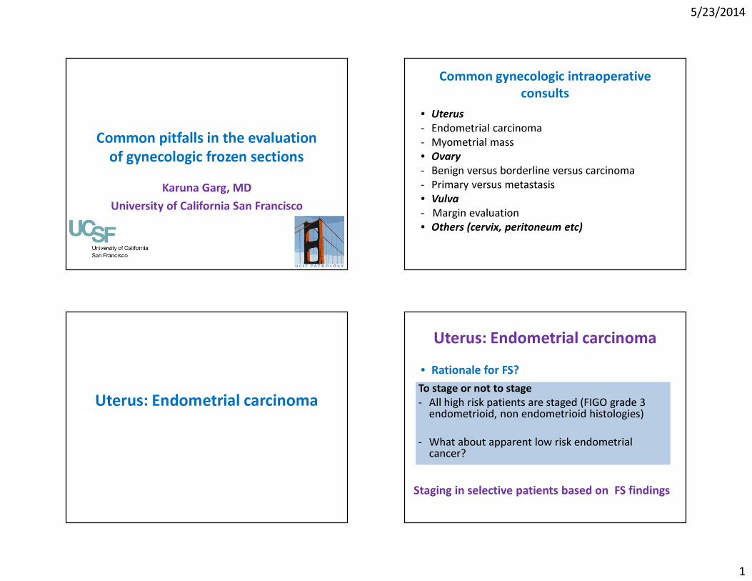
Common pitfalls in the evaluation of gynecologic frozen sections
Karuna Garg, MDUniversity of California San Francisco
Common gynecologic intraoperative consults
• Uterus- Endometrial carcinoma- Myometrial mass• Ovary- Benign versus borderline versus carcinoma- Primary versus metastasis• Vulva- Margin evaluation• Others (cervix, peritoneum etc)
Uterus: Endometrial carcinoma
Uterus: Endometrial carcinoma• Rationale for FS?To stage or not to stage- All high risk patients are staged (FIGO grade 3
endometrioid, non endometrioid histologies)
- What about apparent low risk endometrial cancer?
Staging in selective patients based on FS findings
5/23/2014
2
Endometrial carcinomaTreatment decisions based on FS- Lymphadenectomy or not- Extent of lymphadenectomy- Omentectomy and/or pelvic biopsies
- Sentinel lymph nodes for endometrial cancer
Endometrial carcinomaAccuracy of frozen sections:- Variable (from very good to very poor)
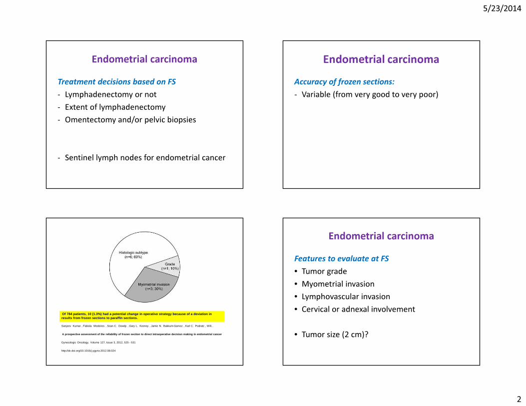
Of 784 patients, 10 (1.3%) had a potential change in operative strategy because of a deviation in results from frozen sections to paraffin sections.
Sanjeev Kumar , Fabiola Medeiros , Sean C. Dowdy , Gary L. Keeney , Jamie N. Bakkum-Gamez , Karl C. Podratz , Will...
A prospective assessment of the reliability of frozen section to direct intraoperative decision making in endometrial cancer
Gynecologic Oncology, Volume 127, Issue 3, 2012, 525 - 531
http://dx.doi.org/10.1016/j.ygyno.2012.08.024
Endometrial carcinomaFeatures to evaluate at FS• Tumor grade• Myometrial invasion• Lymphovascular invasion• Cervical or adnexal involvement
• Tumor size (2 cm)?
5/23/2014
3
Endometrial carcinoma: Treatment decisions?
1. Hysterectomy alone:- Grade 1 endometrioid, no myoinvasion or LVI2. Hysterectomy + pelvic LNs:- Grade 1 endometrioid with myoinvasion3. Hysterectomy + pelvic LNs + para-aortic LNs:- Grade 1-2 endometrioid, myoinvasive, with LVI or cervical
invasion- Grade 3 endometrioid or clear cell4. Hysterectomy + pelvic and para-aortic LNs + omentum:- Serous carcinoma or MMMT
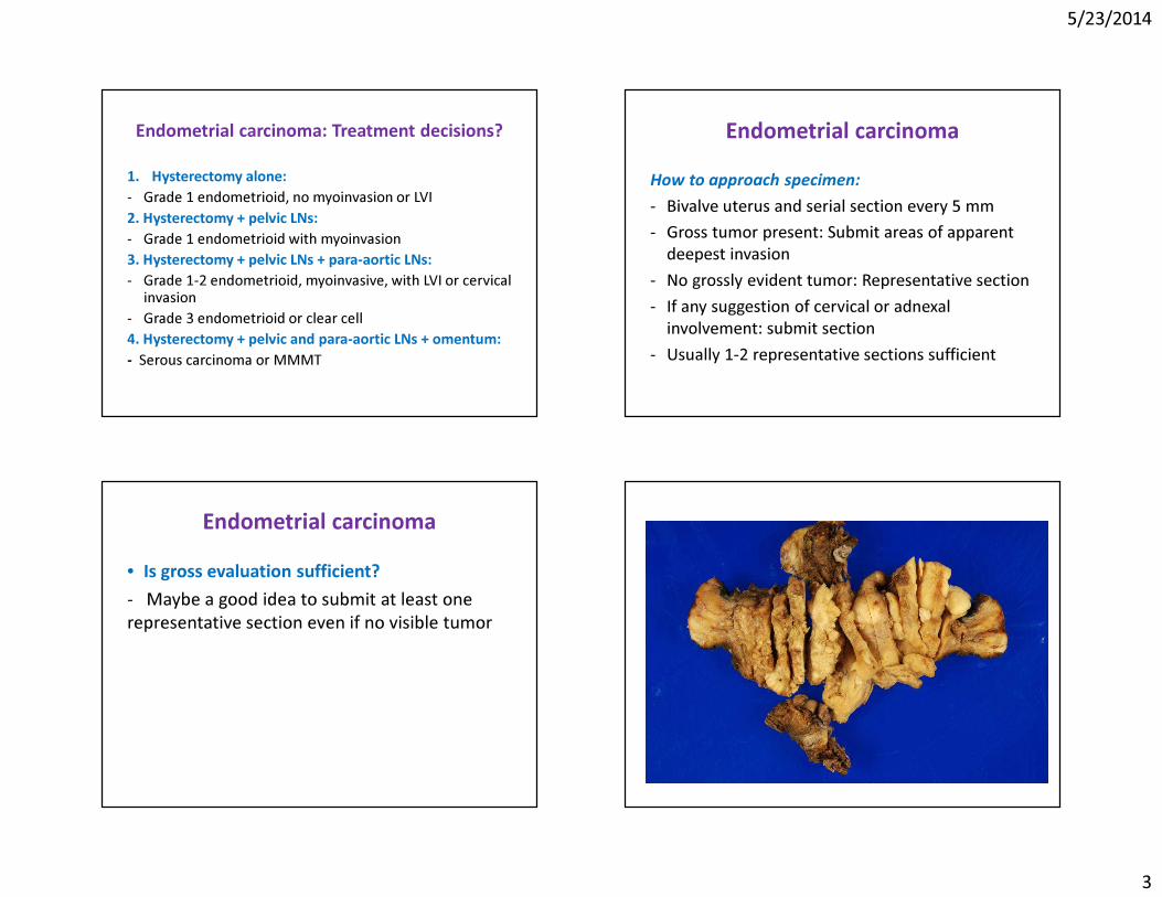
Endometrial carcinomaHow to approach specimen:- Bivalve uterus and serial section every 5 mm- Gross tumor present: Submit areas of apparent
deepest invasion- No grossly evident tumor: Representative section- If any suggestion of cervical or adnexal
involvement: submit section- Usually 1-2 representative sections sufficient
Endometrial carcinoma• Is gross evaluation sufficient?- Maybe a good idea to submit at least one representative section even if no visible tumor
5/23/2014
4
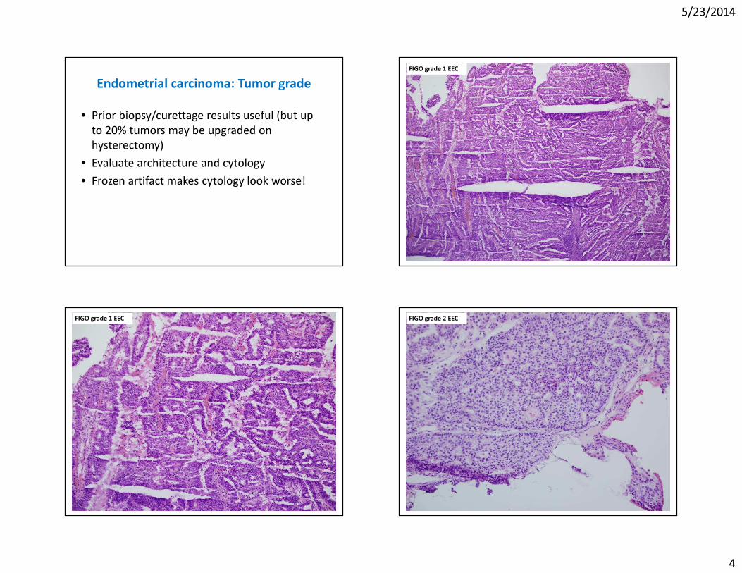
Endometrial carcinoma: Tumor grade• Prior biopsy/curettage results useful (but up
to 20% tumors may be upgraded on hysterectomy)
• Evaluate architecture and cytology• Frozen artifact makes cytology look worse!
FIGO grade 1 EEC
FIGO grade 1 EEC FIGO grade 2 EEC
5/23/2014
5
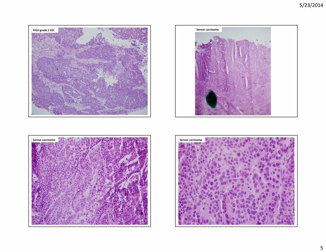
FIGO grade 2 EEC Serous carcinoma
Serous carcinoma Serous carcinoma
5/23/2014
6
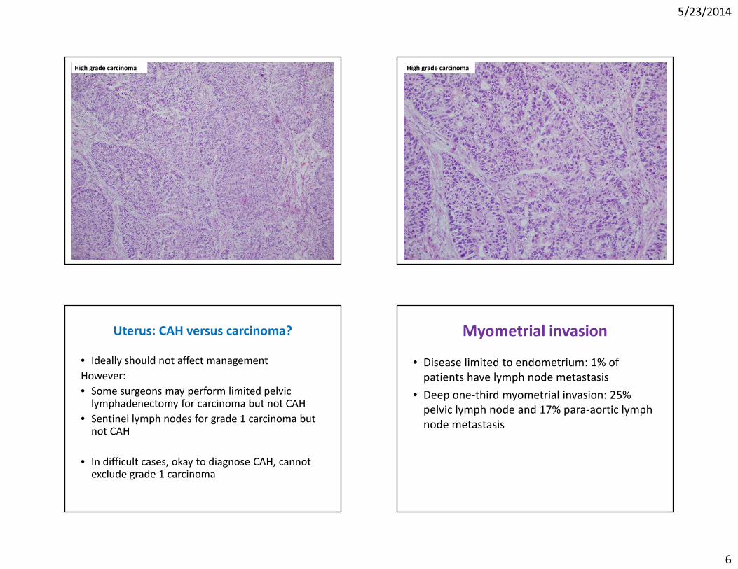
High grade carcinoma High grade carcinoma
• Ideally should not affect managementHowever:• Some surgeons may perform limited pelvic
lymphadenectomy for carcinoma but not CAH• Sentinel lymph nodes for grade 1 carcinoma but
not CAH
• In difficult cases, okay to diagnose CAH, cannot exclude grade 1 carcinoma
Uterus: CAH versus carcinoma? Myometrial invasion• Disease limited to endometrium: 1% of
patients have lymph node metastasis• Deep one-third myometrial invasion: 25%
pelvic lymph node and 17% para-aortic lymph node metastasis
5/23/2014
7
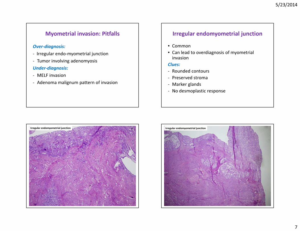
Myometrial invasion: PitfallsOver-diagnosis:- Irregular endo-myometrial junction- Tumor involving adenomyosisUnder-diagnosis:- MELF invasion- Adenoma malignum pattern of invasion
Irregular endomyometrial junction• Common• Can lead to overdiagnosis of myometrial
invasionClues:- Rounded contours- Preserved stroma- Marker glands- No desmoplastic response
Irregular endomyometrial junction Irregular endomyometrial junction
5/23/2014
8
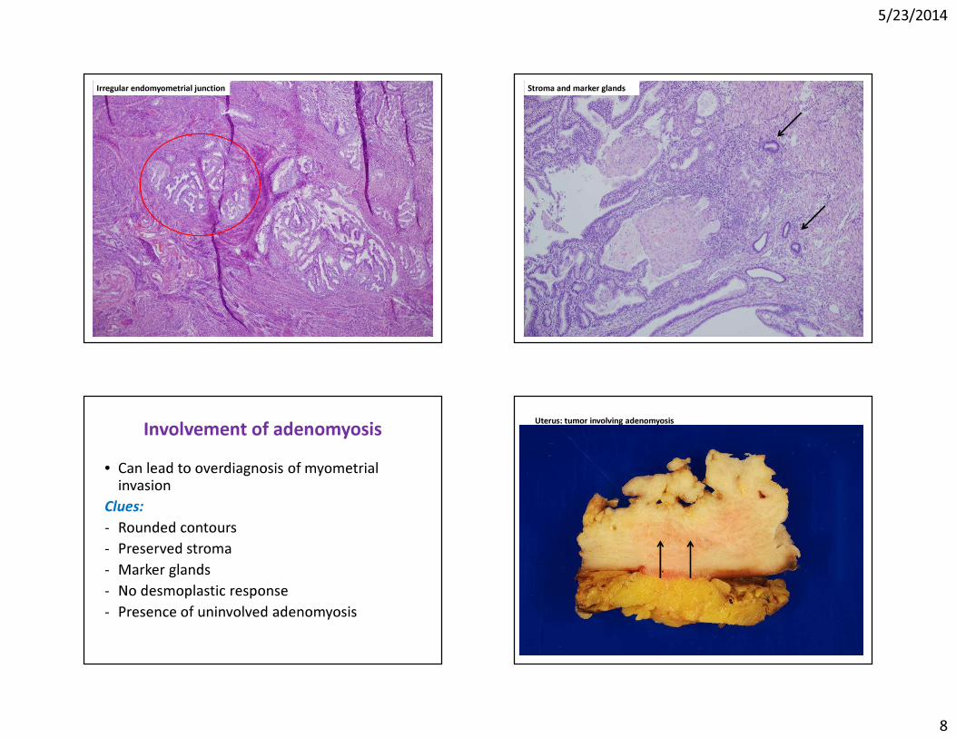
Irregular endomyometrial junction Stroma and marker glands
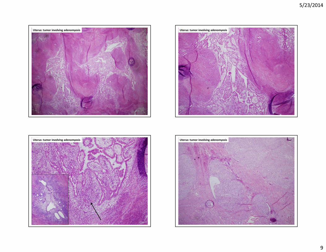
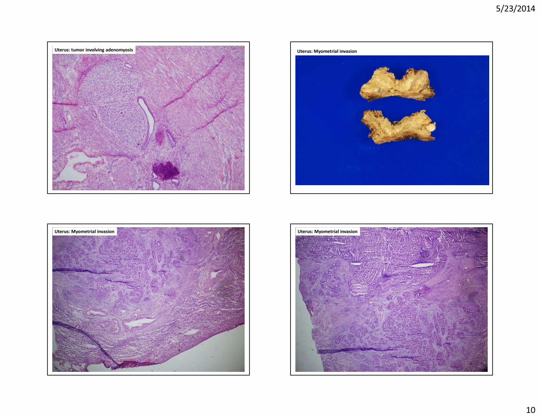
Involvement of adenomyosis• Can lead to overdiagnosis of myometrial
invasionClues:- Rounded contours- Preserved stroma- Marker glands- No desmoplastic response- Presence of uninvolved adenomyosis
Uterus: tumor involving adenomyosis
5/23/2014
9
Uterus: tumor involving adenomyosis Uterus: tumor involving adenomyosis
Uterus: tumor involving adenomyosis Uterus: tumor involving adenomyosis
5/23/2014
10
Uterus: tumor involving adenomyosis Uterus: Myometrial invasion
Uterus: Myometrial invasion Uterus: Myometrial invasion
5/23/2014
11
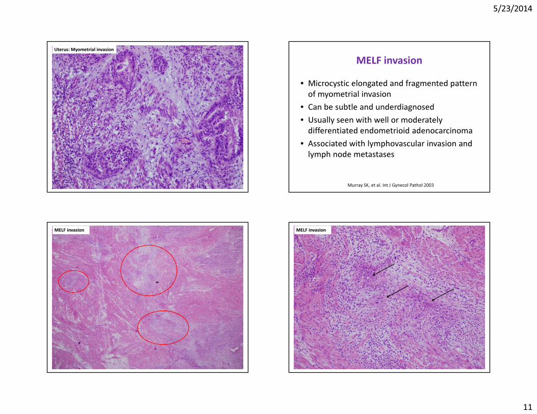
Uterus: Myometrial invasion
MELF invasion• Microcystic elongated and fragmented pattern
of myometrial invasion• Can be subtle and underdiagnosed• Usually seen with well or moderately
differentiated endometrioid adenocarcinoma• Associated with lymphovascular invasion and
lymph node metastases
Murray SK, et al. Int J Gynecol Pathol 2003
MELF invasion MELF invasion
5/23/2014
12
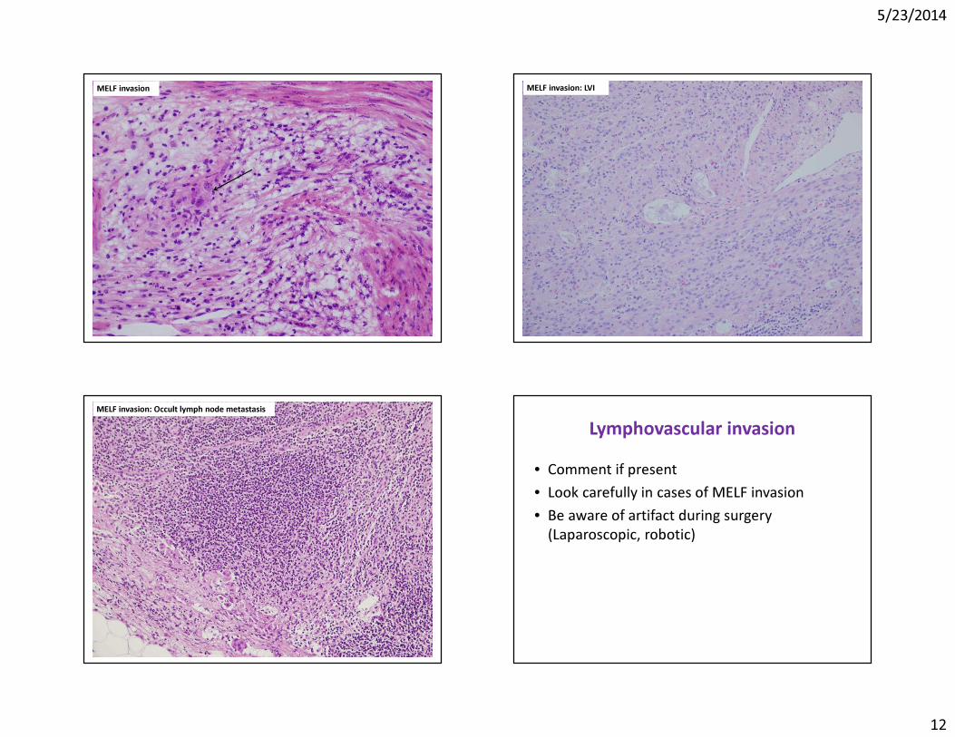
MELF invasion MELF invasion: LVI
MELF invasion: Occult lymph node metastasis
Lymphovascular invasion• Comment if present• Look carefully in cases of MELF invasion• Be aware of artifact during surgery
(Laparoscopic, robotic)
5/23/2014
13
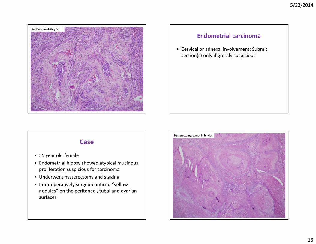
Artifact-simulating LVI
Endometrial carcinoma• Cervical or adnexal involvement: Submit
section(s) only if grossly suspicious
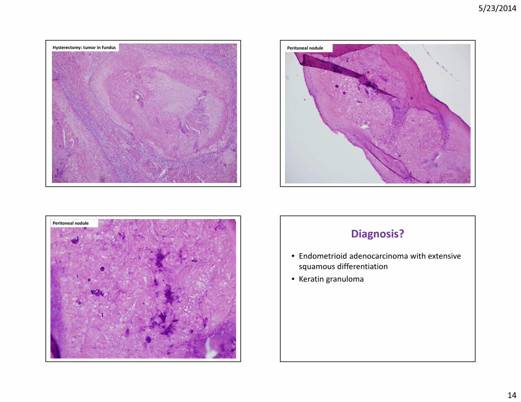
Case• 55 year old female• Endometrial biopsy showed atypical mucinous
proliferation suspicious for carcinoma• Underwent hysterectomy and staging• Intra-operatively surgeon noticed “yellow
nodules” on the peritoneal, tubal and ovarian surfaces
Hysterectomy: tumor in fundus
5/23/2014
14
Hysterectomy: tumor in fundus Peritoneal nodule
Peritoneal nodule
Diagnosis?• Endometrioid adenocarcinoma with extensive
squamous differentiation• Keratin granuloma
5/23/2014
15
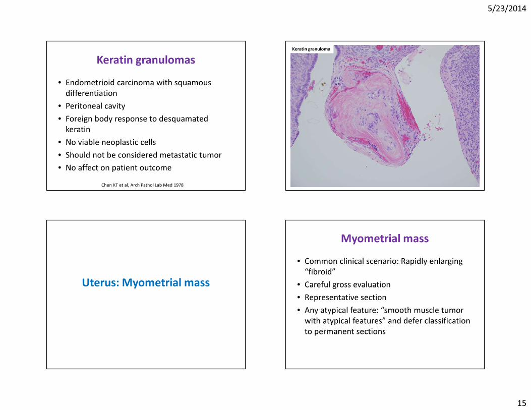
Keratin granulomas• Endometrioid carcinoma with squamous
differentiation• Peritoneal cavity• Foreign body response to desquamated
keratin• No viable neoplastic cells• Should not be considered metastatic tumor• No affect on patient outcome
Chen KT et al, Arch Pathol Lab Med 1978
Keratin granuloma
Uterus: Myometrial mass
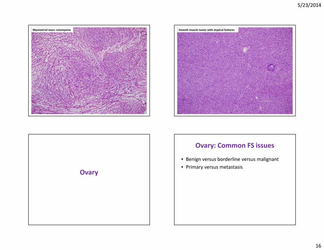
Myometrial mass• Common clinical scenario: Rapidly enlarging
“fibroid”• Careful gross evaluation• Representative section• Any atypical feature: “smooth muscle tumor
with atypical features” and defer classification to permanent sections
5/23/2014
16
Myometrial mass: Leiomyoma Smooth muscle tumor with atypical features
Ovary
Ovary: Common FS issues• Benign versus borderline versus malignant• Primary versus metastasis
5/23/2014
17
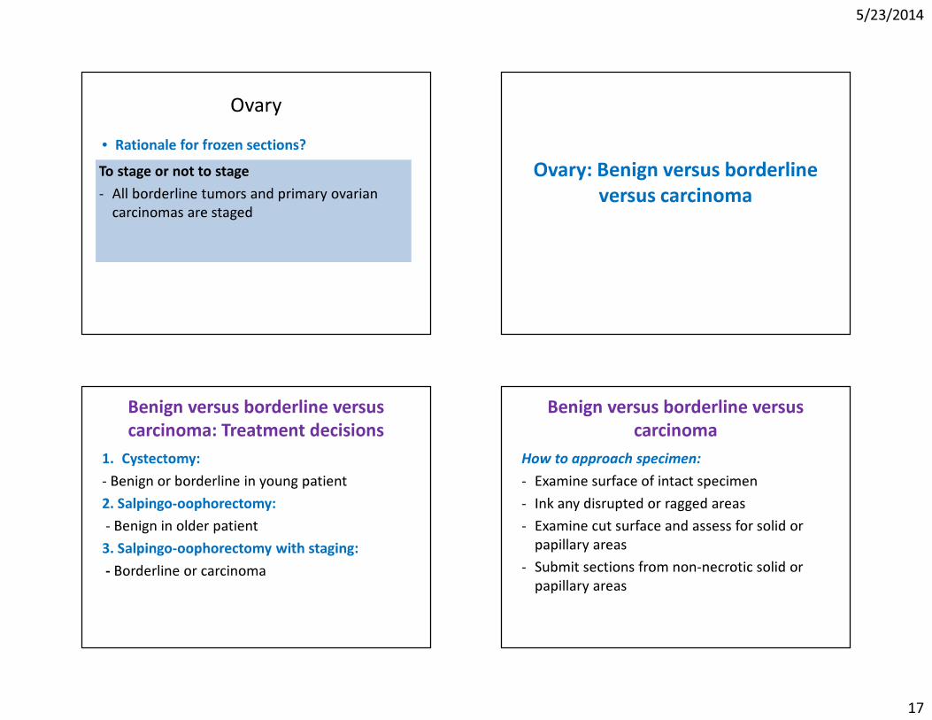
Ovary• Rationale for frozen sections?To stage or not to stage- All borderline tumors and primary ovarian
carcinomas are staged
Ovary: Benign versus borderline versus carcinoma
Benign versus borderline versus carcinoma: Treatment decisions
1. Cystectomy:- Benign or borderline in young patient 2. Salpingo-oophorectomy: - Benign in older patient
3. Salpingo-oophorectomy with staging: - Borderline or carcinoma
Benign versus borderline versus carcinoma
How to approach specimen:- Examine surface of intact specimen- Ink any disrupted or ragged areas- Examine cut surface and assess for solid or
papillary areas- Submit sections from non-necrotic solid or
papillary areas
5/23/2014
18
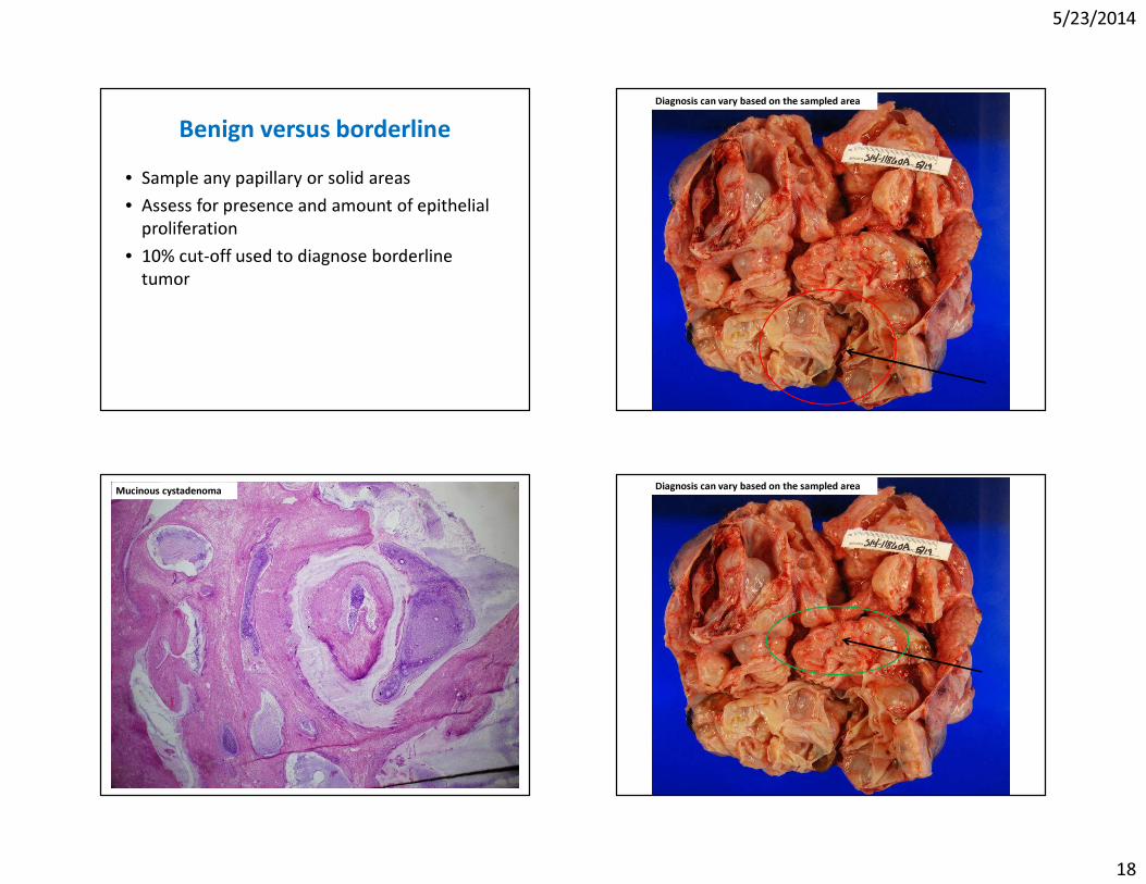
Benign versus borderline• Sample any papillary or solid areas• Assess for presence and amount of epithelial
proliferation• 10% cut-off used to diagnose borderline
tumor
Diagnosis can vary based on the sampled area
Mucinous cystadenoma Diagnosis can vary based on the sampled area
5/23/2014
19
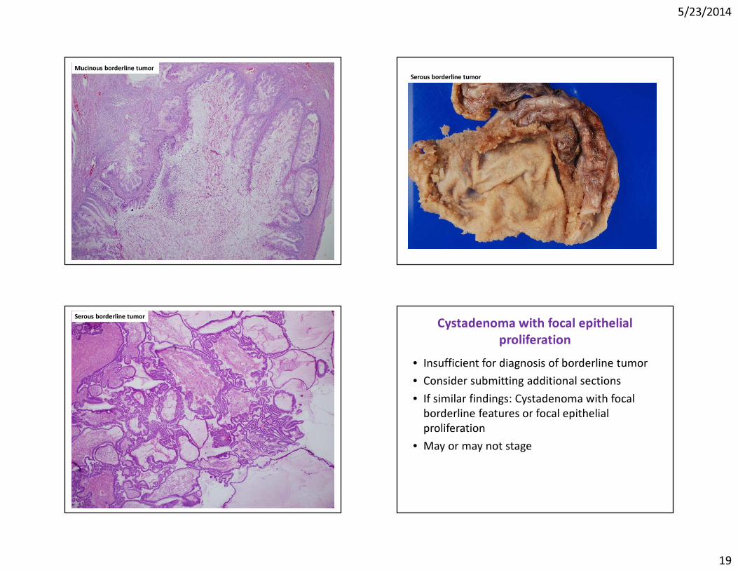
Mucinous borderline tumorSerous borderline tumor
Serous borderline tumor Cystadenoma with focal epithelial proliferation
• Insufficient for diagnosis of borderline tumor• Consider submitting additional sections• If similar findings: Cystadenoma with focal
borderline features or focal epithelial proliferation
• May or may not stage
5/23/2014
20
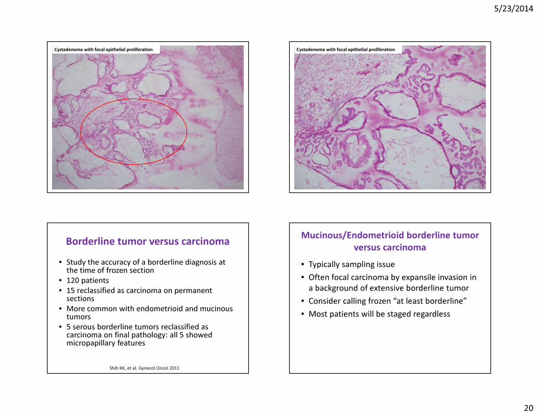
Cystadenoma with focal epithelial proliferation Cystadenoma with focal epithelial proliferation
Borderline tumor versus carcinoma• Study the accuracy of a borderline diagnosis at
the time of frozen section• 120 patients • 15 reclassified as carcinoma on permanent
sections• More common with endometrioid and mucinous
tumors• 5 serous borderline tumors reclassified as
carcinoma on final pathology: all 5 showed micropapillary features
Shih KK, et al. Gynecol Oncol 2011
Mucinous/Endometrioid borderline tumor versus carcinoma
• Typically sampling issue • Often focal carcinoma by expansile invasion in
a background of extensive borderline tumor• Consider calling frozen “at least borderline”• Most patients will be staged regardless
5/23/2014
21
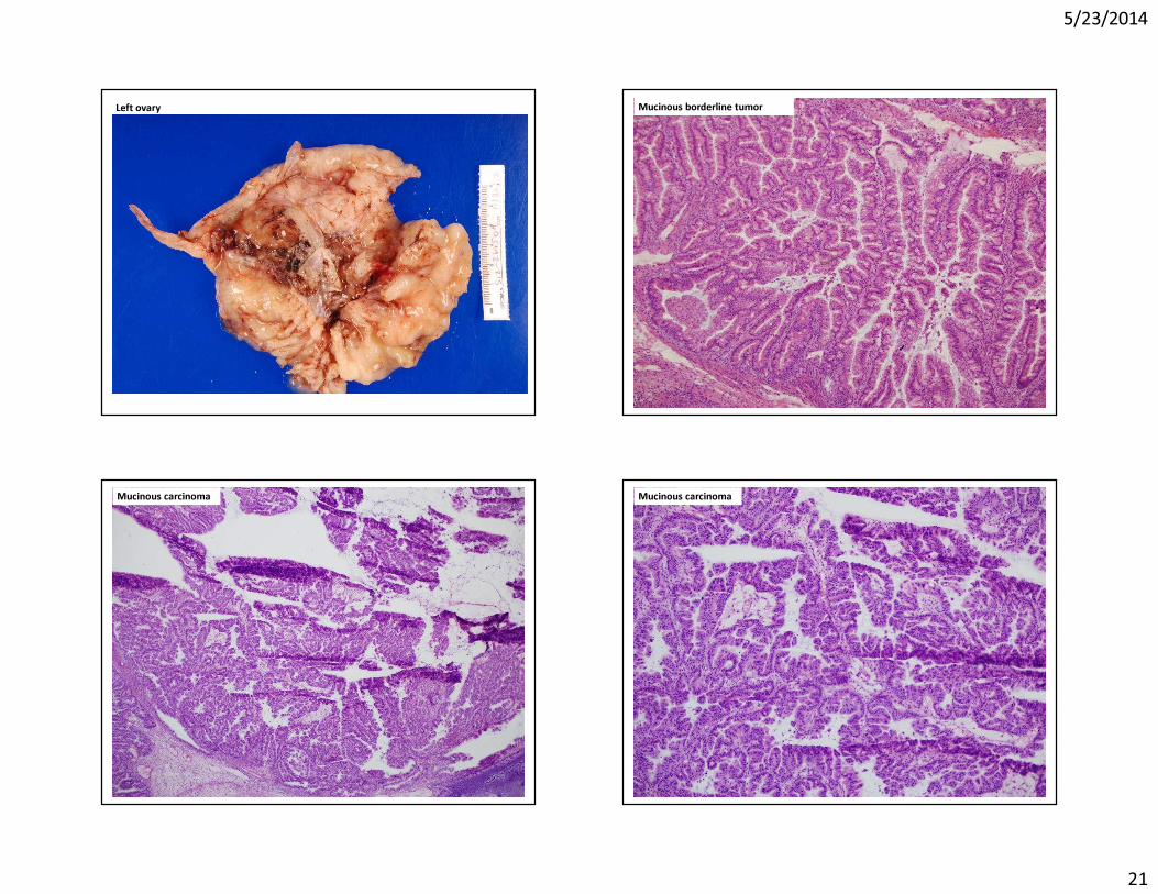
Left ovary Mucinous borderline tumor
Mucinous carcinoma Mucinous carcinoma
5/23/2014
22
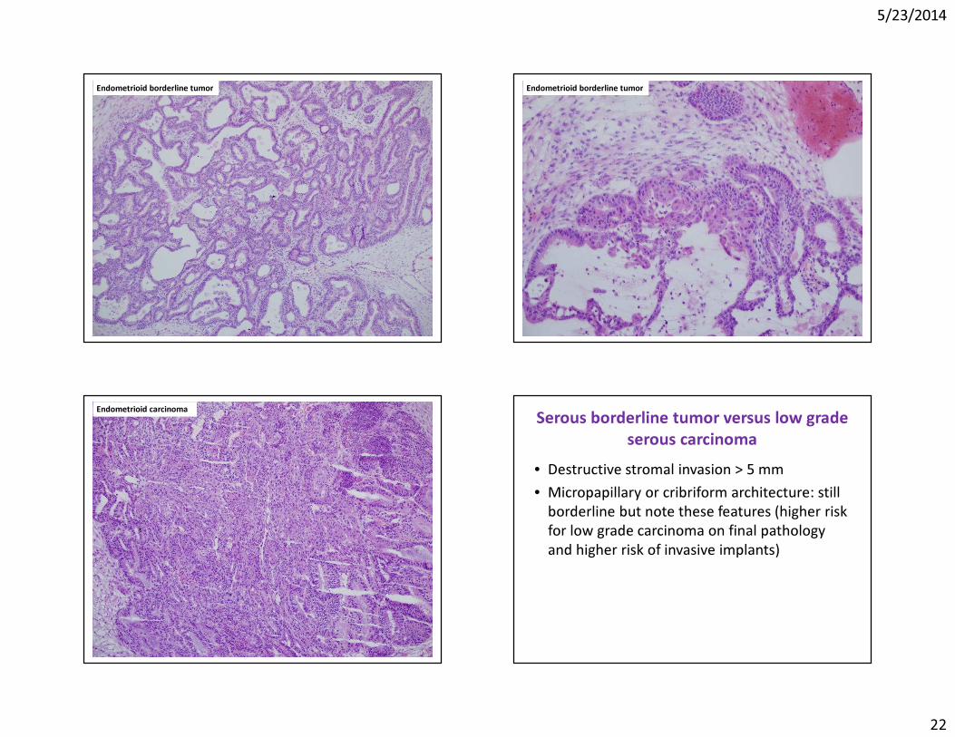
Endometrioid borderline tumor Endometrioid borderline tumor
Endometrioid carcinoma Serous borderline tumor versus low grade serous carcinoma
• Destructive stromal invasion > 5 mm• Micropapillary or cribriform architecture: still
borderline but note these features (higher risk for low grade carcinoma on final pathology and higher risk of invasive implants)
5/23/2014
23
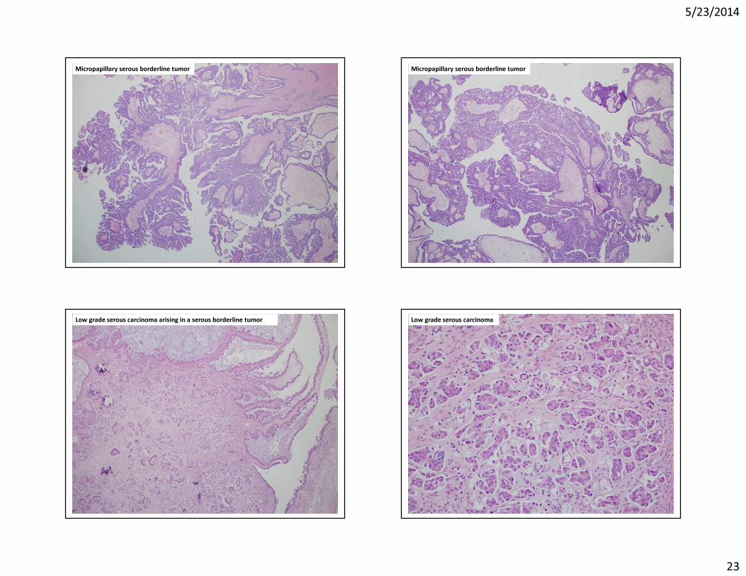
Micropapillary serous borderline tumor Micropapillary serous borderline tumor
Low grade serous carcinoma arising in a serous borderline tumor Low grade serous carcinoma
5/23/2014
24
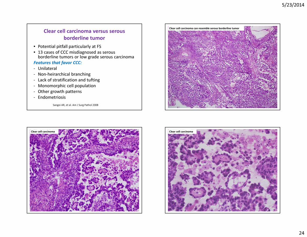
Clear cell carcinoma versus serous borderline tumor
• Potential pitfall particularly at FS• 13 cases of CCC misdiagnosed as serous
borderline tumors or low grade serous carcinomaFeatures that favor CCC:- Unilateral- Non-heirarchical branching- Lack of stratification and tufting- Monomorphic cell population- Other growth patterns- Endometriosis
Sangoi AR, et al. Am J Surg Pathol 2008
Clear cell carcinoma can resemble serous borderline tumor
Clear cell carcinoma Clear cell carcinoma
5/23/2014
25
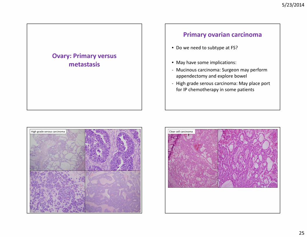
Ovary: Primary versus metastasis
Primary ovarian carcinoma• Do we need to subtype at FS?
• May have some implications:- Mucinous carcinoma: Surgeon may perform
appendectomy and explore bowel- High grade serous carcinoma: May place port
for IP chemotherapy in some patients
High grade serous carcinoma Clear cell carcinoma
5/23/2014
26
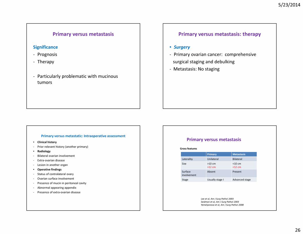
Primary versus metastasisSignificance- Prognosis- Therapy
- Particularly problematic with mucinous tumors
Primary versus metastasis: therapy• Surgery- Primary ovarian cancer: comprehensive
surgical staging and debulking- Metastasis: No staging
Primary versus metastatic: Intraoperative assessment• Clinical history- Prior relevant history (another primary)• Radiology- Bilateral ovarian involvement- Extra-ovarian disease- Lesion in another organ • Operative findings- Status of contralateral ovary- Ovarian surface involvement- Presence of mucin in peritoneal cavity- Abnormal appearing appendix- Presence of extra-ovarian disease
Primary MetastasisLaterality Unilateral BilateralSize >10 cm
>12 cm<10 cm<12 cm
Surface involvement
Absent Present
Stage Usually stage I Advanced stage
Primary versus metastasisGross features
Lee et al, Am J Surg Pathol 2003Seidman et al, Am J Surg Pathol 2003Yemelyanova et al, Am J Surg Pathol 2008
5/23/2014
27
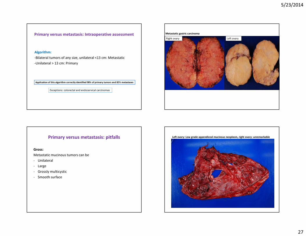
Primary versus metastasis: Intraoperative assessment
Algorithm: -Bilateral tumors of any size, unilateral <13 cm: Metastatic-Unilateral > 13 cm: Primary
Application of this algorithm correctly identified 98% of primary tumors and 82% metastases
Exceptions: colorectal and endocervical carcinomas
Right ovary Left ovaryMetastatic gastric carcinoma
Primary versus metastasis: pitfalls
Gross:Metastatic mucinous tumors can be - Unilateral- Large- Grossly multicystic - Smooth surface
Left ovary: Low grade appendiceal mucinous neoplasm, right ovary: unremarkable
5/23/2014
28
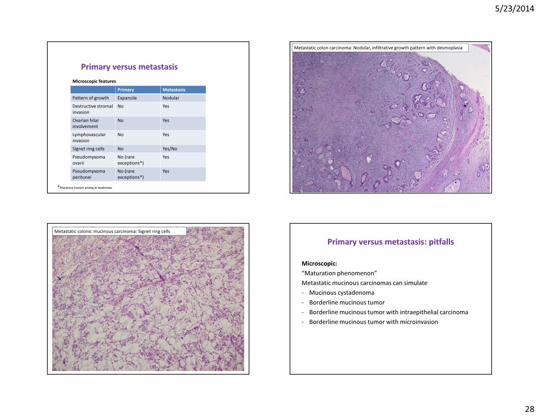
Primary MetastasisPattern of growth Expansile NodularDestructive stromal invasion
No Yes
Ovarian hilar involvement
No Yes
Lymphovascularinvasion
No Yes
Signet ring cells No Yes/NoPseudomyxomaovarii
No (rareexceptions*)
Yes
Pseudomyxomaperitonei
No (rare exceptions*)
Yes
Primary versus metastasisMicroscopic features
*Mucinous tumors arising in teratomas
Metastatic colon carcinoma: Nodular, infiltrative growth pattern with desmoplasia
Metastatic colonic mucinous carcinoma: Signet ring cellsPrimary versus metastasis: pitfalls
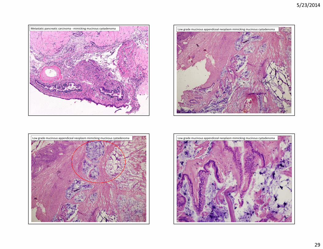
Microscopic:“Maturation phenomenon”Metastatic mucinous carcinomas can simulate- Mucinous cystadenoma- Borderline mucinous tumor- Borderline mucinous tumor with intraepithelial carcinoma - Borderline mucinous tumor with microinvasion
5/23/2014
29
Metastatic pancreatic carcinoma - mimicking mucinous cystadenoma Low grade mucinous appendiceal neoplasm mimicking mucinous cystadenoma
Low grade mucinous appendiceal neoplasm mimicking mucinous cystadenoma Low grade mucinous appendiceal neoplasm mimicking mucinous cystadenoma
5/23/2014
30
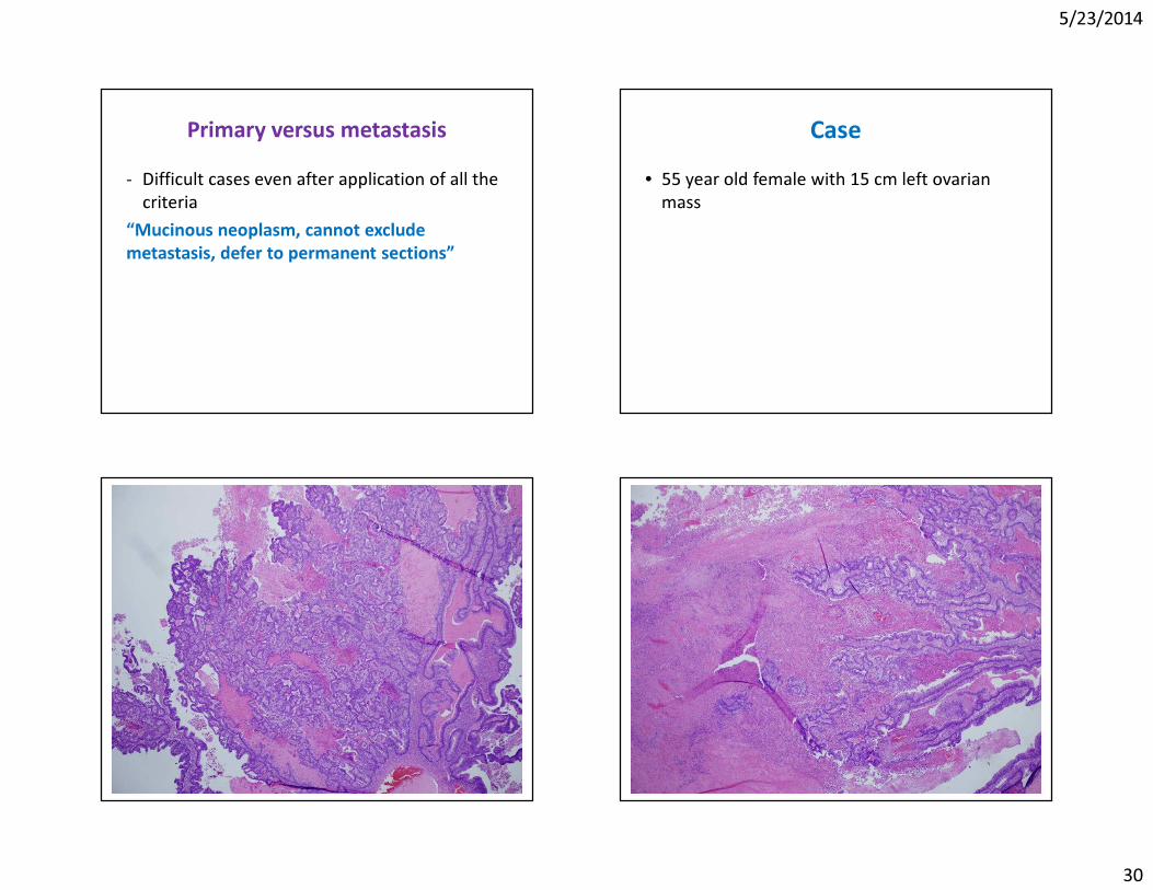
Primary versus metastasis- Difficult cases even after application of all the
criteria“Mucinous neoplasm, cannot exclude metastasis, defer to permanent sections”
Case• 55 year old female with 15 cm left ovarian
mass
5/23/2014
31
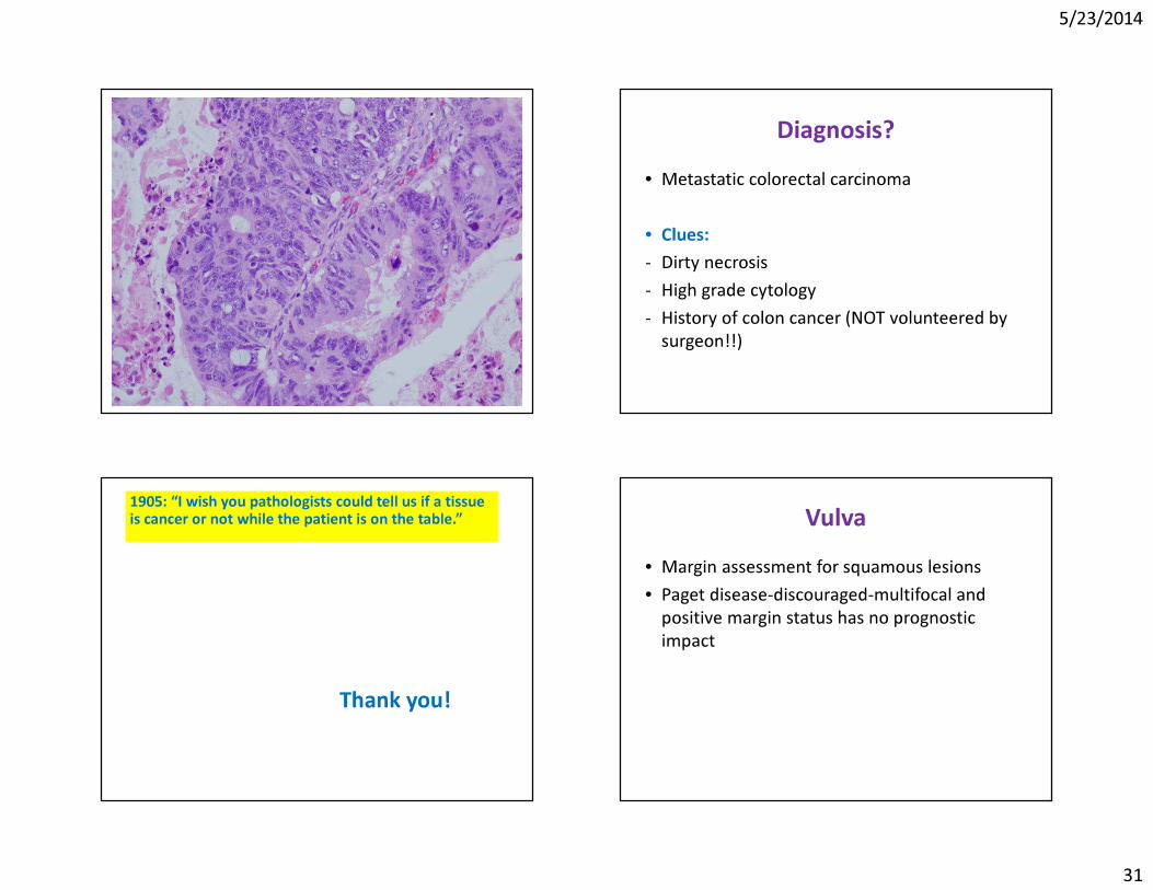
Diagnosis?• Metastatic colorectal carcinoma
• Clues:- Dirty necrosis- High grade cytology- History of colon cancer (NOT volunteered by
surgeon!!)
1905: “I wish you pathologists could tell us if a tissue is cancer or not while the patient is on the table.”
Thank you!
Vulva• Margin assessment for squamous lesions• Paget disease-discouraged-multifocal and
positive margin status has no prognostic impact
5/23/2014
32
Pregnancy/Postpartum
Ectopic pregnancy• Endometrial curettage• Assess grossly for villi (spongy) and submit
suspicious area for frozen• Preferable to handle as a rush specimen for
permanent sections if possible
Pregnancy/Postpartum: Common scenarios
• Diffuse peritoneal studding at the time of cesarean section
• Ovarian mass at the time of cesarean section
Disseminated peritoneal leiomyomatosis (DPL)
• Can look like peritoneal carcinomatosis to the surgeon
• Multiple small granular nodules on the peritoneal surfaces
• Women of reproductive age particularly in pregnancy
• Do not mistake for metastatic sarcoma• Small (<1 cm), no atypia, mitoses or necrosis
5/23/2014
33
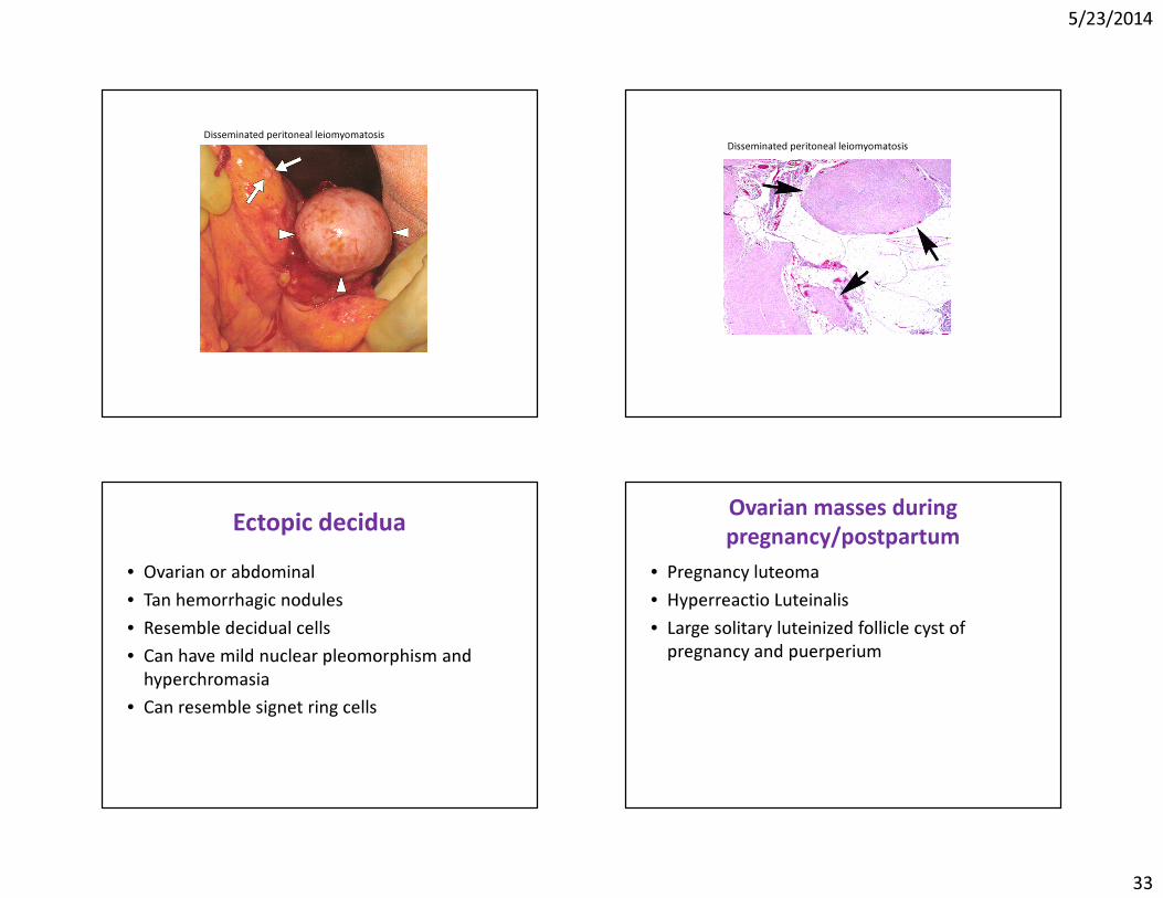
Disseminated peritoneal leiomyomatosisDisseminated peritoneal leiomyomatosis
Ectopic decidua• Ovarian or abdominal• Tan hemorrhagic nodules• Resemble decidual cells• Can have mild nuclear pleomorphism and
hyperchromasia• Can resemble signet ring cells
Ovarian masses during pregnancy/postpartum
• Pregnancy luteoma• Hyperreactio Luteinalis• Large solitary luteinized follicle cyst of
pregnancy and puerperium
5/23/2014
34
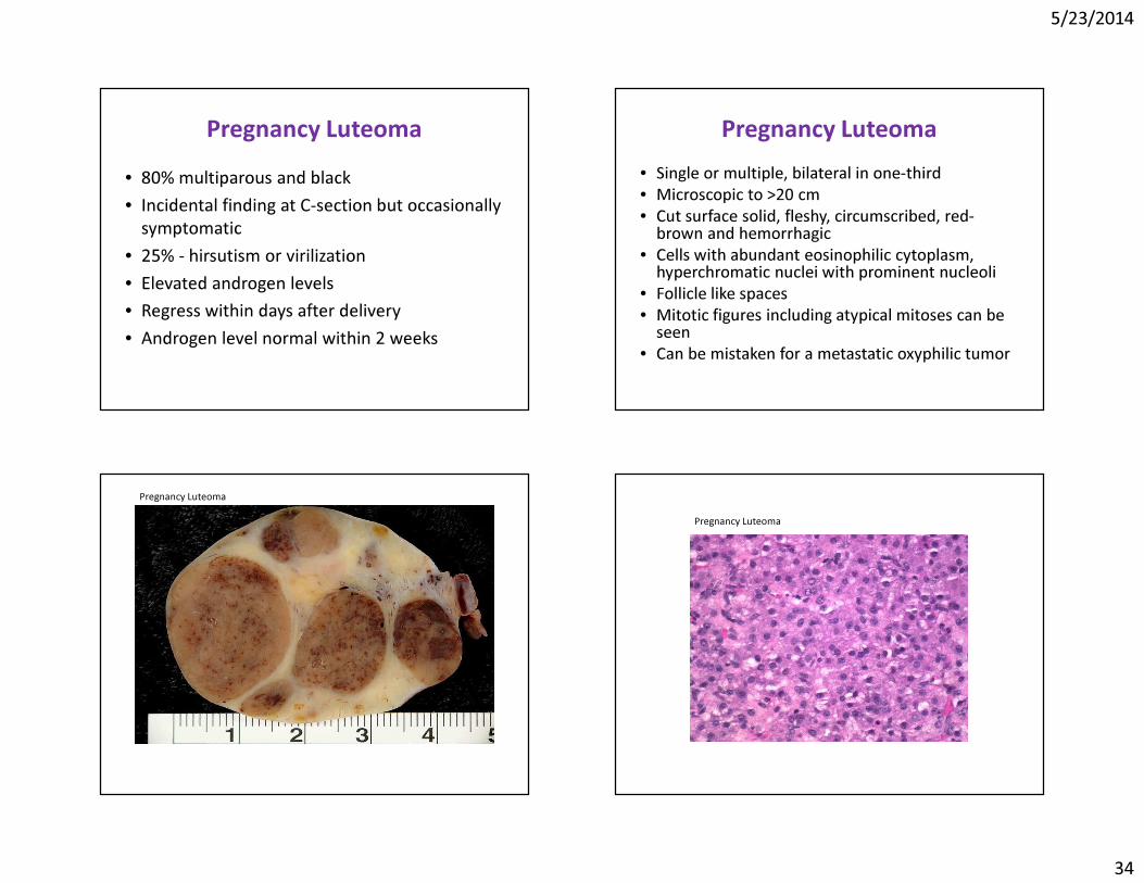
Pregnancy Luteoma• 80% multiparous and black• Incidental finding at C-section but occasionally
symptomatic• 25% - hirsutism or virilization• Elevated androgen levels• Regress within days after delivery• Androgen level normal within 2 weeks
Pregnancy Luteoma• Single or multiple, bilateral in one-third• Microscopic to >20 cm• Cut surface solid, fleshy, circumscribed, red-
brown and hemorrhagic• Cells with abundant eosinophilic cytoplasm,
hyperchromatic nuclei with prominent nucleoli• Follicle like spaces• Mitotic figures including atypical mitoses can be
seen• Can be mistaken for a metastatic oxyphilic tumor
Pregnancy LuteomaPregnancy Luteoma
5/23/2014
35
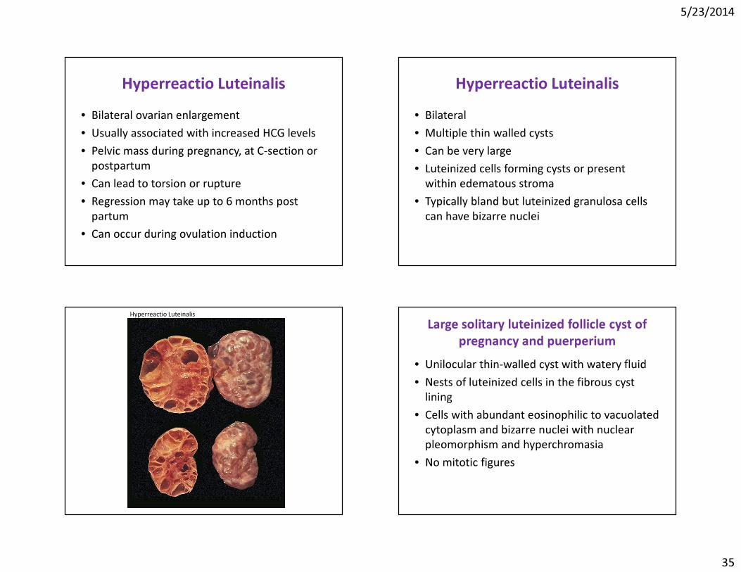
Hyperreactio Luteinalis• Bilateral ovarian enlargement• Usually associated with increased HCG levels• Pelvic mass during pregnancy, at C-section or
postpartum• Can lead to torsion or rupture• Regression may take up to 6 months post
partum• Can occur during ovulation induction
Hyperreactio Luteinalis• Bilateral• Multiple thin walled cysts• Can be very large• Luteinized cells forming cysts or present
within edematous stroma• Typically bland but luteinized granulosa cells
can have bizarre nuclei
Hyperreactio Luteinalis Large solitary luteinized follicle cyst of pregnancy and puerperium
• Unilocular thin-walled cyst with watery fluid• Nests of luteinized cells in the fibrous cyst
lining• Cells with abundant eosinophilic to vacuolated
cytoplasm and bizarre nuclei with nuclear pleomorphism and hyperchromasia
• No mitotic figures
5/23/2014
36
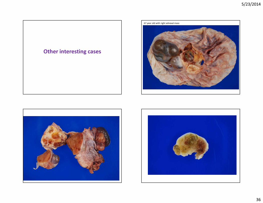
Other interesting cases
67 year old with right adnexal mass
5/23/2014
37
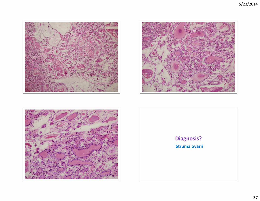
Diagnosis?Struma ovarii

5/23/2014
38
Thank you