Using Qualitative Disease Risk Analysis for Herpetofauna … · Using Qualitative Disease Risk...
14
Using Qualitative Disease Risk Analysis for Herpetofauna Conservation Translocations Transgressing Ecological and Geographical Barriers Mariana Bobadilla Suarez, 1,2 John G. Ewen, 1 Jim J. Groombridge, 2 K. Beckmann, 1 J. Shotton, 1 N. Masters, 1 T. Hopkins, 1 and Anthony W. Sainsbury 1 1 Institute of Zoology, Zoological Society of London, Regent’s Park, London NW1 4RY, UK 2 Durrell Institute of Conservation and Ecology, University of Kent, Giles Lane, Canterbury CT2 7NZ, UK Abstract: Through the exploration of disease risk analysis methods employed for four different UK her- petofauna translocations, we illustrate how disease hazards can be identified, and how the risk of disease can be analysed. Where ecological or geographical barriers between source and destination sites exist, parasite pop- ulations are likely to differ in identity or strain between the two sites, elevating the risk from disease and increasing the number and category of hazards requiring analysis. Simplification of the translocation pathway through the avoidance of these barriers reduces the risk from disease. The disease risk analysis tool is intended to aid conservation practitioners in decision making relating to disease hazards prior to implementation of a translocation. Keywords: Translocations, Reintroductions, Reptiles, Amphibians, Disease management, Conservation INTRODUCTION The types of translocations employed for wildlife conser- vation include reintroduction, population reinforcement, assisted colonisation and ecological replacement (Seddon et al. 2014). Translocations have become increasingly commonplace in conservation, with an increasing variety of taxonomic groups being moved (Seddon et al. 2005) including within the herpetofauna (Germano and Bishop 2009; Ewen et al. 2014). Recovery efforts for many amphibian species have been reliant on translocation as a recovery tool (Griffiths and Pavajeau 2008). One concern associated with wildlife translocations is that the released individuals, or other individuals within the wider destina- tion ecosystem, may suffer from disease linked to the translocation process. This may be a particular concern in amphibians, where close to 25 % of all extinct and threatened species on the IUCN Red List cite disease as a possible cause of decline (Heard et al. 2011) High-profile emerging infectious diseases that have impacted free-living herpetofauna include, Batrachochytrium dendrobatidis (Bd)-associated disease and Batrachochytrium salaman- drivorans (Bsal)-associated disease, ranaviral disease and Snake Fungal Disease (SFD) (Pounds et al. 2006; Teacher et al. 2010; Allender et al. 2011; Miller and Gray 2010; USGS 2013; Hyatt et al. 2002; Martel et al. 2013). There- fore, in undertaking translocations of herpetofauna, as in any other taxonomic group, it is crucial to assess and manage risk from disease. Published online: December 22, 2015 Correspondence to: Mariana Bobadilla Suarez, e-mail: [email protected] EcoHealth 14, S47–S60, 2017 DOI: 10.1007/s10393-015-1086-4 Original Contribution Ó 2015 The Author(s). This article is published with open access at Springerlink.com
Transcript of Using Qualitative Disease Risk Analysis for Herpetofauna … · Using Qualitative Disease Risk...

Using Qualitative Disease Risk Analysis for HerpetofaunaConservation Translocations Transgressing Ecological andGeographical Barriers
Mariana Bobadilla Suarez,1,2 John G. Ewen,1 Jim J. Groombridge,2 K. Beckmann,1
J. Shotton,1 N. Masters,1 T. Hopkins,1 and Anthony W. Sainsbury1
1Institute of Zoology, Zoological Society of London, Regent’s Park, London NW1 4RY, UK2Durrell Institute of Conservation and Ecology, University of Kent, Giles Lane, Canterbury CT2 7NZ, UK
Abstract: Through the exploration of disease risk analysis methods employed for four different UK her-
petofauna translocations, we illustrate how disease hazards can be identified, and how the risk of disease can be
analysed. Where ecological or geographical barriers between source and destination sites exist, parasite pop-
ulations are likely to differ in identity or strain between the two sites, elevating the risk from disease and
increasing the number and category of hazards requiring analysis. Simplification of the translocation pathway
through the avoidance of these barriers reduces the risk from disease. The disease risk analysis tool is intended
to aid conservation practitioners in decision making relating to disease hazards prior to implementation of a
translocation.
Keywords: Translocations, Reintroductions, Reptiles, Amphibians, Disease management, Conservation
INTRODUCTION
The types of translocations employed for wildlife conser-
vation include reintroduction, population reinforcement,
assisted colonisation and ecological replacement (Seddon
et al. 2014). Translocations have become increasingly
commonplace in conservation, with an increasing variety of
taxonomic groups being moved (Seddon et al. 2005)
including within the herpetofauna (Germano and Bishop
2009; Ewen et al. 2014). Recovery efforts for many
amphibian species have been reliant on translocation as a
recovery tool (Griffiths and Pavajeau 2008). One concern
associated with wildlife translocations is that the released
individuals, or other individuals within the wider destina-
tion ecosystem, may suffer from disease linked to the
translocation process. This may be a particular concern in
amphibians, where close to 25 % of all extinct and
threatened species on the IUCN Red List cite disease as a
possible cause of decline (Heard et al. 2011) High-profile
emerging infectious diseases that have impacted free-living
herpetofauna include, Batrachochytrium dendrobatidis
(Bd)-associated disease and Batrachochytrium salaman-
drivorans (Bsal)-associated disease, ranaviral disease and
Snake Fungal Disease (SFD) (Pounds et al. 2006; Teacher
et al. 2010; Allender et al. 2011; Miller and Gray 2010;
USGS 2013; Hyatt et al. 2002; Martel et al. 2013). There-
fore, in undertaking translocations of herpetofauna, as in
any other taxonomic group, it is crucial to assess and
manage risk from disease.Published online: December 22, 2015
Correspondence to: Mariana Bobadilla Suarez, e-mail: [email protected]
EcoHealth 14, S47–S60, 2017DOI: 10.1007/s10393-015-1086-4
Original Contribution
� 2015 The Author(s). This article is published with open access at Springerlink.com
Parasites may cause disease in released animals
resulting in establishment failure (a failed translocation) or,
if novel to the release site, may cause other species at the
release site to decline through disease. In this manuscript,
we define parasites as infectious agents including viruses,
bacteria, fungi, protozoa, helminths and ectoparasites.
Parasites that are novel and introduced to release sites are a
type of alien species (sensu Blackburn and Ewen submitted
this volume). Release of animals may also alter the trans-
mission dynamics of endemic parasites at the destination
site (due to host aggregation) and increase the probability
of an infectious disease outbreak (Aiello et al. 2014). At its
heart, therefore, the occurrence of disease in a translocation
often relates to differing host–parasite communities be-
tween source and destination locations, potentially influ-
enced by stressors acting on the individuals moved
(Sainsbury and Vaughan-Higgins 2012). For example, the
reintroduction of Mallorcan midwife toads (Alytes
muletensis) reared in captive breeding facility is likely to
have accidentally co-introduced an alien parasite, Bd, to the
recipient environment which was associated with disease
outbreaks in native Mallorcan amphibian populations
(Walker et al. 2008). Furthermore, the critically endangered
mountain chicken frog (Leptodactylus fallax) reintroduc-
tion programme in the island nation of Montserrat has
been hindered by the continued presence of Bd during the
reintroduction (Adams et al. 2014). Empirical evidence
from across taxonomic groups demonstrates that as a
consequence of translocations, alien parasites have caused
major epidemics, with adverse effects at the ecosystem level
(Sainsbury and Vaughan-Higgins 2012; Viggers et al. 1993;
Dobson and Foufopoulos 2001).
In conservation translocations, the risks of disease
from some select parasites may be known and management
measures can be employed to mitigate them. However, the
geographical distribution and/or pathogenicity of other
parasites may not be apparent until after the translocation
(Sainsbury and Vaughan-Higgins 2012; Ewen et al. 2012).
Additionally, some parasites may be unknown or uniden-
tified. There is a range of tools available to help identify
infectious and non-infectious health hazards and to assess
their level of risk from disease against various objectives
(e.g. risk to the individuals being moved, to populations of
the species at the destination sites and to the wider
ecosystem including environmental and human health).
Collectively, these tools are components of disease risk
analysis (DRA). However, it remains unclear how widely
these DRA methods are applied to conservation translo-
cations. Although DRA guidelines have been published for
wild animal translocations since 1992 (Davidson and Net-
tles 1992; De With et al. 1998; Corn and Nettles 2001;
Neimanis and Leighton 2004; Hartley 2010), the literature
has only recently started reporting how the methods have
been applied to translocations for conservation purposes
(e.g. Armstrong et al. 2003; Miller 2007; Hartley and Gill
2010; Sainsbury and Vaughan-Higgins 2012; Jakob-Hoff
et al. 2014a) and how they may be integrated more closely
with decision analysis (Ewen et al. 2015). We are unaware
of any peer-reviewed publication of DRA application in the
herpetofauna, a knowledge gap which may in part be due to
a lack of guidance on implementation. Therefore, our focus
here is to show how DRA has been applied in real case
studies to provide worked examples to biodiversity man-
agers.
Here we will briefly describe the qualitative disease risk
analysis (DRA) method developed by Sainsbury and
Vaughan-Higgins (2012) for conservation translocations.
Our intention is not to review DRA methods and their
development, but rather present a series of four case studies
to show managers how these tools have been applied in
translocations of herpetofauna in the UK. We focus par-
ticularly on defining the translocation pathway and explore
how increasing the number of geographical and ecological
barriers crossed on this pathway increases the complexity of
risk. The four case studies include: the smooth snake
(Coronella austriaca), the common European adder (Vipera
berus) (hereafter: the adder), the pool frog (Pelophylax
lessonae) and the sand lizard (Lacerta agilis). Each case
study faced unique challenges, in particular, there are
interesting contrasts between the influence of ecological
and/or geographical barriers within the actual or proposed
(as in the case of the adder) translocation pathway.
QUALITATIVE DISEASE RISK ANALYSIS
The importance of a qualitative DRA lies in endeavouring
to tackle the problem of infectious and non-infectious
agent hazards in translocations in the face of uncertainty,
including the scarcity of baseline data on the number,
identity, pathogenicity and geographical distribution of
parasite hazards (Sainsbury et al. 2012). To ensure appro-
priate judgement of risk is made by decision makers, it is
important that a DRA is undertaken transparently (e.g. it
must explicitly state any assumptions made due to gaps in
knowledge).
S48 M. Bobadilla Suarez et al.

The Sainsbury and Vaughan-Higgins (2012) method of
conducting a DRA has been developed from previous
qualitative DRA methods for wildlife (Davidson and Net-
tles 1992; Leighton 2002) and domestic animals (Murray
et al. 2004). The DRA process described here involves
completing a series of steps that follow a similar structure
to the World Organization for Animal Health (OIE)
guidelines for DRA in domestic animal movements be-
tween countries (Murray et al. 2004) but are tailored to the
needs of wildlife translocations as described by Sainsbury
and Vaughan-Higgins (2012). These steps consist of: (1)
mapping out the translocation pathway, (2) hazard iden-
tification, (3) risk assessment, (4) risk management and (5)
risk communication.
Translocation Pathway
A translocation pathway is a visual representation of the
route of the translocated animals that illustrates the points
at which different types of hazards may potentially harm
translocated individuals or the recipient ecosystem
(Fig. 1a–d). Hazards can either be infectious or non-in-
Figure. 1. Translocation pathways. a The smooth snake translocation pathway included the collection of individuals over the course of several
seasons from five to eight different sites. Smooth snakes were then moved to sites with assumed connectivity (i.e. no geographical or ecological
barriers) at which the species was deemed locally extinct but was present historically. b The adder translocation pathway began with the initial
capture of free-living female adders for captive breeding purposes, followed by the capture of males for captive breeding. It was intended that
juvenile adders either be returned to sites where they were present, reintroduced to sites where historical records of adders existed and where
there was suitable habitat, or introduced to similar sites outside the study area but with suitable habitat. c The pool frog translocation pathway
involved moving individuals from wild populations in Sweden to the UK taking into consideration the geographical barriers. d The sand lizard
reintroduction pathway used captive-bred stock for reintroduction into historical sand lizard habitat. Blue arrows represent movement of
individuals rather than movement of hazards. Hazards in blue boxes are placed on the segments of the translocation pathway where they would
have an effect.
DRA for Herpetofauna S49

fectious. Infectious hazards are parasites that are either
known to be associated with disease in the translocated
species or species at the destination, and/or are novel to the
destination, and/or are novel to the translocated species.
Non-infectious hazards include nutritional deficiencies,
toxins or objects capable of inflicting traumatic injury. We
will largely focus on infectious hazards and their classifi-
cation within the DRA method.
Infectious agents can be categorised into one or more
hazard types according to the stage of the translocation
pathway at which they act, and/or their novelty to the
host’s immune system, and are termed as source, destina-
tion, transport, carrier, population or zoonotic hazards (see
Table 1 for hazard definitions). Understanding which of
these hazards, or combination of hazards, may be operating
in a given translocation pathway allows practitioners to
more clearly see why a hazard has been identified and en-
ables them to consider either alternative translocation
pathways in order to avoid a high risk of disease or to
implement actions to manage those disease risks.
A major consideration in a given translocation path-
way is whether any geographical or ecological barriers are
to be crossed. ‘Geographical barriers’ are natural and
environmental barriers that prevent natural movement
between populations (e.g. rivers, mountain ranges, seas).
‘Ecological barriers’ are those characteristics (for example:
physical, behavioural, reproductive) that prevent interac-
tion between populations, in the absence of geographical
barriers, e.g. the populations may occupy different eco-
logical niches. If either geographical or ecological barriers
are crossed, source and destination hazards come into play,
and the probability that translocated or recipient popula-
tions are exposed to novel parasites is increased. The dis-
tinction between a translocation pathway with and without
geographical and ecological barriers is crucial, because
empirical evidence shows that the major epidemics of
disease associated with translocations have primarily arisen
from source hazards (Sainsbury and Vaughan-Higgins
2012; Cunningham 1996; Dobson and Foufopoulos 2001).
An assumption that there is a high probability that such
alien (source and destination) hazards are absent or min-
imal in a given translocation gives the translocation man-
ager confidence that the overall risk from disease of a given
translocation is markedly reduced. Therefore, if source and
destination environments are not separated by barriers,
source and destination hazards do not require considera-
tion and overall risk from disease is reduced.
Hazard Identification
Through literature review, elicitation of expert opinion
from ecologists, epidemiologists and pathologists, and/or
screening of populations, infectious and non-infectious
agents are identified that have potential to cause disease
through novelty, and/or known pathogenicity, to the
individuals that are translocated, and/or to the populations
of the same or closely related species at the destination,
and/or potentially to the wider ecosystem. To be defined as
a population hazard, there must be evidence that a parasite
or non-infectious agent has an effect on population num-
bers and is capable of causing the population to decline,
with an understanding that translocated populations may
Table 1. Hazard Types and Definitions According to Sainsbury and Vaughan-Higgins (2012) and Masters and Sainsbury (2011).
Hazard type Definition
Source hazard The infectious agents or strains of these agents, carried by translocated individuals which are novel (alien) to the
release environment (Sainsbury and Vaughan-Higgins 2012)
Destination hazard The infectious agents found at the release environment to which the translocated animals are naıve (Sainsbury
and Vaughan-Higgins 2012)
Carrier hazard Those commensal organisms that cause disease when stressors reduce host immunocompetence and alter the
host–parasite relationship (Sainsbury and Vaughan-Higgins 2012)
Transport hazard Those hazards that may be encountered during the transport (between the source and destination) which are
novel to the translocated animals and/or the release environment (Sainsbury and Vaughan-Higgins 2012)
Population hazard Those non-infectious and infectious agents present at the release site that could potentially have a negative
impact on a population as a whole but which are not necessarily novel to them (Sainsbury and Vaughan-
Higgins 2012)
Zoonotic hazard The infectious agents carried by the translocated species which can be transmitted to humans and potentially
harm the latter (Masters and Sainsbury 2011)
S50 M. Bobadilla Suarez et al.

be small and more vulnerable to impacts from diseases.
Searches in the literature can include diseases and/or par-
asites in the translocated species and closely related species.
As each hazard is identified, it must also be classified into a
hazard type (see step 1) and this classification justified
using evidence to ensure a transparent process. Where
parasites are of similar taxonomy and/or epidemiological
parameters, they can be identified as a group, for example
‘gram-negative bacteria’. Searches in the literature and use
of expert opinion enable an evaluation of the geographical
and ecological barriers crossed in the translocation pathway
and therefore enable the hazard type to be defined.
Risk Assessment
This component involves four steps, namely (1) release
assessment, (2) exposure assessment, (3) consequence
assessment, and (4) risk estimation (Murray et al. 2004).
The release assessment explains the pathway through which
a translocated animal could be exposed and infected, or
contaminated with the hazard and estimates the probability
of infection in translocated individuals. It is important to
note that for destination and population hazards, the re-
lease assessment does not apply as these hazards are, by
definition, already present at the destination. The exposure
assessment explains (1) how individuals of the same or
closely related species at the destination could be exposed
to and infected, or contaminated, with the hazard (source,
transport, carrier hazards), (2) the probability of this
exposure and infection occurring and (3) the probability of
dissemination of the hazard through populations at the
destination. For destination and population hazards, the
exposure assessment explains how the translocated animals
would become exposed to and infected with the hazard,
and the probability of both this exposure and infection, and
also dissemination occurring through populations at the
destination. In the case of non-infectious agents, which can
only be population hazards, the exposure assessment ex-
plains how the translocated animals would be exposed and
the probability of occurrence of this exposure.
The consequence assessment then determines the
consequences of exposure, and the probability that these
will occur, contrary to the objectives that the translocation
attempts to fulfil. The objectives against which conse-
quences are estimated will most likely include consequences
for the individuals moved, for the population of the same
species if it is already present at the destination, and for the
wider ecosystem at the destination. A zoonotic hazard can
be additionally designated to any of the other hazard types
and is an occupational health hazard. Finally, risk estima-
tion integrates the results from all three assessments de-
scribed above to provide an overall combined measure of
the risks of disease that each hazard poses (Murray et al.
2004). These estimates will be influenced by the informa-
tion available, values (e.g. risk attitude) of the specialist(s)
undertaking the DRA and can vary from negligible risk to
very high risk (Table 2). Guidelines for assigning these
estimates are difficult to generalise and tend to change on a
case by case basis. Overall, a reasoned, informed and
transparent discussion of the risks of disease from each
hazard is made and included within the DRA to justify each
risk probability.
Risk Management
Risk management consists of identifying and evaluating
management actions that may reduce the risks of diseases
identified in previous steps. The need to identify manage-
ment options is only required for those hazards deemed to
be above negligible risk within any given translocation
project. These decisions are normally made by the DRA
specialist and project managers. Here, any specific man-
agement actions proposed may also include a review and
monitoring component to improve the project teams’
knowledge of disease-related hazards and their level of risk
(Murray et al. 2004).
Risk Communication
Finally, risk communication is essential to ensure that
decision makers have clear and transparent justification for
each hazard’s risk status and risk management options. At
the start of a DRA, a meeting is held with stakeholders to
explain and discuss the DRA process and to agree a
description of the translocation pathway. Further meetings
Table 2. Terminology Used to Describe the Likelihood or
Probability Estimates When Undertaking a Disease Risk Analysis
(Adapted From Murray et al. 2004).
Term Definition
Negligible Not worth considering, insignificant
Low Less than average, coming below the normal level
Medium Average, the usual amount, extent or rate
High Extending above the normal level
DRA for Herpetofauna S51

may be held to discuss results as they become available. A
written report is presented to the decision maker and
stakeholders (those involved in the translocation planning
and those affected by the translocation). The report clearly
presents each of the preceding steps and provides all the
information required for decision makers to make choices
about how and whether a translocation should proceed.
The DRA document should not tell the stakeholders what
to do, but rather elucidate the options available to the
managers, and the risk from disease presented by these
options such that costs and benefits of the translocation can
be considered alongside other evaluations, prior to pro-
ceeding. These management options and their associated
risks are essentially dependent on the species being
translocated, the translocation pathway and the quality,
quantity and breadth of information available to construct
and analyse them. In the following case studies, we omit the
risk communication section as it simply summarises the
previous four steps of the DRA process.
The following four case studies all use the above DRA
method. Here we will present the details of each using the
steps outlined above, while highlighting the major diffi-
culties faced with each one. We will also describe how these
difficulties were managed within the DRA, how they
influenced the recommendations made and how the
experience of conducting a DRA in one species can help
inform its application to other species.
QUALITATIVE DISEASE RISK ANALYSIS IN
PRACTICE
Crossing No Ecological or Geographical Barriers:
Smooth Snakes (Coronella austriaca)
In 2010, a proposal was made to undertake a wild-to-wild
translocation of smooth snakes to restock a dwindling
remnant population in West Sussex from a nearby and
more abundant source in Dorset.
Translocation Pathway
It was believed that the translocation pathway for smooth
snakes did not cross barriers (Fig. 1a) because the indi-
viduals were being taken from a source area with assumed
connectivity of parasite assemblages through sympatric
reptiles to their destination. This inference was made by the
specialist wildlife veterinarians who completed the DRA in
consultation with expert reptile ecologists.
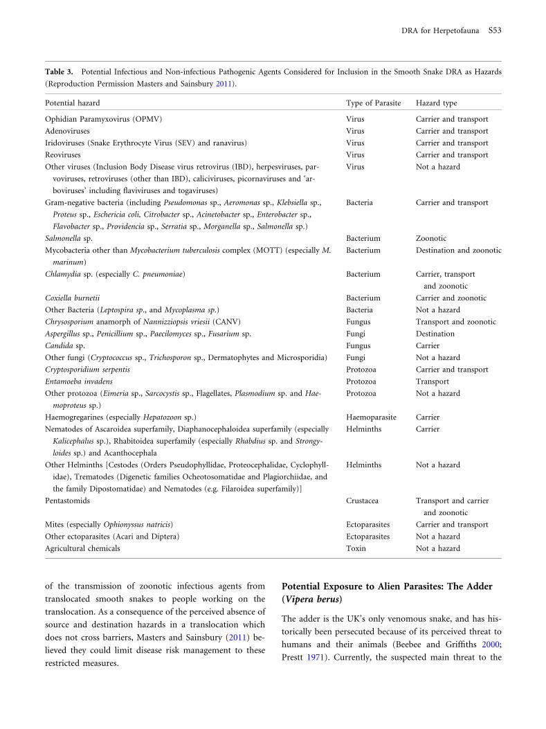
Hazard Identification
Parasites known to be present in smooth snakes, and other
ophidian species, were identified following a detailed re-
view of the published literature (found using keywords
‘infectious disease’ and ‘snakes’) and reptile disease and
medicine textbooks, then evaluated for hazard type and
their inclusion as hazards justified. Table 2 shows the list of
pathogenic agents (infectious and non-infectious) evalu-
ated and those which were identified as hazards (Table 3).
No source or destination hazards were identified because
the translocation pathway was not believed to cross geo-
graphical or ecological barriers.
Risk Assessment
Of the disease hazards identified in the DRA, the viruses
identified were generally found to be the highest risk haz-
ards, but only when evaluated as transport hazards. These
high-risk hazards included ophidian paramyxovirus, ade-
noviruses, iridoviruses and reoviruses. In general, these
hazards were deemed of high risk because of the likelihood
that the smooth snakes could be exposed to alien strains of
these viruses, through direct or indirect contact with exotic
snakes, en route to their destination, and the high proba-
bility that they could then transmit these exotic strains to
naıve animals at the destination. The other high-risk hazard
was Salmonella spp., a carrier, transport, and zoonotic
hazard (also included in the gram-negative bacteria hazard
grouping) (Table 3).
Risk Management
Three general management measures were proposed, which
included (a) biosecurity: in order to reduce the probability
of infection of smooth snakes with any novel infectious
agent during the translocation (e.g. reducing the risk of
disease from transport hazards through the establishment
of quarantine barriers at every stage of the translocation,
for example by designating a specific quarantine zone
within the transporting vehicle in which only smooth-
snake-dedicated tools and equipment, such as vivaria, were
used), (b) husbandry: in order to reduce the probability for
any stress-induced immuno-suppressive effect that may
precipitate disease in smooth snakes being translocated, for
example in association with carrier hazards and (c) occu-
pational health measures: in order to reduce the probability
S52 M. Bobadilla Suarez et al.
of the transmission of zoonotic infectious agents from
translocated smooth snakes to people working on the
translocation. As a consequence of the perceived absence of
source and destination hazards in a translocation which
does not cross barriers, Masters and Sainsbury (2011) be-
lieved they could limit disease risk management to these
restricted measures.
Potential Exposure to Alien Parasites: The Adder
(Vipera berus)
The adder is the UK’s only venomous snake, and has his-
torically been persecuted because of its perceived threat to
humans and their animals (Beebee and Griffiths 2000;
Prestt 1971). Currently, the suspected main threat to the
Table 3. Potential Infectious and Non-infectious Pathogenic Agents Considered for Inclusion in the Smooth Snake DRA as Hazards
(Reproduction Permission Masters and Sainsbury 2011).
Potential hazard Type of Parasite Hazard type
Ophidian Paramyxovirus (OPMV) Virus Carrier and transport
Adenoviruses Virus Carrier and transport
Iridoviruses (Snake Erythrocyte Virus (SEV) and ranavirus) Virus Carrier and transport
Reoviruses Virus Carrier and transport
Other viruses (Inclusion Body Disease virus retrovirus (IBD), herpesviruses, par-
voviruses, retroviruses (other than IBD), caliciviruses, picornaviruses and ‘ar-
boviruses’ including flaviviruses and togaviruses)
Virus Not a hazard
Gram-negative bacteria (including Pseudomonas sp., Aeromonas sp., Klebsiella sp.,
Proteus sp., Eschericia coli, Citrobacter sp., Acinetobacter sp., Enterobacter sp.,
Flavobacter sp., Providencia sp., Serratia sp., Morganella sp., Salmonella sp.)
Bacteria Carrier and transport
Salmonella sp. Bacterium Zoonotic
Mycobacteria other than Mycobacterium tuberculosis complex (MOTT) (especially M.
marinum)
Bacterium Destination and zoonotic
Chlamydia sp. (especially C. pneumoniae) Bacterium Carrier, transport
and zoonotic
Coxiella burnetii Bacterium Carrier and zoonotic
Other Bacteria (Leptospira sp., and Mycoplasma sp.) Bacteria Not a hazard
Chrysosporium anamorph of Nannizziopsis vriesii (CANV) Fungus Transport and zoonotic
Aspergillus sp., Penicillium sp., Paecilomyces sp., Fusarium sp. Fungi Destination
Candida sp. Fungus Carrier
Other fungi (Cryptococcus sp., Trichosporon sp., Dermatophytes and Microsporidia) Fungi Not a hazard
Cryptosporidium serpentis Protozoa Carrier and transport
Entamoeba invadens Protozoa Transport
Other protozoa (Eimeria sp., Sarcocystis sp., Flagellates, Plasmodium sp. and Hae-
moproteus sp.)
Protozoa Not a hazard
Haemogregarines (especially Hepatozoon sp.) Haemoparasite Carrier
Nematodes of Ascaroidea superfamily, Diaphanocephaloidea superfamily (especially
Kalicephalus sp.), Rhabitoidea superfamily (especially Rhabdius sp. and Strongy-
loides sp.) and Acanthocephala
Helminths Carrier
Other Helminths [Cestodes (Orders Pseudophyllidae, Proteocephalidae, Cyclophyll-
idae), Trematodes (Digenetic families Ocheotosomatidae and Plagiorchiidae, and
the family Dipostomatidae) and Nematodes (e.g. Filaroidea superfamily)]
Helminths Not a hazard
Pentastomids Crustacea Transport and carrier
and zoonotic
Mites (especially Ophionyssus natricis) Ectoparasites Carrier and transport
Other ectoparasites (Acari and Diptera) Ectoparasites Not a hazard
Agricultural chemicals Toxin Not a hazard
DRA for Herpetofauna S53
adder’s persistence is habitat fragmentation and degrada-
tion (JNCC 2010). In 2010, a reintroduction project was
proposed for supplementing adder populations in west-
central England. The proposal involved the release of cap-
tive-bred offspring of wild-caught adults, which had been
captured and taken into captivity prior to the DRA being
conducted. These individuals were housed within a zoo
which possessed exotic species of wide geographical origin
including non-native vipers. It was the intention to release
the progeny of these adders into the same forest whence the
breeding adults had been collected.
Translocation Pathway
It was assessed that the proposed translocation pathway in-
cluded the transfer of the adders across ecological and geo-
graphical barriers because there was potential contact and
transmission of parasites between non-native vipers and the
adders in the zoological collection (Fig. 1b) (Beckmann et al.
2014). By breeding adders in the zoo setting, project man-
agers had inadvertently created the conditions for parasites
to cross geographical and ecological barriers and hence there
was a risk of disease from source hazards.
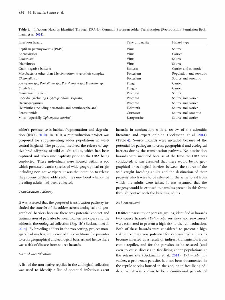
Hazard Identification
A list of the non-native reptiles in the zoological collection
was used to identify a list of potential infectious agent
hazards in conjunction with a review of the scientific
literature and expert opinion (Beckmann et al. 2014)
(Table 4). Source hazards were included because of the
potential for pathogens to cross geographical and ecological
barriers during the translocation pathway. No destination
hazards were included because at the time the DRA was
conducted; it was assumed that there would be no geo-
graphical or ecological barriers between the source of the
wild-caught breeding adults and the destination of their
progeny which were to be released in the same forest from
which the adults were taken. It was assumed that the
progeny would be exposed to parasites present in this forest
through contact with the breeding adults.
Risk Assessment
Of fifteen parasites, or parasite groups, identified as hazards
two source hazards (Entamoeba invadens and reoviruses)
were estimated to present a high risk to the reintroduction.
Both of these hazards were considered to present a high
risk, since there was potential for captive-bred adders to
become infected as a result of indirect transmission from
exotic reptiles, and for the parasites to be released (and
even to cause disease) in free-living adder populations at
the release site (Beckmann et al. 2014). Entamoeba in-
vadens, a protozoan parasite, had not been documented in
the reptile species housed in the zoo, or in free-living ad-
ders, yet it was known to be a commensal parasite of
Table 4. Infectious Hazards Identified Through DRA for Common European Adder Translocation (Reproduction Permission Beck-
mann et al. 2014).
Infectious hazard Type of parasite Hazard type
Reptilian paramyxovirus (PMV) Virus Source
Adenoviruses Virus Carrier
Reoviruses Virus Source
Iridoviruses Virus Source
Gram-negative bacteria Bacteria Carrier and zoonotic
Mycobacteria other than Mycobacterium tuberculosis complex Bacterium Population and zoonotic
Chlamydia sp. Bacterium Source and zoonotic
Aspergillus sp., Penicillium sp., Paecilomyces sp., Fusarium sp. Fungi Carrier
Candida sp. Fungus Carrier
Entamoeba invadens Protozoa Source
Coccidia (including Cryptosporidium serpentis) Protozoa Source and carrier
Haemogregarines Protozoa Source and carrier
Helminths (including nematodes and acanthocephalans) Helminth Source and carrier
Pentastomids Crustacea Source and zoonotic
Mites (especially Ophionyssus natricis) Ectoparasite Source and carrier
S54 M. Bobadilla Suarez et al.

captive reptiles (Barnard and Upton 1994; Wilson and
Carpenter 1996). Reoviruses were also deemed high risk, in
light of their ability to ‘‘jump’’ to other species. In fact, two
wild-caught adult adders in the captive breeding pro-
gramme at the zoological collection died and tested positive
for reovirus at post-mortem examination; the pathological
significance of the infection was unclear in each case. The
origin of reovirus infection was uncertain, and in the ab-
sence of data regarding the presence/prevalence of reovirus
in wild adders and sympatric reptiles in the UK, there was
concern that the infection may have originated from cap-
tive exotic reptile species and may therefore be alien to
native herpetofauna (Beckmann et al. 2014).
Risk Management
The DRA report proposed two alternative translocation
strategies as a means to reduce the risk from disease for the
adder, namely (1) a direct wild-to-wild translocation, or (2)
establishment of a dedicated captive breeding facility at the
destination site (rather than within the zoological collec-
tion). These alternatives were predicted to greatly reduce
the ecological and geographical barriers in the translocation
pathway (Beckmann et al. 2014) and therefore reduce
source and destination hazards. Both these approaches are
currently under consideration. In preparing for a future
wild-to-wild translocation, analysis of the literature
(Shotton and Sainsbury 2014) showed that it cannot be
assumed that adder populations within England are con-
tiguous because (1) long-term population studies of adders
have shown strong site philopatry and high hibernacula
fidelity (Phelps 2004); (2) vipers show a low migration
potential (Hand 2013); and (3) high genetic differentiation
exists between adder populations in Europe (Durrant 2014;
Ursenbacher et al. 2009) and therefore population struc-
turing may reveal ecological barriers and hence conditions
where source and destination hazards may be present.
Crossing Geographical and Ecological Barriers: Pool
Frogs (Pelophylax lessonae)
In the late 1990s, the northern clade pool frog became extinct
in England (Beebee 2013; Beebee et al. 2005). Following
extensive planning (Buckley and Foster 2005), reintroduc-
tion of wild-caught pool frogs collected from Sweden and
transported to England occurred between 2005 and 2008
(Baker and Foster 2015). As this was one of the first
translocations we were involved with, our method of DRA at
that time was more rudimentary (Sainsbury et al., in press).
Translocation Pathway
This translocation crossed geographical and ecological
barriers when moving pool frogs from mainland Sweden to
an isolated landmass (England) (Fig. 1c), and therefore
source and destination hazards were of greatest concern.
Hazard Identification
Hazard identification was achieved through detailed liter-
ature review and screening for parasites in the source
population of pool frogs in Sweden and four native
amphibian species at the destination site in England (Ta-
ble 5). The literature review revealed a relative lack of
information regarding parasites of amphibians in Sweden
(Sainsbury et al., in press), and in the context of the global
amphibian decline and its association with infectious dis-
ease, it was decided that it was important to obtain better
information on parasites through screening of pool frogs in
Sweden and amphibians at the reintroduction site in
England. Cunningham et al. (2001) expressed concerns that
‘‘potentially catastrophic epidemic ranavirus disease or
cutaneous chytridiomycosis’’ could be co-introduced with
any translocation of amphibians, including pool frogs, and
therefore an emphasis was placed on identifying the pres-
ence and absence of ranaviruses and Bd in Swedish pool
Table 5. Infectious Hazards for the Pool Frog DRA (Adapted From Sainsbury et al., in press).
Infectious hazard Type of parasite Hazard type
Ranaviruses Virus Destination
Batrachochytrium dendrobatidis Fungus Destination
Amphibiocystidium ranae Mesomycetozoea Destination
Unidentified intestinal protozoa Protozoa Destination
Trypanosoma rotatorium Protozoa Source
Unidentified intestinal Opalinid cysts Protozoa Source and transport
DRA for Herpetofauna S55

frogs. Source and destination hazards were identified
because geographical and ecological barriers were present
between Sweden and England. The results of screening
showed no ranaviruses or Bd in the Swedish pool frog
populations sampled, but these agents were known to be
present in England and therefore they were identified as
destination hazards. Two protozoan parasites Trypanosoma
rotatorium and unidentified intestinal opalinid cysts were
detected in pool frogs from Sweden and identified as source
hazards.
Risk Assessment
The DRA process in this case study focussed on source
hazards (by estimating the probability of co-introduction
and the likelihood of consequences based on pathogenic
capabilities) and destination hazards (by estimating the
probability of establishment of a parasite in the released
population of pool frogs and the probability that an
established parasite would be pathogenic). Two high-risk
hazards were analysed: ranaviruses and Bd in England as
destination hazards.
Risk Management
When the pool frogs were translocated, strict biosecurity
was adopted to try to protect the small reintroduced pool
frog population from these destination hazards until the
pool frog population could become established. The disease
risk management protocol included using amphibian-proof
fencing at the release site to create a quarantine barrier to
try to prevent ingress of destination hazards, health
examinations of pool frogs before and after translocation,
and pathological examination of any dead animals found
(see Vaughan-Higgins et al. 2015, this volume).
Captive Breeding in Multiple Locations: Sand Lizard
Reintroduction (Lacerta agilis)
Translocations of sand lizards began in 1968 (Moulton
et al. 2011) and were mostly carried out from wild-to-wild
for mitigation purposes, driven by impending habitat dis-
turbances due to development and without DRA. Begin-
ning in the 1990s, the focus of conservation translocations
for sand lizards shifted to a preference for reintroduction
using captive-bred stock. A post hoc DRA was requested by
Natural England in order to assess the risks from disease
associated with this long-term captive breeding and release
programme (Lloyd and Sainsbury 2003). It is important to
note that several of the captive-breeders held collections
which included exotic reptile species and which did not
have biosecurity measures in place (Lloyd and Sainsbury
2003).
Translocation Pathway
The reintroduction of sand lizards into existing and his-
torical habitats was carried out using captive-bred stock
from several breeders (Fig. 1d). This pathway included the
crossing of geographical and ecological barriers because
non-native reptiles were present without biosecurity.
Hazard Identification
Similar to the adder scenario, special attention was placed
on the possibility of direct and indirect contact with exotic
reptile species that were in shared captive breeding facilities
(Lloyd and Sainsbury 2003). Visits to breeders and/or
surveys of their facilities were invaluable when further
developing the DRA. A full list of hazards considered for
this DRA can be found in Table 6.
Risk Assessment
The DRA found iridoviruses, paramyxoviruses, Entamoeba
invadens and mycobacteria to be of highest risk, mainly
because of evidence suggesting catastrophic consequences
through epidemic disease, should these parasites be released
into the destination ecosystem as novel agents (contracted
from exotic reptiles).
Risk Management
In this case it was logistically difficult to call for a relocation
of the captive breeding facility to the release site. Therefore,
the captive breeding facilities were to be placed under
permanent quarantine, which would allow for strict
biosecurity practices to be established, including barrier
methods to minimise exposure to non-native species and
their parasites.
DISCUSSION
We have illustrated how DRA can help during the planning
stages of translocations to better identify which infectious
agents may be hazards and what options may be available to
S56 M. Bobadilla Suarez et al.

manage the risk of disease they might present. DRA is, in
essence, a process for working with uncertainty in hazard
identification and consequence assessment and making risk-
sensitive management decisions based on this information
(Ewen et al. 2015). Importantly, the method chosen in all of
these case studies was made transparent by justifying which
hazards were considered and why. Ideally, potential man-
agement actions are also supported by evidence. Whilst this
approach does not mean risks are removed and all hazards
are identified, it does provide a practical and rational ap-
proach to assessing disease-related risks.
Through the use of these four examples, we have shown
that the more complicated a translocation pathway is (i.e. the
more barriers involved), the more complicated the DRA
process will be where barriers are crossed source and desti-
nation hazards must be analysed and the hazard list will be
lengthened. Examples of complicated translocation pathways
resulting in more complex DRAs can also be seen in the
Eurasian crane (Grus grus) (Vaughan and Sainsbury 2010;
Sainsbury and Vaughan-Higgins 2012) and short-haired
bumblebee (Bombus subterraneus) (Brown et al. this volume)
reintroduction to England, Regent honeyeaters (Xanthomyza
Phrygia) in Australia (Jakob-Hoff et al. 2014a), and Eastern
wild turkeys (Meleagris gallopavo silvestris) to Canada (Nei-
manis and Leighton 2004). Themost straightforward solution
to reduce disease risks in these cases is simplification of the
translocation pathway, for example by avoiding holding ani-
mals for translocation in multi-species, multi-origin captive
facilities. Alternatives may be feasible with options to place
captive breeding facilities at the release site. This method was
successfully used for reintroduction of cirl buntings (Emberiza
cirlus) in south west England (McGill et al. 2010). Special care
must be taken to ensure that when trying to eliminate obvious
geographical and ecological barriers, similar to those in the
pool frog scenario, one also considers those obscure ecological
barriers as identified in the adder scenario. Epidemiological
principles also support a reduction in duration and distance of
transport during a translocation, which likely also reduce
stress. Transport and carrier hazards are more likely to be
associated with disease when these transport durations are
longer and more complex.
This DRA approach (Sainsbury and Vaughan-Higgins
2012) follows a reasoned, methodical and widely accepted
set of guidelines (Jakob-Hoff et al. 2014b). Its most valu-
able asset is the ability to identify and evaluate those
sometimes overlooked disease risks with transparency. In
the face of increasingly apparent parasite threats to global
biodiversity (Daszak et al. 2000), this tool can help to
consider the risks from disease in translocation. While the
challenges we face when compiling a DRA are many (e.g.
often including a lack of information on parasite presence,
identity, geographical distribution and virulence), further
Table 6. Infectious Hazards for the Sand Lizard DRA.
Infectious hazard Type of parasite Hazard type
Adenovirus Virus Source, carrier and transport
Herpesviruses Virus Not a hazard
Reovirus Virus Source, carrier and transport
Iridoviruses Virus Source, carrier and transport
Paramyxovirus (PMV) Virus Source, carrier and transport
Gram-negative bacteria (Aeromonas spp., Corynebacterium spp.,
Klebsiella spp., Proteus spp., Pseudomonas spp., Salmonella spp.)
Bacteria Carrier and zoonotic
Mycobacteria Bacteria Zoonotic
Trichomonads Protozoa Transport and carrier
Entamoeba invadens Protozoa Source and transport
Coccidia (Eimeria, sp. Isospora sp. and Cryptosporidia sp.) Protozoa Source, transport and carrier
Haemogregarina, Hepatozoon, Haemoproteus, Plasmodium, Trypanosomes Haemoparasites Source and carrier
Non-native nematodes Mesocestoides spp., Oswaldocruzia filiformis,
Metaplagiorchis molini, Oochoristica tuberculata
Helminths Source
Non-native cestodes Helminths Source
Non-native trematodes Helminths Source
Pentastomids Crustacea Source and zoonotic
Ophionyssus saurarum, Ixodes ricinus, Uropoda sp. Ectoparasites Destination
DRA for Herpetofauna S57

application and critical evaluation can help to continually
improve our application of these tools.
ACKNOWLEDGMENTS
The authorswould like to thankChris Lloyd, Jim Foster, Paul
Edgar, Fieke Molenaar, Kat Walsh, John Baker, Andrew
Cunningham, Stephen Price, Glyn Davies, Lucy Stead,
MatthewPerkins, Becki Lawson, Chris Pollard, JulianDrewe,
Richard Ssuna, Ntombi Mudenda, David Martinez Jimenez,
Mike Hart, John Buckley, Brian Banks, Clyde Hutchinson,
Iain McGill, Katie Macdonald, Jamie Wood, the Committee
members of the Reinforcing Wyre’s Reptiles, Francis Flana-
gan, Katharine Woods; Rachel Marschang and staff at the
Institute of Environmental and Animal Hygiene, University
of Hohenheim and Laboklin, Bad Kissingen, Germany; Ann
Pocknell and staff at Finn Pathologists; Shaheed Karl
Macgregor, Shinto John, the APHA, Eileen Harris, Gabriela
Peniche, Chris Durrant, FayeWillman andNickMoulton for
their assistance with this work. ZSl acknowledges the
financial support of Natural England and CONACYT
(Scholarship Number 312985).
FUNDING
This study was funded by CONACYT (Consejo Nacional de
Ciencia y Tecnologia), Scholarship Number 312985. The
Zoological Society of London acknowledges the financial
contribution of Natural England.
OPEN ACCESS
This article is distributed under the terms of the Creative
Commons Attribution 4.0 International License (http://
creativecommons.org/licenses/by/4.0/), which permits un-
restricted use, distribution, and reproduction in any med-
ium, provided you give appropriate credit to the original
author(s) and the source, provide a link to the Creative
Commons license, and indicate if changes were made.
REFERENCES
Adams SL, Morton MN, Gray G, Terry A, Hudson, M, Martin L(2014) Enabling Montserrat to save the Critically EndageredMountain Chicken. Darwin Initiative Final report Available:http://www.darwininitiative.org.uk/documents/18018/23105/18-018%20FR%20Edited.pdf
Aiello CM, Nussear KE, Walde AD, Esque TC, Emblidge PG, SahP, Bansal S, Hudson SJ (2014) Disease dynamics during wildlifetranslocations: disruptions to the host population and potentialconsequences for transmission in desert tortoise contact net-works. Animal Conservation 17(S1):27–39
Allender MC, Dreslik M, Wylie S, Phillips C, Wylie DB, MaddoxC, Delaney MA, Kinsel MJ (2011) Chrysosporium sp. infection ineastern massasauga rattlesnakes. Emerging infectious diseases17:2383–2384
Armstrong D, Jakob-Hoff R, Seal US (2003) Animal movementsand disease risk – a workbook, Conservation Breeding SpecialistGroup (SSC/IUCN): Apple Valley, Minnesota
Baker JMR, Foster J (2015) Pool Frog Reintroduction Plan forThompson Common, Norfolk. Version: 20 March 2015.Unpublished report. Amphibian and Reptile Conservation,Bournemouth.
Barnard S, Upton S (1994) A Veterinary Guide to the Parasites ofReptiles Volume I: Protozoa. Krieger Publishing Company.pp154.
Beebee TJ (2013) Effects of road mortality and mitigation mea-sures on amphibian populations. Conservation Biology 27:657–668
Beebee TJ, Buckley J, Evans I, Foster JP, Gent AH, Gleed-OwenCP, Kelly G, Rowe G, Snell C, Wycherley JT, Zeisset I (2005)Neglected native or undesirable alien? Resolution of a conser-vation dilemma concerning the pool frog (Rana lessonae) Bio-diversity & Conservation 14:1607–1626
Beebee, TJC, Griffiths RA (2000). Amphibians and reptiles: Anatural history of the British herpetofauna. The New Naturalistseries. pp 270.
Beckmann K, Hopkins T, Sainsbury AW (2014) Disease riskanalysis for the translocation of captive European common adder(Vipera berus) from a Worcestershire zoo to sites in the WyreForest, UK: Report to the Zoological Society of London andNatural England, pp 83
Blackburn T & Ewen JG. (2015) this issue Submitted to Ecohealth.
Buckley, J. & Foster, J. (2005). Reintroduction strategy for thepool frog Rana lessonae in England. English Nature ResearchReport 642. English Nature, Peterborough. pp 56.
Brown MJF, Sainsbury AW, Vaughan-Higgins RJ, Measures GH,Jones CM, Gammans N (2015) Bringing back a healthy buzz?Invertebrate parasites and re-introductions: a case study inbumblebees. Submitted to Ecohealth.
Corn JL, Nettles VF (2001) Health protocol for translocation offree-ranging elk. Journal of Wildlife Diseases 37:413–426
Cunningham AA (1996) Disease risks of wildlife translocations.Conservation Biology 10:349–353
Cunningham AA, Daszak P, Hyatt AD (2001) Amphibia. In:Quarantine and Health Screening Protocols for Wildlife prior toTranslocation and Release into the Wild. MH Woodford (Ed).Office International des Epizooties. Paris. pp 74-79
Daszak P, Cunningham AA, Hyatt AD (2000) Emerging infectiousdiseases of wildlife–threats to biodiversity and human health.Science 287:443–449
Davidson WR, Nettles VF (1992) Relocation of wildlife: identi-fying and evaluating disease risks. Transactions of the NorthAmerican Wildlife and Natural Resources Conference, 466-473
De With N, Ribble C, Aramini JJ, Leighton FA, Wobeser G (1998)Risk Assessment for the Importation of Farmed Elk (Cervuselaphus canadensis) to Saskatchewan from Ontario (Canada)with respect to the Nematode Parasites Elaphostrongylus cervi
S58 M. Bobadilla Suarez et al.

and Parelaphostrongylus tenuis. Canadian Cooperative WildlifeHealth Centre. Available: http://fr.cwhc-rcsf.ca/wildlife_health_topics/risk_analysis/era_step1.php
Dobson A, Foufopoulos J (2001) Emerging infectious pathogensof wildlife. Philosophical Transactions of the Royal Society ofLondon. Series B, Biological sciences 356:1001–1012
Durrant (unpublished data, 2014). Confidential report: Popula-tion genetic analysis for the conservation of the commonEuropean adder (Vipera berus) in the United Kingdom. Zoo-logical Society of London.
Ewen JG, Acevedo-Whitehouse K, Alley MR, Carraro C, SainsburyAW, Swinnerton K, Wodroffe R (2012) In:ReintroductionBiology: Integrating Science and management, Ewen JG, Arm-strong DP, Parker KA, Seddon PJ (editors), Oxford: BlackwellPress, pp291- 335
Ewen JG, Sainsbury AW, Jackson B, Canessa S (2015)In: Advancesin Reintroduction Biology of Australian and New ZealandFauna, Armstrong D, Hayward M, Moro D, Seddon P (editors)Clayton: CSIRO Publishing, pp 43-57
Ewen JG, Soorae PS, Canessa S (2014) Reintroduction objectives,decisions and outcomes: global perspectives from the herpeto-fauna. Animal Conservation 17:74–81
Germano JM, Bishop PJ (2009) Suitability of amphibians andreptiles for translocation. Conservation Biology 23:7–15
Griffiths RA, Pavajeau L (2008) Captive breeding, reintroduction,and the conservation of amphibians. Conservation Biology22:852–861
Hand N (2013) Make space for the Adder. Ranger 104:10–13
Hartley M (2010) Qualitative risk assessment of the role of theferal wild boar (Sus scrofa) in the likelihood of incursion and theimpacts on effective disease control of selected exotic diseases inEngland. European Journal of Wildlife Research 56:401–410
Hartley M, Gill E (2010) Assessment and mitigation processes fordisease risks associated with wildlife management and conser-vation interventions. The Veterinary Record 166:487–490
Heard M, Smith KF, Ripp K (2011) Examining the evidence forchytridiomycosis in threatened amphibian species. PloS One6:e23150
Hyatt AD, Williamson M, Coupar BEH, Middleton D, Hengst-berger SG, Gould AR, Selleck P, Wise TJ, Kattenbelt J, Cun-ningham AA, Lee J (2002) First identification of a ranavirusfrom green pythons (Chondropython viridis). Journal of Wild-life Diseases 38:239–252
Jakob-Hoff RIn: Carraro CSainsbury AEwen JCanessa S (editors)(2014) Regent Honeyeater Disease Risk Analysis, Apple Valley,MN: IUCN SSC Conservation Breeding Specialist Group
Jakob-Hoff RM, MacDiarmid SC, Lees C, Miller PS, Travis D, KockR (2014b) Manual of procedures for wildlife disease risk analysis.Manual of procedures for wildlife disease risk analysis. pp 149
JNCC (2010) UK priority species data collation: Vipera berusversion 2, Joint Nature Conservation Committee. available:http://jncc.defra.gov.uk/_speciespages/2695.pdf
Leighton FA (2002) Health risk assessment of the translocation ofwild animals. Revue scientifique et technique-Office internationaldes epizooties 21:187–216
Lloyd C, Sainsbury AW (2003) Disease Risk Analysis for the Sandlizard (Lacerta agilis agilis) Reintroduction. Zoological Societyof London and Natural England. pp11
Martel A, Spitzen-van der Sluijs A, Blooi M, Bert W, Ducatelle R,Fisher MC, Woeltjes A, Bosman W, Chiers K, Bossuyt F, Pas-
mans F (2013) Batrachochytrium salamandrivorans sp. nov.causes lethal chytridiomycosis in amphibians. Proceedings of theNational Academy of Sciences 110:15325–15329
Masters N, Sainsbury AW (2011) Disease risk analysis for the wildto wild translocation of the smooth snake within the UK.Zoological Society of London and Natural England. pp 62
McGill I, Feltrer Y, Jeffs C, Sayers G, Marshall RN, Peirce MA,Stidworthy MF, Pocknell A, Sainsbury AW (2010) Isosporoidcoccidiosis in translocated cirl buntings (Emberiza cirlus). Ve-terinary Record 167:656–660
Miller PS (2007) Tools and techniques for disease risk assessmentin threatened wildlife conservation programmes. InternationalZoo Yearbook 41:38–51
Miller DL, Gray MJ (2010) Amphibian decline and mass mor-tality: The value of visualizing ranavirus in tissue sections. TheVeterinary Journal 186:133–134
Moulton, M, Wilkinson, J, Davis, C, Foster, J & Howe, L (2011)Sand lizard translocation in the UK. In: Soorae, P. S. (ed.)(2011). Global Re-introduction Perspectives: 2011. More casestudies from around the globe. Gland, Switzerland: IUCN/SSCRe-introduction Specialist Group and Abu Dhabi, UAE: Envi-ronment Agency-Abu Dhabi. xiv + 250 pp.
Murray N, Macdiarmid S, Wooldridge M, Gummow B, Morley R,Weber S, Giovannini A, Wilson D (2004) Handbook on importrisk analysis for animals and animal products, Office of Inter-national Epizootics (OIE), Paris
Neimanis AS, Leighton FA (2004) Health risk assessment for theintroduction of Eastern wild turkeys (Meleagris gallopavo sil-vestris) into Nova Scotia.
Phelps T (2004) Population dynamics and spatial distribution ofthe adder Vipera berus in southern Dorset, England. Mertensiella15:241–258
Pounds JA, Bustamante MR, Coloma LA, Consuegra JA, FogdenMP, Foster PN, La Marca E, Masters KL, Merino-Viteri A,Puschendorf R, Ron SR, Sanchez-Azofeifa GA, Still CJ, YoungBE (2006) Widespread amphibian extinctions from epidemicdisease driven by global warming. Nature 439:161–167
Prestt I (1971) An ecological study of the viper Vipera berus insouthern Britain, J. Zool. Lond. 164:373–418
Sainsbury AW, Armstrong DP, Ewen JG (2012) Methods of dis-ease risk analysis for reintroduction programmes. In: Reintro-duction Biology: Integrating Science and Management, Ewen JG,Armstrong DP, Parker KA, Seddon PJ (editors), Oxford:Blackwell Publishing Ltd., pp 337–359
Sainsbury, AW, Chang YM, Agren E, Vaughan-Higgins RJ, McGillIS, Molenaar FM, Peniche G and Foster J. 2015. Disease riskanalysis and post-release health surveillance for a reintroductionprogramme: the pool frog Pelophylax lessonae, in press
Sainsbury AW, Vaughan-Higgins RJ (2012) Analyzing diseaserisks associated with translocations. Conservation Biology26:442–452
Seddon PJ, Soorae PS, Launay F (2005) Taxonomic bias in rein-troduction projects. Animal Conservation 8:51–58
Seddon PJ, Griffiths CJ, Soorae PS, Armstrong DP (2014) Rev-ersing defaunation: Restoring species in a changing world. Sci-ence 345:406–412
Shotton J, Sainsbury AW 2014. Adder (Vipera berus) wild to wildtranslocations: disease risk management and post-release healthsurveillance. Report to Natural England and the ZoologicalSociety of London. 42 pp
DRA for Herpetofauna S59

Teacher AGF, Cunningham AA, Garner TWJ (2010) Assessing thelong-term impact of Ranavirus infection in wild common frogpopulations. Animal Conservation 13:514–522
Ursenbacher S, Monney JC, Fumagalli L (2009) Limited geneticdiversity and high differentiation among the remnant adder(Vipera berus) populations in the Swiss and French JuraMountains. Conservation Genetics 10:303–315
USGS, (2013) Snake Fungal Disease. National Wildlife HealthCenter. Available: http://www.nwhc.usgs.gov/disease_information/other_diseases/snake_fungal_disease.jsp
Vaughan R & Sainsbury AW (2010) Disease risk analysis forthe reintroduction of the Eurasian crane (Grus grus) toEngland. Zoological Society of London and Natural England.pp 59
Vaughan-Higgins RJ, Masters N, Sainsbury AW, (2015) this volume.Biosecurity for translocations: Fisher’s estuarine moth (Gortynaborelii lunata), short-haired bumblebee (Bombus subterraneus),pool frog (Pelophylax lessonae) and cirl bunting (Emberiza cirlus)translocations as case studies. Ecohealth submitted
Viggers KL, Lindenmayer DB, Spratt DM (1993) The importanceof disease in reintroduction programmes. Wildlife Research20:687–698
Walker SF, Bosch J, James TY, Litvintseva AP, Oliver Valls JA,Pina S, Garcia G, Rosa GA, Cunningham AA, Hole S, GriffithsRA, Fisher MC (2008) Invasive parasites threaten speciesrecovery programs. Current Biology 18:R853–R854
Wilson S, Carpenter J (1996) Endoparasitic Diseases of Reptiles.Seminars in Avian and Exotic Pet Medicine 5:64–74
S60 M. Bobadilla Suarez et al.