USING PROMOGRAN®/ PROMOGRAN PRISMA® on · PDF fileelevated protease activity: case...
12
USING PROMOGRAN®/ PROMOGRAN PRISMA® on wounds with elevated protease activity: CASE STUDIES INTERNATIONAL CASE STUDIES CASE STUDIES SERIES 2012
Transcript of USING PROMOGRAN®/ PROMOGRAN PRISMA® on · PDF fileelevated protease activity: case...

USING PROMOGRAN®/PROMOGRAN PRISMA® on wounds with elevated protease activity: CASE STUDIES
INTERNATIONALCASE STUDIESC
ASE
ST
UD
IES
SER
IES
20
12

ii | INTERNATIONAL CASE STUDIES
PROMOGRAN®/ PROMOGRAN PRISMA®
This document has been jointly developed by Wounds International and Systagenix with financial support from Systagenix
For further information about Systagenix please visit:www.systagenix.com
The case reports presented in this document are the work of the authors and do not necessarily reflect the opinions of Systagenix.
Published by:Wounds International Enterprise House 1–2 Hatfields London SE1 9PG, UK Tel: + 44 (0)20 7627 1510 Fax: +44 (0)20 7627 1570 [email protected] www.woundsinternational.com
How to cite this document: International case series: Using PROMOGRAN®/PROMOGRAN PRISMA® on wounds with elevated protease activity: case studies. London: Wounds International, 2013.
ABOUT THIS DOCUMENTThis document contains a series of case reports describing the use of PROMOGRAN® and PROMOGRAN PRISMA® (Systagenix) on patients with non-healing chronic wounds with elevated protease activity (EPA), assessed using a WOUNDCHEK™ Protease Status (Systagenix) test. All patients were treated for a minimum of four weeks and the decision to continue with PROMOGRAN®/PROMOGRAN PRISMA® was based on continual assessment. A formal assessment was performed weekly, although patients were instructed to carry out dressing changes more regularly in accordance with product labelling.
All patients were assessed for:
■■ clinical signs of improvement, including granulation extent, wound bed quality and reduction in wound area/size
■■ elevated protease activity (EPA)■■ infection (based on clinical assessment of signs of infection).
Photographs were taken weekly in the majority of cases to document wound progression. Relevant additional wound treatments, such as compression therapy, antibiotic therapy, analgesia, etc, were reported.
The clinicians undertaking the study were also asked to rate the dressings (from highly satisfied to dissatisfied).
The weekly assessment outcomes are cited for each case in the following format:
Wound size† Area (Xmm/cm x Ymm/cm) x Depth (mm/cm) OR Area (cm2)
Granulation % granulation or (increasing granulation) OR (decreasing granluation) OR ü (presence of granulation) OR û (absence of granulation) OR a comment on quality
Protease activity EPA (elevated protease activity) or LOW (low protease activity)
Infection Y (presence of infection) OR N (absence of infection)
† The percentage reduction in wound area over four weeks is a good prognostic indicator of healing status1-4.References1. Sheehan P, Jones P, Caselli A, et al. Percent change in wound area of diabetic foot ulcers over a 4-week
period is a robust predictor of complete healing in a 12-week prospective trial. Diabetes Care 2003; 26(6): 1879-82
2. Coerper S, Beckert S, Küper MA, et al. Fifty percent area reduction after 4 weeks of treatment is a reliable indicator for healing – analysis of a single-center cohort of 704 diabetic patients. J Diabetes Complications 2009; 23(1): 49–53
3. Snyder RJ, Cardinal M, Dauphinée DM, et al. A post-hoc analysis of reduction in diabetic foot ulcer size at 4 weeks as a predictor of healing by 12 weeks. Ostomy Wound Manage 2010; 56(3): 44–50
4. Gelfand JM, Hoffstad O, Margolis DJ. Surrogate endpoints for the treatment of venous leg ulcers. J Invest Dermatol 2002; 119(6): 1420–25
Note: '—' means no observation/information recorded
PROMOGRAN®/ PROMOGRAN PRISMA® CASE STUDIES | 1
PROMOGRAN®/ PROMOGRAN PRISMA®
BOX 2: PRECAUTIONS AND CONTRAINDICATIONS TO THE USE OF PROMOGRAN®/PROMOGRAN PRISMA®
■ Patients with known hypersensitivity to the components of this product, ie ORC, collagen and, in the case of PROMOGRAN PRISMA®, silver. Discon-tinue use if signs of sensitivity appear
■ PROMOGRAN PRISMA®: patients with extensive burns
■ PROMOGRAN PRISMA®: systemic anti-microbial therapy should be considered when wound infection is evident (note: may be used, under medical supervision, in conjunction with systemic antibiotics)
■ PROMOGRAN®: if infection is suspected during treatment, an appropriate anti-microbial dressing or systemic therapy should be used
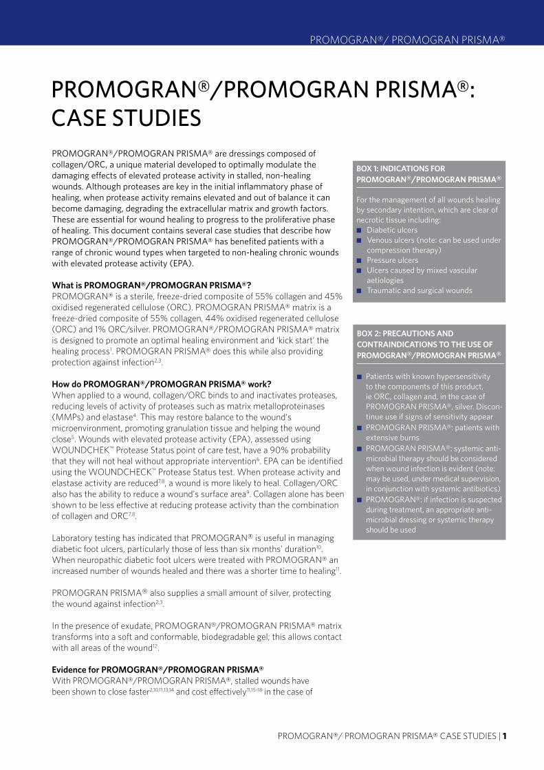
PROMOGRAN®/PROMOGRAN PRISMA®: CASE STUDIESPROMOGRAN®/PROMOGRAN PRISMA® are dressings composed of collagen/ORC, a unique material developed to optimally modulate the damaging effects of elevated protease activity in stalled, non-healing wounds. Although proteases are key in the initial inflammatory phase of healing, when protease activity remains elevated and out of balance it can become damaging, degrading the extracellular matrix and growth factors. These are essential for wound healing to progress to the proliferative phase of healing. This document contains several case studies that describe how PROMOGRAN®/PROMOGRAN PRISMA® has benefited patients with a range of chronic wound types when targeted to non-healing chronic wounds with elevated protease activity (EPA).
What is PROMOGRAN®/PROMOGRAN PRISMA®?PROMOGRAN® is a sterile, freeze-dried composite of 55% collagen and 45% oxidised regenerated cellulose (ORC). PROMOGRAN PRISMA® matrix is a freeze-dried composite of 55% collagen, 44% oxidised regenerated cellulose (ORC) and 1% ORC/silver. PROMOGRAN®/PROMOGRAN PRISMA® matrix is designed to promote an optimal healing environment and ‘kick start’ the healing process1. PROMOGRAN PRISMA® does this while also providing protection against infection2,3.
How do PROMOGRAN®/PROMOGRAN PRISMA® work?When applied to a wound, collagen/ORC binds to and inactivates proteases, reducing levels of activity of proteases such as matrix metalloproteinases (MMPs) and elastase4. This may restore balance to the wound's microenvironment, promoting granulation tissue and helping the wound close5. Wounds with elevated protease activity (EPA), assessed using WOUNDCHEK™ Protease Status point of care test, have a 90% probability that they will not heal without appropriate intervention6. EPA can be identified using the WOUNDCHECK™ Protease Status test. When protease activity and elastase activity are reduced7,8, a wound is more likely to heal. Collagen/ORC also has the ability to reduce a wound's surface area9. Collagen alone has been shown to be less effective at reducing protease activity than the combination of collagen and ORC7,8.
Laboratory testing has indicated that PROMOGRAN® is useful in managing diabetic foot ulcers, particularly those of less than six months' duration10. When neuropathic diabetic foot ulcers were treated with PROMOGRAN® an increased number of wounds healed and there was a shorter time to healing11.
PROMOGRAN PRISMA® also supplies a small amount of silver, protecting the wound against infection2,3.
In the presence of exudate, PROMOGRAN®/PROMOGRAN PRISMA® matrix transforms into a soft and conformable, biodegradable gel; this allows contact with all areas of the wound12.
Evidence for PROMOGRAN®/PROMOGRAN PRISMA®With PROMOGRAN®/PROMOGRAN PRISMA®, stalled wounds have been shown to close faster2,10,11,13,14 and cost effectively11,15-18 in the case of
BOX 1: INDICATIONS FOR PROMOGRAN®/PROMOGRAN PRISMA®
For the management of all wounds healing by secondary intention, which are clear of necrotic tissue including:■ Diabetic ulcers■ Venous ulcers (note: can be used under
compression therapy)■■■ Pressure ulcers■■■ Ulcers caused by mixed vascular
aetiologies■ Traumatic and surgical wounds

2 | INTERNATIONAL CASE STUDIES
PROMOGRAN®/ PROMOGRAN PRISMA®
PROMOGRAN PRISMA®, while providing protection against infection. The clinical efficacy of PROMOGRAN®/PROMOGRAN PRISMA® compared with standard care has been shown in seven randomised controlled trials10,11,19-22, 23. Further, a recent study has confirmed that the clinical efficacy of PROMOGRAN®/PROMOGRAN PRISMA® is increased, when targeted to wounds with EPA. Seventy-seven percent of venous leg ulcers with EPA responded to PROMOGRAN®/PROMOGRAN PRISMA® treatment by week four24.
WOUNDCHEK™ Protease Status testWOUNDCHEK™ Protease Status test was developed to aid wound assessment and help clinicians target advanced wound care therapies more effectively. WOUNDCHEK™ Protease Status test is able to detect EPA. A chronic wound with EPA has a 90% probability that it won't heal without appropriate intervention. As there are no visual cues for EPA25, it has so far gone undetected. However, a study has shown that 28% of non-healing chronic wounds have EPA6. A WOUNDCHEK™ Protease Status test can help clinicians establish within 15 minutes which wounds may most benefit from a protease-modulating therapy24, ensuring appropriate and targeted use of these therapies.
A WOUNDCHEK™ Protease Status test is: ■■ A rapid point of care diagnostic■■ Easy to use■■ Able to detect EPA in 15 minutes■■ Able to identify which wounds to treat with protease-modulating therapy.
More research is needed to understand what the test results obtained following targeted PROMOGRAN®/PROMOGRAN PRISMA® treatment tell a clinician about treatment effectiveness. For example, how soon a low result indicates that it would be appropriate to stop treatment or after how long an elevated result is indicative of a lack of treatment effectiveness. In the interim, this information should be considered in the context of traditional indicators of healing progress, such as wound area/size reduction and visual signs of healing.
Appropriate use of silver dressingsA recent consensus on the appropriate use of silver dressings described the main roles of silver dressings in the management of wounds to be the reduction of bioburden and to act as an antimicrobial barrier26. Whenever a silver-containing dressing, such as PROMOGRAN PRISMA®, is used to improve healing or to prevent infection, the rationale should be fully documented in the patient's health records and a schedule for review should be specified.
ABOUT PROMOGRAN®/PROMOGRAN PRISMA®
■ For further information about PROMOGRAN®/PROMOGRAN PRISMA® please go to: http://www.systagenix.com/our- products/lets-promote
AND http://www.woundsinternational.com/made-easys/promogran-and- promogran-prisma-made-easy
Tips on using PROMOGRAN®/PROMOGRAN PRISMA®■■ Before treatment, dry necrotic tissue must first be removed by surgical, enzymatic or autolytic debridement.■■ For optimal effect, apply matrix directly to the entire wound bed.■■ For a wound with low or no exudate apply matrix and hydrate with saline or Ringer’s solution. Alternatively, the matrix can be
pre-wetted with saline or Ringer’s solution before application using the tray it is pre-packaged in.■■ The matrix can be cut or folded and ‘packed’ into deeper wounds.■■ The matrix must be covered with either gauze, a non-adhering or a hydropolymer dressing.■■ After hydration the matrix forms a gel. This biodegradable gel is naturally absorbed over time.■■ It is not necessary to remove any residual matrix/gel. Reapply the matrix up to every 72 hours depending on the volume of
exudate.■■ For heavily exuding or, in the case of PROMOGRAN PRISMA®, infected wounds it may be necessary to re-treat the wound
every 24 hours.

PROMOGRAN®/ PROMOGRAN PRISMA® CASE STUDIES | 3
PROMOGRAN®/ PROMOGRAN PRISMA®
INTERNATIONAL CONSENSUS APPROPRIATE USE OF SILVER DRESSINGS IN WOUNDS
■ To download a copy of the consensus document please go to: http://www.woundsinternational.com
Reducing bioburdenThe consensus document recommends that silver dressings be used initially for a two week 'challenge' period. At the end of the two weeks, the wound, the patient and the management approach should be re-evaluated26.
Prophylactic useSilver-containing dressings, such as PROMOGRAN PRISMA®, may be used as an antimicrobial barrier in wounds at high risk of infection or re-infection (see the consensus document for further information).
References1. Cullen B, Ivins N. PROMOGRAN® and PROMOGRAN PRISMA® Made Easy. Wounds International 2010;
1(3). Available from: www.woundsinternational.com.
2. Cullen, B Nisbet L, Gibson M, et al. A clinical study examining the effect of ORC/collagen/silver-ORC on healing and wound biochemistry. Presented at: Symposium of Advanced Wound Care (SAWC), Dallas, TX, 2009.
3. Gottrup F, Karlsmark T, Bishoff-Mikkelsen M, et al. Comparative clinical study to determine the effects of collagen/ORC + silver on wound healing of diabetic foot ulcers. European Wound Management Association (EWMA), Geneva, Switzerland, 2010.
4. Gibson D, Cullen B, Legerstee R, et al. MMPs Made Easy. Wounds International 2009; 1(1). Available from www.woundsinternational.com.
5. Cullen B, Smith R, McCulloch E, et al. Mechanism of action of collagen/ORC, a protease modulating matrix for treatment of diabetic foot ulcers. Wound Rep Regen 2002; 10: 16-25.
6. Serena T, Cullen B, Bayliff S, et al. Protease activity levels associated with healing status of chronic wounds. Poster presented at: Wounds UK, Harrogate, 2011.
7. Cullen B, Donnelly T, Boyle C, et al. A comparison of collagen containing dressings to modify the chronic wound environment. Poster presented at: European Wound Management Association (EWMA), Glasgow, UK, 2007.
8. Cullen B, Boyle C, Webb Y. Modulation of the chronic wound environment; an in vitro evaluation of advanced wound therapies. Presented at: Symposium of Advanced Wound Care (SAWC), Tampa FL, USA, 2007.
9. Cullen B, Kemp L, Essler L. Rebalancing wound biochemistry improves healing: a clinical study examining the effect of PROMOGRAN. Wound Repair Regen 2004; 12(2): A4.
10. Veves A, Sheenan P, Pham HT. A randomized , controlled trial of PROMOGRAN (a collagen/ORC vs standard treatments in the management of diabetic foot ulcers. Arch Surg 2002; 137: 822-7.
11. Lazaro-Martinez JL, Garcia-Morales E, Beneit-Montesinos JV, et al. Randomized comparative trial of a collagen/oxidized regenerated cellulose dressing in the treatment of neuropathic diabetic foot ulcers. Article in Spanish. Circ Esp 2007; 82(1): 27-31.
12. World Union of Wound Healing Societies (WUWHS). Principles of best practice: Wound exudate and the role of dressings. A consensus document. London: MEP Ltd, 2007. Available from: www.woundsinternational.com.
13. Cullen B, Rennison T, Boyle C. ORC/collagen matrix containing silver controls bacterial bioburden and retains dermal cell viability. Poster presented at: European Wound Management Association (EWMA), Prague, May 2006.
14. Gregory S, Boothman S, Delbono M, et al. The Influence of ORC/collagen biomaterials. Poster presented at European Tissue Repair Society (ETRS) Stuttgart, September 2005.
15. Hart J, Silcock D, Gunnigle S, et al. The role of oxidized regenerated cellulose/collagen in wound repair: effects in vitro on fibroblast biology and in vivo in a model of compromised healing. Int J Biochem Cell Biol 2002; 34: 1557-70.
16. Snyder R, Richter D, Hill ME. Sequential therapies and advanced wound care products as a standard of care in the home care setting. Proceedings of the 9th Annual New Cardiovascular Horizons, New Orleans, 2008.
17. Ghatenekar O, Willis M, Persson U. Cost effectiveness of treating deep diabetic foot ulcers with collagen/ORC in four European countries. J Wound Care 2002; 11(2): 70-74.
18. Tacconi G, Vagnoni E. Clinical experiences and cost effective analysis of collagen/ORC/silver. European Wound Management Association (EWMA), Helsinki, 2009.
19. Nisi G, Brandi C, Grimaldi L, et al. Use of a protease-modulating matrix in the treatment of pressure sores. Chir Ital 2005; 57(4): 465-8.
20. Vin F, Teot L, Meaume S. The healing properties of PROMOGRAN in venous leg ulcers. J Wound Care 2002; 11(9): 335-41.
21. Wollina U, Schmidt WD, Kronert C, et al. Some effects of a topical collagen-based matrix on the microcirculation and wound healing in patients with chronic venous leg ulcers: preliminary observations. Int J Low Extrem Wounds 2005; 4(4): 214-24.
22. Gottrup F, Karlsmark T, Bishoff-Mikkelsen M, et al. Comparative clinical study to show the combined mode of action of collagen/ORC/silver in controlling bioburden and modulating the wound microenvironment to promote healing. Poster presented at: Wounds International Conference, Cape Town, 2011.
23. Lanzara S, Tacconi G, Gianesini S, et al. A pilot randomised trial to determine the effects of a new active dressing on wound healing of venous leg ulcers. Presented at European Wound Management Association (EWMA), Lisbon, 2008.
24. Cullen B, Gibson M, Nesbit, L. Targeted use of protease modulating dressings improves clinical outcomes. Poster presented at: Wounds UK, Harrogate, 2011.
25. Snyder R, Cullen B, Nisbet L, Serena T. The importance of proteases in wound healing and wound assessment. Poster presented at: Wounds UK, Harrogate, 2011.
26. International consensus. Appropriate use of silver dressings in wounds. An expert working group consensus. London: Wounds International, 2012. Available from www.woundsinternational.com.

4 | INTERNATIONAL CASE STUDIES
PROMOGRAN®/ PROMOGRAN PRISMA®
Assessment Wk 0
Wk 1
Wk 2
Wk 3
Wk 4
Wound size (cm2)
3.4 2.66 2.1 1.32 0.64
Protease activity
EPA EPA EPA EPA Low
Granulation ü ü —
Infection N — N — —
Case 1BackgroundMr F, a 38-year-old man, presented with a pressure ulcer of 14 months' duration. Congenital spina bifida had resulted in loss of mobility and altered protein metabolism. Mr F was wheelchair bound and, due to anatomical abnormalities caused by congenital malformation, had previously sustained pressure ulcers on the left and right ischial tuberosities. The current wound had been previously treated with negative pressure wound therapy, silver impregnated carbon dressings, Hydrofiber‰ dressings, topical antibiotics (mupirocin) and, most recently, with a bioactive ion-releasing dressing (TRIONIC®, Systagenix) to stimulate granulation.
TreatmentThe wound was on the left ischial tuberosity. It measured 20mm x 17mm x 14mm (depth). It had a granulating base but the wound edges were ill-defined. There was no evidence of infection, but due to lack of progress with previous treatments a WOUNDCHEK™ Protease Status test was performed which showed elevated protease activity (EPA). The wound was dressed with PROMOGRAN® and TIELLE® as the secondary dressing. The community healthcare team also ensured appropriate pressure redistributing surfaces were in place in Mr F’s home.
Week 1: Seven days later, Mr F returned to the clinic. The dressing was removed and the wound was cleansed with saline to remove debris and exudate. There was an increase in granulation tissue and the wound had decreased in size (19mm x 14mm x 14mm). A WOUNDCHEK™ Protease Status test indicated EPA. The wound was re-dressed with PROMOGRAN® and TIELLE® as a secondary dressing.
Week 2: Improvement had continued. The wound bed was composed of healthy granulation tissue. An ultrasound probe indicated the tissue was stable and well organised with no dead space. The wound had reduced in size (14m x 15mm x 12mm) and remained free from infection. A WOUNDCHEK™ Protease Status test showed EPA. The wound dressing regimen remained the same.
Week 3: The wound appeared dramatically improved. Granulation tissue had increased, the wound had reduced in size (11mm x 12mm x 8mm) and the epithelial borders were advancing well. A WOUNDCHEK™ Protease Status test indicated protease activity remained elevated, confirming that treatment needed to be continued. The treatment regimen was maintained.
Week 4: The wound had greatly decreased in size (8mm x 8mm x 3mm). A WOUNDCHEK™ Protease Status test indicated protease activity was low.
OutcomeDuring the evaluation, the clinical staff rated the dressing as satisfactory or highly satisfactory in terms of ease of use. Mr F was very satisfied. This challenging wound had been present over a prolonged period. Local wound treatment along with appropriate pressure redistributing surfaces in the home led to improvement in the ulcer. The quality of new granulation tissue that formed in the wound bed particularly impressed staff.
By: Juan Carlos Alvarez Vazquez, Hospital Universitario Lucus Augusti, Lugo, España
0 1 2 3 4
Case 1 - PROMOGRAN, PU, 14m, ES
1
0
3
4
2
Wou
nd s
ize
(cm
2 )
Time (weeks)
Figure 3: Graph showing changes in wound size over the evaluation period. The wound reduced in size by 81%.
Figures 1-2: The wound reduced in size and the epithelial borders advanced well.
Baseline
Week 3

PROMOGRAN®/ PROMOGRAN PRISMA® CASE STUDIES | 5
PROMOGRAN®/ PROMOGRAN PRISMA®
Case 2BackgroundMr T was a 91-year-old man with a venous leg ulcer on the medial aspect of the left lower leg. He had experienced multiple episodes of leg ulceration. The current wound had been present for seven months.
TreatmentThe wound measured 10cm2 (4cm x 2.5cm x 2mm). The base was estimated to be 25–50% granulation tissue, and there was maceration of the surrounding skin. There was no evidence of infection. As there had been a lack of progress with treatments prior to presentation a WOUNDCHEK™ Protease Status test was carried out and indicated elevated protease activity (EPA). The wound was dressed with PROMOGRAN® and TIELLE® as a secondary dressing. A three-layer reduced compression bandage system was applied toe to knee.
Week 1: The dressing was removed and the wound cleansed with water at body temperature. Improvement was noted. The wound bed was composed of 50–75% healthy granulation tissue. The wound had also decreased in size (5.5cm2) and the condition of the periwound skin had improved. A WOUNDCHEK™ Protease Status test indicated EPA. The wound was dressed with PROMOGRAN® and TIELLE® as a secondary dressing. A three-layer reduced compression bandage system was applied toe to knee.
Week 2: The wound bed was composed of 50–75% healthy granulation tissue. Wound size was 4.5cm2. The wound remained free from infection but a WOUNDCHEK™ Protease Status test confirmed protease activity remained elevated. The treatment regimen was continued. A three-layer reduced compression bandage system was applied toe to knee.
Week 3: The wound continued to improve. The wound base was composed of 50–75% granulation tissue. It had decreased in size to 4cm2 and the epithelial border had advanced well. The wound remained free from infection but a WOUNDCHEK™ Protease Status test indicated that protease activity was elevated. The treatment regimen was continued unaltered.
Week 4: The wound had become painful and was thought to be infected. There had been a small increase in size 4.1cm2 , a reduction in granulation tissue in the wound base, and the patient reported pain for the first time. PROMOGRAN® and TIELLE® were discontinued and an antimicrobial dressing was chosen to manage the infection. A WOUNDCHEK™ Protease Status test indicated protease activity remained elevated.
OutcomeIn recalcitrant wounds such as this it is not unusual for the bacterial burden to fluctuate and, subsequently, for the wound to deteriorate. However, taking into account the real improvement observed from baseline to week 3, clinicians said they would consider treatment with PROMOGRAN® when infection had resolved. PROMOGRAN PRISMA® might have been useful as an alternative since it provides protection against infection. During the course of treatment, the clinical staff rated the dressing as satisfactory or highly satisfactory in terms of ease of use.
By: Jane Megson, Wound Care Research Nurse, Bradford Royal Infirmary, Bradford, UK
Figures 1-2: Wound size decreased and granulation tissue increased until week 4 when the wound was found to have increased in size and PROMOGRAN® was discontinued due to infection.
Baseline
Week 3
Assessment Wk 0
Wk 1
Wk 2
Wk 3
Wk 4
Wound size (cm2)
10 5.5 4.5 4 4.1
Protease activity
EPA EPA EPA EPA EPA
Granulation (%)
25-50
50-75
50-75
50-75
0- 25
Infection N N N N Y
Case 3 - PROMOGRAN, VLU, 7m, UK
0 1 2 3 40
10
5
Wou
nd s
ize
(cm
2 )
Time (weeks)
Figure 3: Graph showing changes in wound size over the evaluation period. The wound reduced 59% in size over the evaluation period.

6 | INTERNATIONAL CASE STUDIES
PROMOGRAN®/ PROMOGRAN PRISMA®
Case 6 - PRISMA, DFU, 8w, UK
0 1 2 3 40
4
2
Wou
nd s
ize
(cm
2 )
Time (weeks)
Figure 4: Graph showing changes in wound size over the evaluation period. The wound decreased in size by 71.5% over three weeks.
Case 3
Background Mr G, a 42-year-old man with type 2 diabetes and neuropathy, presented with a diabetic foot ulcer of eight weeks’ duration on his left heel. The wound had been treated with ACTISORB® Silver (Systagenix) for two weeks because it was infected and malodorous. The wound was blistered and the skin had been picked at. The wound measured 2.1cm2 (15mm x 14mm) and was 4mm deep.
TreatmentElevated protease levels were detected by a WOUNDCHEK™ Protease Status test. PROMOGRAN PRISMA® was chosen because there had been recent infection and the dressing offered protection against the wound becoming infected again. TIELLE® was used as a secondary dressing.
Week 1: After a week of treatment the wound was debrided. The patient had been taking antibiotics (co-amoxiclav) for three weeks. The course was nearly finished and there were signs of healing. The wound bed was not visible but appeared to be less deep. The wound width and length (15mm x 14mm) remained unchanged. The wound was not infected or critically colonised. A WOUNDCHEK™ Protease Status test showed elevated protease activity (EPA). Both the nurse and patient were highly satisfied with the dressing so it was decided to continue using it along with TIELLE®.
Week 2: The wound was assessed after debridement. There were signs of healing and the wound bed was improving. Granulation tissue coverage was estimated to be 50–75%. The wound now measured 10mm x 7mm x 3mm. There was no infection or critical colonisation. Protease activity was still elevated and this was confirmed using a WOUNDCHEK™ Protease Status test. The patient and nurse were both satisfied with the PROMOGRAN PRISMA® dressing and so it was continued.
Week 3: The wound was debrided. There were signs of healing and the wound had continued to improve, now measuring 10mm x 6mm x 3mm. The wound was not infected or critically colonised, with evidence of 50–75% granulation tissue. A WOUNDCHEK™ Protease Status test showed that protease activity was low so PROMOGRAN PRISMA® was discontinued.
Outcome PROMOGRAN PRISMA® was discontinued after three weeks because the wound had decreased in size and was healing well. A WOUNDCHEK™ Protease Status test indicated low protease activity. The dressing regimen was changed to ALLEVYN™ Heel (Smith & Nephew). After treatment with PROMOGRAN PRISMA®, the wound had reduced in size considerably. The clinical staff did not note any problems with the dressing.
By: Paul Chadwick, Principal Podiatrist, Salford Royal (NHS) Foundation, Salford, UK
Assessment Wk 0 Wk 1 Wk 2 Wk 3
Wound size (cm2)
2.1 2.1 0.7 0.6
Protease activity
EPA EPA EPA Low
Granulation (%)
50 50 50-75 50-75
Infection N N N N
Figures 1-3: The wound decreased in size and the wound bed improved.
Week 1
Week 2
Week 3

PROMOGRAN®/ PROMOGRAN PRISMA® CASE STUDIES | 7
PROMOGRAN®/ PROMOGRAN PRISMA®
BackgroundMs G, a 72-year-old woman, presented with a venous leg ulcer measuring 11.5cm x 7.5cm (86.25cm2) with a depth of 0.5cm that had been present for 18 months. She had no comorbidities but she had had chronic venous ulcers several years previously, which had healed with standard treatment.
The current wound had occurred after minimal trauma to her lower left leg. The wound was not progressing, there were clinical signs of critical colonisation and the wound bed colour was bright red.
TreatmentA WOUNDCHEKTM Protease Status test showed elevated protease activity (EPA). The wound had been treated with occlusive bandaging and multi-layer compression bandages. PROMOGRAN PRISMA® and TIELLE® were selected to treat the EPA and bacterial load in the wound bed.
Week 1: After a week of treatment and compression therapy the wound began to show signs of healing. It measured 9cm x 6.5cm. Granulation tissue was estimated at 50–75% and the edges had improved. Both nurse and patient were satisfied with the dressing (the patient reported being ‘highly satisfied’). The wound was not thought to be criticially colonised or infected. A WOUNDCHEKTM Protease Status test showed that protease activity remained elevated. PROMOGRAN PRISMA® and TIELLE® were continued.
Week 2: There were further signs of healing, but as a result of debridement, the wound measured 10.7cm x 6.5cm and granulation remained at 50–75%. The wound was not infected or critically colonised. The WOUNDCHEKTM Protease Status test showed EPA. The dressing regimen was continued.
Week 3: The wound was 10cm x 6cm with 50–75% granulation tissue. There was no infection or critical colonisation. A WOUNDCHEKTM Protease Status test was not conducted. The dressing regimen was continued.
Week 4: The wound had again reduced in size and measured 9cm x 5.6cm and there was evidence of 50–75% granulation tissue. A WOUNDCHEKTM Protease Status test showed that protease activity was low. It was decided to continue to use PROMOGRAN® after the case study period to avoid an increase in protease activity (eg matrix metalloproteinases).
OutcomePROMOGRAN PRISMA® was able to kick-start healing of the patient’s wound after 18 months of non-healing. At the end of the study period protease levels were low, the wound had reduced in size and there was a high percentage of healthy granulation tissue. The aims of using the dressing had been achieved and staff found it very easy to use.
By: Professor Marco Romanelli, Consultant Dermatologist, University of Pisa, Italy
Case 4
Figures 1-3: The wound reduced in size over the course of the study period and protease activity lowered.
Baseline
Week 1
Week 4
Assessment Wk 0
Wk 1
Wk 2
Wk 3
Wk 4
Wound size (cm2)
86.25 58.5 69.55 60 50.4
Protease activity
EPA EPA EPA — Low
Granulation (%)
Poor 50-75
50-75
50-75
50-75
Infection N N N N N
Case 7 - PRISMA, VLU, 18m, IT
0 1 2 3 4
200
6080
100
40
Wou
nd s
ize
(cm
2 )
Time (weeks)
Figure 4: Graph showing changes in wound size over the evaluation period. The wound reduced in size by 42% over the course of the evaluation.

8 | INTERNATIONAL CASE STUDIES
PROMOGRAN®/ PROMOGRAN PRISMA®
Case 5
Background The patient was an 83-year-old woman who had presented with a venous leg ulcer on the lateral side of the lower leg that had occurred after a minor trauma at home and rapidly enlarged. The wound measured 5.25cm2 (3.5cm x 1.5cm) and had been present for 12 years. She had a history of venous thrombosis.
TreatmentAdvanced dressings and compression therapy had been used for several months. The wound bed was granulating but stable. The wound was not infected and a WOUNDCHEKTM Protease Status test showed that protease activity was elevated. PROMOGRAN PRISMA®, TIELLE® and two-layer compression bandaging were applied.
Week 1: After one week of treatment the wound showed signs of healing. There was 75–100% granulation tissue and it looked to have reduced slightly in size (although it measured the same as it did at baseline: 3.5cm x 1.5cm). The nurse and patient were both highly satisfied with the dressing’s performance. The wound was not infected or critically colonised. It was decided to continue with PROMOGRAN PRISMA®, TIELLE® and compression bandaging.
Week 2: The wound had reduced in size and the edges were advancing. The wound comprised 50–75% granulation tissue and measured 3.5cm x 1cm. The nurse and patient were both satisfied with the progress. The wound was not infected or critically colonised. A WOUNDCHEKTM Protease Status test showed that protease activity was low. The wound was dressed with PROMOGRAN PRISMA®. A secondary dressing was considered necessary, but TIELLE® was not used as a gentler adhesive was considered more appropriate for the patient's fragile skin. Compression bandaging was continued.
Week 3: The wound continued to show signs of healing. The wound bed had good granulation tissue coverage (50–75%) and measured 3.5cm x 1cm. The nurse was satisfied with progress and the patient reported being highly satisfied. The wound was free from infection and was not critically colonised. PROMOGRAN PRISMA® was applied to maintain low protease activity, with two-layer compression bandaging.
Week 4: After four weeks of treatment using PROMOGRAN PRISMA® and compression bandaging the wound continued to heal. It comprised 50–75% granulation tissue and now measured 3cm x 1cm. Both nurse and patient were satisfied with the dressing. The wound remained free from infection and was not critically colonised. At the end of the evaluation period there appeared to be no need to continue using PROMOGRAN PRISMA® as the wound had begun to heal.
Outcome PROMOGRAN PRISMA® helped to activate healing in this wound, which had not healed for 12 years. The wound was kept infection-free during the study period and protease activity, which was initially found to be elevated, was lowered. Clinical staff felt the dressing had achieved very good results, as shown by the clinical results. By: Professor Marco Romanelli, Consultant Dermatologist, University of Pisa, Italy
Assessment Wk 0
Wk 1
Wk 2
Wk 3
Wk 4
Wound size (cm2)
5.25 5.25 3.5 3.5 3
Protease activity
EPA — Low — —
Granulation (%)
ü 75-100
50-75
50-75
50-75
Infection N N N N N
Case 9 - PRISMA, VLU, 12y, IT
0 1 2 3 4
2
0
6
4
Wou
nd s
ize
(cm
2 )
Time (weeks)
Figure 4: Graph showing changes in wound size over the evaluation period. The wound reduced in size by 43% over the study period.
Week 2
Figures 1-3: An improvement was seen in the wound during the evaluation period and it remained free of infection throughout.
Week 4
Baseline

PROMOGRAN®/ PROMOGRAN PRISMA® CASE STUDIES | 9
PROMOGRAN®/ PROMOGRAN PRISMA®
Case 6
Background The patient, a 70-year-old man, presented with a venous leg ulcer on the left leg of 12 years' duration, which had recurred after eight months of him being ulcer-free. The wound measured 5cm x 1cm x 0.8cm. He had a history of multiple wounds, which had healed and recurred during the previous nine years. Several treatments had been attempted, including autologous skin grafting. He had had a history of venous insufficiency since the age of 45, and had found it difficult to be concordant with compression stockings.
TreatmentThis particular wound had recurred over a period of seven years. It had been treated with four-layer compression bandaging and a foam dressing. The wounds had not been improving but there was evidence of granulation tissue. It was not infected and a WOUNDCHEKTM Protease Status test showed that protease activity was elevated. PROMOGRAN PRISMA® was applied with the aim of promoting healing.
Week 1: Granulation tissue was estimated to cover 50–75% of the wound and exudate levels were reduced. The edges of the wound were a healthy colour and beginning to advance, although the wound still measured 5cm x 1cm (the depth was not recorded). The wound was not infected and a WOUNDCHEKTM Protease Status test showed low protease activity. PROMOGRAN PRISMA® was continued without a secondary dressing due to the reduced exudate levels and to maintain low MMP activity. Two-layer compression bandaging was applied.
Week 2: The wound had reduced in size and granulation tissue coverage was greater than 75%. The wound measured 3.5cm x 1cm and remained free from infection and critical colonisation. A WOUNDCHEKTM Protease Status test showed protease activity to be low. It was decided to continue with PROMOGRAN PRISMA® to maintain low protease activity. Two-layer compression bandaging was also continued.
Week 3: The wound had reduced in size to 3.2cm x 1cm. Granulation coverage continued to be greater than 75% and the wound was not infected or critically colonised. A further WOUNDCHEKTM Protease Status test showed that protease activity was low.
Week 4: After four weeks of treatment there was 50–75% granulation tissue and the surrounding skin was described as regular. The wound measured 3cm x 0.8cm. The wound was not infected and a WOUNDCHEKTM Protease Status test showed that protease activity was low.
DiscussionBoth the nurse and patient reported being satisfied or highly satisfied during the evaluation period based on the dressing's ease of use and performance. PROMOGRAN PRISMA® proved to be an appropriate treatment for this previously non-healing wound. The elevated protease activity was reduced to low after one week and the wound continued to heal over the four-week period. Treatment with PROMOGRAN PRISMA® was continued to avoid recurrence.
By: Professor Marco Romanelli, Consultant Dermatologist, University of Pisa, Italy
Week 2
Baseline
Figures 1-3: Granulation tissue increased over the study period and EPA reduced.
Week 4
Assessment Wk 0
Wk 1
Wk 2
Wk 3
Wk 4
Wound size (cm2)
5 5 3.5 3.2 2.4
Protease activity
EPA Low Low Low Low
Granulation (%)
Some 50-75
>75 >75 50-75
Infection N N N N N
Case 10 - PRISMA, VLU, ?, IT
0 1 2 3 4
20
68
10
4
Wou
nd s
ize
(cm
2 )
Time (weeks)
Figure 4: Graph showing changes in wound size over the evaluation period. The wound decreased in size by 52% over the evaluation period.

10 | INTERNATIONAL CASE STUDIES
PROMOGRAN®/ PROMOGRAN PRISMA®
A Wounds International publicationwww.woundsinternational.com