Useof Human Surfactant Low Molecular Weight Apoproteins in ...
8
Use of Human Surfactant Low Molecular Weight Apoproteins in the Reconstitution of Surfactant Biologic Activity Susan D. Revak,* T. Allen Merritt,* Eric Degryse,1 Lorette Stefani,1 Michael Courtney,1 Mikko Hallman,11 and Charles G. Cochrane* *Department ofImmunology, Research Institute of Scripps Clinic, La Jolla, California 92037; tDivision of Neonatal/Perinatal Medicine, Department ofPediatrics, University of California at San Diego (UCSD), San Diego, California 92103; §Department ofMolecular and Cellular Biology, Transgene, 67082 Strasbourg Cedex France; and I1Children's Hospital, The University ofHelsinki, Helsinki, Finland Abstract Two low molecular weight (LMW) apoproteins were isolated from human pulmonary surfactant. SDS polyacrylamide gel analysis showed one protein (SP 18) to have an apparent mo- lecular weight of 18,000 when unreduced and 9,000 D after reduction. The second protein (SP 9) migrated at - 9,000 D in the presence or absence of reducing agents. Both proteins con- tain a high number of hydrophobic amino acids. The NH2-ter- minal sequence of SP 18 was determined to be: NH2-phe-pro- ile-pro-leu-pro-tyr-. A cDNA clone isolated from a human adult lung cDNA library contained a long open reading frame encoding at an internal position the human SP 18 amino-termi- nal sequence. Mixtures of phospholipids (PL) and SP 9 and SP 18 were assessed for their capacity to reduce surface tensions on a pulsating bubble surfactometer. The addition of 1% apoprotein resulted in a reduction of surface tension after 15 s from 42.9 dyn/cm for PL alone to 16.7 and 6.3 dyn/cm for preparations containing SP 9 and SP 18, respectively. In vivo assessment of reconstituted surfactant activity was performed in fetal rabbits. Reconstituted surfactant consisting of PL + 0.5% SP 18 in- stilled intratracheally at delivery resulted in a marked increase in lung compliance, while the incorporation of 0.5% SP 9 yielded a moderate increase. These data show the ability to produce biologically active surfactant by the addition of iso- lated LMW apoproteins to defined PL. Introduction Pulmonary surfactant, which lines the alveolar epithelium of mature mammalian lungs, has been shown to be a lipoprotein complex capable of reducing surface tension at the air-liquid interface (1, 2). The apoproteins present in this complex have been the subject of many studies over the past few years which have helped to elucidate the physiologic role they play in sur- factant activity. While initial interest seemed focused primar- ily on a 35,000-D major glycoprotein (3-17), more recent studies have described hydrophobic 5,000-18,000-D apopro- teins (18-27) which may, in fact, be more important in the This is publication No. 4720-IMM from the Department of Immunol- ogy, Research Institute of Scripps Clinic. Address reprint requests to Dr. Revak. Receivedfor publication 25 June 1987 and in revisedform 28 Sep- tember 1987. expression of surfactant surface tension reduction activity. These low molecular weight (LMW)' apoproteins, one of which has been described as a "proteolipid" (21), can be found in several organic surfactant lipid extracts being tested clini- cally (Tokyo Tanabe's "surfactant TA," calf lung surfactant extract) that have also been shown not to contain the 35,000-D apoprotein (20, 23, 25). That the 35,000-D and the LMW apoproteins of surfactant are distinct has been shown immu- nologically (22-26), and different amino acid compositions (4, 8, 14, 16, 17, 19, 21, 23, 25) have been reported. In the canine system, two unique amino acid sequences have been derived from cDNA clones (27, 28). While it is apparent that the LMW apoproteins represent a group of proteins clearly distinguish- able from the 35,000-D surfactant apoprotein, it is not clear as to whether this group is comprised of one or more apoproteins and what differences exist in their characteristics and functions if there are multiple proteins. We have previously reported the isolation of the 35,000-D protein from human amniotic fluid surfactant and studied its ability when added to phospholipids to lower the surface ten- sion of a pulsating bubble and to increase lung compliance and alveolar expansion when instilled into fetal rabbits (17). In the current study we have isolated two LMW apoproteins from human amniotic fluid surfactant, partially characterized them, and used the same pulsating bubble and fetal rabbit models to show that each can be recombined with synthetic phospho- lipids (PL) to yield a functionally active reconstituted surfac- tant. Furthermore, a cDNA clone corresponding to the larger apoprotein has been isolated and its nucleotide sequence de- termined. We will refer to these two distinct LMW apoproteins as surfactant protein (SP) 18 and SP 9, with the number fol- lowing SP indicating the molecular weight (in kilodaltons) of the protein as it appears in unreduced SDS-PAGE. Methods Purification of LMW apoproteins. Human pulmonary surfactant was isolated from full-term amniotic fluid and applied to a column of DEAE-Sephacel A-50 (Pharmacia Fine Chemicals, Uppsala, Sweden) using 4 ml packed volume per 200 mg surfactant, in a Tris-EDTA buffer containing 1% n-octyl-beta-D-glucopyranoside as previously de- scribed (17, 29). This particular column and conditions were used to isolate the 35,000-D apoprotein (for use in other studies) without ex- posing it to potentially denaturing organic solvents. The void volume, containing the lipids and proteins that did not bind to the column under these conditions, was pooled and extracted with an equal vol- 1. Abbreviations used in this paper: DPPC, dipalmitoylphosphatidyl- choline; LMW, low molecular weight; PG, phosphatidylglycerol; PL, phospholipid(s); SP, surfactant protein. 826 Revak, Merritt, Degryse, Stefani, Courtney, Hallman, and Cochrane J. Clin. Invest. © The American Society for Clinical Investigation, Inc. 0021-9738/88/03/0826/08 $2.00 Volume 81, March 1988, 826-833
Transcript of Useof Human Surfactant Low Molecular Weight Apoproteins in ...

Use of HumanSurfactant Low Molecular Weight Apoproteins in theReconstitution of Surfactant Biologic Activity
Susan D. Revak,* T. Allen Merritt,* Eric Degryse,1 Lorette Stefani,1 Michael Courtney,1Mikko Hallman,11 and Charles G. Cochrane**Department of Immunology, Research Institute of Scripps Clinic, La Jolla, California 92037; tDivision of Neonatal/PerinatalMedicine, Department of Pediatrics, University of California at San Diego (UCSD), San Diego, California 92103;
§Department of Molecular and Cellular Biology, Transgene, 67082 Strasbourg Cedex France;and I1Children's Hospital, The University of Helsinki, Helsinki, Finland
Abstract
Two low molecular weight (LMW) apoproteins were isolatedfrom human pulmonary surfactant. SDS polyacrylamide gelanalysis showed one protein (SP 18) to have an apparent mo-lecular weight of 18,000 when unreduced and 9,000 D afterreduction. The second protein (SP 9) migrated at - 9,000 Dinthe presence or absence of reducing agents. Both proteins con-tain a high number of hydrophobic amino acids. The NH2-ter-minal sequence of SP 18 was determined to be: NH2-phe-pro-ile-pro-leu-pro-tyr-. A cDNA clone isolated from a humanadult lung cDNA library contained a long open reading frameencoding at an internal position the human SP 18 amino-termi-nal sequence.
Mixtures of phospholipids (PL) and SP9 and SP 18 wereassessed for their capacity to reduce surface tensions on apulsating bubble surfactometer. The addition of 1%apoproteinresulted in a reduction of surface tension after 15 s from 42.9dyn/cm for PL alone to 16.7 and 6.3 dyn/cm for preparationscontaining SP9 and SP 18, respectively. In vivo assessment ofreconstituted surfactant activity was performed in fetal rabbits.Reconstituted surfactant consisting of PL + 0.5% SP 18 in-stilled intratracheally at delivery resulted in a marked increasein lung compliance, while the incorporation of 0.5% SP 9yielded a moderate increase. These data show the ability toproduce biologically active surfactant by the addition of iso-lated LMWapoproteins to defined PL.
Introduction
Pulmonary surfactant, which lines the alveolar epithelium ofmature mammalian lungs, has been shown to be a lipoproteincomplex capable of reducing surface tension at the air-liquidinterface (1, 2). The apoproteins present in this complex havebeen the subject of many studies over the past few years whichhave helped to elucidate the physiologic role they play in sur-factant activity. While initial interest seemed focused primar-ily on a 35,000-D major glycoprotein (3-17), more recentstudies have described hydrophobic 5,000-18,000-D apopro-teins (18-27) which may, in fact, be more important in the
This is publication No. 4720-IMM from the Department of Immunol-ogy, Research Institute of Scripps Clinic. Address reprint requests to
Dr. Revak.Receivedfor publication 25 June 1987 and in revisedform 28 Sep-
tember 1987.
expression of surfactant surface tension reduction activity.These low molecular weight (LMW)' apoproteins, one ofwhich has been described as a "proteolipid" (21), can be foundin several organic surfactant lipid extracts being tested clini-cally (Tokyo Tanabe's "surfactant TA," calf lung surfactantextract) that have also been shown not to contain the 35,000-Dapoprotein (20, 23, 25). That the 35,000-D and the LMWapoproteins of surfactant are distinct has been shown immu-nologically (22-26), and different amino acid compositions (4,8, 14, 16, 17, 19, 21, 23, 25) have been reported. In the caninesystem, two unique amino acid sequences have been derivedfrom cDNAclones (27, 28). While it is apparent that the LMWapoproteins represent a group of proteins clearly distinguish-able from the 35,000-D surfactant apoprotein, it is not clear asto whether this group is comprised of one or more apoproteinsand what differences exist in their characteristics and functionsif there are multiple proteins.
Wehave previously reported the isolation of the 35,000-Dprotein from human amniotic fluid surfactant and studied itsability when added to phospholipids to lower the surface ten-sion of a pulsating bubble and to increase lung compliance andalveolar expansion when instilled into fetal rabbits (17). In thecurrent study we have isolated two LMWapoproteins fromhuman amniotic fluid surfactant, partially characterized them,and used the same pulsating bubble and fetal rabbit models toshow that each can be recombined with synthetic phospho-lipids (PL) to yield a functionally active reconstituted surfac-tant. Furthermore, a cDNAclone corresponding to the largerapoprotein has been isolated and its nucleotide sequence de-termined. Wewill refer to these two distinct LMWapoproteinsas surfactant protein (SP) 18 and SP 9, with the number fol-lowing SP indicating the molecular weight (in kilodaltons) ofthe protein as it appears in unreduced SDS-PAGE.
Methods
Purification of LMWapoproteins. Humanpulmonary surfactant was
isolated from full-term amniotic fluid and applied to a column ofDEAE-Sephacel A-50 (Pharmacia Fine Chemicals, Uppsala, Sweden)using 4 ml packed volume per 200 mg surfactant, in a Tris-EDTAbuffer containing 1% n-octyl-beta-D-glucopyranoside as previously de-scribed (17, 29). This particular column and conditions were used to
isolate the 35,000-D apoprotein (for use in other studies) without ex-
posing it to potentially denaturing organic solvents. The void volume,containing the lipids and proteins that did not bind to the columnunder these conditions, was pooled and extracted with an equal vol-
1. Abbreviations used in this paper: DPPC, dipalmitoylphosphatidyl-choline; LMW, low molecular weight; PG, phosphatidylglycerol; PL,phospholipid(s); SP, surfactant protein.
826 Revak, Merritt, Degryse, Stefani, Courtney, Hallman, and Cochrane
J. Clin. Invest.© The American Society for Clinical Investigation, Inc.0021-9738/88/03/0826/08 $2.00Volume 81, March 1988, 826-833
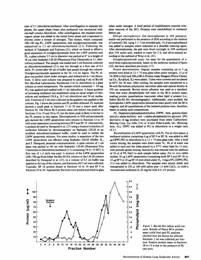
ume of 2:1 chloroform/methanol. After centrifugation to separate thephases, the upper phase (water plus methanol) was reextracted withone-half volume chloroform. After centrifugation, the resultant lowerorganic phase was added to the initial lower phase and evaporated todryness under a stream of nitrogen. This extract, which contained100-180 mg PL, LMWapoproteins, and octylglucopyranoside, wasredissolved in 2.5 ml chloroform/methanol (2:1). Following themethod of Takahashi and Fujiwara (21), which we found to afford agood separation of octylglucopyranside from the LMWproteins andPL, a glass column 2.5 cm in diameter was packed at 40 to a height of38 cm with Sephadex LH-20 (Pharmacia Fine Chemicals) in 2:1 chlo-roform/methanol. The sample was loaded and 2-ml fractions collectedas chloroform/methanol (2:1) was run through at a flow rate of 8.5ml/h. PL eluted after 40 ml of buffer had passed through the column.Octylglucopyranoside appeared at the 56-116 ml region. The PL re-gion was pooled, dried under nitrogen, and redissolved in I ml chloro-form. A silicic acid column was prepared by packing 9 ml of Bio-SilHA (Bio-Rad Laboratories, Richmond, CA) in chloroform in a glasscolumn at room temperature. The sample (which contained - 50 mgPL) was applied and washed with 11 ml chloroform. A linear gradientof increasing methanol was established using an equal weight of chlo-roform and methanol (38.8 g, 26.5 ml chloroform and 50 ml metha-nol). Fractions of 2 ml were collected as the gradient was applied to thecolumn. Fig. 1 shows the protein and PL profiles obtained. PL analysesshowed a small peak in fractions 17 to 20 and a major peak afterfraction 30. The Pierce BCAprotein assay (see below) was positive infractions 12 to 19 and 28 to 33, but the latter peak is likely to be due tothe PL present in this region. Electrophoresis in SDSpolyacrylamidegels showed the LMWapoproteins were present in fractions 13 to 19with some separation occurring between SP9 and SP 18. Alternatively,a method devised by Hawgood et al. (27) using a butanol extraction ofsurfactant followed by chromatography on Sephadex LH-20 in anacidified chloroform/methanol buffer, could be used to isolate theLMWapoprotein mixture. For some studies, a separation of the twoLMWapoproteins was effected using Sephadex LH-60 (Shiffer, K.,and S. Hawgood, personal communication). A glass column of 1 cmdiam was packed to 40 cm with Sephadex LH-60 (Pharmacia FineChemicals) in chloroform/methanol (1:1) containing 5%0.1 NHCI. Aflow rate of 1-2 ml/h was used. A mixture of the LMWapoproteins(200-700 Mg) from either the Bio-Sil HAcolumn or the LH-20 columndescribed by Hawgood et al. (27), in a volume of 0.5 ml buffer wasapplied to the top of the column, and fractions of 0.5 ml were collected.Typically, SP 18 protein eluted in fractions 16 to 19 and SP 9 infractions 24 to 29. Appropriate fractions were pooled and dried in glass
EI.,0a
%t0
L.*0.
100
90
80
70
60
50
40
30
20
10
tubes under nitrogen. A brief period of lyophilization ensured com-plete removal of the HCI. Proteins were resolubilized in methanolbefore use.
SDS-gel electrophoresis. Gel electrophoresis in 16% polyacryl-amide was performed in the presence of SDSaccording to the methodof Laemmli (30), using 3 X 7-cm minislab gels. 1%fl-mercaptoethanolwas added to samples where indicated as a disulfide reducing agent.After electrophoresis, the gels were fixed overnight in 50% methanolplus 12% acetic acid, washed in water for 2 h, and silver-stained ac-cording to the method of Wray et al. (31).
Octylglucopyranoside assay. An assay for the quantitation of n-octyl-beta-D-glucopyranoside, based on the anthrone method of Spiro(32), has been described previously (17).
Protein determinations. Organic samples containing up to 5 Migprotein were dried in 12 X 75-mm glass tubes under nitrogen. 15 Al of1%SDSin H20 and 300 Ml BCAProtein Assay Reagent (Pierce Chemi-cal Co., Rockford, IL) were added. Tubes were covered and incubatedat 60°C for 30 min. After cooling, the samples were transferred to a96-well flat-bottom polystyrene microtiter plate and optical density at550 nm measured. Bovine serum albumin was used as a standard.Note that some phospholipids will react in the BCA protein assay,making protein quantitations inaccurate when lipid is present (i.e.,before Bio-Sil HA chromatography). Additionally, once purified, thehydrophobic LMWapoproteins themselves react poorly with the BCAreagents, and all quantitations of the isolated proteins were, therefore,based on amino acid compositions.
PL. Dipalmitoylphosphatidylcholine (DPPC, beta, gamma-dipal-mitoyl-L-alpha-lecithin) and L-alpha-phosphatidyl-DL-glycerol (PG,derivative of egg lecithin) were purchased from either Calbiochem-Behring Corp. (La Jolla, CA) or Avanti Polar-Lipids, Inc. (Birming-ham, AL). DPPCwas added to PG in chloroform in a weight ratioof 3:1.
Recombination ofLMWapoproteins with PL. For in vitro assays, amethanol solution containing 4 Mg of SP 9 or SP 18, was added to 400MgDPPC/PGin chloroform in a 12 X 75-mm glass tube. After a briefvortex mixing, the samples were dried under N2. 90 Ml of water wasadded to each and the tubes placed in a 37°C water bath for 15 min,with periodic gentle mixing. Isotonicity was restored with the additionof 10 Ml of 9% NaCl to each sample before assay. For in vivo rabbitstudies, 50 ,g LMWapoproteins (containing both SP 9 and SP 18) or25Mg SP 9 or 25g SP 18 were dried under N2. 5 mgof PL (DPPC/PG,3:1) was added in chloroform. The samples were mixed, dried, andresuspended in 250 Ml 100 mMsaline plus 1.5 mMCaCl2, to yield areconstituted surfactant at 20 mg/ml with 0.5-1% protein.
_n 2.4
2.2
2.0
1.8
1.6
1.4
1.2
1.0
.8
.6
.4
.2
0
E
co
~0._Q
CL)0
0~._
Figure 1. Bio-Sil HA (silicic acid) col-umn. Results of Pierce BCAproteinassay (solid line) and PL analyses(dashed line) are shown for selectedfractions. 2 ml was collected per frac-tion. Positive protein assay in fractions28 to 33 is due to the presence of PL(see text).
Reconstitution of HumanLung Surfactant Activity 827
2 4 6 8 10 12 14 16 18 20 22 24 26 28 30 32 34 36 38 40
Fraction Number

Surfactant activity assays. In vitro assays of surfactant activity,assessed as its ability to lower the surface tension of a pulsating bubble,and in vivo assays using fetal rabbits, have both been described in detailpreviously (17).
Morphometric analyses. Fetal rabbit lungs, inflated to 30 cm H20and then deflated to 10 cm H20, were submerged in 10O% formalin for72 h. Parafin sections were oriented from apex to base and 5-Mmsections taken anterior to posterior. After hematoxylin and eosinstaining, 10 fields (X 100) were point-counted from apex to base onmultiple sections. Standardized morphometric methods (33) were usedto determine ratios of lung interstitium to air spaces for each treatmentgroup. Intersections of alveolar perimeters were also determined.
PL phosphorus assays. PL were quantitated according to themethod of Bartlett (34).
Amino acid analysis. Triplicate samples for amino acid composi-tions were hydrolyzed with HCl at 1 10C for 24 h, with HCI at 150'Cfor 24 h, or in performic acid at 1 10C for 24 h, followed by HClhydrolysis at 1 10C for 24 h. Analyses were performed on an aminoacid analyzer (model 121-M; Beckman Instruments, Inc., Fullerton,CA). Tryptophan was not determined.
Amino acid sequencing. Vapor-phase protein sequencing was per-formed on an amino acid sequencer (470A; Applied Biosystems, Inc.,Foster City, CA) with an on-line model 120A HPLC.
Isolation of cDNA clones for human SP 18. RNAwas preparedaccording to Chirgwin et al. (35) from a sample of unaffected adultlung tissue obtained during surgical removal of a neoplastic lesion.Preparation of double-stranded cDNAwas carried out using standardtechniques (36, 37) and a library was constructed in lambda NM607asdescribed (38). SP 18 clones were identified by screening phage plaqueswith synthetic oligonucleotide probes (39) that were prepared using anautomated synthesizer (Applied Biosystems, Inc.) and purified byHPLC. Initial candidate clones were obtained using probe TG996(5'CATTGCCTGTGGTATGGCCTGCTC3'), which was derivedfrom the partial nucleotide sequence of a small human surfactantapoprotein cDNA(40). Larger clones (up to 1.5 kb) were isolated usingprobe TGl 103 (5'TCGAGCAGGATGACGGAGTAGCGC3'),which was based on the 5' sequence of one of the original clones. Thenucleotide sequence of the cDNAclones was determined by the chaintermination method (41) using Eco RI restriction fragments subclonedin an appropriate M13 vector.
Results
Characteristics of the LMWapoproteins. The LMWapopro-teins isolated from human amniotic fluid appeared after silicicacid chromatography, or after the Sephadex LH-20 columnchromatography described by Hawgood et al. (27), as two pro-tein bands in SDS-PAGEunder nonreducing conditions. Theupper band, having a weight of 18,000 Dand therefore termedSP 18, is a dimer, and with the addition of f-mercaptoethanol,reduced to 9,000 D (Fig. 2). The other LMWapoprotein,which we will call SP 9,2 appears as a diffuse band between9,000 and 12,000 D in the presence or absence of reducingagents. These two proteins could be separated by chromatogra-phy on Sephadex LH-60. The resultant purified proteins areshown in Fig. 2.
Amino acid compositions were determined for SP 18 andSP 9. Because of the extremely hydrophobic nature of theseproteins, HCOhydrolysis was performed at 150'C for 24 h, inaddition to the standard 1 10C 24-h hydrolysis, and values forvaline, leucine, and isoleucine were calculated from analyses
2. This is probably the same protein as that designated SAP-6 byWhitsett and co-workers (24), SP 5-8 by Hawgood and co-workers(27), and PSP-6 by Phelps et al. (25).
UNREDUCED
LMW SP18 SP9
REDUCED
LMW SP18 SP9 STlSImaw- .1
Mot Wt
_il _-18K-
adz " < 12~~~-K m e
A B C D E F G
Figure 2. Silver-stained SDS-PAGEof LMWapoproteins. Lanes Aand D show a sample after silicic acid or Sephadex LH-20 chroma-tography; both LMWproteins are present. Lanes B, C, E, and Fshow the resolution of SP 18 (lanes B and E) and SP 9 (lanes CandF) after chromatography on Sephadex LH-60. Molecular weightstandards are shown in lane G. Lanes A-C are unreduced samples,and lanes D-F contain identical samples reduced with fl-mercap-toethanol before electrophoresis.
of the hydrolysates done under the extreme conditions. Asshown in Table I, both proteins are extremely hydrophobic,with high levels of valine and leucine.
Amino-terminal sequence analysis of SP 18 yielded thefollowing sequence: NH2-phe-pro-ile-pro-leu-pro-tyr-.
Repeated sequencing of the purified SP 9 protein showedmultiple peptides, all rich in leucine and containing at least sixconsecutive valines. NH2-terminal analysis showed phenylala-nine, glycine, and isoleucine, with the relative amounts of eachvarying from preparation to preparation.
Nucleotide sequence analysis of SP 18 cDNA. The nucleo-tide sequence of an SP 18 cDNAclone is presented in Fig. 3.The sequence displays 83% homology with the canine SP 18cDNA(27). A sequence within a large open reading frame wasidentified which matches perfectly with the amino terminus ofSP 18, as determined by Edman degradation of the isolatedprotein (underlined in Fig. 3). This suggests that mature SP 18arises by processing of a larger precursor molecule. In the ma-ture sequence there is a single potential N-glycosylation site(Asn 110), no sites for tyrosine sulfation, and no G-X-Y re-peats as found in the 35,000-D apoprotein (15). The molecularweight of 9,000 obtained by SDS-PAGEof reduced SP 18 islower than that predicted for the complete sequence withamino terminus NH2-Phe-Pro-Ile-Pro-Leu-Pro-Tyr (19,772D), implying further processing in the region of amino acids 70to 90. In support of this, the theoretical amino acid composi-tion (column 4, Table I) of a putative 9,000-D protein com-prising residues 1 to 81 compares well with the determinedvalues for purified SP 18. Note, however, that the carboxyterminus is, at this time, unknown. The amino-terminal por-tion of the protein (residues 1 to 81) is alkaline and morehydrophobic than the COOH-terminal portion (residues 82 to181): the Kyte-Doolittle index for residues 1 to 81 is 9,100 (pl,8.6), and is -3,000 (pl, 5.91) for residues 82 to 181 (42). Theamino terminus (residues 1 to 81) is, as in the canine sequence(27), composed of three hydrophobic domains: residues 1 to11, 22 to 49, and 53 to 74. These are interspersed with acharged domain (residues 12 to 21) and two hydrophilic andcharged stretches (residues 47 to 54 and 72 to 81).
828 Revak, Merritt, Degryse, Stefani, Courtney, Hallman, and Cochrane

Table I. Amino Acid Composition of HumanSP 9 and SP 18and a Comparison with the Theoretical Composition of SP 18*
SP9 SP 18 SP 18*Amino acid (residues/100) (residues/100) (residues/100)
Aspartic acid (or asparagine) 1.1 3.4 3.7Threonine 0.8 1.5 1.2Serine 1.8 2.7 2.5Glutamic acid (or glutamine) 1.5 6.7 6.2Proline 8.3 7.8 7.4Glycine 10.6 6.1 4.9Alanine 4.9 10.2 9.9Cysteinet 9.1 7.2 8.6Valinel 12.2 11.7 11.1Methionine 3.4 3.2 3.7IsoleucineO 6.8 6.4 7.4Leucineo 22.4 17.4 17.3Tyrosine 0.7 2.2 2.5Phenylalanine 2.6 1.5 1.2Histidine 5.4 0 0Lysine 4.7 3.0 2.5Arginine 3.9 9.0 8.6Tryptophan ND ND 1.2
Tryptophan was not determined.* Theoretical composition based on sequence data through residue 81.
Determined after performic acid and HCOhydrolyses.§ Determined after 24 h HC1 hydrolysis at 1500C.
Reconstitution of surfactant activity with LMWapopro-teins. Samples were prepared containing 400 ,ug/l00 1Al PL(DPPG/PG, 3:1 by weight), PL plus 4 ,g SP 9, or PL plus 4 ,gSP 18. Each sample was assayed in the pulsating bubble sur-factometer for the ability to lower surface tension. The resultsare shown in Table II as the mean minimal surface tension at15 s, 1 min, and 5 min. Natural human surfactant, isolatedfrom term amniotic fluid, diluted to 4 mg/ml, is shown forcomparison. While neither PL nor LMWapoproteins alonehad significant surface tension-lowering capacities, a mixtureof PL with either SP 9 or SP 18 showed significant activity.Recombining the PL with 1% by weight of SP 18 lowered thesurface tensions measured to levels comparable with those ob-tained with an equal amount of natural human surfactant (6.3+ 0.2 dyn/cm for PL plus SP 18 at 15 s, 2.0 + 1.2 dyn/cm fornatural surfactant). On an equal weight basis, SP 9 loweredsurface tension less effectively (16.7 ± 0.8 dyn/cm at 15 s).
In vivo assays of recombinant surfactant activity were per-formed by instilling into the airways of immature fetal rabbitssaline solutions containing Ca++ alone or with the addition ofPL, PL plus LMWapoproteins, or natural human surfactant.The animals were ventilated for 30 min and then degassed byplacement in a bell jar under vacuum. The lungs were theninflated to given pressures and the volume of air required foreach pressure was noted. The volumes required for given pres-sures during deflation from 30 cm H20 were likewise deter-mined. The resulting pressure/volume curves are shown inFig. 4 for animals that received reconstituted surfactant madewith purified SP 9 or SP 18 (0.5% by weight compared withtotal phospholipid concentration) and appropriate control an-imals. Improved lung compliance is apparent in those animalstreated with natural or either reconstituted surfactant as com-pared with those receiving saline or PL, with the SP 18 ap-
CAC CTG GGC CTG TGC AAA TCC CGG CAG CCA GAG CCA GAG CAG GAGHis Leu Gly Leu Cys Lys Ser Arg Gln Pro Glu Pro Glu Gln Glu-62
CCA GGG ATG TCA GAC CCC CTG CCC AAA CCT CTG CGG GAC CCT CTGPro Gly Met Ser Asp Pro Leu Pro Lys Pro Leu Arg Asp Pro Leu
CCA GAC CCT CTG CTG GAC AAG CTC GTC GTC CCT GTG CTG CCC GGGPro Asp Pro Leu Leu Asp Lys Leu Val Val Pro Val Leu Pro Gly
GCC CTC CAG GCG AGG CCT GGG CCT CAC ACA CAG GAT CTC TCC GAGAla Leu Gln Ala Arg Pro Gly Pro His Thr Gln Asp Leu Ser Glu
CAG CAA TTC CCC ATT CCT CTC CCC TAT TGC TGG CTC TGC AGG GCTGln Gln Phe Pro Ile Pro Leu Pro Tyr Cys Trp Leu Cys Arg Ala
-1 1
CTG ATC AAG CGG ATC CAA GCC ATG ATT CCC AAG GGT GCG CTA GCTLeu Ile Lys Arg Ile Gin Ala Met Ile Pro Lys Gly Ala Leu Ala
GTG GCA GTG GCC CAG GTG TGC CGC GTG GTA CCT CTG GTG GCG GGCVal Ala Val Ala Gin Val Cys Arg Val Val Pro Leu Val Ala Gly
GGC ATC TGC CAG TGC CTG GCT GAG CGC TAC TCC GTC ATC CTG CTCGly Ile Cys Gln Cys Leu Ala Glu Arg Tyr Ser Val Ile Leu Leu
GAC ACG CTG CTG GGC CGC ATG CTG CCC CAG CTG GTC TGC CGC CTCAsp Thr Leu Leu Gly Arg Met Leu Pro Gln Leu Val Cys Arg Leu
GTC CTC CGG TGC TCC ATG GAT GAC AGC GCT GGC CCA AGG TCG CCGVal Leu Arg Cys Ser Met Asp Asp Ser Ala Gly Pro Arg Ser Pro
81
ACA GGA GAA TGG CTG CCG CGA GAC TCT GAG TGC CAC CTC TGC ATGThr Gly Glu Trp Leu Pro Arg Asp Ser Glu Cys His Leu Cys Met
TCC GTG ACC ACC CAG GCCGGG AAC AGC AGC GAG CAG GCC ATA CCASer Val Thr Thr Gln Ala Gly Asn Ser Ser Glu Gln Ala Ile Pro
110
CAG GCA ATG CTC CAG GCC TGT GTT GGC TCC TGG CTG GAC AGG GAAGln Ala Met Leu Gin Ala Cys Val Gly Ser Trp Leu Asp Arg Glu
AAG TGC AAG CAA TTT GTG GAG CAG CAC ACG CCC CAG CTG CTG ACCLys Cys Lys Gin Phe Val Glu Gln His Thr Pro Gln Leu Leu Thr
CTG GTG CCC AGG GGC TGG GAT GCC CAC ACC ACC TGC CAG GCC CTCLeu Val Pro Arg Gly Trp Asp Ala His Thr Thr Cys Gin Ala Leu
GGA GTG TGT GGG ACC ATG TCC AGC CCT CTC CAG TGT ATC CAC AGCGly Val Cys Gly Thr Met Ser Ser Pro Leu Gln Cys Ile His Ser
CCC GAC CTT TGATGAGAACTCAGCTGTCCAPro Asp Leu
181
45
90
135
180
225
270
315
360
405
450
495
540
585
630
675
720
750
Figure 3. Sequence of the human SP 18 cDNAclone and the de-duced amino acid sequence. The NH2-terminal amino acid sequencedetermined by Edman degradation is underlined.
pearing more effective than SP 9 on an equal weight basis. Asimilar experiment was performed using a mixture of SP 9 andSP 18 for reconstitution. The results were almost identical tothe PL plus SP 18 curve presented in Fig. 4. After compliancemeasurements, the lungs were inflated to 30 cm H20, deflatedback to 10 cm H20, clamped, excised, and fixed in formalin.Thin sections were stained with hematoxylin and eosin andexamined microscopically. As shown in Fig. 5, lungs treatedwith saline (A) or PL (C) appeared atelectatic, while those fromanimals that received natural (B) or reconstituted (D) surfac-
Table II. Minimum Surface Tensions in the Pulsating Bubble*
15 s mmin 5 min
PLt 42.9±1.4 41.6±1.6 34.9±4.9PL + SP 9§ 16.7±0.8 14.1±1.2 12.2±1.0PL + SP 18§ 6.3±0.2 5.1± 1.0 4.9±0.6Natural human surfactant'l 2.0±1.2 2.4±1.4 0.4±0.4
* Pulsation of 20 cycles/min started 10 s after bubble formation. Allvalues are in dyn * cm-l and are the average of at least three determi-nations.8PL DPPC:PG, 3:1, 4 mg/ml.§ 1%by weight compared with PL.1Diluted to 4 mg/ml.
Reconstitution of HumanLung Surfactant Activity 829

000
c
E.5
A_--& Natural Human Surfactant*--U PL + SPI8D---e PL + SP9* -* PL0---O Saline
5 10 15 20 25 30
Pressure In cm H20
tant showed normal alveolar expansion. Morphometric analy-ses of the thin sections showed an interstitium to air space ratioof 4.70 for saline treatment and 3.29 for phospholipids alone,as compared with 0.498 for natural surfactant and 0.538 forreconstituted surfactant. These data are shown in Table III andcorroborate the significant (P < 0.001; Mann-Whitney U test)increase in air space seen in Fig. 5. A comparison of alveolarperimeters similarly demonstrated a significantly (P < 0.003)greater number of intersections of the alveolar boundaries insaline- or PL-treated fetuses compared with surfactant-treatedanimals.
Figure 4. Inflation (A) and deflation (B)pressure/volume curves of fetal rabbit
21TP4'4 , ,ilungs30 min after intratracheal instillationof 100 ,d of saline (o), 2 mgPL DPPC:PG,
*,,. 3:1 (o), PL + 10,ug SP 9 (o), PL + 10 gg... - ve - -D---..... SP 18 (u), or2 mgnatural human surfac-5 10 15 20 25 30 tant (&). Data are expressed as the mean of
Pressure in cm H20 four animals±l SD.
Discussion
This report describes two LMWapoproteins isolated fromhuman amniotic fluid surfactant that can be added to knownPL to produce a biologically active surfactant. While we havereferred to these proteins in the current study as SP 18 and SP9, it is apparent from the recent literature that multiple no-menclature and an assortment of reported molecular weights(ranging from 5,000 to 18,000) exist (18-27). The apparentdifferences in physical properties may be explained by a vari-ety of factors including species differences, varying purifica-
16...
k ')..- f.
4i ;' wt-.1
.il-
B.
T.. * 'fr Se
P. u
I4 i'
,r7o
Figure 5. Fetal rabbitlungs (X 125, hematoxy-lin-eosin stain) after treat-ment with saline (A), nat-ural human surfactant(B), PL DPPC:PG(C), orPL plus LMWapopro-teins (SP 9 + SP 18) (D).
830 Revak, Merritt, Degryse, Stefani, Courtney, Hallman, and Cochrane
tA
I--,"41
.
N4:1,

Table III. Morphometric Analysis of Airspaceafter Fetal Rabbit Treatment
Tracheal instillation Interstitium/air space
Saline 4.70PL* 3.29PL* + LMWapoproteinst 0.538Natural human surfactantO 0.498
*2 mgof 3:1 DPPC:PGper animal.*20 Ag of LMWapoproteins added to PL.§ 2 mgper animal.
tion and handling techniques, varying determinations of lowmolecular weights based on standards in SDS-polyacrylamidegels, and potential interference by lipids of LMWproteinbands in gels. Comparisons of amino acid compositions andsequences and immunologic analyses using monospecific anti-bodies will help to sort out the LMWapoproteins. Wefeel thatthe SP 9 protein described here, giving a diffuse band onSDS-polyacrylamide gels from 9,000-12,000 D under reduc-ing or nonreducing conditions, is probably the same protein asthat designated SAP-6 by Whitsett and co-workers (24), SP 5-8by Hawgood and co-workers (27), PSP-6 by Phelps et al. (25),and the 5-kD proteolipid of Takahashi and Fujiwara (21). Theextremely hydrophobic nature of this protein is apparent fromits amino acid composition (Table I) and sequence data, whichshow at least six consecutive valine residues preceded by aleucine-rich region. The presence of three amino-terminal resi-dues (phenylalanine, glycine, and isoleucine) in our prepara-tions of SP 9 derived from amniotic fluid surfactant suggests acollection of peptides having an identical sequence but havinghad one or two residues removed from the amino-terminus.Phelps et al. (25) have recently reported a similar finding withbovine PSP-6 apoprotein.
SP 18 appears to be a disulfide-linked dimer of two identi-cal 9,000-D peptides (but different from the 9,000-D peptideof SP 9). A single NH2-terminal sequence, phe-pro-ile-pro-leu-pro-tyr-, was found. This sequence, with the exception ofthe NH2-terminal phenylalanine, is identical to that predictedby the canine SP 18 cDNA clone isolated by Hawgood et al.(27). Amino acid composition (Table I) shows a high numberof hydrophobic residues. When unreduced SDS-PAGEwereoverloaded with SP 18 protein, sequentially less intenselystaining bands were seen at 36,000 and 56,000 D, suggestingoligomeric forms of the protein; upon reduction, only a single9,000-D band was seen (unreported observations).
The nucleotide sequence of an SP 18 cDNA clone pre-dicted that the apoprotein is synthesised as a large precursorthat is cleaved to release a 19,722-D protein carrying the NH2-terminal sequence determined by Edmandegradation. (Fig. 3).To obtain the 9,000-D mature polypeptide, a further cleavagenear the center of the molecule would be necessary. Interest-ingly, if cleavage occurs between residues 72 and 98, an un-even number of cysteines would be present in either peptide,one of which may therefore be available for intermolecularbonding.
Comparison of the deduced amino acid sequence of theentire human SP 18 with the canine SP 18 sequence displaysan overall homology of 71%, with many of the differences
involving conservative changes. There are two additional resi-dues in canine SP 18 (molecular weight, 20,060) but no differ-ence in pl (7.8). Significantly, all the cysteine and 13 of the 14proline residues, both of which may be considered as majorstructural determinants, are conserved and occupy the sameposition in both proteins. Moreover, the amino-terminal re-gion of the molecule is more conserved (83% homology) be-tween dog and man than the COOH-terminus (69% homol-ogy). This is consistent with the first half of the molecule har-boring the biological activity.
Both SP 9 and SP 18 apoproteins, isolated as describedabove, could be shown to have biophysical activity after re-combination with PL. The addition of 1% by weight of SP 18to DPPC/PG resulted in an immediate increase in surfacepressure causing surface tensions of < 10 dyn/cm by 15 s. Theaddition of 1% SP 9 to DPPC/PGwas slightly less effective,lowering surface tensions to 16.7, 14.1, and 12.2 dyn/cm at 15s, 1 and 5 min, respectively. Mixtures of both SP 18 and SP 9were also effective, but further studies will be required to de-termine whether the combined effect is additive or synergistic.
In vivo studies of reconstituted surfactant using the fetalrabbit model (43) were performed using mixtures of SP 18 andSP 9 as well as each protein individually. A marked improve-ment in lung compliance was seen in animals treated withnatural surfactant or reconstituted surfactant prepared with SP18 apoprotein, as compared with those receiving PL alone orsaline (Fig. 4). A moderate improvement was seen when SP 9was used. Identical studies using a mixture of SP 18 and SP 9to prepare the reconstituted surfactant showed results verysimilar to those obtained with SP 18 alone (solid squares, Fig.4); however, the exact ratio of SP 18 and SP 9 in those studiescould not be accurately ascertained. Fig. 5 shows representa-tive microscopic alvelor fields, indicating the lack of atelectasisafter surfactant instillation.
Suzuki et al. ( 19) have reported a reduction in surface ten-sion (measured on the Wilhelmy balance or in a pulsatingbubble), and a fivefold increase in tidal volumes of prema-turely delivered rabbits at insufflation pressures of 25 cm H20when porcine LMW(< 15,000 D) surfactant apoproteins areadded to mixtures of DPPC:DPPG)at a weight ratio of 5:80:20(protein/DPPC/DPPG). Whether one or multiple proteins arepresent in this system is unclear.
Our previous studies using the 35,000-D apoprotein (17)also showed a moderate reduction in surface tension, similarto that obtained with SP 9 in the current studies. Clearly,further studies must be done using various combinations andconcentrations of SP 18, SP 9, and the 35,000-D apoprotein, aswell as Ca++ and perhaps various PL to elucidate the interac-tions between these various components of surfactant and todetermine the best conditions for a biologically active recom-binant surfactant. Hawgood et al. (27) have shown in the ca-nine system a synergistic, calcium-dependent effect on thestimulation of PL surface film formation by the addition of the35,000-D apoprotein and the LMWapoproteins.
Improvements in lung function as measured by a decreasein mean airway pressure and oxygen requirements is an imme-diate effect also seen in human preterm infants treated withnatural human surfactant (29, 44-46) or lipid extracts (con-taining LMWapoproteins) of bovine surfactant (47-52). Theability to reproduce the essential components of these surfac-tants via synthetic means (i.e., genetically engineered proteinscombined with synthetic PL) would permit their use in the
Reconstitution of HumanLung Surfactant Activity 831

treatment of not only infant respiratory distress syndrome, butin other pathologic conditions as well, where an abnormalityor shortage of pulmonary surfactant may play a crucial role.
Acknowledgments
The authors wish to acknowledge the support and cooperation of theSan Diego Amniotic Fluid Bank, which is sponsored by the March ofDimes, and to thank the many mothers who donated amniotic fluidand the obstetricians and nurses who collected it at the following insti-tutions: UCSDMedical Center, Grossmont Hospital, Naval RegionalMedical Center, Scripps Memorial Hospital, Kaiser Hospital, andSharp Hospital in San Diego, and University Central Hospital, Hospi-tal of the Midwifery Institute, and the Jorvi Hospital in Helsinki. Wethank also Ms. Katherine Holcomb and Ms. Ellen Riihela for theirexpert technical assistance in the isolation of surfactant from the amni-otic fluid, Ms. Joan Sorg for phospholipid analyses, Mr. Mike Ballardfor amino acid analyses, Mr. Tim Burke and Dr. Tom Edgington foramino acid sequencing, Dr. J. P. Lecocq for helpful discussions, Fran-cine Jaeger and Gerlinde Lenzen for nucleotide sequencing, and Mrs.Monica Bartlett for manuscript preparation.
This work was supported by grants HD-16292, FDA-PHS000112,and HL-35036 from National Institutes of Health (NIH), by the Marchof Dimes, the General Clinical Research Center, UCSD, Division ofResearch Resources grant RR-00827 (NIH) (T. A. Merritt), The Fin-nish Academy, and by the Sigrid Juselius Foundation (M. Hallman).
Note added in proof. After submission of this manuscript two groupspublished similar cloning and sequence analyses of human SP 18:Glasser et al. (53) and Jacobs et al. (54).
References
1. Goerke, J. 1974. Lung surfactant. Biochim. Biophys. Acta.344:241-261.
2. Shelley, S. A., J. U. Balis, J. E. Paciga, C. G. Espenoza, and A. V.Richmond. 1982. Biochemical composition of adult human lung sur-factant. Lung. 160:195-206.
3. Benson, B. J., S. Hawgood, and M. C. Williams. 1984. Role ofapoprotein and calcium ions in surfactant function. Exp. Lung. Res.6:223-236.
4. Bhattacharyya, S. N., and W. S. Lynn. 1978. Isolation and char-acterization of a pulmonary glycoprotein from human amniotic fluid.Biochim. Biophys. Acta. 537:329-335.
5. Bhattacharyya, S. N., and W. S. Lynn. 1979. Structural charac-terization of a glycoprotein isolated from alveoli of patients with al-veolar proteinosis. J. Biol. Chem. 254:5191-5198.
6. Floros, J. F., D. S. Phelps, and H. W. Taeusch. 1985. Biosynthe-sis and in vitro translation of the major surfactant-associated proteinfrom human lung. J. Biol. Chem. 260:495-500.
7. Hawgood, S., B. J. Benson, and R. L. Hamilton, Jr. 1985. Effectsof a surfactant-associated protein and calcium ions on the structureand surface activity of lung surfactant lipids. Biochemistry. 24:184-190.
8. Katyal, S. L., and G. Singh. 1984. Structural and ontogenicrelationships of rat lung surfactant apoproteins. Exp. Lung Res.6:175-189.
9. Katyal, S. L., and G. Singh. 1979. An immunologic study of theapoproteins of rat lung surfactant. Lab. Invest. 40:562-567.
10. King, R. J., and M. C. MacBeth. 1981. Interactions of the lipidand protein components of pulmonary surfactant: role of phosphatidylglycerol and calcium. Biochim. Biophys. Acta. 647:159-168.
11. King, R. J., M. C. Carmichael, and P. M. Horowitz. 1983.Reassembly of lipid protein complexes of pulmonary surfactant. Pro-posed mechanism of interaction. J. Biol. Chem. 258:10672-10680.
12. Metcalfe, I. L., G. Enhorning, and F. Possmayer. 1980. Pulmo-
nary surfactant-associated proteins: their role in the expression of sur-face activity. J. Appl. Physiol. Respir. Environ. Exercise Physiol.49:34-41.
13. Singh, G., and S. L. Katyal. 1984. Surfactant apoproteins: im-munohistochemistry. In Advances in Immunohistochemistry. Vol. 7.R. A. De Leltis, editor. Mason Publishing Co., New York. 263-275.
14. Sueishi, K., and B. J. Benson. 1981. Isolation of a major apoli-poprotein of canine and murine pulmonary surfactant. Biochemicaland immunochemical characteristics. Biochim. Biophys. Acta.665:442-453.
15. White, R. T., D. Damm, J. Miller, K. Spratt, J. Schilling, S.Hawgood, B. Benson, and B. Cordell. 1985. Isolation and characteriza-tion of the human pulmonary surfactant apoprotein gene. Nature(Lond.). 317:361-363.
16. Whitsett, J. A., W. Hull, G. Ross, and T. Weaver. 1985. Char-acteristics of human surfactant-associated glycoproteins A. Pediatr.Res. 19:501-508.
17. Revak, S. D., T. A. Merritt, M. Hallman, and C. G. Cochrane.1986. Reconstitution of surfactant activity using purified human apo-protein and phospholipids measured in vitro and in vivo. Am. Rev.Respir. Dis. 134:1258-1265.
18. Phizackerley, P. J. R., M. H. Town, and G. E. Newman. 1979.Hydrophobic proteins of lamellated osmiophilic bodies isolated frompig lung. Biochem. J. 183:731-736.
19. Suzuki, Y., T. Curstedt, G. Grossmann, T. Kobayashi, R. Nils-son, K. Nohara, and B. Robertson. 1986. The role of the low-molecu-lar weight (. 15000 daltons) apoproteins of pulmonary surfactant.Eur. J. Respir. Dis. 69:336-345.
20. Taeusch, H. W., K. M. W. Keough, M. Williams, R. Slavin, E.Steele, A. S. Lee, D. Phelps, N. Kariel, J. Floros, and M. E. Avery.1986. Characterization of bovine surfactant for infants with Respira-tory Distress Syndrome. Pediatrics. 77:572-581.
21. Takahashi, A., and T. Fujiwara. 1986. Proteolipid in bovinelung surfactant: its role in surfactant function. Biochem. Biophys. Res.Commun. 135:527-532.
22. Yu, S.-H., and F. Possmayer. 1986. Reconstitution of surfac-tant activity by using the 6kDa apoprotein associated with pulmonarysurfactant. Biochem. J. 236:85-89.
23. Whitsett, J. A., B. L. Ohning, G. Ross, J. Meuth, T. Weaver,B. A. Holm, D. L. Shapiro, and R. H. Notter. 1986. Hydrophobicsurfactant-associated protein in whole lung surfactant and its impor-tance for biophysical activity in lung surfactant extracts used for re-placement therapy. Pediatr. Res. 20:460-467.
24. Whitsett, J. A., W. M. Hull, B. Ohning, G. Ross, and T. E.Weaver. 1986. Immunologic identification of a pulmonary surfactant-associated protein of molecular weight = 6000 daltons. Pediatr. Res.20:744-749.
25. Phelps, D. S., L. M. Smith, and H. W. Taeusch. 1987. Charac-terization and partial amino acid sequence of a low molecular weightsurfactant protein. Am. Rev. Respir. Dis. 135:1112-1117.
26. Suzuki, Y., K. Kogishi, Y. Fujita, T. Kina, and S. Nishikawa.1986. A monoclonal antibody to 15,000 dalton protein associated withporcine pulmonary surfactant. Exp. Lung Res. 11:61-73.
27. Hawgood, S., B. J. Benson, J. Schilling, D. Damm,J. A. Clem-ents, and R. T. White. 1987. Nucleotide and amino acid sequences ofpulmonary surfactant protein SP18 and evidence for cooperation be-tween SP18 and SP28-36 in surfactant lipid adsorption. Proc. Natl.Acad. Sci. USA. 85:66-70.
28. Benson, B., S. Hawgood, J. Schilling, J. Clements, D. Damm,B. Cordell, and R. T. White. 1985. Structure of canine pulmonarysurfactant apoprotein: cDNA and complete amino acid sequence.Proc. Natl. Acad. Sci. USA. 82:6379-6383.
29. Hallman, M., T. A. Merritt, H. Schneider, B. L. Epstein, F.Mannino, D. K. Edwards, and L. Gluck. 1983. Isolation of humansurfactant from amniotic fluid and a pilot study of its efficacy inrespiratory distress syndrome. Pediatrics. 71:473-482.
30. Laemmli, U. K. 1970. Cleavage of structural proteins during
832 Revak, Merritt, Degryse, Stefani, Courtney, Hallman, and Cochrane

the assembly of the head of bacteriophage T4. Nature (Lond.).227:680-685.
31. Wray, W., T. Boulikas, V. P. Wray, and R. Hancock. 1981.Silver staining of proteins in polyacrylamide gels. Anal. Biochem.118:197-203.
32. Spiro, R. 1966. Analysis of sugars found in glycoproteins.Methods Enzymol. 8:3-5.
33. Weibel, E. R. 1979. Lung morphometrics. In StereologicalMethods. Vol. I. Academic Press, Inc. NewYork. 33-58.
34. Bartlett, G. R. 1959. Phosphorus assay in column chromatogra-phy. J. Biol. Chem. 234:466-468.
35. Chirgwin, J. M., A. E. Przbyla, R. J. MacDonald, and W. J.Rutter. 1979. Isolation of biologically active ribonucleic acid fromsources enriched in ribonuclease. Biochemistry. 18:5294-5299.
36. Kupper, H., W. Keller, C. Kurz, S. Forss, H. Schaller, R.Frauze, K. Strohmaier, 0. Marquardt, V. G. Zaslavsky, and P. H.Hofschneider. 1981. Cloning of cDNA of major antigen of foot-and-mouth disease virus and expression in E. coli. Nature (Lond.).289:555-559.
37. Efstratiadis, A., and L. Villa-Komaroff. 1979. Cloning of dou-ble stranded DNA. In Genetic Engineering. J. K. Stelow and A. Hol-laender, editors. Plenum Publishing Corp., NewYork. 1: 15-49.
38. Le Bouc, Y., D. Dreyer, F. Jaeger, M. Binoux, and P. Sonder-meyer. 1986. Complete characterization of the human IGF-I nucleo-tide sequence isolated from newly constructed adult cDNA library.FEBS(Fed. Eur. Biochem. Soc.) Letts. 196:108-112.
39. Benton, W. D., and R. W. Davis. 1977. Screening lambda gtrecombinant clones by hybridisation to single plaques in situ. Science(Wash. DC). 196:180-182.
40. Schilling, J. W., R. T. White, B. Cordell, and B. J. Benson.1986. Recombinant alveolar surfactant protein. International patentapplication. WO86/03408.
41. Sanger, F., S. Nicklen, and A. R. Coulson. 1977. DNAse-quencing with chain terminating inhibitors. Proc. Natl. Acad. Sci.USA. 74:5463-5467.
42. Kyte, J., and R. F. Doolittle. 1982. A simple method for dis-playing the hydropathic character of a protein. J. Mol. Biol. 157:105-132.
43. Schneider, M., M. Hallman, K. Benirschke, and L. Gluck.1982. Human surfactant: a therapeutic trial in premature rabbits. J.Pediatrics. 100:619-622.
44. Merritt, T. A., M. Hallman, K. Holcomb, D. Strayer, B. Bloom,S. Revak, and C. G. Cochrane. 1986. Human surfactant treatment of
severe respiratory distress syndrome: pulmonary effluent indicators oflung inflammation. J. Pediatr. 108:741-745.
45. Hallman, M., T. A. Merritt, A. L. Jarvenpaa, B. Boynton, F.Mannino, L. Gluck, T. Moore, and D. Edwards. 1985. Exogenoushuman surfactant for treatment of severe respiratory distress syn-drome: a randomized prospective clinical trial. J. Pediatr. 106:963-969.
46. Merritt, T. A., M. Hallman, B. T. Bloom, C. Berry, K. Ben-irschke, D. Sahn, T. Key, D. Edwards, A. L. Jarvenpaa, M. Pohjavuori,K. Kankaanpaa, M. Kunnas, M. Paatero, J. Rapola, and J. Jaaske-lainen. 1986. Prophylactic treatment of very premature infants withhuman surfactant. N. Engl. J. Med. 315:785-790.
47. Smyth, J. A., I. L. Metcalfe, P. Duffly, F. Possmayer, M. H.Bryan, and G. Enhorning. 1983. Hyaline membrane disease treatedwith bovine surfactant. Pediatrics. 71:913-917.
48. Enhorning, G., A. Shennan, F. Possmayer, M. Dunn, C. P.Chee, and J. Milligan. 1985. Prevention of neonatal respiratory distresssyndrome by tracheal instillation of surfactant: a randomized clinicaltrial. Pediatrics. 76:145-153.
49. Fujiwara, T., H. Maeta, S. Chida, T. Morita, Y. Watabe, and T.Abe. 1980. Artificial surfactant therapy in hyaline-membrane disease.Lancet. i:55-59.
50. Kwong, M. S., E. A. Egan, R. H. Notter, and D. L. Shapiro.1985. A double blind clinical trial of calf lung lipid for the preventionof hyaline membrane disease in extremely premature infants. Pediat-rics. 76:585-592.
51. Shapiro, D. L., R. H. Notter, F. C. Morin, K. Deluga, L. M.Golub, R. Sinkin, K. Weiss, and C. Cox. 1985. A double blind, ran-domized trial of a calf lung surfactant extract administered at birth tovery premature infants for the prevention of respiratory distress syn-drome. Pediatrics. 76:593-599.
52. Fujiwara, T. 1984. Surfactant replacement in neonatal RDS. InPulmonary Surfactant. B. Robertson, L. M. G. Van Golde, and J.Batenburg, editors. Elsevier Science Publishers, Amsterdam. 479-503.
53. Glasser, S. W., T. R. Korfhagen, T. Weaver, T. Pilot-Matias, J.L. Fox, and J. A. Whitsett. 1987. cDNA and deduced amino acidsequence of human pulmonary surfactant-associated proteolipid SPL(Phe). Proc. Nati. Acad. Sc. USA. 84:4007-401 1.
54. Jacobs, K. A., D. S. Phelps, R. Steinbrink, J. Fisch, R. Kriz, L.Mitsock, J. P. Dougherty, H. W. Taeusch, and J. Floros. 1987. Isola-tion of a cDNAclone encoding a high molecular weight precursor to a6-kDa pulmonary surfactant-associated protein. J. Biol. Chem.262:9808-9811.
Reconstitution of HumanLung Surfactant Activity 833