Michael Sawaya ACA Meeting Thursday, July 27, 2006, 4:35 PM Honolulu/Kahuku
Upload
ricardo-tunnellCategory
view
215download
0
Use of Solvent Iodide Ions as an Effective In-house Tool for Crystallographic Phasing
Michael R. Sawaya & Duilio Cascio
January 17, 2002

Phasing by solvent halide ions promises to be a revolutionary new way to phase protein structures
Quick soak in halide (30 seconds)Easy to performApplicable to any proteinNon-toxic, no heavy atom wasteHigh quality phasing
Acta Cryst (2000). D56, 232-237
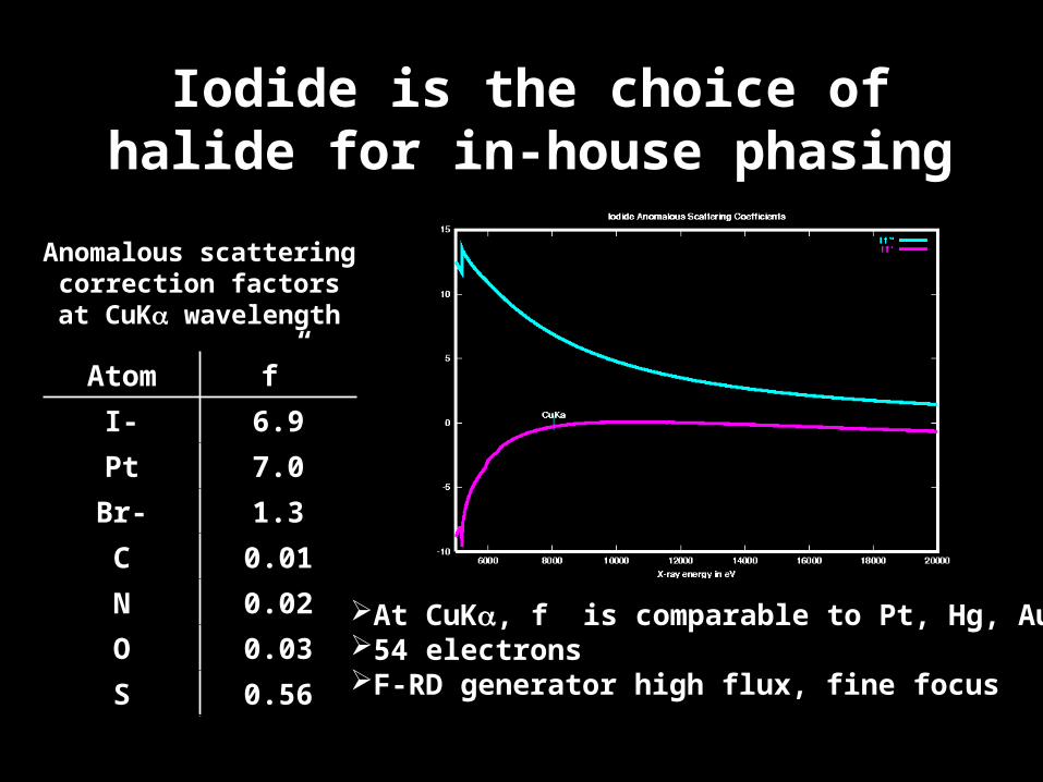
Iodide is the choice of halide for in-house phasing
Atom f”
I- 6.9
Pt 7.0
Br- 1.3
C 0.01
N 0.02
O 0.03
S 0.56
Anomalous scatteringcorrection factors
at CuK wavelength
At CuK, f” is comparable to Pt, Hg, Au54 electronsF-RD generator high flux, fine focus

How successful is phasing by iodide at UCLA?
Original study performed with only 4 proteins using a synchrotron source. Will this method prove to be generally applicable in a real academic research lab with real proteins with real problems?
What are the limitations? (e.g. resolution limits, lack of isomorphism,data quality, soaking conditions, number of iodide sites required for good phases)
Will all proteins bind a sufficient number of iodides to generate good phases? Dauter et al., suggest that the number of iodides bound is simply proportional to the surface area of the protein. Is this always true?
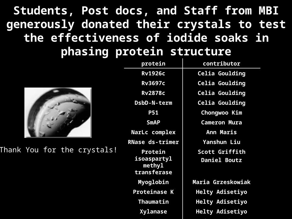
Students, Post docs, and Staff from MBI generously donated their crystals to test the effectiveness of
iodide soaks in phasing protein structure
protein contributor
Rv1926c Celia Goulding
Rv3697c Celia Goulding
Rv2878c Celia Goulding
DsbD-N-term Celia Goulding
P51 Chongwoo Kim
SmAP Cameron Mura
NarLc complex Ann Maris
RNase ds-trimer Yanshun Liu
Protein isoaspartyl methyl transferase
Scott Griffith
Daniel Boutz
Myoglobin Maria Grzeskowiak
Proteinase K Helty Adisetiyo
Thaumatin Helty Adisetiyo
Xylanase Helty Adisetiyo
lysozyme Students of M230B (2001)
Thank You for the crystals!

Experimental methods flow chart
SOAKING PROCEDURES•Weigh 0.008g KI (one medium sized grain)
•Dissolve KI in 100 uL of reservoir solution•Add appropriate % of glycerol (for cryoprotection)
•Soak crystal 30 seconds•Mount on cold stream
DATA COLLECTIONPROCEDURES
•Collect data on F-RD when possible•Collect 360 degrees of data
•Process with Denzo/Scalepack
DETERMINE IODIDE SUBSTRUCTURE Import data with xprepx (Bruker)
•Calculate difference Patterson coefficients using SAS, SIRAS, SIR data•Locate Iodide sites with ShelxD
•Verify quality of sites by overlapping predicted Patterson peaks on Patterson map
PHASING•CCP4 suite: scalepack2mtz, truncate, cad, scaleit, mlphare, dm
MODEL BUILDING•Arp/wArp

12/14 crystals soaked showed clear evidence of iodide binding

8/14 Iodide soaks led to complete structure determination1 structure had not been previously determined

Example of electron density generated by SIRAS phasing based on 11 iodide sites
DsbD N-term
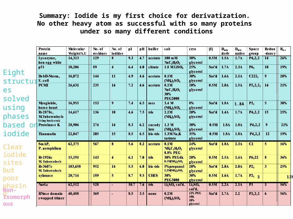
Summary: Iodide is my first choice for derivatization. No other heavy atom as successful with so many proteins
under so many different conditions
Eightstructuressolved using phasesbased on iodide
Cleariodidesites butpoorphasing
Non-Isomorphous
3 13%
1.8A

Tips for successful phasing with iodide
Poor peak heights in difference Patterson map? High redundancy of intensity measurements is crucial to locating heavy atom sites and phasing. Collect 360 degrees of data. Not just iodide, but any derivative would benefit.
Iodide soak is non isomorphous with native? Non-isomorphorism can be reduced by a quick back-soak in cryo-conditions lacking iodide. (eg. Rv2878c)
Iodide sites not convincing? ShelxD often succeeds at finding iodide sites based on anomalous differences alone. But, If the solution is not clear, try using isomorphous differences (SIR) or a combination of isomorphous and anomalous differences (SIRAS) output by xprepx.

Data collected using FR-D generator can produce better quality maps than RU200 generator
I/ (2.0 A) 7.6 26.4Rsym (2.0 A) 37.3% 7.1%

Poor phasing is a direct consequence of too few iodides/surface area
Need 1 iodide bound per 10-20 residues

Why do some proteins bind disproportionately fewer
iodides/surface area?Two possibilities
1) Soaking conditions (e.g. pH, salt, buffer) disfavor or compete with iodide binding. If true then we could search for conditions that favor iodide binding.
or2) Residue composition of the protein surface disfavors
iodide binding. Make predictions about iodide binding based on amino acid composition.

Iodide binding appears insensitive to the composition of the cryo-solvent
Thaumatin1.3M Na,K tartrate
35% glycerolBis-Tris pH 6.5
0.5M KI
1 iodide/14 residues
Rv1926c0.1M (NH4)2SO4
30% PEG 4000Tris pH 7.0
0.5M KI
1 iodide/47 residues
Soaking a Rv1926c crystal in thaumatin’s cryo-conditions did not increase the number of iodides bound.
But, why expect conditions that are optimal for iodide binding to one protein to also be optimal for another protein? Thaumatin is a more basic protein (pI=8.5) than Rv1926c (pI=6.1). Perhaps if I tried a more substantial change in pH to
change the electrostatic potential of the surface…
Experiment to test effects of cryo-solvent on iodide substitution

Higher pH appears to weaken iodide binding
Proteinase K0.1M (NH4)2SO4
30% glycerolCacodylate pH 6.5
0.5M KI
Proteinase K20% PEG 8000
20% glycerolCHES pH 9.5
0.5M KI
Top 3 negative peaks in Fobs(pH9.5) –Fobs(pH6.5) difference Fourier map correspond to iodide sites.
Experiment to test effects of pH on iodide substitution

Tally of side chains in contact with102 iodide sites
Note: Arginine and lysine are the two residues most frequently found in iodide binding sites.

The amino acid composition favored by iodide is significantly depleted in negatively charged side chains compared to the average
amino acid composition on the surface of most proteins
Red data points taken from The Atomic Structure of Protein-Protein Recognition Sites by Lo Conte, Chothia & Janin, J. Mol. Biol., 285,2177-2198
CC=0.77
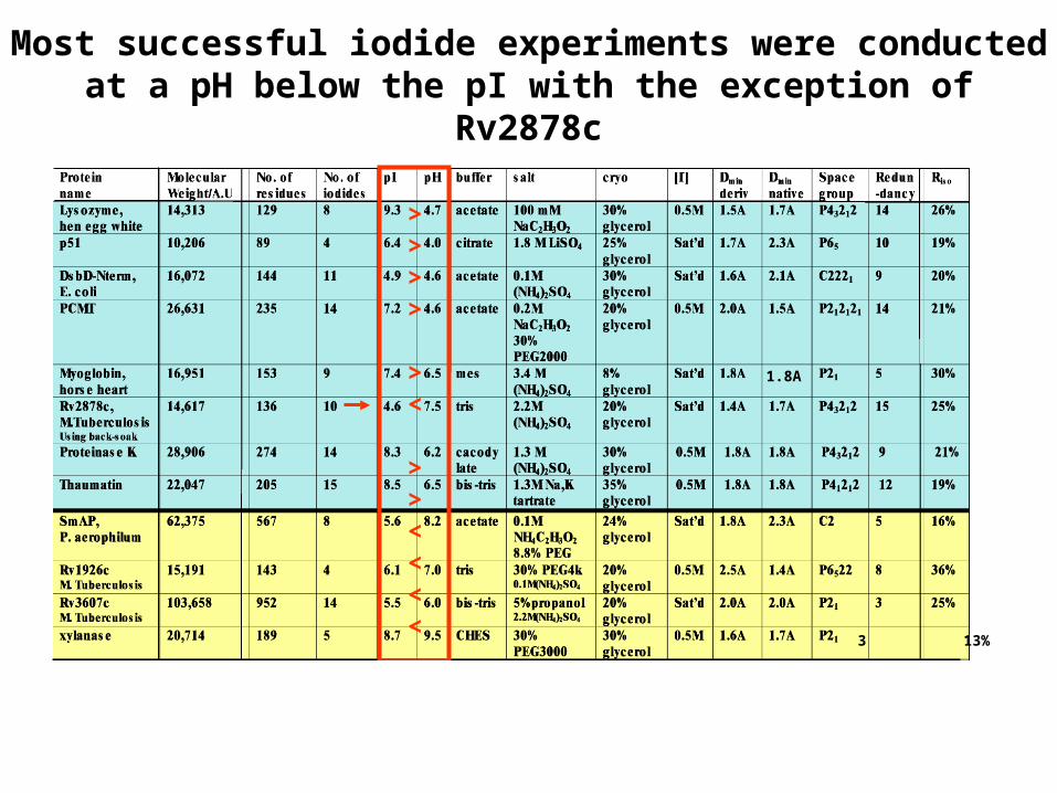
Most successful iodide experiments were conducted at a pH below the pI with the exception of Rv2878c
3 13%
1.8A
>>>>
><
>><<<<

A protein may still bind iodide even if pH > pI since iodide binding sites are often non-polar
No consensus coordinationgeometry
Polar & nonpolarHydrogens at a radius3.5-4 Angstroms
Peptide planes
Could involve any of the20 amino acids
ConclusionsSIRAS phasing from iodide soaks in-house is effective, quick, easy, and non-toxic. 8/14 structures could be determined at UCLA
Even in cases where there are too few iodide sites to produce a good map, iodide sites could be used in combination with other derivatives (e.g. CsCl).
High redundancy, high resolution, and a bright, focused x-ray source (F-RD) are important factors for success.
Soaking at pH < pI improves chances of success
Future: Lower the pH of cryo-conditions of Rv1926c orxylanase to increase iodide binding and solve another structure.

Acknowledgements
Duilio Cascio- partner in experiments, advice, inspiration
CRYSTALSCelia GouldingChongwoo KimCam MuraAnn MarisYanshun LiuScott GriffithDaniel BoutzMaria GrzeskowiakHelty Adisetiyo
SUPPORTDavid Eisenberg
Todd YeatesRichard Dickerson
James Bowie
Zbigniew Dauter-advice onback-soaking, shelxD, xprepx.Peter Muller-xprep connectionsKim Ma –X-ray maintenance
STATISTICSGary KleigerTodd Norcross


![arXiv:1606.03630v1 [q-bio.BM] 11 Jun 2016 · 2018-08-07 · crystallographic expertise of Dan Anderson, Duilio Cascio, and Mike Sawaya has been indispensable without their help I](https://static.fdocuments.us/doc/165x107/5ed96d74f59b0f56f45f78d2/arxiv160603630v1-q-biobm-11-jun-2016-2018-08-07-crystallographic-expertise.jpg)