Use of Continuous Electronic Fetal Monitoring in a Preterm Fetus
8
Hindawi Publishing Corporation Journal of Pregnancy Volume 2011, Article ID 848794, 7 pages doi:10.1155/2011/848794 Review Article Use of Continuous Electronic Fetal Monitoring in a Preterm Fetus: Clinical Dilemmas and Recommendations for Practice Karolina Afors and Edwin Chandraharan St. George’s Healthcare NHS Trust, Blackshaw Road, London SW17 0QT, UK Correspondence should be addressed to Edwin Chandraharan, [email protected] Received 1 April 2011; Accepted 24 May 2011 Academic Editor: Yves Jacquemyn Copyright © 2011 K. Afors and E. Chandraharan. This is an open access article distributed under the Creative Commons Attribution License, which permits unrestricted use, distribution, and reproduction in any medium, provided the original work is properly cited. The aim of intrapartum continuous electronic fetal monitoring using a cardiotocograph (CTG) is to identify a fetus exposed to intrapartum hypoxic insults so that timely and appropriate action could be instituted to improve perinatal outcome. Features observed on a CTG trace reflect the functioning of somatic and autonomic nervous systems and the fetal response to hypoxic or mechanical insults during labour. Although, National Guidelines on electronic fetal monitoring exist for term fetuses, there is paucity of recommendations based on scientific evidence for monitoring preterm fetuses during labour. Lack of evidence-based recommendations may pose a clinical dilemma as preterm births account for nearly 8% (1 in 13) live births in England and Wales. 93% of these preterm births occur after 28 weeks, 6% between 22–27 weeks, and 1% before 22 weeks. Physiological control of fetal heart rate and the resultant features observed on the CTG trace differs in the preterm fetus as compared to a fetus at term making interpretation difficult. This review describes the features of normal fetal heart rate patterns at different gestations and the physiological responses of a preterm fetus compared to a fetus at term. We have proposed an algorithm “ACUTE” to aid management. 1. CTG Monitoring of a Preterm Fetus: The Current Status The cardiotocograph (CTG) is a continuous electronic re- cord of the fetal heart rate obtained either via an ultrasound transducer placed on the mother’s abdomen or via an electrode attached to the fetal scalp. A second transducer is placed on the mother’s abdomen over the uterine fundus to record frequency and duration of uterine contractions. Both components are then traced simultaneously on a paper strip. Based on current scientific evidence, a CTG is not recom- mended in the UK as a method of routine fetal assessment of the preterm fetus (<37 weeks gestation) and currently no clinical practice guidelines on intrapartum monitoring of the preterm fetus exist in the UK The International Federation of Gynaecologists and Obstetricians (FIGO) guidelines for interpretation of intrapartum cardiotocogram distinguish 2 levels of abnormalities, suspicious and pathological, how- ever, the gestation to which such criteria can be applied has not been specified. The American College of Obstetricians and Gynaecologists (ACOG) published a practice bulletin on intrapartum fetal heart rate monitoring in 2009. Within this guideline, the decision to monitor the preterm fetus remains vague with recommendations that each case requires discussion between obstetric and neonatal input, in addition to weighing up likelihood of severe morbidity of the preterm fetus (based on gestational age and fetal weight) and issues related to mode of delivery [1]. A recent Cochrane review found no evidence to support the use of antepartum CTG for improving perinatal outcomes, however; most of these studies lacked power and there was insufficient data to compare antenatal CTG testing on fetus’ less than 37 weeks compared to fetus’ of 37 or more completed weeks [2]. Due to the lack of research and evidence that exists on electronic fetal monitoring (EFM) of the preterm fetus the definition of a normal fetal heart pattern also presents a chal- lenge. Several characteristics of FHR patterns are dependant on gestational age as they reflect the development and matu- rity of cardiac centres in the central nervous system as well as the cardiovascular system and, hence, differ greatly between
Transcript of Use of Continuous Electronic Fetal Monitoring in a Preterm Fetus

Hindawi Publishing CorporationJournal of PregnancyVolume 2011, Article ID 848794, 7 pagesdoi:10.1155/2011/848794
Review Article
Use of Continuous Electronic Fetal Monitoring in a PretermFetus: Clinical Dilemmas and Recommendations for Practice
Karolina Afors and Edwin Chandraharan
St. George’s Healthcare NHS Trust, Blackshaw Road, London SW17 0QT, UK
Correspondence should be addressed to Edwin Chandraharan, [email protected]
Received 1 April 2011; Accepted 24 May 2011
Academic Editor: Yves Jacquemyn
Copyright © 2011 K. Afors and E. Chandraharan. This is an open access article distributed under the Creative CommonsAttribution License, which permits unrestricted use, distribution, and reproduction in any medium, provided the original work isproperly cited.
The aim of intrapartum continuous electronic fetal monitoring using a cardiotocograph (CTG) is to identify a fetus exposed tointrapartum hypoxic insults so that timely and appropriate action could be instituted to improve perinatal outcome. Featuresobserved on a CTG trace reflect the functioning of somatic and autonomic nervous systems and the fetal response to hypoxicor mechanical insults during labour. Although, National Guidelines on electronic fetal monitoring exist for term fetuses, there ispaucity of recommendations based on scientific evidence for monitoring preterm fetuses during labour. Lack of evidence-basedrecommendations may pose a clinical dilemma as preterm births account for nearly 8% (1 in 13) live births in England and Wales.93% of these preterm births occur after 28 weeks, 6% between 22–27 weeks, and 1% before 22 weeks. Physiological control offetal heart rate and the resultant features observed on the CTG trace differs in the preterm fetus as compared to a fetus at termmaking interpretation difficult. This review describes the features of normal fetal heart rate patterns at different gestations andthe physiological responses of a preterm fetus compared to a fetus at term. We have proposed an algorithm “ACUTE” to aidmanagement.
1. CTG Monitoring of a Preterm Fetus:The Current Status
The cardiotocograph (CTG) is a continuous electronic re-cord of the fetal heart rate obtained either via an ultrasoundtransducer placed on the mother’s abdomen or via anelectrode attached to the fetal scalp. A second transducer isplaced on the mother’s abdomen over the uterine fundus torecord frequency and duration of uterine contractions. Bothcomponents are then traced simultaneously on a paper strip.Based on current scientific evidence, a CTG is not recom-mended in the UK as a method of routine fetal assessmentof the preterm fetus (<37 weeks gestation) and currently noclinical practice guidelines on intrapartum monitoring of thepreterm fetus exist in the UK The International Federationof Gynaecologists and Obstetricians (FIGO) guidelines forinterpretation of intrapartum cardiotocogram distinguish 2levels of abnormalities, suspicious and pathological, how-ever, the gestation to which such criteria can be applied hasnot been specified. The American College of Obstetricians
and Gynaecologists (ACOG) published a practice bulletinon intrapartum fetal heart rate monitoring in 2009. Withinthis guideline, the decision to monitor the preterm fetusremains vague with recommendations that each case requiresdiscussion between obstetric and neonatal input, in additionto weighing up likelihood of severe morbidity of the pretermfetus (based on gestational age and fetal weight) and issuesrelated to mode of delivery [1]. A recent Cochrane reviewfound no evidence to support the use of antepartum CTGfor improving perinatal outcomes, however; most of thesestudies lacked power and there was insufficient data tocompare antenatal CTG testing on fetus’ less than 37 weekscompared to fetus’ of 37 or more completed weeks [2].
Due to the lack of research and evidence that exists onelectronic fetal monitoring (EFM) of the preterm fetus thedefinition of a normal fetal heart pattern also presents a chal-lenge. Several characteristics of FHR patterns are dependanton gestational age as they reflect the development and matu-rity of cardiac centres in the central nervous system as well asthe cardiovascular system and, hence, differ greatly between
2 Journal of Pregnancy
a preterm and a term fetus. Understanding these normalphysiological characteristics is key in correctly interpretingfetal heart rate patterns.
2. Factors That Affect Fetal Heart Rateduring Labour
During labour, uterine contractions gradually build up andincrease in intensity and frequency and may cause com-pression of the umbilical cord and/or the fetal head. These“mechanical compressions” may result in decelerations infetal heart resulting in early and variable decelerations,respectively. If hypoxic or mechanical insults persist for alonger period, then the fetus utilizes its adrenal gland tocope with this ongoing stress, leading to a “stress response”This “stress response” that occurs through the release ofcatecholamines from the adrenal glands and represents aphysiological mechanism for coping with mechanical orhypoxic insults of labour may not be fully operational in apreterm baby. This may also be the case when the normalphysiological reserves of the fetus may be impaired (intra-uterine growth restriction, fetal infection). Inability of apreterm or growth restricted fetus to mount a requiredstress response may lead to maladaptive responses resultingin permanent hypoxic insult on the fetal brain occurringat a lower threshold than in the term fetus. Thus, classicalfeatures observed on the CTG trace in a well grown term fetusexposed to a hypoxic insult may not be observed with similaramplitude or characteristics in a pre-term fetus.
Fetal heart rate is regulated by the autonomic nervoussystem consisting of 2 branches; the parasympathetic andsympathetic branch which exerts opposing influences onthe FHR. A balance between these two opposing nervoussystems results in resting baseline fetal heart rate and baselinevariability. During fetal development, the sympathetic ner-vous system that is responsible for survival (“fight or flight”response) develops much earlier than the parasympatheticnervous system (“rest and sleep”) that develops during thethird trimester. Hence, a preterm fetus may have a higherbaseline fetal heart rate with apparent reduction of baselinevariability due to unopposed action of sympathetic nervoussystem.
2.1. Baroreceptors. The parasympathetic nervous system isactivated by stimulation of baroreceptors situated in thecarotid sinus or aortic arch secondary to increase in fetal sys-temic blood pressure, leading to a fall in heart rate mediatedthrough the vagus nerve. This is illustrated by a decelerationon a CTG. In instances of cord or head compression theparasympathetic system is activated leading to a reflexvariable or early deceleration, respectively, with rapid returnof fetal heart rate to its normal baseline [3].
2.2. Chemoreceptors. Chemo-receptors are located peripher-ally within the aortic and carotid bodies and centrally inthe medulla oblongata. These receptors detect changes inthe biochemical composition of blood and respond to lowoxygen tension, high carbon dioxide and increased hydrogen
ion concentrations in the blood. In cases of utero-placentalinsufficiency, where carbon dioxide and hydrogen ion accu-mulate with resultant decrease in oxygen concentrations,the chemo-receptors are activated. This results in parasym-pathetic activation leading to a fall in heart rate, which isprotracted and takes longer to recover to baseline rate. Thesetypes of decelerations are termed “late” decelerations anddue to the accumulation of carbon dioxide and hydrogenions are more suggestive of metabolic acidosis [3].
2.3. Somatic Nervous System. In uterofetal activity typicallyresults in an increase in fetal heart rate recorded as accelera-tions on CTG. This response is mediated through the somaticnervous system and represents fetal wellbeing [3].
2.4. Fetal Adrenal Glands. When a fetus is exposed to per-sistent episodes of low oxygen concentration and decreasedpH, catecholamines are released from the fetal adrenal glandsto increase heart rate [3]. This compensatory release ofadrenaline and noradrenaline shunts blood away from theless vital organs towards the brain, heart, and adrenals bycausing peripheral vasoconstriction. This clinical scenario ofdecelerations, followed by loss of accelerations, subsequentrise in baseline heart rate and gradual loss of variability istypical of a gradually evolving hypoxia (Figure 1).
3. Characteristics of Fetal Heart Rate ina Preterm Fetus
When assessing well-being of a term fetus during labour, fourfeatures are evaluated for classification of the CTG. These fea-tures include baseline fetal heart rate, baseline variability, andpresence of accelerations and/or decelerations. According toNational Institute of Health and Clinical Excellence (NICE)guidelines on electronic fetal monitoring in labour, thesefeatures, which are present in labour, are further categorizedinto reassuring and nonreassuring as outlined in Table 1below.
Characteristics of antepartum and intrapartum fetalheart rate tracings differ in the preterm fetus as comparedto a term fetus. Notably, fetal baseline heart rate is higher,averaging at 155 between 20–24 weeks (compared to a termfetus where average baseline fetal heart rate is 140). Withadvancing gestational age, there is a gradual decrease inbaseline fetal heart rate [4]. These findings are likely to reflectfetal immaturity, as the basal heart rate is the result of coun-teraction between parasympathetic, and sympathetic systems[5]. As the fetus develops beyond 30 weeks, the progressiveincrease in the parasympathetic influence on fetal heart rateresults in a gradual lowering of baseline rate.
Fetal heart rate accelerations are also noted to changewith advancing gestational age. Accelerations of fetal heartrate in association with fetal movements occur as a resultof fetal somatic activity and are first apparent in the 2ndtrimester. Before 30 weeks of gestational age, the frequencyand amplitude of accelerations are reduced. Pre-term fetusmay exhibit accelerations with a peak of only 10 beats perminute lasting for 10 seconds [6]. With subsequent increase

Journal of Pregnancy 3
- Sympathetic-- Parasympathetic
Peripheral
- Aortic bodies
BP
Stress
CO2
H+
PO2
Central chemoreceptorsMedulla oblongata
Adrenal glands
Kidneys
Right commoncarotid artery
Right subclavian artery
Brachiocephalic artery
Right coronary artery
Left coronary artery
Left
LeftsubclavianarteryArch
ofaorta
Asc
endi
ng
aort
a
Descen
ding
aorta
CNS cardiac centres
chemoreceptors
Baroreceptors
- Aortic arch
- Carotid sinus
Adrenal gland Catecholamines and cortisol
commoncarotid artery
Figure 1: Pathophysiology of fetal heart rate changes.
Table 1: Categorizing individual features of CTG according to NICE guidelines.
FeatureBaseline(bpm)
Variability(bpm)
Decelerations Accelerations
ReassuringNonreassuring
110–160100–109161–180
>5<5 for 40–90
minutes
NoneTypical variable decelerations with >50%of contractions for over 90 minutes.Single-prolonged deceleration for up to 3minutes.
PresentThe absence of acceleration withotherwise normal trace is ofuncertain significance
Abnormal
<100>180
Sinusoidal pattern>10 minutes
<5 for 90 minutes
Either atypical variable decelerations with>50% of contractions or latedecelerations, both for over 30 minutes.Single-prolonged deceleration for morethan 3 minutes.
in gestational age, the frequency of accelerations increasesalong with amplitude over the baseline value [6].
Fetal heart rate decelerations in the absence of uterinecontractions often occur in the normal preterm fetus be-tween 20 and 30 weeks gestation. As described by Sorokinet al. these decelerations have a lower depth and duration,but can be seen frequently on intrapartum CTG tracings[4]. Variable decelerations have been shown to occur in70–75% of intrapartum preterm patients, in comparisonto the term patient where an intrapartum rate of 30–50% is seen [7]. Several theories have been proposed as apotential explanation for this fetal heart rate pattern, notablydecreased amount of amniotic fluid, reduced the Wharton
jelly component in the cord of the preterm fetus and lackof development of the fetal myocardium and, therefore, theresultant reduced force of contraction.
Baseline variability may be affected due to incompletedevelopment of autonomic nervous system and subsequentinterplay between parasympathetic and sympathetic systems.Variability may also be decreased secondary to the effectof fetal tachycardia present in preterm fetuses. Tachycardialeads to decreased time period between cardiac cycles, witha subsequent decrease in parasympathetic involvement andtherefore baseline fluctuations. Reduction in fetal baselinevariability in the preterm fetus has been described, howeverthis has not been quantified. Some studies report a higher

4 Journal of Pregnancy
200
180
160
140
120
100
100
80
80
60
60
4020
0
TOCO
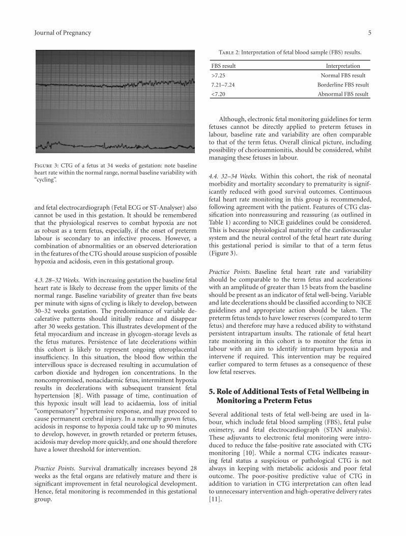
Figure 2: CTG of a fetus at 26 weeks of gestation: note higherbaseline heart rate, apparent reduction in baseline variability, and“shallow” variable decelerations.
incidence of adverse outcome following a tracing withreduced variability compared to the presence of decelerations[8].
One of the hallmarks of fetal wellbeing is considered to be“cycling” of the fetal heart rate [3]. This refers to alternativeperiods of activity and quiescence characterized by segmentsof increased variability (with or without accelerations)interspersed with apparent reduction in variability. Theseare believed to reflect Rapid Eye Movement (REM) andnon-REM sleep. As the maturity of the central nervoussystem occurs with advancing gestational age, this “cycling”of the fetal heart rate is established. Hence, in an extremepreterm infant, cycling may be absent and this may be due tofunctional immaturity of the central nervous system, ratherthan hypoxic insult.
4. Interpreting Intrapartum CTG atDifferent Gestations
4.1. 24–26 Weeks. Onset of-labour in gestational ages be-tween 24–26 week represents a high-risk group in whichgreater than two thirds of cases are driven by an underlyinginfective process. Other possible factors that may contributeto onset of labour in this group include multiple gestationsmaternal risk factors such as increased maternal age, raisedbody mass index (BMI), or pregnancies conceived throughin-vitro fertilization (IVF). At this gestation, there is a highrisk of neonatal morbidity and mortality, and survival isdependant more on fetal weight and maturity rather thanmode of delivery. Hence, continuous monitoring of thefetus during labour, with the view to recognizing featuresof suspected fetal compromise on CTG and instituting anoperative intervention, should be considered with caution.The use of CTG monitoring in this group is contentiousand each case should be considered individually with a planof care agreed following discussion between the patient,obstetrician, and neonatologists. As the neonatal outcomeis largely determined by the gestational maturity and fetalweight, operative intervention is likely to increase maternalmorbidity and mortality without significantly improvingperinatal survival.
Practice Points. Baseline fetal heart rate in this cohort offetuses is likely to remain at the higher end of normal(between 150–160) due to the unopposed effect of thesympathetic nervous system. Although, the baseline heartrate is expected to be higher, any rate greater than 160 shouldbe still considered to be tachycardic. Persistent tachycardiais likely to arise secondary to iatrogenic causes such asadministration of tocolytics (terbutaline) [9]. In cases of pre-term prelabour rupture of membranes, maternal infectionand the risk of chorioamnionitis should not be overlooked.
Baseline variability and cycling may be reduced at thisgestation as a result of impaired development of the para-sympathetic component of the autonomic nervous system.Medications such as pethidine, magnesium sulphate andeven steroids have also been associated with reduced fetalheart rate variability. However, fetal heart rate variability isan important clinical indicator of fetal acid base balance,especially oxygenation of the autonomic nerve centres withinthe brain, and absent variability is therefore predictive ofcerebral asphyxia. A thorough history of each case shouldbe determined prior to CTG interpretation, and instanceswhere variability is persistently reduced without explanation,should be viewed with caution.
Accelerations at this gestation may not be present or maybe significantly reduced with a lower amplitude (rise of 10beats from the baseline rather than 15 beats). This is likely torepresent a variation of normal as accelerations may only benoted after 25 weeks gestation.
Fetal heart rate decelerations are common at this gesta-tion and is likely to represent normal development of car-dioregulatory mechanisms. In the presence of other reassur-ing features of the CTG (as outlined above), these decel-erations should not be considered as indicative of hypoxia,and interventions should be avoided based on this parameteralone. Figure 2 shows CTG of a preterm fetus at 26 weeks.
4.2. 26–28 Weeks. Within this group, fetal heart rate tracingswill show many similarities to the 24–26 week gestationcohort. After 27 weeks gestation, the frequency of variabledecelerations observed is generally reduced [5]. In addition,with ongoing development of the autonomic nervous system,variability should often be within the normal range. Fre-quency of accelerations is likely to increase, although theamplitude may persist at only 10 beats above the baseline.Likely, iatrogenic causes of fetal heart rate abnormalities (asmentioned above) should also be noted and documented.
Practice Points. Survival in this group is significantly higherthan those between 24–26 weeks as survival improvesapproximately 10% every week during this period. Approxi-mately half of those babies who survive may develop long-term neurological or developmental defects. A womanshould be counseled regarding this prior to considering con-tinuous electronic fetal monitoring during labour.
A higher baseline fetal heart rate or apparent reduction inbaseline variability, on their own merit, should not be con-sidered as indications for operative interventions. Additionaltests of fetal well-being such as fetal blood sampling (FBS)
Journal of Pregnancy 5
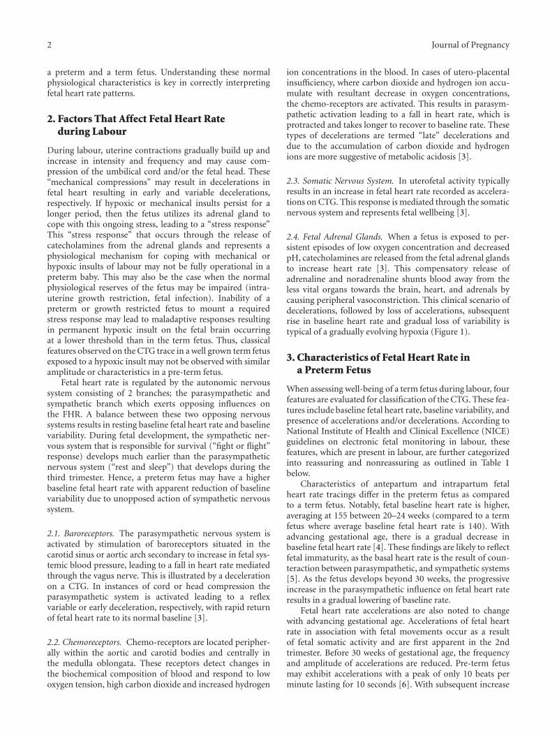
Figure 3: CTG of a fetus at 34 weeks of gestation: note baselineheart rate within the normal range, normal baseline variability with“cycling”.
and fetal electrocardiograph (Fetal ECG or ST-Analyser) alsocannot be used in this gestation. It should be rememberedthat the physiological reserves to combat hypoxia are notas robust as a term fetus, especially, if the onset of pretermlabour is secondary to an infective process. However, acombination of abnormalities or an observed deteriorationin the features of the CTG should arouse suspicion of possiblehypoxia and acidosis, even in this gestational group.
4.3. 28–32 Weeks. With increasing gestation the baseline fetalheart rate is likely to decrease from the upper limits of thenormal range. Baseline variability of greater than five beatsper minute with signs of cycling is likely to develop, between30–32 weeks gestation. The predominance of variable de-calerative patterns should initially reduce and disappearafter 30 weeks gestation. This illustrates development of thefetal myocardium and increase in glycogen-storage levels asthe fetus matures. Persistence of late decelerations withinthis cohort is likely to represent ongoing uteroplacentalinsufficiency. In this situation, the blood flow within theintervillous space is decreased resulting in accumulation ofcarbon dioxide and hydrogen ion concentrations. In thenoncompromised, nonacidaemic fetus, intermittent hypoxiaresults in decelerations with subsequent transient fetalhypertension [8]. With passage of time, continuation ofthis hypoxic insult will lead to acidaemia, loss of initial“compensatory” hypertensive response, and may proceed tocause permanent cerebral injury. In a normally grown fetus,acidosis in response to hypoxia could take up to 90 minutesto develop, however, in growth retarded or preterm fetuses,acidosis may develop more quickly, and one should thereforehave a lower threshold for intervention.
Practice Points. Survival dramatically increases beyond 28weeks as the fetal organs are relatively mature and there issignificant improvement in fetal neurological development.Hence, fetal monitoring is recommended in this gestationalgroup.
Table 2: Interpretation of fetal blood sample (FBS) results.
FBS result Interpretation
>7.25 Normal FBS result
7.21–7.24 Borderline FBS result
<7.20 Abnormal FBS result
Although, electronic fetal monitoring guidelines for termfetuses cannot be directly applied to preterm fetuses inlabour, baseline rate and variability are often comparableto that of the term fetus. Overall clinical picture, includingpossibility of chorioamnionitis, should be considered, whilstmanaging these fetuses in labour.
4.4. 32–34 Weeks. Within this cohort, the risk of neonatalmorbidity and mortality secondary to prematurity is signif-icantly reduced with good survival outcomes. Continuousfetal heart rate monitoring in this group is recommended,following agreement with the patient. Features of CTG clas-sification into nonreassuring and reassuring (as outlined inTable 1) according to NICE guidelines could be considered.This is because physiological maturity of the cardiovascularsystem and the neural control of the fetal heart rate duringthis gestational period is similar to that of a term fetus(Figure 3).
Practice Points. Baseline fetal heart rate and variabilityshould be comparable to the term fetus and accelerationswith an amplitude of greater than 15 beats from the baselineshould be present as an indicator of fetal well-being. Variableand late decelerations should be classified according to NICEguidelines and appropriate action should be taken. Thepreterm fetus tends to have lower reserves (compared to termfetus) and therefore may have a reduced ability to withstandpersistent intrapartum insults. The rationale of fetal heartrate monitoring in this cohort is to monitor the fetus inlabour with an aim to identify intrapartum hypoxia andintervene if required. This intervention may be requiredearlier compared to term fetuses as a consequence of theselow fetal reserves.
5. Role of Additional Tests of Fetal Wellbeing inMonitoring a Preterm Fetus
Several additional tests of fetal well-being are used in la-bour, which include fetal blood sampling (FBS), fetal pulseoximetry, and fetal electrocardiograph (STAN analysis).These adjuvants to electronic fetal monitoring were intro-duced to reduce the false-positive rate associated with CTGmonitoring [10]. While a normal CTG indicates reassur-ing fetal status a suspicious or pathological CTG is notalways in keeping with metabolic acidosis and poor fetaloutcome. The poor-positive predictive value of CTG inaddition to variation in CTG interpretation can often leadto unnecessary intervention and high-operative delivery rates[11].

6 Journal of Pregnancy
Table 3: Proposed Management Algorithm “ACUTE” for intrapartum fetal monitoring (CTG) in preterm gestations (<34 weeks).
A Assess survival and long-term outcome at the given gestational age.
CConsider the wider clinical picture:presence of co-existing infection, maternal age, condition of the fetus (severe growthrestriction, congenital malformations), wishes of the woman (e.g., request to “do everything possible” in view of IVFconception, previous preterm losses) in formulating management plan.
U Understand normal fetal cardiovascular and nervous system physiology at the given gestation in interpreting the CTG.
T Treatment of underlying predisposing factors of uterine irritability (infection, antepartum haemorrhage) and treatment ofpreterm labour (tocolytics and steroids, if appropriate) to optimise maternal and fetal outcome.
EEvaluate maternal risks of operative interventions (classical C. section, haemorrhage, infections, increased risk of uterinerupture in future pregnancies) and potential fetal benefits (survival and long-term morbidity) due to commencingcontinuous electronic fetal monitoring at the given gestation and counsel appropriately.
5.1. Fetal Blood Sampling. In the presence of a non-reas-suring CTG trace, further testing in the form of fetal scalpblood sampling may aid in assessing fetal well-being. Afterrupture of membranes and once the cervix is adequatelydilated (>3 cm), sampling a small amount of blood fromthe fetal scalp can be used to measure pH or lactate andthus detect acidosis. It is not recommended in fetuses withbleeding disorders and is contraindicated in pregnanciescomplicated with HIV, Hepatitis B or C as it may increasevertical transmission. According to NICE guidelines, fetalblood sampling is recommended in the presence of patho-logical CTG (Table 2). If the pH value is <7.20, immediatedelivery is recommended, whereas a pH of 7.20–7.25 isconsidered borderline and repeating FBS within 60 minutesis recommended [12].
With regards to the pre-term fetus, fetal blood samplinghas not been validated in this group. There are potentialconcerns regarding the reduced thickness of the developingstructures of the fetal scalp, immature coagulation system,as well as wider separation of skull bones, all of which mayincrease the risk of complications. Moreover, studies haveshown fetal acidosis to occur more often in pre-term fetusesdelivered before 34 weeks than those delivered between 34–36 weeks [5]. Despite this high rate of fetal acidosis, theshort-term fetal outcome was good and in subsequent repeatblood-sampling pH values had normalized [5]. This highrate of dramatic fetal acidosis in the preterm may repre-sent an alternative intrapartum compensatory mechanism.Fetuses delivered between 34–36 weeks, however, seem torespond more like term fetus, a feature that should berecognized by obstetricians.
5.2. Fetal Pulse Oximetry. Fetal pulse oximetry was firstintroduced in clinical practice in the 1980s. It provided ameans of monitoring fetal oxygen saturation of fetal hae-moglobin that is measured optically (similar technology forpulse oximetry in adults) during labour. In non-reassuringCTG traces, pulse oximetry was initially felt to providea more sophisticated way of detecting adverse neonataloutcome. Several studies defined a critical threshold of <30%SpO2 persisting for greater than ten minutes as a predictorof fetal acidosis and poor neonatal outcome [13]. This cutoff value yielded a sensitivity of 81% and specificity of100% to predict scalp pH of <7.2 [14]. Recent large RCT’s,however, have demonstrated no reduction in operative
delivery rate or in predicting adverse neonatal outcome [15].This mode of fetal monitoring now remains obsolete and themanufacturers have ceased production.
5.3. Fetal ECG (ST Analyser or STAN). This technology isbased on analyzing the ST segment of the fetal myocardiumfor ischaemic changes during fetal hypoxia as well asdetermining the ratio between the T wave and QRS complex(T/QRS Ratio) of the fetal ECG. The latter is alteredsecondary to release of potassium during glyocogenolysis inthe fetal myocardium mediated through that catecholoaminesurge, which occurs during hypoxic stress. Myocardium of apreterm fetus has less stored glycogen with increased watercontent and also the epicardial-endocardial interphase ismuch smaller than a term fetus. Hence, ST analyser is notrecommended prior to 36 weeks of gestation as it may notbe reliable due to changes in the myocardial compositiondescribed above.
5.4. Preterminal Trace. A fetus that demonstrates features ofpreterminal trace has exhausted all its reserves to combathypoxia and hence immediate delivery is recommended [16].However, caution should be exercised in fetuses prior to 28weeks that demonstrate such features as perinatal outcome ispoor in this group. Hence, a woman should be counseled thatthe risks of operative intervention may outweigh the benefits.
6. Conclusion
Continuous electronic fetal monitoring of preterm fetusesposes a clinical dilemma to clinicians caring for these fetusesduring labour. Although, clinical evidence-based guidelinesand recommendations exist for monitoring term fetusesduring labour, there is paucity of scientific evidence inthe preterm group. Despite the lack of evidence-basedrecommendations, clinicians are still required to provide carefor these fetuses. Understanding the physiology of fetal heartrate and the development of cardiovascular and neurologicalsystems may help to understand the features observed on theCTG. It is important to realize that physiological reservesavailable to combat hypoxia are less than those availableto a term fetus. Hence, a preterm fetus may suffer ahypoxic insult sooner than its term counterpart. It is vitalto counsel women prior to instituting continuous electronic
Journal of Pregnancy 7
fetal monitoring, especially in extreme preterm fetuses (24–26 weeks) as survival in this group is largely determinedby fetal maturity than the mode of delivery. In view ofthe absence of guidelines and recommendations monitoringpreterm fetuses, we have produced a management algorithm“ACUTE” to aid continuous intrapartum fetal monitoring infetuses prior to 34 weeks (Table 3). Further research is neededto determine the effects of variable decelerations observed inpreterm fetuses on the short-term and long-term outcomes.
Conflict of Interests
The authors declare no conflict of interests.
References
[1] ACOG, “Intrapartum fetal heart rate monitoring: nomen-clature, interpretation, and general management principles,”ACOG Practice Bulletin, vol. 106, pp. 192–202, 2009.
[2] R. M. Grivell, Z. Alfirevic, G. M. Gyte, and D. Devane, “An-tenatal cardiotocography for fetal assessment,” CochraneDatabase of Systematic Reviews, no. 1, Article ID CD007863,2010.
[3] E. Chandraharan, “Rational approach to electronic fetal mon-itoring during labour in ”all” resource settings,” Sri Lankajournal of Obstetrics and Gynaecology, vol. 32, pp. 77–84, 2010.
[4] Y. Sorokin, L. J. Dierker, S. K. Pillay, I. E. Zador, M. L. Shreiner,and M. G. Rosen, “The association between fetal heart ratepatterns and fetal movements in pregnancies between 20and 30 weeks’ gestation,” American Journal of Obstetrics andGynecology, vol. 143, no. 3, pp. 243–249, 1982.
[5] M. Westgren, P. Holmquist, N. W. Svenningsen, and I. Inge-marsson, “Intrapartum fetal monitoring in preterm deliveries:prospective study,” Obstetrics and Gynecology, vol. 60, no. 1,pp. 99–106, 1982.
[6] T. Wheeler and A. Murrills, “Patterns of fetal heart rate duringnormal pregnancy,” British Journal of Obstetrics and Gynaecol-ogy, vol. 85, no. 1, pp. 18–27, 1978.
[7] B. Zanini, R. H. Paul, and J. R. Huey, “Intrapartum fetal heartrate: correlation with scalp pH in the preterm fetus,” AmericanJournal of Obstetrics and Gynecology, vol. 143, no. 1, pp. 952–957, 1980.
[8] F. Goupil, H. Legrand, and J. Vaquier, “Antepartum fetal heartrate monitoring. II. Deceleration patterns,” European Journalof Obstetrics Gynecology and Reproductive Biology, vol. 11, no.4, pp. 239–249, 1981.
[9] E. Chandraharan and S. Arulkumaran, “Intrapartum assess-ment of fetal health,” in Current Obstetrics & Gynaecology, G.M. Mukherjee, Ed., Jaypee Brothers, 2007.
[10] C. E. East and P. B. Colditz, “Intrapartum oximetry of thefetus,” Anesthesia & Analgesia, vol. 105, pp. S59–S65, 2007.
[11] S. M. Baird and D. J. Ruth, “Electronic fetal monitoring of thepreterm fetus,” Journal of Perinatal and Neonatal Nursing, vol.16, no. 1, pp. 12–24, 2002.
[12] National Institute of Clinical Health and Excellence, “Intra-partum care—Clinical guideline 55,” 2007, http://www.nice.org.uk/CG055.
[13] A. Tekin, S. Ozkan, E. Caliskan, S. Ozeren, A. Corakci, and I.Yucesoy, “Fetal pulse oximetry: correlation with intrapartumfetal heart rate patterns and neonatal outcome,” Journal ofObstetrics and Gynaecology Research, vol. 34, no. 5, pp. 824–831, 2008.
[14] B. Langer, B. Carbonne, F. Goffinet, F. Le Goueff, N. Berkane,and M. Laville, “Fetal pulse oximetry and fetal heart ratemonitoring during stage II of labour,” European Journal ofObstetrics Gynecology and Reproductive Biology, vol. 72, pp.73–79, 1997.
[15] C. E. East, S. P. Brennecke, J. F. King, F. Y. Chan, and P. B.Colditz, “The effect of intrapartum fetal pulse oximetry, inthe presence of a nonreassuring fetal heart rate pattern, onoperative delivery rates: a multicenter, randomized, controlledtrial (the FOREMOST trial),” American Journal of Obstetricsand Gynecology, vol. 194, no. 3, p. 606, 2006.
[16] E. Chandraharan and S. Arulkumaran, “Prevention of birthasphyxia: responding appropriately to cardiotocograph (CTG)traces,” Best Practice and Research: Clinical Obstetrics andGynaecology, vol. 21, no. 4, pp. 609–624, 2007.

Submit your manuscripts athttp://www.hindawi.com
Stem CellsInternational
Hindawi Publishing Corporationhttp://www.hindawi.com Volume 2014
Hindawi Publishing Corporationhttp://www.hindawi.com Volume 2014
MEDIATORSINFLAMMATION
of
Hindawi Publishing Corporationhttp://www.hindawi.com Volume 2014
Behavioural Neurology
EndocrinologyInternational Journal of
Hindawi Publishing Corporationhttp://www.hindawi.com Volume 2014
Hindawi Publishing Corporationhttp://www.hindawi.com Volume 2014
Disease Markers
Hindawi Publishing Corporationhttp://www.hindawi.com Volume 2014
BioMed Research International
OncologyJournal of
Hindawi Publishing Corporationhttp://www.hindawi.com Volume 2014
Hindawi Publishing Corporationhttp://www.hindawi.com Volume 2014
Oxidative Medicine and Cellular Longevity
Hindawi Publishing Corporationhttp://www.hindawi.com Volume 2014
PPAR Research
The Scientific World JournalHindawi Publishing Corporation http://www.hindawi.com Volume 2014
Immunology ResearchHindawi Publishing Corporationhttp://www.hindawi.com Volume 2014
Journal of
ObesityJournal of
Hindawi Publishing Corporationhttp://www.hindawi.com Volume 2014
Hindawi Publishing Corporationhttp://www.hindawi.com Volume 2014
Computational and Mathematical Methods in Medicine
OphthalmologyJournal of
Hindawi Publishing Corporationhttp://www.hindawi.com Volume 2014
Diabetes ResearchJournal of
Hindawi Publishing Corporationhttp://www.hindawi.com Volume 2014
Hindawi Publishing Corporationhttp://www.hindawi.com Volume 2014
Research and TreatmentAIDS
Hindawi Publishing Corporationhttp://www.hindawi.com Volume 2014
Gastroenterology Research and Practice
Hindawi Publishing Corporationhttp://www.hindawi.com Volume 2014
Parkinson’s Disease
Evidence-Based Complementary and Alternative Medicine
Volume 2014Hindawi Publishing Corporationhttp://www.hindawi.com