

Use of chest imaging in COVID-19 - WHO
56
A RAPID ADVICE GUIDE 11 JUNE 2020 Use of chest imaging in COVID-19 COVID-19
Transcript of Use of chest imaging in COVID-19 - WHO
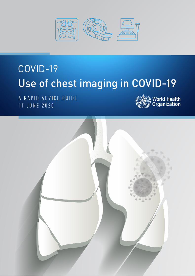
A R A P I D A D V I C E G U I D E1 1 J U N E 2 0 2 0
Use of chest imaging in COVID-19 COVID-19

WHO/2019-nCoV/Clinical/Radiology_imaging/2020.1
A R A P I D A D V I C E G U I D E1 1 J U N E 2 0 2 0
Use of chest imaging in COVID-19 COVID-19
WHO/2019-nCoV/Clinical/Radiology_imaging/2020.1
© World Health Organization 2020
Some rights reserved. This work is available under the Creative Commons Attribution-NonCommercial-ShareAlike 3.0 IGO licence (CC BY-NC-SA 3.0 IGO; https://creativecommons.org/licenses/by-nc-sa/3.0/igo).
Under the terms of this licence, you may copy, redistribute and adapt the work for non-commercial purposes, provided the work is appropriately cited, as indicated below. In any use of this work, there should be no suggestion that WHO endorses any specific organization, products or services. The use of the WHO logo is not permitted. If you adapt the work, then you must license your work under the same or equivalent Creative Commons licence. If you create a translation of this work, you should add the following disclaimer along with the suggested citation: “This translation was not created by the World Health Organization (WHO). WHO is not responsible for the content or accuracy of this translation. The original English edition shall be the binding and authentic edition”. Any mediation relating to disputes arising under the licence shall be conducted in accordance with the mediation rules of the World Intellectual Property Organization. (http://www.wipo.int/amc/en/mediation/rules/)
Suggested citation. Use of chest imaging in COVID-19: a rapid advice guide. Geneva: World Health Organization; 2020 (WHO/2019-nCoV/Clinical/Radiology_imaging/2020.1). Licence: CC BY-NC-SA 3.0 IGO.
Cataloguing-in-Publication (CIP) data. CIP data are available at http://apps.who.int/iris.
Sales, rights and licensing. To purchase WHO publications, see http://apps.who.int/bookorders. To submit requests for commercial use and queries on rights and licensing, see http://www.who.int/about/licensing. Third-party materials. If you wish to reuse material from this work that is attributed to a third party, such as tables, figures or images, it is your responsibility to determine whether permission is needed for that reuse and to obtain permission from the copyright holder. The risk of claims resulting from infringement of any third-party-owned component in the work rests solely with the user.
General disclaimers. The designations employed and the presentation of the material in this publication do not imply the expression of any opinion whatsoever on the part of WHO concerning the legal status of any country, territory, city or area or of its authorities, or concerning the delimitation of its frontiers or boundaries. Dotted and dashed lines on maps represent approximate border lines for which there may not yet be full agreement.
The mention of specific companies or of certain manufacturers’ products does not imply that they are endorsed or recommended by WHO in preference to others of a similar nature that are not mentioned. Errors and omissions excepted, the names of proprietary products are distinguished by initial capital letters.All reasonable precautions have been taken by WHO to verify the information contained in this publication. However, the published material is being distributed without warranty of any kind, either expressed or implied. The responsibility for the interpretation and use of the material lies with the reader. In no event shall WHO be liable for damages arising from its use.
Design by L’IV Com Sàrl
ContentsAcknowledgements . . . . . . . . . . . . . . . . . . . . . . . . . . . . . . . . . . . . . . . . . . . . . . . . . . . . . . . . . . . . . . . . v
Abbreviations . . . . . . . . . . . . . . . . . . . . . . . . . . . . . . . . . . . . . . . . . . . . . . . . . . . . . . . . . . . . . . . . . . . . vii
Executive summary . . . . . . . . . . . . . . . . . . . . . . . . . . . . . . . . . . . . . . . . . . . . . . . . . . . . . . . . . . . . . . viii
1. Introduction . . . . . . . . . . . . . . . . . . . . . . . . . . . . . . . . . . . . . . . . . . . . . . . . . . . . . . . . . . . . . . . . . . . . 11.1 Background . . . . . . . . . . . . . . . . . . . . . . . . . . . . . . . . . . . . . . . . . . . . . . . . . . . . . . . . . . . . . . . . . . 11.2 Purpose . . . . . . . . . . . . . . . . . . . . . . . . . . . . . . . . . . . . . . . . . . . . . . . . . . . . . . . . . . . . . . . . . . . . . 11.3 Scope . . . . . . . . . . . . . . . . . . . . . . . . . . . . . . . . . . . . . . . . . . . . . . . . . . . . . . . . . . . . . . . . . . . . . . . 11.4 Clinical perspective and health care settings . . . . . . . . . . . . . . . . . . . . . . . . . . . . . . . . . . . . . . . . . 21.5 Target audience . . . . . . . . . . . . . . . . . . . . . . . . . . . . . . . . . . . . . . . . . . . . . . . . . . . . . . . . . . . . . . . 3
2. Guideline development . . . . . . . . . . . . . . . . . . . . . . . . . . . . . . . . . . . . . . . . . . . . . . . . . . . . . . . . . . . 42.1 Contributors to the guide . . . . . . . . . . . . . . . . . . . . . . . . . . . . . . . . . . . . . . . . . . . . . . . . . . . . . . . 42.2 Management of declaration of interests . . . . . . . . . . . . . . . . . . . . . . . . . . . . . . . . . . . . . . . . . . . . 52.3 Identification of the key questions . . . . . . . . . . . . . . . . . . . . . . . . . . . . . . . . . . . . . . . . . . . . . . . . . 52.4 Identification of the critical outcomes . . . . . . . . . . . . . . . . . . . . . . . . . . . . . . . . . . . . . . . . . . . . . . 62.5 Evidence identification and retrieval, quality assessment and synthesis of evidence . . . . . . . . . . 72.6 Stakeholder survey . . . . . . . . . . . . . . . . . . . . . . . . . . . . . . . . . . . . . . . . . . . . . . . . . . . . . . . . . . . . . 72.7 Additional data . . . . . . . . . . . . . . . . . . . . . . . . . . . . . . . . . . . . . . . . . . . . . . . . . . . . . . . . . . . . . . . . 82.8 Formulation of the recommendations . . . . . . . . . . . . . . . . . . . . . . . . . . . . . . . . . . . . . . . . . . . . . . 82.9 Document preparation and review . . . . . . . . . . . . . . . . . . . . . . . . . . . . . . . . . . . . . . . . . . . . . . . . 92.10 Update of the guide . . . . . . . . . . . . . . . . . . . . . . . . . . . . . . . . . . . . . . . . . . . . . . . . . . . . . . . . . . . 9
3. Recommendations . . . . . . . . . . . . . . . . . . . . . . . . . . . . . . . . . . . . . . . . . . . . . . . . . . . . . . . . . . . . . . 103.1 Recommendation 1 . . . . . . . . . . . . . . . . . . . . . . . . . . . . . . . . . . . . . . . . . . . . . . . . . . . . . . . . . . 113.2 Recommendation 2 . . . . . . . . . . . . . . . . . . . . . . . . . . . . . . . . . . . . . . . . . . . . . . . . . . . . . . . . . . 123.3 Recommendation 3 . . . . . . . . . . . . . . . . . . . . . . . . . . . . . . . . . . . . . . . . . . . . . . . . . . . . . . . . . . 153.4 Recommendation 4 . . . . . . . . . . . . . . . . . . . . . . . . . . . . . . . . . . . . . . . . . . . . . . . . . . . . . . . . . . 173.5 Recommendation 5 . . . . . . . . . . . . . . . . . . . . . . . . . . . . . . . . . . . . . . . . . . . . . . . . . . . . . . . . . . 193.6 Recommendation 6 . . . . . . . . . . . . . . . . . . . . . . . . . . . . . . . . . . . . . . . . . . . . . . . . . . . . . . . . . . 21
4. Monitoring and evaluation . . . . . . . . . . . . . . . . . . . . . . . . . . . . . . . . . . . . . . . . . . . . . . . . . . . . . . . 234.1 Relevant to both diagnostic and management recommendations . . . . . . . . . . . . . . . . . . . . . . . 234.2 Relevant to diagnostic recommendations . . . . . . . . . . . . . . . . . . . . . . . . . . . . . . . . . . . . . . . . . . 234.3 Relevant to management recommendations . . . . . . . . . . . . . . . . . . . . . . . . . . . . . . . . . . . . . . . 23
5. Research priorities . . . . . . . . . . . . . . . . . . . . . . . . . . . . . . . . . . . . . . . . . . . . . . . . . . . . . . . . . . . . . . 245.1 Relevant to both diagnostic and management recommendations . . . . . . . . . . . . . . . . . . . . . . . 245.2 Relevant to diagnostic recommendations . . . . . . . . . . . . . . . . . . . . . . . . . . . . . . . . . . . . . . . . . . 245.3 Relevant to management recommendations . . . . . . . . . . . . . . . . . . . . . . . . . . . . . . . . . . . . . . . 25
6. Publication and dissemination . . . . . . . . . . . . . . . . . . . . . . . . . . . . . . . . . . . . . . . . . . . . . . . . . . . . 26
iiiContents
References . . . . . . . . . . . . . . . . . . . . . . . . . . . . . . . . . . . . . . . . . . . . . . . . . . . . . . . . . . . . . . . . . . . . . . 27
Annex 1: Infection prevention and control for chest imaging in patients with suspected or confirmed COVID-19 . . . . . . . . . . . . . . . . . . . . . . . . . . . . . . . . . . . . . . . . . . . . . . . . . . . . . . . . . . . . 29
A1 Introduction . . . . . . . . . . . . . . . . . . . . . . . . . . . . . . . . . . . . . . . . . . . . . . . . . . . . . . . . . . . . . . . . . 29A2 General considerations . . . . . . . . . . . . . . . . . . . . . . . . . . . . . . . . . . . . . . . . . . . . . . . . . . . . . . . . . 29A3 Specific considerations . . . . . . . . . . . . . . . . . . . . . . . . . . . . . . . . . . . . . . . . . . . . . . . . . . . . . . . . . 33References . . . . . . . . . . . . . . . . . . . . . . . . . . . . . . . . . . . . . . . . . . . . . . . . . . . . . . . . . . . . . . . . . . . . . 36
Annex 2: List of contributors . . . . . . . . . . . . . . . . . . . . . . . . . . . . . . . . . . . . . . . . . . . . . . . . . . . . . . . . 38
Annex 3: Summary and management of declared interests from GDG members . . . . . . . . . . . . . . 42
Web Annex A: Imaging for COVID-19: a rapid reviewWHO/2019-nCoV/Clinical/Radiology_imaging/Web_Annex_A/2020.1
Web Annex B: GRADE evidence-to-decision tablesWHO/2019-nCoV/Clinical/Radiology_imaging/Web_Annex_B/2020.1
Use of chest imaging in COVID-19: a rapid advice guideiv
AcknowledgementsThe Radiation and Health Unit of the World Health Organization (WHO) gratefully acknowledges the contributions by many individuals and organizations to the development of this rapid advice guide.
Guideline development groupWe appreciate the expert contributions from several stakeholders during the development of these recommendations. The following individuals served as members of the guideline development group: John Adabie Appiah, Ivana Blazic, Mansoor Fatehi, Nicola Flor, Eveline Hitti, Hussain Jaffri, Zheng-Yu Jin, Hans Ulrich Kauczor, Michael Kawooya, Ella Annabelle Kazerooni, Jane Ko, Rami Mahfouz, Valdair Muglia, Rose Nyabanda, Marcelo Sanchez, Priya Shete, Marina Ulla, Huadan (Danna) Xue and Chuansheng Zheng. Special thanks to Guy Frija for chairing the guideline development group and Elie Akl for serving as vice-chair and for providing methodological guidance.
External review groupWe thank the following members of the external review group for peer reviewing the rapid advice guide and providing valuable inputs: Deniz Akata, Jocelyne Basseal, Salah Bendib, Jeffrey Burns, Bin Cao, Luis Donoso, David Hui, Dina Husseiny Salama, David Koff, Boudjema Mansouri, Stephanie Newell, Deepak Patkar, Mathias Prokop, Francesco Sardanelli, Arthur Soares Souza Jr, Jacob Sosna, Evangelina Vazquez Curiel, Mingxing Xie, and Hwan Seok Yong.
Systematic review teamWe extend our sincere thanks to the systematic review team for their prompt assistance and collaboration in preparing this work: Roger Chou from the Evidence-based Practice Center at Oregon Health and Science University, United States of America, and his team, including David Buckley, Tracy Dana, Elaine Graham, Erica Hart, Marian McDonagh, Heidi Nelson, Miranda Pappas, Annette Totten and Ngoc Wasson. The contributions provided by Nicola Flor and Francesco Sardanelli from Italy as technical resource experts are greatly acknowledged. WHO also acknowledges Xuan Yu from Lanzhou University (WHO Collaborating Centre for Guideline Implementation and Knowledge Translation) from China, who assisted in searching the Chinese language databases and translation.
External partnersWe acknowledge the International Society of Radiology, a non-state actor in official relations with WHO, which provided technical support for data collection on imaging practices in COVID-19 management including the development and dissemination of a survey to inform this guide. We would also like to thank the staff of the European Society of Radiology for their provision of virtual meeting assistance and rapporteur services, in particular Martina Szucsich and Monika Hierath. We extend our thanks to the American University of Beirut (AUB), in particular to Sally Yaacoub for assisting with the design and analysis of surveys on contextual factors and outcome rating, and for facilitating the virtual GRADEpro sessions in collaboration with Joanne Khabsa.
vAcknowledgements
Contributors to Annex 1We wish to thank the International Society of Radiographers and Radiological Technologists (ISRRT), the World Federation for Ultrasound in Medicine and Biology (WFUMB) and the International Society of Radiology (ISR), three non-state actors in official relations with WHO, for their technical assistance in the development of the guidance on infection prevention and control in imaging practices presented in Annex 1. We particularly thank the ISRRT members Donna Newman and Stewart Whitley, the WFUMB member Jacques Abramowicz and the ISR member Ivana Blazic. We would like to acknowledge the WHO colleagues from the Infection Prevention and Control pillar, led by April Baller, for their technical advice. In particular we express our appreciation to Victoria Willet and Fernanda Lessa, members of the WHO IPC team at headquarters and to Jocelyne Basseal, member of the WHO Infection Prevention and Control team in the Regional Office for the Western Pacific, for their review and valuable suggestions on Annex 1.
WHO steering groupMaria del Rosario Perez (Radiation and Health Unit) oversaw the work on this guide, with contributions from members of the WHO guideline steering group - Anshu Banerjee (Maternal, Newborn, Child and Adolescent Health and Ageing), Zhanat Carr (Radiation and Health Unit), Neelam Dhingra-Kumar (Quality of Care), Janet Diaz (Health Care Readiness), Ivan Ivanov (Environment, Climate Change and Health), Pablo Jimenez (Pan American Health Organization Radiology and Radiation Protection), Mark Perkins (Emerging Diseases and Zoonoses), Judith van Andel (Digital Health and Innovation), Emilie van Deventer (Radiation and Health Unit), Adriana Velazquez Berumen (Medical Devices and Diagnostics) and Wilson Milton Were (Child Health and Development).
We would like to acknowledge Susan Norris and her colleagues from the WHO Guideline Review Committee Secretariat, for their technical advice.
Core groupThe core group included Maria del Rosario Perez, Emilie van Deventer, Guy Frija, Elie Akl, Ivana Blazic and Sally Yaacoub.
Guide writingIvana Blazic was the lead writer of this guide under the overall guidance and leadership of Maria del Rosario Perez and Emilie van Deventer. Technical editing was provided by Kai Lashley, Further Consulting, the Netherlands.
FundingThis project has been funded by a voluntary contribution from the Government of Japan. The views of the funders have not influenced the development and content of this guide.
Use of chest imaging in COVID-19: a rapid advice guidevi
AbbreviationsARDS acute respiratory distress syndrome
CDR computed digital radiography
COVID-19 coronavirus disease 2019
CT computed tomography
DDR direct digital radiography
GDG guideline development group
GRADE Grading of Recommendations Assessment, Development and Evaluation (methodology)
ICU intensive care unit
ISR International Society of Radiology
ISRRT International Society of Radiographers and Radiological Technologists
PACS picture archiving and communication system
PAHO Pan American Health Organization
PICO population, intervention, comparator, outcomes (question format)
RT-PCR reverse transcriptase polymerase chain reaction
SARS-CoV-2 severe acute respiratory syndrome coronavirus-2 SpO2 oxygen saturation
WFUMB World Federation for Ultrasound in Medicine and Biology
WHO World Health Organization
viiAbbreviations
Executive summarySince its identification in China in December 2019, the novel coronavirus COVID-19 has rapidly evolved into a pandemic. COVID-19 manifests with non-specific respiratory symptoms of variable severity and may require advanced respiratory support. The diagnosis of COVID-19 is currently confirmed by laboratory testing through identification of viral RNA in reverse transcriptase polymerase chain reaction (RT-PCR). Chest imaging has been considered as part of the diagnostic workup of patients with suspected or probable COVID-19 disease where RT-PCR is not available, or results are delayed or are initially negative in the presence of symptoms suggestive of COVID-19. Imaging has been also considered to complement clinical evaluation and laboratory parameters in the management of patients already diagnosed with COVID-19.
Prior to initiating the development of this guide, several Member States requested advice from WHO on the role of chest imaging in patients with suspected or confirmed COVID-19. A review of imaging practices in patients with suspected or confirmed COVID-19 across the world found wide variations. This motivated the development of global guidance on the use of chest imaging to support Member States in their response to the COVID-19 pandemic.
This rapid advice guide examines the evidence and makes recommendations for the use of chest imaging in acute care of adult patients with suspected, probable or confirmed COVID-19, including chest radiography, computed tomography (CT) and lung ultrasound. It is intended to be a practical guide for health care professionals involved in the care pathway of COVID-19, from the time of presentation to a health facility to home discharge. The guidance is relevant to patients with different levels of disease severity, from asymptomatic individuals to critically ill patients.
This rapid advice guide was developed in accordance with the WHO handbook for guideline development, supported by a core group, a WHO steering group, a guideline development group and an external review group of international experts. Scoping thematic discussions determined the focus areas and the key questions to be addressed. The relevant evidence was systematically reviewed, and the quality of the evidence for key outcomes was assessed using the Grading of Recommendations Assessment, Development and Evaluation (GRADE) approach. Evidence-to-decision tables were used to interpret health and contextual evidence relating to each of the key questions. A set of online technical consultations of the guideline development group took place between 30 April and 8 May 2020. Prior to the technical consultation, all contributors declared any potential conflicts of interest, and their declared interest forms were reviewed and managed in accordance with the relevant WHO procedures. The guideline development group and external reviewers reviewed the draft rapid advice guide prior to executive clearance of the final version and publication.
This guide provides recommendations for six different clinical scenarios. Due to the limited available evidence the guideline development group made conditional recommendations, which implies that the balance between benefits and harms of chest imaging may vary in different situations. Therefore, remarks are included to describe the circumstances under which each recommendation would benefit patients. In addition, the document provides considerations about implementation of the recommendations and suggestions for monitoring and evaluation (i.e. some outcome and performance measures were identified for assessing the impact of the adoption of the recommendations). The guideline development group and the external review group identified knowledge gaps meriting further research, which are included in this guide as well.
Use of chest imaging in COVID-19: a rapid advice guideviii
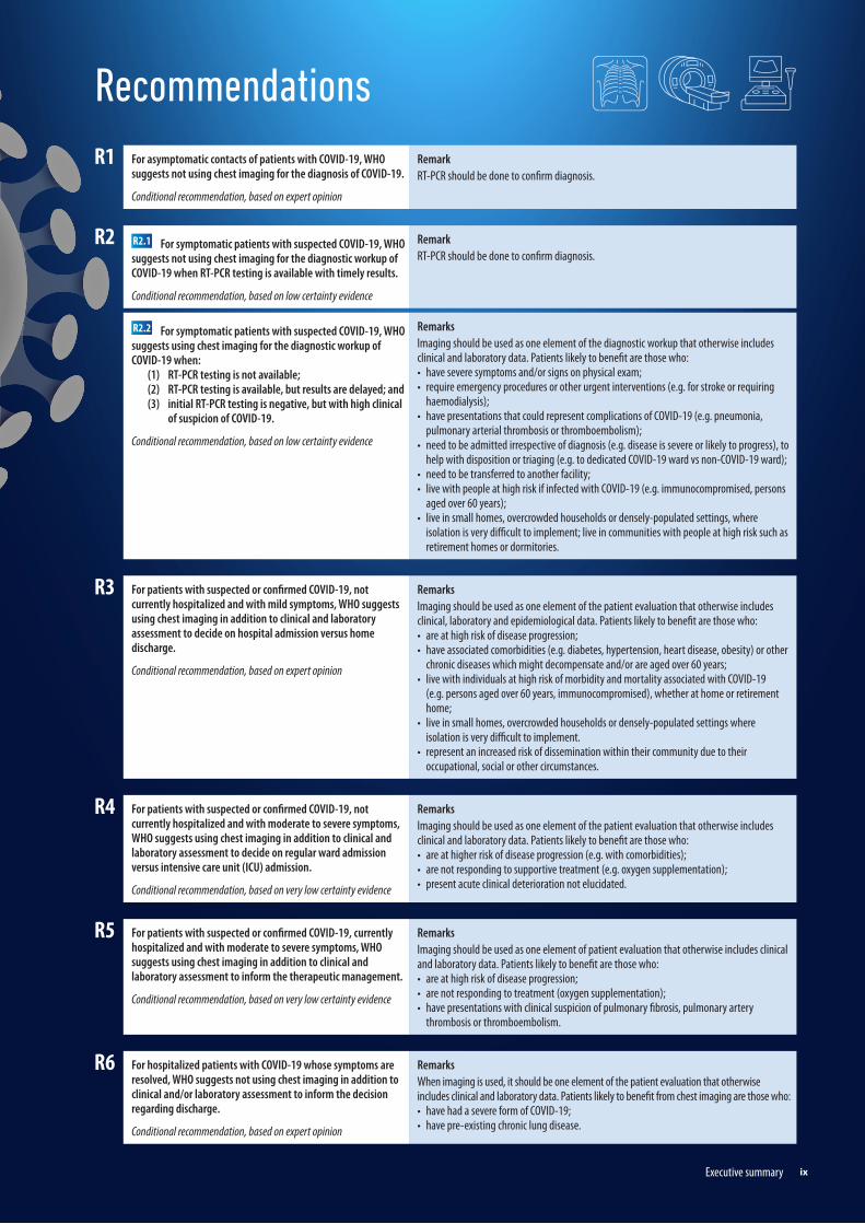
RecommendationsR1 For asymptomatic contacts of patients with COVID-19, WHO
suggests not using chest imaging for the diagnosis of COVID-19.
Conditional recommendation, based on expert opinion
RemarkRT-PCR should be done to confirm diagnosis.
R2 R2.1 For symptomatic patients with suspected COVID-19, WHO suggests not using chest imaging for the diagnostic workup of COVID-19 when RT-PCR testing is available with timely results.
Conditional recommendation, based on low certainty evidence
RemarkRT-PCR should be done to confirm diagnosis.
R2.2 For symptomatic patients with suspected COVID-19, WHO suggests using chest imaging for the diagnostic workup of COVID-19 when:
(1) RT-PCR testing is not available; (2) RT-PCR testing is available, but results are delayed; and (3) initial RT-PCR testing is negative, but with high clinical
of suspicion of COVID-19.
Conditional recommendation, based on low certainty evidence
RemarksImaging should be used as one element of the diagnostic workup that otherwise includes clinical and laboratory data. Patients likely to benefit are those who:• have severe symptoms and/or signs on physical exam;• require emergency procedures or other urgent interventions (e.g. for stroke or requiring
haemodialysis);• have presentations that could represent complications of COVID-19 (e.g. pneumonia,
pulmonary arterial thrombosis or thromboembolism); • need to be admitted irrespective of diagnosis (e.g. disease is severe or likely to progress), to
help with disposition or triaging (e.g. to dedicated COVID-19 ward vs non-COVID-19 ward);• need to be transferred to another facility;• live with people at high risk if infected with COVID-19 (e.g. immunocompromised, persons
aged over 60 years);• live in small homes, overcrowded households or densely-populated settings, where
isolation is very difficult to implement; live in communities with people at high risk such as retirement homes or dormitories.
R3 For patients with suspected or confirmed COVID-19, not currently hospitalized and with mild symptoms, WHO suggests using chest imaging in addition to clinical and laboratory assessment to decide on hospital admission versus home discharge.
Conditional recommendation, based on expert opinion
RemarksImaging should be used as one element of the patient evaluation that otherwise includes clinical, laboratory and epidemiological data. Patients likely to benefit are those who: • are at high risk of disease progression;• have associated comorbidities (e.g. diabetes, hypertension, heart disease, obesity) or other
chronic diseases which might decompensate and/or are aged over 60 years;• live with individuals at high risk of morbidity and mortality associated with COVID-19
(e.g. persons aged over 60 years, immunocompromised), whether at home or retirement home;
• live in small homes, overcrowded households or densely-populated settings where isolation is very difficult to implement.
• represent an increased risk of dissemination within their community due to their occupational, social or other circumstances.
R4 For patients with suspected or confirmed COVID-19, not currently hospitalized and with moderate to severe symptoms, WHO suggests using chest imaging in addition to clinical and laboratory assessment to decide on regular ward admission versus intensive care unit (ICU) admission.
Conditional recommendation, based on very low certainty evidence
RemarksImaging should be used as one element of the patient evaluation that otherwise includes clinical and laboratory data. Patients likely to benefit are those who:• are at higher risk of disease progression (e.g. with comorbidities);• are not responding to supportive treatment (e.g. oxygen supplementation);• present acute clinical deterioration not elucidated.
R5 For patients with suspected or confirmed COVID-19, currently hospitalized and with moderate to severe symptoms, WHO suggests using chest imaging in addition to clinical and laboratory assessment to inform the therapeutic management.
Conditional recommendation, based on very low certainty evidence
RemarksImaging should be used as one element of patient evaluation that otherwise includes clinical and laboratory data. Patients likely to benefit are those who:• are at high risk of disease progression;• are not responding to treatment (oxygen supplementation);• have presentations with clinical suspicion of pulmonary fibrosis, pulmonary artery
thrombosis or thromboembolism.
R6 For hospitalized patients with COVID-19 whose symptoms are resolved, WHO suggests not using chest imaging in addition to clinical and/or laboratory assessment to inform the decision regarding discharge.
Conditional recommendation, based on expert opinion
RemarksWhen imaging is used, it should be one element of the patient evaluation that otherwise includes clinical and laboratory data. Patients likely to benefit from chest imaging are those who:• have had a severe form of COVID-19;• have pre-existing chronic lung disease.
ixExecutive summary

1. Introduction1.1 BackgroundThe World Health Organization (WHO) developed this rapid advice guide on the use of medical imaging in the context of the COVID-19 pandemic. A cluster of pneumonia cases in Wuhan, China was first reported to the WHO Country Office in China on 31 December 2019 (1). Soon thereafter, a novel coronavirus was identified as the causative agent (2–4). This virus was named severe acute respiratory syndrome coronavirus-2 (SARS-CoV-2) and the associated disease was named coronavirus disease 2019 (COVID-19) (5). Since December 2019, COVID-19 has rapidly spread from Wuhan to other parts of China and throughout the world. On 30 January 2020, WHO declared the outbreak a public health emergency of international concern (6) and on 11 March 2020, WHO characterized the outbreak as a pandemic (7).
COVID-19 manifests with non-specific respiratory symptoms of variable severity, ranging from mild to life threatening, which may demand advanced respiratory assistance and artificial ventilation. The diagnosis of COVID-19 is currently confirmed by identification of viral RNA in reverse transcriptase polymerase chain reaction (RT-PCR). In settings where laboratory testing (RT-PCR) is not available or results are delayed or are initially negative in the presence of symptoms attributable to COVID-19, chest imaging has been considered as part of the diagnostic workup of patients with suspected or probable COVID-19 (8). Imaging has been also considered to complement clinical evaluation and laboratory parameters in the management of patients already diagnosed with COVID-19 (9).
Several Member States requested advice from WHO on the role of chest imaging for the diagnostic workup of patients with suspected or probable COVID-19 disease and to inform clinical management of COVID-19. Important variations in imaging practices related to COVID-19 across the world have been highlighted in a recent survey conducted by the International Society of Radiology and the European Society of Radiology. In response to this, WHO undertook the development of this rapid advice guide.
1.2 PurposeTo support Member States in their response to the COVID-19 pandemic this rapid advice guide provides up-to-date guidance on use of chest imaging in patients with suspected or confirmed COVID-19. This guide is also expected to promote the quality and safety of radiation use in health facilities, thus enhancing protection and safety of patients and health workers. It is not intended to replace clinical judgment or specialist consultation but rather to support care providers for the clinical management of these patients.
1.3 ScopeThis document contains recommendations for the use of chest imaging in acute care of adult1 patients with COVID-19, including chest radiography, computed tomography (CT) and lung ultrasound. It is intended to
1 While the recommendations apply to adult patients, some considerations about chest imaging in children are included in this guide.
11. Introduction

be a practical guide for health care professionals involved in the care pathway of patients with suspected, probable or confirmed COVID-19, from outpatient facility or hospital entry to home discharge. The guidance is provided for patients with different levels of disease severity, from asymptomatic individuals to critically ill patients. The document is structured around key questions relevant to the various clinical stages of the disease and different clinical scenarios. Additional guidance on infection prevention and control in medical imaging procedures for COVID-19 management is provided in Annex 1. Infection prevention and control measures include both general measures for all imaging procedures and specific precautions for chest radiography, chest CT and lung ultrasound. Imaging of other body sites (e.g. brain, heart, abdomen, kidney) and imaging follow-up of discharged patients with COVID-19 (e.g. pulmonary fibrosis and other sequelae) are outside of the scope of this guide.
1.4 Clinical perspective and health care settingsA variety of chest imaging findings have been described in patients with COVID-19. Imaging could be useful for the diagnostic workup of patients with suspected COVID-19 and for the management of patients diagnosed with COVID-19.
This guide provides recommendations on imaging procedures and, when relevant, considers different levels of COVID-19 probability (Table 1) and disease severity (Table 2). It also provides implementation considerations for different resource settings, within and across low- and middle-income countries as well as high-income countries.
Table 1. COVID-19 infection probability and case definitions1
Contact A person who experienced any one of the following exposures from 2 days before to 14 days after the onset of symptoms of a probable or confirmed case: (1) face-to-face contact with a probable or confirmed case within 1 meter and for more than 15 minutes; (2) direct physical contact with a probable or confirmed case; (3) direct care for a patient with probable or confirmed COVID-19 disease without using proper personal protective
equipment; OR (4) other situations as indicated by local risk assessments (for confirmed asymptomatic cases, the period of contact
is measured as the 2 days before through the 14 days after the date on which the sample was taken which led to confirmation).
Suspected case (A) A patient with acute respiratory illness (fever and at least one sign/symptom of respiratory disease, e.g. cough, shortness of breath), AND a history of travel to or residence in a location reporting community transmission of COVID-19 disease during the 14 days prior to symptom onset; OR
(B) A patient with any acute respiratory illness AND having been in contact with a confirmed or probable COVID-19 case in the past 14 days prior to symptom onset; OR
(C) A patient with severe acute respiratory illness (fever and at least one sign/symptom of respiratory disease, e.g. cough, shortness of breath – AND requiring hospitalization) in the absence of an alternative diagnosis that fully explains the clinical presentation.
Confirmed case A person with laboratory confirmation of COVID-19 infection, irrespective of clinical signs and symptoms.
1 See the WHO website for the most up-to-date case definitions: https://www.who.int/emergencies/diseases/novel-coronavirus-2019/technical-guidance.
Use of chest imaging in COVID-19: a rapid advice guide2
To support the implementation of the recommendations, consideration was given to various risk factors for disease progression, such as age over 60 years (increasing with age), comorbidities (e.g. hypertension, cardiovascular disease, cerebrovascular disease, cancer, diabetes, obesity, chronic pulmonary disease, tuberculosis), immunosuppressive conditions (e.g. HIV/AIDS), smoking and special groups (pregnancy, children). Additional implementation considerations include the availability of human resources (health workforce and qualified staff) and physical resources (personal protective equipment and other infection prevention and control measures, laboratory testing, hospital beds and imaging equipment/devices).
1.5 Target audienceThis document is primarily intended for health professionals working in emergency departments, imaging departments, clinical departments, intensive care units (ICUs) and other health care settings involved in the diagnosis of COVID-19 and in the management of COVID-19 patients. These health professionals include clinicians, radiologists, radiographers, sonographers, nurses and other health care providers. The document can also be useful for hospital managers and planners, policy-makers, hospital architects, biomedical engineers, medical physicists, logistics staff, water/sanitation and infection prevention and control officers. Health authorities and radiation regulators can use the guide to develop specific national standards relevant to COVID-19 outbreak preparedness, readiness and response in different contexts. Finally, it can be useful to funders that wish to donate equipment and devices as well as funding priority research, such as that discussed in Chapter 5.
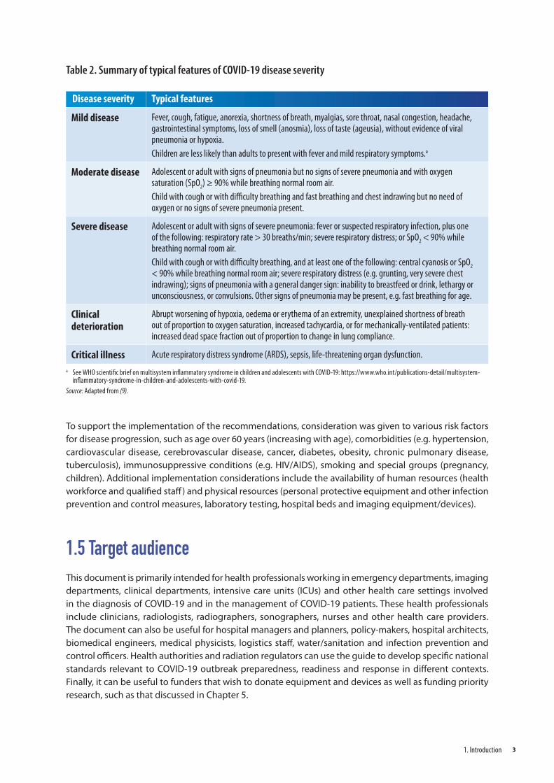
Table 2. Summary of typical features of COVID-19 disease severity
Disease severity Typical features
Mild disease Fever, cough, fatigue, anorexia, shortness of breath, myalgias, sore throat, nasal congestion, headache, gastrointestinal symptoms, loss of smell (anosmia), loss of taste (ageusia), without evidence of viral pneumonia or hypoxia. Children are less likely than adults to present with fever and mild respiratory symptoms.a
Moderate disease Adolescent or adult with signs of pneumonia but no signs of severe pneumonia and with oxygen saturation (SpO2) ≥ 90% while breathing normal room air. Child with cough or with difficulty breathing and fast breathing and chest indrawing but no need of oxygen or no signs of severe pneumonia present.
Severe disease Adolescent or adult with signs of severe pneumonia: fever or suspected respiratory infection, plus one of the following: respiratory rate > 30 breaths/min; severe respiratory distress; or SpO2 < 90% while breathing normal room air. Child with cough or with difficulty breathing, and at least one of the following: central cyanosis or SpO2 < 90% while breathing normal room air; severe respiratory distress (e.g. grunting, very severe chest indrawing); signs of pneumonia with a general danger sign: inability to breastfeed or drink, lethargy or unconsciousness, or convulsions. Other signs of pneumonia may be present, e.g. fast breathing for age.
Clinical deterioration
Abrupt worsening of hypoxia, oedema or erythema of an extremity, unexplained shortness of breath out of proportion to oxygen saturation, increased tachycardia, or for mechanically-ventilated patients: increased dead space fraction out of proportion to change in lung compliance.
Critical illness Acute respiratory distress syndrome (ARDS), sepsis, life-threatening organ dysfunction.a See WHO scientific brief on multisystem inflammatory syndrome in children and adolescents with COVID-19: https://www.who.int/publications-detail/multisystem-
inflammatory-syndrome-in-children-and-adolescents-with-covid-19.Source: Adapted from (9).
31. Introduction
2. Guideline developmentThe development of this rapid advice guide followed the process outlined in the WHO handbook for guideline development (10). Given the nature of the emergency, the process was implemented within a time frame of two months1. The process included identifying priority questions and outcomes, retrieving and synthesizing the evidence, assessing the certainty of evidence, formulating the recommendations, and planning for dissemination and implementation. The guideline development process considered resource use and cost implications of implementing the recommendations from a public health perspective.
2.1 Contributors to the guideIn conformity with the WHO process, the following bodies were established: a WHO steering group, a guideline development group (GDG) and an external review group. In addition, a systematic review team was contracted to conduct a rapid systematic review of the evidence and a core group oversaw the prompt management of the project. The members of the different groups are listed in Annex 2, which also includes a list of contributors to the development of the guidance on infection prevention and control provided in the Annex 1.
WHO steering groupThe WHO steering group was composed of relevant staff members from WHO headquarters, including from the departments of Environment, Climate Change and Health (ECH), Maternal, Newborn, Child and Adolescent Health and Ageing (MCA), Integrated Health Services (IHS), Health Care Readiness (HCR), Emerging Diseases and Zoonoses (EZD), Health Product Policy and Standards (HPS), Business Relationship Management (BRM), as well as the Regional Advisor on Radiological Health in the WHO Regional Office for the Americas. The WHO steering group helped identify the GDG and external review group members. It contributed to the formulation of the key questions and reviewed the recommendations and the final document.
Guideline development groupThe GDG included experts and relevant stakeholders from multiple disciplines: a guideline methodologist, experts in the field of medical imaging, emergency medicine, intensive care, pulmonology and molecular diagnostics, as well as a representative from a patient advocacy organization. The GDG provided input at all stages of the process and played the main role in development of recommendations. The composition of the GDG ensured geographic representation from five of the six WHO regions, gender balance and absence of conflicts of interest.
External review groupThe external review group was composed of experts in the field of medical imaging and pulmonary diseases, and representatives of patient advocacy groups and civil society. The experts reviewed the
1 Reports on the use of chest imaging published shortly after the COVID-19 outbreak were reviewed during February 2020; preliminary project scoping was done in early March; Member State requests for advice on use of chest imaging were received from mid-March; steering group established on 19 March; establishment of the guideline development group (GDG) and scoping meeting occurred on 27 March; rapid reviews conducted between 13 April and 1 May; GRADEpro webinar occurred on 30 April; five consecutive GDG working meetings were held between 1 May and 8 May; peer review of the draft recommendations occurred from 6 to 19 May until final draft was submitted for executive clearance on 24 May 2020 (total of 67 days: 19 March to 24 May).
Use of chest imaging in COVID-19: a rapid advice guide4

recommendations developed by the GDG and the final document, and commented on the technical accuracy, clarity of language, contextual issues and implications for implementation. The group was asked not to modify the recommendations that were formulated by the GDG.
Systematic review teamThe systematic review team was composed of experts in the field of systematic reviews with clinical background in internal medicine and content experts in the field of medical imaging. They conducted rapid reviews of the literature and provided a report summarizing the findings and certainty of evidence for each key question (Section 2.3). The systematic review report was shared with members of the GDG. Representatives of the systematic review team attended the GDG meetings to provide an overview of the available evidence and to respond to technical queries from the GDG (11, 12).
Core groupThe development of these recommendations under very compressed timelines during the COVID-19 pandemic represented a challenge in the context of unprecedented demands in terms of global and local public health response. Anticipating this challenge, the WHO Secretariat assembled a core group to assist in project management. This group included two methodologists, the chairperson of the GDG and a radiology consultant who worked in close consultation with the WHO Secretariat and participated in daily planning and coordination meetings held virtually. The core group drafted the key questions using the “population, intervention, comparator and outcome” (PICO) format, supervised the syntheses and retrieval of evidence, convened and facilitated the GDG meetings, liaised with all established groups, and drafted and finalized the rapid advice guide. In addition, the core group facilitated survey implementation and assessment of current imaging practices in different regions of the world.
2.2 Management of declaration of interestsThe disclosure and appropriate management of relevant financial and non-financial conflicts of interest of GDG members and other external experts and contributors is a critical part of guideline development at WHO. According to WHO regulations, all experts must declare their interests prior to participation in WHO guideline development processes and meetings. All GDG members were therefore required to complete a standard WHO declaration of interests form before engaging in the guideline development process. All declarations were reviewed before finalizing the experts’ invitations to participate based on the criteria for assessing the severity of conflicts of interest as outlined in the WHO handbook for guideline development (10) to all participating experts. All findings from the declaration of interests forms received were managed in accordance with the relevant WHO guidelines on a case-by-case basis and communicated to the experts at the start of the first GDG meeting. Annex 3 provides a summary of the declaration of interests and how conflicts of interest declared by invited experts were managed.
2.3 Identification of the key questionsThe core group performed a rapid search for formal consensus statements on the use of chest imaging in COVID-19 management from professional bodies and/or national health authorities, with the assistance of the GDG and the International Society of Radiology. These statements were used to inform the development of the key questions. The core group formulated the key questions in PICO format, with the help of the steering group, the GDG and the systematic review team. These key questions formed the basis of the systematic reviews and of the development of recommendations.
52. Guideline development
The following seven key PICO questions were identified.
1. In asymptomatic contacts of patients with COVID-19, and in contexts where laboratory testing (RT-PCR) is not available/results are delayed/results are initially negative, should chest imaging (including chest radiography, CT scan, lung ultrasound) vs no chest imaging be used for the diagnostic workup of COVID-19?
2. In symptomatic patients with suspected COVID-19, and in contexts where laboratory testing (RT-PCR) is not available/results are delayed/results are initially negative, should chest imaging (including chest radiography, CT scan, lung ultrasound) vs no chest imaging be used for the diagnostic workup of COVID-19?
3. In patients with suspected or confirmed COVID-19, not currently hospitalized and with mild symptoms, should chest imaging (including chest radiography, CT scan, lung ultrasound) vs no chest imaging be used to support the decision on hospital admission versus home discharge?
4. In patients with suspected or confirmed COVID-19, not currently hospitalized and exhibiting moderate to severe symptoms, should chest imaging (including chest radiography, CT scan, lung ultrasound) vs no chest imaging be used to support decision on regular ward admission versus ICU admission?
5. In patients with suspected or confirmed COVID-19, currently hospitalized and exhibiting moderate or severe symptoms, should chest imaging (including chest radiography, CT scan, lung ultrasound) vs no chest imaging be used to modify the therapeutic management?
6. In patients with suspected or confirmed COVID-19 and clinical deterioration and/or suspicion of pulmonary embolism, should imaging (including CT pulmonary angiography) vs no imaging be used to diagnose pulmonary embolism?1
7. In patients with COVID-19 whose symptoms are resolved, should chest imaging (including chest radiography, CT scan, lung ultrasound) be added to vs not added to laboratory criteria to support decisions on hospital discharge vs no discharge?
2.4 Identification of the critical outcomesThe core group drafted a list of outcomes relevant for each PICO question. The list included three types of outcomes:
diagnostic accuracy measures (rates of true positive, true negative, false positive, false negative);
clinical outcomes, including the “core outcomes” developed for COVID-19 (Allison Tong, COVID-19 project, personal communication, 24 April 2020) (mortality, respiratory failure, multi-organ failure, shortness of breath, recovery), adverse effects of imaging (e.g. exposure to radiation) and COVID-19 transmission to health care workers;
health systems outcomes, including service use (length of emergency department stay, length of hospital stay, length of ICU stay), availability of care, access to care and quality of care.
1 This PICO question was addressed in the systematic review report (Web Annex A; published exclusively online) and discussed by the GDG. No study evaluated the diagnostic accuracy of imaging (with or without measurement of d-dimer) for diagnosis of pulmonary arterial thrombosis or thromboembolism in patients with suspected or confirmed COVID-19. Therefore, no recommendation was developed, and the topic was included in the list of research priorities (see Chapter 5).
Use of chest imaging in COVID-19: a rapid advice guide6
The list of outcomes was circulated to the GDG which scored the importance of each outcome on a scale of 1 to 9 (1–3: not important; 4–6: important; and 7–9: critical). The average score for each outcome was used to prioritize the outcomes for each PICO question. The outcomes selected for each question and the scores assessing their importance are included in the evidence-to-decision tables presented in Web Annex B.
2.5 Evidence identification and retrieval, quality assessment and synthesis of evidenceThe systematic review team performed a rapid review of the scientific literature to inform the development of the rapid guidance on the use of chest imaging for patients with COVID-19 (Web Annex A). The core group reviewed and provided input into the protocol and worked closely with the systematic review team to ensure the output of the systematic review met the needs of the guidance development process. The systematic review team produced a table summarizing the evidence and its certainty using the Grading of Recommendations Assessment, Development and Evaluation (GRADE) methodology, for each PICO question (11). The lead author on the systematic review team attended the GDG meetings to provide a summary of the available evidence for each question and to respond to technical queries from GDG members.
According to the GRADE methodology, the certainty of evidence is categorized into “high”, “moderate”, “low” and “very low”. The judgment of certainty is based on the study design, factors that lower the certainty of evidence (risk of bias, indirectness, inconsistency, imprecision, publication bias) and factors that increase the certainty of evidence (12).
A thorough search was initially performed up to 15 April 2020, with subsequent literature surveillance through 29 April 2020. Prior to publication of this guide, the systematic review team updated their search up to 28 May 2020. The systematic review team assessed whether, and to what extent, the newly identified studies modified the body of evidence for each question and judged that the newly identified studies did not impact the main conclusions of their initial review or the certainty of evidence (Web Annex A). Taking this into consideration the core group decided there was no substantial evidence to warrant a reconsideration of the originally drafted recommendations, which were therefore not revised.
2.6 Stakeholder surveyThe core group conducted an online cross-sectional survey of stakeholders asking them to rate (i) the importance of the outcomes and (ii) their views on the acceptability, feasibility, impact on equity and resource use of the relevant chest imaging modalities (chest radiography, chest CT and lung ultrasound) in the different clinical scenarios. The survey was developed by the methodologists at the American University of Beirut, and widely disseminated by the WHO Secretariat with the assistance of the steering group, WHO collaborating centres on radiation and health, and relevant nongovernmental organizations, which have official relationships with WHO. A total of 249 respondents from all WHO regions, including patients and the public, health care workers (i.e. clinicians, radiologists, radiographers/radiological technologists, medical physicists and others), regulators, policy-makers and researchers participated in the survey over a period of five days. A summary of the results of this survey for each PICO question has been included in the evidence-to-decision tables provided in Web Annex B.
72. Guideline development
2.7 Additional dataInformation about the use of chest imaging in patients with suspected, probable or confirmed COVID-19 around the world was gathered at the beginning of the project to assess current imaging practices and identify clinical scenarios for which global guidance was most needed.
Existing guidance on use of chest imaging in patients with COVID-19 was reviewed and summarized. The following eligibility criteria were adopted: national or international/multinational formal consensus statements on use of chest imaging, established for the management of the COVID-19 pandemic, and developed or endorsed by national or international professional societies and/or health authorities. A total of 33 guidance documents from 22 organizations from all WHO regions1 were identified.
A survey conducted by the International Society of Radiology and the European Society Radiology on current imaging practices in the management of COVID-19 received responses from 52 imaging services from 31 countries representing all WHO regions2. The information collected helped to understand current practice heterogeneities and to identify relevant scenarios to formulate the research questions.
2.8 Formulation of the recommendationsOnce the evidence had been identified and synthesized and its quality assessed, the GDG was tasked with formulating the recommendations based on evidence. GRADE provides a framework to accomplish this task, with explicit consideration of specific factors that may affect the direction and strength of each recommendation. The direction (whether “in favour of” or “against” an intervention) and strength (whether “conditional” or “strong”) of the recommendations reflects the GDG’s degree of confidence as to whether the desirable effects of the intervention being considered outweigh the undesirable effects. Table 3 provides the interpretation of strong and conditional recommendations from the perspectives of patients, clinicians and policy-makers.
Table 3. Interpretation of the strength of recommendations for different stakeholders
Strong recommendation Conditional recommendation
Patients Most individuals in this situation would want the recommended course of action; only a small proportion would not.
Most individuals in this situation would want the suggested course of action, but many would not.
Clinicians Most patients should receive the recommended course of action.
Be prepared to help patients to make a decision that is consistent with their own values.
Policy-makers The recommendation can be adopted as a policy in most situations.
Policy-making will require substantial debate and involvement of various stakeholders.
Due to the COVID-19-related lockdown measures in most countries during the development of the rapid advice guide, a physical meeting of the GDG could not be held. Therefore, the members of the GDG were invited to attend a series of five online meetings of around 2 hours each (30 April, 4 May, 5 May, 7 May, 8 May 2020). The first meeting was dedicated to introducing the project and its process. The four subsequent meetings were devoted to formulating the recommendations.
1 46% from the European Region, 32% from the Region of the Americas, 7% from the Western Pacific Region, 7% from the Eastern Mediterranean Region, 4% from the South-East Asia Region, and 4% from multiregional organizations that are based in the African Region and elsewhere in the world.
2 Region of the Americas: 10 services from 2 countries; the African Region: 8 services from 4 countries; the Eastern Mediterranean Region: 3 services from 3 countries: the South-East Asia Region: 1 service from 1 country: the Western Pacific Region: 7 services from 5 countries; and the European Region: 23 services from 16 countries.
Use of chest imaging in COVID-19: a rapid advice guide8
The methodologists developed an evidence-to-decision table for each PICO question using the GRADEpro software. Each table includes sections on the following criteria: benefits and harms, the certainty of the evidence, values and preferences, resource use, equity, acceptability and feasibility (13,14). The tables were pre-populated with the summary of evidence provided in the systematic review report (Web Annex A), and the results of the stakeholders’ survey (Web Annex B).
The GDG developed the recommendations based on the PICO questions, and used the evidence-to-decision tables to guide discussions (15). For each PICO question, the GDG reviewed the information pre-populated in the evidence-to-decision tables. First, the systematic review team leader presented the evidence identified by the systematic review. Then the lead methodologist discussed the interpretation of the evidence with the GDG. Next, the methodologist in charge of the stakeholders’ survey on acceptability, feasibility, impact on equity and resource use of each of the three chest imaging modalities presented the survey results to the GDG.
The GDG then contributed “additional considerations” for each of the evidence-to-decision criteria, which were included in the evidence-to-decision tables (Web Annex B).
The GDG voted on each of the evidence-to-decision factors, then on the direction and strength of the recommendation using an online voting tool (menti.com). The voting results served as the starting point for building consensus. None of the GDG members expressed opposition to the final strength or direction of any of the recommendations. When the systematic review identified no relevant evidence for the PICO question, the recommendation was stated as “based on expert opinion”.
The GDG also contributed remarks and implementation considerations for each of the recommendations. After the meetings, the core group circulated the draft recommendations and the accompanying remarks and implementation considerations to the GDG and the external review group for feedback prior to incorporation into the final version of the rapid advice guide.
2.9 Document preparation and reviewPrior to the online meetings, the core group shared relevant documents and supporting materials with the GDG by email and through shared folders online. Following the virtual meetings, the core group first shared the draft recommendations with the GDG to ascertain that they clearly and accurately reflected the deliberations and decisions made. At that point, the recommendations and remarks were also shared with the steering group and the external review group for their review and input.
In a second step, the core group prepared a full draft of the rapid advice guide. The draft document was sent to the GDG, the steering group and the external review group for review, and then finalized based on the feedback received. Further modifications made to the document consisted only of addition of the updated review of available evidence, corrections of factual errors and language editing to improve clarity. The final draft was professionally edited for clearance and publication.
2.10 Update of the guideThese recommendations have been produced in response to the COVID-19 pandemic. WHO will closely monitor emerging data on relevant topics addressed in this rapid advice guide, which will be updated within the next six months if warranted by evidence. The Radiation and Health Unit in the Department of Environment, Climate Change and Health at WHO headquarters in Geneva will be responsible for the update as appropriate.
92. Guideline development

3. RecommendationsThis chapter presents the recommendations the guideline development group (GDG) developed to answer the “population, intervention, comparator and outcome” PICO questions on the use of chest imaging in the diagnostic workup and clinical management of patients with COVID-19 for different clinical scenarios (including contacts, suspected or confirmed cases). All developed recommendations are conditional, which means that the desirable effects likely outweigh the undesirable effects under certain conditions, some of which are summarized in the remarks following each recommendation. The conditions reflect what the GDG discussed as important to optimizing the benefits of the intervention under consideration. This chapter also provides consideration about implementation of the recommendations. The implementation considerations reflect what the GDG discussed as important for the intervention to translate into the expected benefits when implemented. Membership of the GDG and the external review group included experts from 10 high-income countries and 14 low- and middle-income countries who developed and/or reviewed the implementation considerations linked to each recommendation. They provided comments reflecting the variability of resource settings within and between countries. Availability of resources when choosing the imaging modalities, particularly in low-resource settings and in low- and middle-income countries, was a recurrent theme in the discussion of the different recommendations. Accordingly, this issue was discussed for all recommendations, including its effect on their implementation. Each recommendation is followed by a succinct summary of the supporting evidence. More detailed information is provided in the systematic review report in Web Annex A. The recommendations should be read alongside the remarks and implementation considerations that follow each recommendation.
Use of chest imaging in COVID-19: a rapid advice guide10
3.1 Recommendation 1
R1 For asymptomatic contacts of patients with COVID-19, WHO suggests not using chest imaging for the diagnosis of COVID-19.
Conditional recommendation, based on expert opinion
Remark
RT-PCR should be done to confirm diagnosis of COVID-19.
EvidenceThe systematic review identified no eligible study evaluating the diagnostic accuracy of imaging in asymptomatic contacts of patients with COVID-19.
Implementation considerations1. Consider whether RT-PCR is available and, if the test is performed, whether the results are positive or
negative.2. Consider the use of chest imaging in asymptomatic contacts who progress to develop respiratory
symptoms (body temperature monitoring).3. Consider assessing incidental pulmonary findings suspicious of COVID-19 on imaging performed for
other reasons (e.g. thoracic spine radiography, cardiac CT) in countries/regions with previous or current high COVID-19 prevalence.
113. Recommendations
3.2 Recommendation 2
R2.2 For symptomatic patients with suspected COVID-19, WHO suggests using chest imaging for the diagnostic workup of COVID-19 when: (1) RT-PCR testing is not available; (2) RT-PCR testing is available, but results are delayed; and (3) initial RT-PCR testing is negative, but with high clinical suspicion of COVID-19.
Conditional recommendation, based on low certainty evidence
Remarks
Imaging should be used as one element of the diagnostic workup that otherwise includes clinical and laboratory data. Patients likely to benefit from chest imaging are those who:• have severe symptoms and/or signs on physical exam;• require emergency procedures or other urgent interventions (e.g. for stroke or
requiring haemodialysis);• have presentations that could represent complications of COVID-19 (e.g.
pneumonia, pulmonary arterial thrombosis or thromboembolism); • need to be admitted irrespective of diagnosis (e.g. disease is severe or likely to
progress), to help with disposition or triaging (e.g. to dedicated COVID-19 ward vs non-COVID-19 ward);
• need to be transferred to another facility;• live with people at high risk if infected with COVID-19 (e.g.
immunocompromised, persons aged over 60 years);• live in small homes, overcrowded households or densely-populated settings,
where isolation is very difficult to implement;• live in communities with people at high risk such as retirement homes or
dormitories. When choosing the imaging modalities, consider the following.• Compared to chest CT, chest radiography appears to have lower sensitivity and
might have higher specificity. Chest radiography is less-resource intensive, is associated with lower radiation doses, is easier to repeat sequentially for monitoring disease progression, and can be performed with portable equipment at the point of care (which minimizes the risk of cross-infection related to patient transport).
• Chest CT has a relatively high sensitivity but a relatively low specificity and can be useful in patients with some pre-existing pulmonary diseases. However, the absence of radiological signs of pneumonia cannot completely exclude a viral infection.
• Lung ultrasound has very low-certainty evidence supporting its diagnostic accuracy but might be helpful with the appropriate expertise as a supplemental or alternative modality (e.g. in pregnant women, children). Lung ultrasound can be done at the point of care but requires closer physical proximity of the operator to the patient for a longer period and requires specific infection prevention and control precautions.
• The differential diagnoses and potential complications for each specific case (e.g. CT angiography for pulmonary thromboembolism, ultrasound for pleural effusions and heart conditions) should be considered when choosing imaging modality.
• Choice should be made through shared decision-making involving the referring physician, the radiologist and the patient whenever possible. If feasible, the patient should be provided with information regarding the imaging modality to be used and the likelihood of requiring subsequent imaging procedures.
R2.1 For symptomatic patients with suspected COVID-19, WHO suggests not using chest imaging for the diagnostic workup of COVID-19 when RT-PCR testing is available with timely results.
Conditional recommendation, based on low certainty evidence
Remark
RT-PCR should be done to confirm diagnosis of COVID-19.
Use of chest imaging in COVID-19: a rapid advice guide12
EvidenceThe systematic review (Web Annex A) identified 23 studies that evaluated the diagnostic accuracy of three imaging modalities in symptomatic patients with suspected COVID-19, against a reference standard (Web Annex A), chest radiography (n=3), chest CT (n=19) and lung ultrasound (n=1). None of these studies compared two imaging modalities against each other.
The systematic review team judged those studies to be at either high risk of bias (n=17) or moderate risk of bias (n=6). The studies provided limited information regarding clinical presentation (e.g. the severity of symptoms at presentation) and few reported specific criteria for a positive imaging test for COVID-19. Eleven studies did not describe a reference standard to diagnose COVID-19 that included serial RT-PCR or clinical follow-up. The median sensitivity and specificity reported by the included studies were 0.64 and 0.82 for chest radiography; 0.92 and 0.56 for chest CT; and 0.95 and 0.83 for lung ultrasound. The systematic review team judged the certainty of this evidence to be low for chest radiography, chest CT and lung ultrasound. The corresponding evidence-to-decision table available in Web Annex B provides the counts for true positives, true negatives, false positives and false negatives for four hypothetical prevalence values of COVID-19 infection, which were assumed to be 20%, 40%, 60% and 80% among symptomatic patients with suspected COVID-19.
The update of the review conducted before the publication of the guide identified five new studies that evaluated the diagnostic accuracy of chest radiography, chest CT and lung ultrasound in symptomatic patients with suspected COVID-19. The synthesized evidence as well as its associated certainty was judged to remain unchanged (Web Annex A).
Implementation considerations1. Implement the recommendations based on your equipment availability. Consider the resources
needed (budget, health workforce, personal protective equipment, imaging equipment), the need to adapt the clinical workflow and the need to deprioritize other indications for imaging.
2. Consider the use of locally-developed flow charts, infographics and other decision-support tools to facilitate implementation.
3. Bear in mind that recommendations for imaging depend on severity of symptoms and that chest imaging is an essential investigation in those who develop respiratory symptoms or hypoxia.
4. Monitor respiratory symptoms and physical exam findings to guide timing of chest imaging.5. Consider the use of portable equipment for performing chest radiography at the point of care. In the
case of home health care, combine chest radiography and/or lung ultrasound by portable equipment with RT-PCR testing.
6. Mitigate the risk of infection transmission to health care workers and to other patients associated with patient transport to the imaging department (e.g. use of point of care imaging such as portable equipment). (See infection prevention and control precautions in Annex 1.)
7. Consider the possibility of false negative imaging results in patients for whom chest imaging indicates no findings suspicious of COVID-19 (particularly during the first 2 days after symptom onset).a. If discharged from the emergency department or other outpatient assessment setting, patients
need to abide by the local public health measures (e.g. quarantine, social distancing) until definitive RT-PCR diagnosis is made.
b. If the patient is admitted, health care workers need to consider appropriate clinical precautions until definitive RT-PCR diagnosis is made.
8. When performing chest radiography and chest CT, minimize radiation dose while maintaining diagnostic image quality (e.g. low-dose scanning protocols) and use digital imaging rather than film-screen equipment (16).
133. Recommendations
9. Consider the potential harms from exposure to ionizing radiation, in particular for pregnant women and children.
10. Ensure proper use of personal protective equipment by health care workers and proper disinfection of equipment and devices (see Annex 1).
11. Provide appropriate training of radiologists and technologists on infection prevention and control practices and ensure efficient management of typical imaging findings of COVID-19 through accepted local protocols.
12. Consider the transfer of images for remote reporting (teleradiology) as needed (e.g. settings where radiologists are not available for on-site reporting).
13. Provide information to patients about safety provisions adopted by the facility for infection prevention and control (see Annex 1) as well as for radiation protection (16).
14. Make provisions to ensure that all patients get the imaging services they need without suffering financial hardship.
Use of chest imaging in COVID-19: a rapid advice guide14
3.3 Recommendation 3
R3 For patients with suspected or confirmed COVID-19, not currently hospitalized and with mild symptoms, WHO suggests using chest imaging in addition to clinical and laboratory assessment to decide on hospital admission versus home discharge.
Conditional recommendation, based on expert opinion
Remarks
Imaging should be used as one element of the patient evaluation that otherwise includes clinical, laboratory and epidemiological data. Patients likely to benefit are those who: • are at high risk of disease progression;• have associated comorbidities (e.g. diabetes, hypertension, heart disease,
obesity) or other chronic diseases which might decompensate and/or are aged over 60 years;
• live with individuals at high risk of morbidity and mortality associated with COVID-19 (e.g. persons aged over 60 years, immunocompromised), whether at home or retirement home;
• live in small homes, overcrowded households or densely-populated settings where isolation is very difficult to implement.
• represent an increased risk of dissemination within their community due to their occupational, social or other circumstances.
When choosing the imaging modalities, consider the following.• Compared to chest CT, chest radiography appears to have lower sensitivity
and might have higher specificity. Chest radiography is less resource intensive, is associated with lower radiation doses, is easier to repeat sequentially for monitoring disease progression, and can be performed with portable equipment at the point of care (which minimizes the risk of cross-infection related to patient transport).
• Chest CT has a relatively high sensitivity but a relatively low specificity and can be useful in patients with some pre-existing pulmonary diseases.
• Lung ultrasound has very low-certainty evidence supporting its diagnostic accuracy but might be helpful with the appropriate expertise as a supplemental or alternative modality (e.g. in pregnant women, children). Lung ultrasound can be done at the point of care but requires closer physical proximity of the operator to the patient for a longer period and requires specific infection prevention and control precautions.
• The differential diagnoses and potential complications for each specific case (e.g. CT angiography for pulmonary arterial thrombosis or thromboembolism, ultrasound for pleural effusions and heart conditions) should be considered when choosing imaging modality.
• Choice should be made through shared decision-making involving the referring physician, the radiologist and the patient whenever possible. If feasible, the patient should be provided with information regarding the imaging modality to be used and the likelihood of requiring subsequent imaging procedures.
• When there is a clinical deterioration, the systemic aspect of COVID-19 should be considered, in particular heart, brain, kidney and gastrointestinal localizations.
153. Recommendations
EvidenceThe systematic review identified no eligible study that evaluated any chest imaging modality in patients with suspected or confirmed COVID-19 not yet hospitalized to support decisions on hospital admission versus home discharge on health outcomes.
Implementation considerations1. Implement the recommendations based on your equipment availability. Consider the resources
needed (budget, health workforce, personal protective equipment, imaging equipment), the need to adapt the clinical workflow and the need to deprioritize other indications for imaging.
2. Consider performing RT-PCR tests of suspected cases within 24 hours and implement precautions until results are available.
3. Consider that home isolation may not be feasible in certain settings (e.g. overcrowded households, densely-populated cities).
4. If available, low-dose CT can be performed on adult patients. For paediatric patients, chest radiography would be favoured.
5. Consider the potential harms from exposure to ionizing radiation, in particular for pregnant women and children.
6. Favour the use of portable equipment for performing chest imaging in isolated rooms in the emergency department.
7. Consider the possibility of false negative imaging results in patients for whom chest imaging indicates no findings suspicious of COVID-19 (particularly during the first 2 days after symptom onset).a. If discharged from the emergency department or other outpatient assessment setting, patients
need to abide by the local public health measures (e.g. quarantine, social distancing) until definitive RT-PCR diagnosis is made.
b. If the patient is admitted, health care workers need to consider appropriate clinical precautions until a definitive RT-PCR diagnosis is made.
8. When performing chest radiography and chest CT, minimize radiation dose while maintaining diagnostic image quality (e.g. low-dose scanning protocols), and use digital imaging rather than film-screen equipment (16).
9. When performing chest radiography, consider using portable equipment, and if feasible, a unit dedicated to patients with COVID-19.
10. Ensure proper use of personal protective equipment by health care workers and proper disinfection of equipment and devices (see Annex 1).
11. Provide appropriate training of radiologists and technologists on infection prevention and control practices and ensure efficient management of typical imaging findings of COVID-19 through accepted local protocols.
12. Consider the transfer of images for remote reporting (teleradiology) as needed (e.g. settings where radiologists are not available for on-site reporting).
13. Set policy/pathway for use of imaging related to COVID-19 illustrated with flow charts, infographics and/or other decision-support tools locally developed and accepted.
14. Inform the patient about safety provisions for infection prevention and control (see Annex 1) as well as for radiation protection (16).
15. Make provisions to ensure that all patients get the imaging services they need without suffering financial hardship.
Use of chest imaging in COVID-19: a rapid advice guide16
3.4 Recommendation 4
R4 For patients with suspected or confirmed COVID-19, not currently hospitalized and with moderate to severe symptoms, WHO suggests using chest imaging in addition to clinical and laboratory assessment to decide on regular ward admission versus intensive care unit (ICU) admission.
Conditional recommendation, based on very low certainty evidence
Remarks
Imaging should be used as one element of the patient evaluation that otherwise includes clinical and laboratory data. Patients likely to benefit are those who: • are at higher risk of disease progression (e.g. with comorbidities);• are not responding to supportive treatment (e.g. oxygen supplementation);• present acute clinical deterioration not elucidated.When choosing the imaging modalities, consider the following.• Compared to chest CT, chest radiography appears to have lower sensitivity
and might have higher specificity. Chest radiography is less resource intensive, is associated with lower radiation doses, is easier to repeat sequentially for monitoring disease progression, and can be performed with portable equipment at the point of care (which minimizes the risk of cross-infection related to patient transport).
• Chest CT has a relatively high sensitivity but a relatively low specificity and can be useful in patients with some pre-existing pulmonary diseases. However, the absence of radiological signs of pneumonia cannot completely exclude a viral infection.
• Lung ultrasound has very low-certainty evidence supporting its diagnostic accuracy but might be helpful with the appropriate expertise as a supplemental or alternative modality (e.g. in pregnant women, children, patients on mechanical ventilation). Lung ultrasound can be done at the point of care but requires closer physical proximity of the operator to the patient for a longer period and requires specific infection prevention and control precautions.
• The differential diagnoses and potential complications for each specific case (e.g. CT angiography for pulmonary arterial thrombosis or thromboembolism, ultrasound for pleural effusions and heart conditions) should be considered when choosing imaging modality.
• Choice should be made through shared decision-making involving the referring physician, the radiologist, and the patient whenever possible. If feasible, the patient should be provided with information regarding the imaging modality to be used and the likelihood of requiring subsequent imaging procedures.
• When there is a clinical deterioration, the systemic aspect of COVID-19 should be considered, in particular heart, brain, kidney and gastrointestinal localizations.
173. Recommendations
EvidenceThe systematic review identified no eligible study that evaluated any chest imaging modality in patients with suspected or confirmed COVID-19 not yet hospitalized to support decisions on regular ward admission versus intensive care unit (ICU) admission on health outcomes. The update of the review conducted before the publication of the guide identified one new study that evaluated the use of chest imaging in patients with suspected or confirmed COVID-19 not yet hospitalized (Web Annex A). The certainty of the evidence was judged as very low.
Implementation considerations1. Implement the recommendations based on equipment availability. Consider the resources needed
(budget, health workforce, personal protective equipment, imaging equipment), the need to adapt the clinical workflow, and the need to deprioritize other indications for imaging.
2. If available, low-dose chest CT can support the decision on regular ward admission versus ICU admission. Chest radiographs are preferred for follow-up in regular ward admission. Patients with rapid progression of COVID-19 pneumonia or diffuse lung damage need ICU admission.
3. Consider the possibility of false negative imaging results in patients for whom chest imaging indicates no findings suspicious of COVID-19 (particularly during the first 2 days after symptom onset).
4. Health care workers need to consider appropriate clinical precautions until the definitive RT-PCR diagnosis is made. Ensure proper use of personal protective equipment and proper disinfection of equipment and devices (see Annex 1).
5. When performing chest radiography and chest CT, minimize radiation dose while maintaining diagnostic image quality (e.g. low-dose CT protocols) and use digital imaging rather than film-screen equipment (16).
6. When performing chest radiography, consider using portable equipment, and if feasible, a unit dedicated to patients with COVID-19.
7. Consider the potential harm from exposure to ionizing radiation, in particular for pregnant women and children.
8. Provide appropriate training of radiologists and technologists on infection prevention and control practices and ensure efficient management of typical imaging findings of COVID-19 through accepted local protocols.
9. Consider the transfer of images for remote reporting (teleradiology) as needed (e.g. settings where radiologists are not available for on-site reporting).
10. Set policy/pathway for use of imaging related to COVID-19 illustrated with flow charts or diagrams locally developed and accepted.
11. If clinical condition permits, inform the patient about safety provisions for infection prevention and control (see Annex 1) as well as for radiation protection (16).
12. Make provisions to ensure that all patients get the imaging services they need without suffering financial hardship.
Use of chest imaging in COVID-19: a rapid advice guide18
3.5 Recommendation 5
R5 For patients with suspected or confirmed COVID-19, currently hospitalized and with moderate to severe symptoms, WHO suggests using chest imaging in addition to clinical and laboratory assessment to inform therapeutic management.
Conditional recommendation, based on very low certainty evidence
Remarks
Imaging should be used as one element of patient evaluation that otherwise includes clinical and laboratory data. Patients likely to benefit are those who: • are at high risk of disease progression;• are not responding to treatment (oxygen supplementation);• have presentations with clinical suspicion of pulmonary fibrosis, pulmonary
artery thrombosis or thromboembolismWhen choosing the imaging modalities consider the following.• Compared to chest CT, chest radiography appears to have lower sensitivity
and might have higher specificity. Chest radiography is less resource intensive, is associated with lower radiation doses, is easier to repeat sequentially for monitoring disease progression, and can be performed with portable equipment at the point of care (which minimizes the risk of cross-infection related to patient transport).
• Chest CT has a relatively high sensitivity but a relatively low specificity and can be useful in patients with some pre-existing pulmonary diseases. However, the absence of radiological signs of pneumonia cannot completely exclude a viral infection.
• Lung ultrasound has very low-certainty evidence supporting its diagnostic accuracy but might be helpful with the appropriate expertise as a supplemental or alternative modality (e.g. in pregnant women, children, patients with mechanical ventilation). Ultrasound can be useful when assessing for pleural complications and evaluating the condition of the heart. Lung ultrasound can be done at the point of care but requires closer physical proximity of the operator to the patient for a longer period and requires specific infection prevention and control precautions.
• The differential diagnoses and potential complications for each specific case (e.g. CT angiography for pulmonary artery thrombosis or thromboembolism, lung ultrasound for pleural effusions) should be considered when choosing imaging modality.
• Choice should be made through shared decision-making involving the referring physician, the radiologist and the patient whenever possible. If feasible, the patient should be provided with information regarding the imaging modality to be used and the likelihood of requiring subsequent imaging procedures.
• When there is a clinical deterioration, the systemic aspect of COVID-19 should be considered, in particular heart, brain, kidney and gastrointestinal localizations.
193. Recommendations
EvidenceThe systematic review team identified three studies that evaluated chest imaging in patients currently hospitalized with moderate or severe symptoms and suspected or confirmed COVID-19, for predicting mortality or admission to the ICU. The certainty of evidence was judged to be very low.
Implementation considerations1. Bedside lung ultrasound can be helpful to explain respiratory gas exchange deterioration and to detect
pleural complication in ICU patients. 2. Portable equipment is preferred for follow-up of ICU patients. Bedside chest radiography can be helpful
for dynamic evaluation of COVID-19 pneumonia and its complications. Resolution or progress of lung consolidation seen on a bedside chest radiograph can inform the therapeutic management. Chest imaging can inform management of pneumothorax or pneumomediastinum.
3. Daily chest radiographs in stable patients are not necessary and may increase the risk of viral transmission to health care workers.
4. When complications are suspected, in particular pulmonary arterial thrombosis or thromboembolism, contrast-enhanced CT may be considered, after weighing the potential risks and benefits.
Use of chest imaging in COVID-19: a rapid advice guide20
3.6 Recommendation 6
R6 For hospitalized patients with COVID-19 whose symptoms are resolved, WHO suggests not using chest imaging in addition to clinical and/or laboratory assessment to inform the decision regarding discharge.
Conditional recommendation, based on expert opinion
Remarks1
When imaging is used, it should be one element of the patient evaluation that otherwise includes clinical and laboratory data. Patients likely to benefit from chest imaging are those who:• have had a severe form of COVID-19;• have pre-existing chronic lung disease.When choosing the imaging modalities consider the following.• Compared to chest CT, chest radiography appears to have lower sensitivity
and might have higher specificity. Chest radiography is less resource intensive, is associated with lower radiation doses, is easier to repeat sequentially for monitoring disease recovery, and can be performed with portable equipment at the point of care or home.
• Chest CT has a relatively high sensitivity but a relatively low specificity and can be useful in patients with some pre-existing pulmonary diseases.
EvidenceThe systematic review team identified no study that evaluated any chest imaging modality to support the decision on discharge home.
Implementation considerations1. Consider radiological findings along with clinical and laboratory data.2. Implement the recommendations based on equipment availability. Consider the resources needed
(budget, health workforce, personal protective equipment, imaging equipment), the need to adapt the clinical workflow, and the need to deprioritize other indications for imaging.
3. Decision to discharge should be based more on clinical stability and two negative RT-PCR tests at least 24 hours apart.
4. Implement re-evaluation for patients who had severe form of the disease, to depict fibrotic changes. 5. Keep a record of the explorations carried out.6. When performing chest radiography and chest CT, minimize radiation dose while maintaining
diagnostic image quality (e.g. low-dose scanning protocols) and use digital imaging rather than film-screen equipment (16).
7. When performing chest radiography, consider using portable equipment, and if feasible, a COVID-19 dedicated unit.
8. Consider the potential harm from exposure to ionizing radiation, in particular for pregnant women and children.
9. Ensure proper use of personal protective equipment by health care workers and proper disinfection of equipment and devices (see Annex 1).
10. Provide appropriate training of radiologists and technologists on infection prevention and control practices and ensure efficient management of typical imaging findings of COVID-19 through accepted local protocols.
11. Consider the transfer of images for remote reporting (teleradiology) as needed (e.g. settings where radiologists are not available for on-site reporting).
1 Recommendation 6 corresponds to PICO question 7, as no recommendation was issued for PICO question 6.
213. Recommendations
12. Set policy/pathway for use of imaging related to COVID-19 illustrated with flow charts, infographics and/or other decision-support tools locally developed and accepted.
13. Provide information to patients about safety provisions adopted by the facility for infection prevention and control (see Annex 1) as well as for radiation protection (16).
14. Make provisions to ensure that all patients get the imaging services they need without suffering financial hardship.
Use of chest imaging in COVID-19: a rapid advice guide22
4. Monitoring and evaluationThis chapter identifies some outcome and performance measures that can be used to measure the impact of the recommendations provided in this guide. They include measures that are relevant to all the recommendations provided in Chapter 3 (i.e. for both diagnostic and management recommendations), and others that are relevant for one of these two groups of recommendations. They could help set up baseline data against which to assess changes resulting from the implementation of this guide and provide a framework to facilitate the generation of comparable information in a standardized manner.
4.1 Relevant to both diagnostic and management recommendations
Monitor the number of requested chest imaging investigations related to COVID-19 and judge their adequacy.
Monitor the impact of COVID-19-related chest imaging in different clinical scenarios on institutional and national resources (human and financial).
Monitor the appropriate implementation of workflow and infection prevention and control measures (e.g. personal protective equipment).
Monitor the number of cases of COVID-19 infections among hospital staff attributable to COVID-19-related chest imaging.
4.2 Relevant to diagnostic recommendations Compare the results of COVID-19-related chest imaging with the results of RT-PCR (once available).
Monitor the impact of chest imaging on patient stratification into different COVID-19-related risk profiles.
4.3 Relevant to management recommendations Monitor the use of portable radiography equipment.
Monitor the request of CT pulmonary angiography in suspected and confirmed COVID-19 patients.
234. Monitoring and evaluation
5. Research prioritiesThis chapter identifies some research priorities in areas where the certainty of the available evidence is low or very low, or where evidence is lacking. They are presented as research topics which are relevant for both diagnostic and management recommendations, followed by other topics which are relevant for one of these two groups of recommendations.
5.1 Relevant to both diagnostic and management recommendations
Conduct randomized controlled trials to compare the effects of using the different imaging modalities and using no imaging (in addition to clinical judgement) on clinical and health services outcomes of interest, for the questions addressed in this rapid advice guide.
Evaluate access and health insurance coverage of chest imaging services related to COVID-19 in different settings.
Study the role of artificial intelligence in chest imaging in different settings.
Assess the incidence of COVID-19 infections among hospital staff attributable to chest imaging of patients with COVID-19 (e.g. in radiologists and radiographers).
Evaluate the implementation of workflow developed for COVID-19-related chest imaging.
Evaluate the safety and effectiveness of performing portable chest radiography, with and without RT-PCR testing, at home.
Evaluate the impact of COVID-19-related imaging on institutional and national resources (human and financial).
Evaluate the impact of COVID-19-related imaging on equity.
Assess the values and preferences of different stakeholders for relevant chest imaging modalities in different settings.
5.2 Relevant to diagnostic recommendations Conduct well-designed studies to assess the diagnostic accuracy measures of the different imaging modalities. These studies should ideally be cohort studies of patients with suspected or confirmed COVID-19 that clearly describe the disease severity and use an adequate reference standard (serial RT-PCR and/or clinical follow-up) and clearly defined criteria for positive imaging.
Use of chest imaging in COVID-19: a rapid advice guide24
Study the characteristics of the chest imaging findings in suspected COVID-19 cases who eventually turn out to be positive.
Study the diagnostic value of chest imaging in asymptomatic contacts who eventually become symptomatic.
Assess the frequency of radiological findings of COVID-19 in asymptomatic contacts who are scheduled for urgent or non-urgent interventions (e.g. cardiac catheterization, surgery, endoscopy).
Study the findings of CT pulmonary angiography in patients with COVID-19, particularly those with severe and moderate symptoms.
5.3 Relevant to management recommendations Evaluate the prognostic value of chest imaging findings during hospital admission regarding inpatient clinical outcomes (risk stratification), and duration of hospital stay.
Evaluate the prognostic value of chest imaging findings upon discharge regarding post-discharge clinical outcomes (risk stratification) and readmission rates.
Evaluate the correlation between radiological improvement and clinical improvement in patients with COVID-19.
Assess the proportion of patients with COVID-19 infection who have pulmonary sequelae on follow-up imaging.
Assess the value of different imaging modalities in assessing the short- and long-term complications of COVID-19.
Evaluate the COVID-19 community transmission attributed to patients who are discharged based on negative findings in chest imaging.
255. Research priorities
6. Publication and disseminationThis rapid advice guide is available online and in print. Web Annex A (the systematic review report) and Web Annex B (the evidence-to-decision tables) have been published exclusively online; links to those annexes can be found under their entries at the end of the rapid advice guide. WHO will continue to work closely with its regional offices and with technical partners, professional bodies and other relevant stakeholders to ensure wide dissemination of these recommendations. Key steps in the dissemination include publication and translation into other languages, and development of derivative products to support country adaptation, implementation, monitoring and evaluation (e.g. a toolkit). This will be complemented with organization of webinars, presentations in conferences and publication of articles in peer-reviewed journals. To facilitate effective implementation, the integration of these recommendations in future relevant WHO guidance documents on COVID-19 will be considered.
Use of chest imaging in COVID-19: a rapid advice guide26
ReferencesIn the interests of specificity during the COVID-19 pandemic – during which new data become available by the day – the references below that deal with COVID-19 or SARS-CoV-2 exceptionally include both the day and month of publication (where available). This is meant to assist the reader in quickly determining the exact date of publication.
1. Novel coronavirus - China. Geneva: World Health Organization; 2020 (https://www.who.int/csr/don/12-january-2020-novel-coronavirus-china/en/, accessed 22 May 2020).
2. Lu R, Zhao X, Li J, et al. Genomic characterisation and epidemiology of 2019 novel coronavirus: implications for virus origins and receptor binding. Lancet. 2020 Feb 22; 395(10224):565–74. doi:10.1016/s0140-6736(20)30251-8.
3. Zhou P, Yang XL, Wang XG, et al. A pneumonia outbreak associated with a new coronavirus of probable bat origin. Nature. 2020 Feb 3; 579(7798):270–3. doi:10.1038/s41586-020-2012-7.
4. Zhu N, Zhang D, Wang W, et al. A novel coronavirus from patients with pneumonia in China, 2019. N Engl J Med. 2020 Feb 20; 382(8):727–33. doi:10.1056/NEJMoa2001017.
5. Novel coronavirus (2019-nCov) – Situation Report 22. Geneva: World Health Organization; 11 February 2020 (https://www.who.int/docs/default-source/coronaviruse/situation-reports/20200211-sitrep-22-ncov.pdf?sfvrsn=fb6d49b1_2, accessed 22 May 2020).
6. Statement on the second meeting of the International Health Regulations (2005) Emergency Committee regarding the outbreak of novel coronavirus (2019-nCoV). Geneva: World Health Organization; 2020 (https://www.who.int/news-room/detail/30-01-2020-statement-on-the-second-meeting-of-the-international-health-regulations-(2005)-emergency-committee-regarding-the-outbreak-of-novel-coronavirus-(2019-ncov), accessed 22 May 2020).
7. WHO Director-General’s opening remarks at the media briefing on COVID-19 – 11 March 2020. Geneva: World Health Organization; 2020 (https://www.who.int/dg/speeches/detail/who-director-general-s-opening-remarks-at-the-media-briefing-on-covid-19---11-march-2020, accessed 22 May 2020).
8. Manna S, Wruble J, Maron S, et al. COVID-19: a multimodality review of radiologic techniques, clinical utility, and imaging features. Radiol Cardiothorac Imaging. 2020 June 1; 2(3): [issue being compiled] (https://pubs.rsna.org/doi/10.1148/ryct.2020200210, accessed 3 June 2020).
9. Clinical management of COVID-19. Interim guidance. Geneva: World Health Organization; 27 May 2020 (https://www.who.int/publications-detail/clinical-management-of-covid-19, accessed 28 May 2020).
10. WHO handbook for guideline development. Geneva: World Health Organization; 2014. (https://www.who.int/publications/guidelines/handbook_2nd_ed.pdf?ua=1, accessed 22 May 2020).
27References
11. Guyatt GH, Thorlund K, Andrew DO, Stephen DW, Patrick D, Furukawa TA, et al. GRADE guidelines: 13. Preparing summary of findings tables and evidence profiles—continuous outcomes. J Clin Epidemiol. 2013; 66(2):173–183. (https://www.sciencedirect.com/science/article/abs/pii/S0895435612002405, accessed 22 May 2020).
12. Guyatt GH, Oxman AD, Akl EA, Kunz R, Vist G, Brozek J, et al. GRADE guidelines: 1. Introduction—GRADE evidence profiles and summary of findings tables. J Clin Epidemiol. 2011; 64(4):383–394 (https://www.sciencedirect.com/science/article/abs/pii/S0895435610003306, accessed 22 May 2020).
13. Alonso-Coello P, Schünemann HJ, Moberg J, Brignardello-Petersen R, Akl EA, Davoli M, et al. GRADE evidence to decision (EtD) frameworks: a systematic and transparent approach to making well informed healthcare choices. 1: Introduction. BMJ. 2016; 353:i2016 (https://pubmed.ncbi.nlm.nih.gov/27353417/, accessed 22 May 2020).
14. Alonso-Coello P, Oxman AD, Moberg J, Brignardello-Petersen R, Akl EA, Davoli M, et al. GRADE evidence to decision (EtD) frameworks: a systematic and transparent approach to making well informed healthcare choices. 2: Clinical practice guidelines. BMJ. 2016; 353:i2016 (https://pubmed.ncbi.nlm.nih.gov/27365494/, accessed 22 May 2020).
15. Andrews J, Guyatt GH, Oxman AD, Alderson P, Dahm P, Falck-Ytter Y, et al. GRADE guidelines: 14. Going from evidence to recommendations: the significance and presentation of recommendations. J Clin Epidemiol. 2013; 66(7):719–725 (https://www.sciencedirect.com/science/article/abs/pii/S0895435612001382, accessed 22 May 2020).
16. Radiation protection and safety in medical uses of ionizing radiation. IAEA Safety Standards Series No. SSG-46. Vienna: International Atomic Energy Agency; 2018 (https://www.iaea.org/publications/11102/radiation-protection-and-safety-in-medical-uses-of-ionizing-radiation, accessed 22 May 2020).
Use of chest imaging in COVID-19: a rapid advice guide28
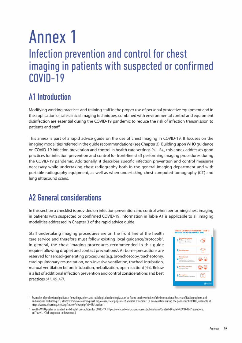
Annex 1Infection prevention and control for chest imaging in patients with suspected or confirmed COVID-19
A1 IntroductionModifying working practices and training staff in the proper use of personal protective equipment and in the application of safe clinical imaging techniques, combined with environmental control and equipment disinfection are essential during the COVID-19 pandemic to reduce the risk of infection transmission to patients and staff.
This annex is part of a rapid advice guide on the use of chest imaging in COVID-19. It focuses on the imaging modalities referred in the guide recommendations (see Chapter 3). Building upon WHO guidance on COVID-19 infection prevention and control in health care settings (A1–A4), this annex addresses good practices for infection prevention and control for front-line staff performing imaging procedures during the COVID-19 pandemic. Additionally, it describes specific infection prevention and control measures necessary while undertaking chest radiography both in the general imaging department and with portable radiography equipment, as well as when undertaking chest computed tomography (CT) and lung ultrasound scans.
A2 General considerationsIn this section a checklist is provided on infection prevention and control when performing chest imaging in patients with suspected or confirmed COVID-19. Information in Table A1 is applicable to all imaging modalities addressed in Chapter 3 of the rapid advice guide.
Staff undertaking imaging procedures are on the front line of the health care service and therefore must follow existing local guidance/protocols1. In general, the chest imaging procedures recommended in this guide require following droplet and contact precautions2. Airborne precautions are reserved for aerosol-generating procedures (e.g. bronchoscopy, tracheotomy, cardiopulmonary resuscitation, non-invasive ventilation, tracheal intubation, manual ventilation before intubation, nebulization, open suction) (A5). Below is a list of additional infection prevention and control considerations and best practices (A1, A6, A7).
1 Examples of professional guidance for radiographers and radiological technologists can be found on the website of the International Society of Radiographers and Radiological Technologists, at https://www.elearning.isrrt.org/course/view.php?id=12 and its CT webinar: CT examination during the pandemic COVID19, available at https://www.elearning.isrrt.org/course/view.php?id=13#section-1.
2 See the WHO poster on contact and droplet precautions for COVID-19: https://www.who.int/csr/resources/publications/Contact-Droplet-COVID-19-Precautions.pdf?ua=1. (Click on poster to download.)
CONTACT AND DROPLET PRECAUTIONS - COVID-19PERSONAL PROTECTIVE EQUIPMENT (PPE)
2 Put on the gown
3
1Perform hand hygieneAlcohol based handrubRub hands for20–30 seconds.
Water and soapWash hands for 40–60 seconds.
or
Put on eye protection Put on face shield or goggles.
5 Put on glovesEnsure gloves are placed over the cu� of the gown.
oror
4
Put on the maskMedical mask
orRespirator mask (N95, FFP2, FFP3, or equivalent). Only use if performing aerosol generating procedures.
oror
29Annexes

Table A1. Infection prevention and control checklist when performing chest imaging in patients with suspected or confirmed COVID-19
Imaging personnel tasks Patient considerations Equipment considerations (fixed and portable)
Environmental considerations of imaging room
Preparation• Explore whether the imaging procedure
would change patient management, and/or assess if the procedure could be delayed.
• Assess whether portable imaging is an option for suspected and confirmed COVID-19 cases.
• Evaluate risk factors (age > 60 years, comorbidities, serious underlying medical conditions, immunosuppressive condition, pregnancy, mental health concerns, etc.).
• Perform hand hygiene and don personal protective equipment following all appropriate steps.
• Use personal protective equipment during transfer to department when portable imaging equipment is unavailable.
• Ensure that the imaging protocol and patient identification procedures are followed.
• Verify imaging request and check whether imaging is required urgently.
• Determine whether patient will come to imaging department or whether portable imaging is possible/necessary.
• Inform all patients of the need for hand hygiene, and the use of tissues or elbow when coughing or sneezing.
• Supply medical masks to patients (and caregivers, if present) upon their arrival for chest imaging, if available and if patient is able to tolerate.
• Ensure infection prevention and control measures are employed when managing the imaging equipment.
• Subject the imaging equipment to regular cleaning and disinfection, consistent with local infection prevention and control guidance and complete, sign and date cleaning schedules.
• Remove unnecessary equipment from imaging room.
• Determine whether the examination can be performed with portable imaging equipment.
• Cover equipment that cannot be moved with plastic or other suitable material.
• Ensure infection prevention and control measures are employed when managing the imaging room.
• Subject the imaging room to regular cleaning and disinfection, consistent with local infection prevention and control guidance and complete, sign and date cleaning schedules.
• Verify that terminal cleaning and disinfection of the imaging room occurred at end of the previous day. If not done (or not verifiable) ensure that terminal cleaning and disinfection of the imaging room is performed before starting.
During• Ensure appropriate personal protective
equipment is worn.• Employ contact and non-contact
radiographer/technologist technique for chest radiography, chest CT and lung ultrasound.
• Ensure one patient attends the imaging department at a time wherever possible – and undertake further imaging, if this is required.
• Provide medical mask to patient (if feasible), as well as comfort and reassurance.
• Ensure standard operating procedures for infection prevention and control according to local guidance are in place, including contact minimization and barrier precautions (e.g. suitable covers, whenever possible).
• Control access to imaging room or patient area during the portable radiography procedure.
• Consider use of appropriate signage/visual alerts in front of imaging room (e.g. patient inside/arriving, ongoing cleaning/disinfection, time of last cleaning/disinfection).
Post procedure• Ensure imaging review is made appropriately
and apply local protocols to follow up clinical and infection prevention and control actions, if/as required.
• If the chest imaging procedure was performed at the imaging department, wear personal protective equipment during patient transfer.
• Ensure personal protective equipment is doffed appropriately, if used.
• Ensure rapid delivery of the imaging results to guide management.
• Ensure appropriate decontamination of medical equipment between patients (applicable to both fixed and portable equipment).
• Ensure appropriate environmental cleaning and disinfection (focus on high-touch surfaces) between patients. Staff performing this task should be trained in cleaning and disinfection and should wear appropriate personal protective equipment.
• Be aware that, if bedside imaging was performed using portable equipment, room cleaning and disinfection should occur following the protocols applicable for the specific setting (e.g. emergency room, regular ward, intensive care unit).
Use of chest imaging in COVID-19: a rapid advice guide30
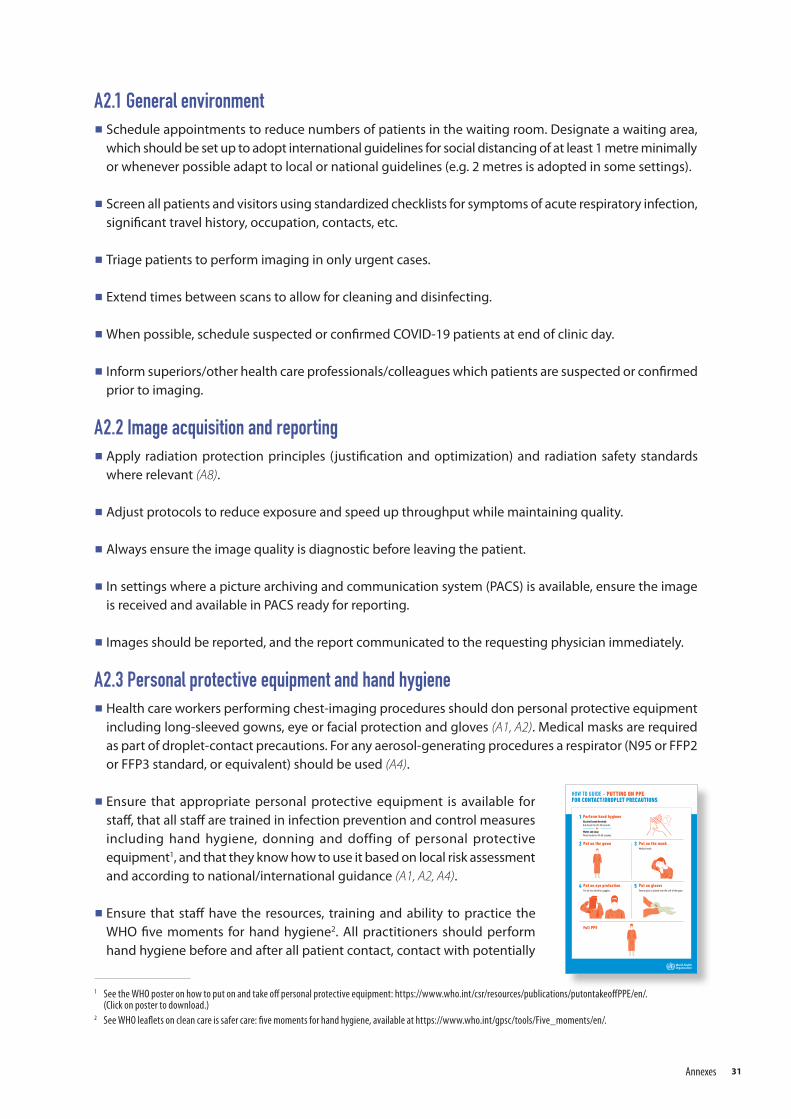
A2.1 General environment Schedule appointments to reduce numbers of patients in the waiting room. Designate a waiting area, which should be set up to adopt international guidelines for social distancing of at least 1 metre minimally or whenever possible adapt to local or national guidelines (e.g. 2 metres is adopted in some settings).
Screen all patients and visitors using standardized checklists for symptoms of acute respiratory infection, significant travel history, occupation, contacts, etc.
Triage patients to perform imaging in only urgent cases.
Extend times between scans to allow for cleaning and disinfecting.
When possible, schedule suspected or confirmed COVID-19 patients at end of clinic day.
Inform superiors/other health care professionals/colleagues which patients are suspected or confirmed prior to imaging.
A2.2 Image acquisition and reporting Apply radiation protection principles (justification and optimization) and radiation safety standards where relevant (A8).
Adjust protocols to reduce exposure and speed up throughput while maintaining quality.
Always ensure the image quality is diagnostic before leaving the patient.
In settings where a picture archiving and communication system (PACS) is available, ensure the image is received and available in PACS ready for reporting.
Images should be reported, and the report communicated to the requesting physician immediately.
A2.3 Personal protective equipment and hand hygiene Health care workers performing chest-imaging procedures should don personal protective equipment including long-sleeved gowns, eye or facial protection and gloves (A1, A2). Medical masks are required as part of droplet-contact precautions. For any aerosol-generating procedures a respirator (N95 or FFP2 or FFP3 standard, or equivalent) should be used (A4).
Ensure that appropriate personal protective equipment is available for staff, that all staff are trained in infection prevention and control measures including hand hygiene, donning and doffing of personal protective equipment1, and that they know how to use it based on local risk assessment and according to national/international guidance (A1, A2, A4).
Ensure that staff have the resources, training and ability to practice the WHO five moments for hand hygiene2. All practitioners should perform hand hygiene before and after all patient contact, contact with potentially
1 See the WHO poster on how to put on and take off personal protective equipment: https://www.who.int/csr/resources/publications/putontakeoffPPE/en/. (Click on poster to download.)
2 See WHO leaflets on clean care is safer care: five moments for hand hygiene, available at https://www.who.int/gpsc/tools/Five_moments/en/.
HOW TO GUIDE - PUTTING ON PPE FOR CONTACT/DROPLET PRECAUTIONS
Perform hand hygieneAlcohol based handrubRub hands for 20–30 seconds.
Water and soapWash hands for 40–60 seconds.
or
Put on the gown Put on the maskMedical mask.
Put on eye protection Put on face shield or goggles.
Full PPE
Put on glovesEnsure glove is placed over the cu� of the gown.
1
2
4
3
5
31Annexes
infectious material (e.g. linen from patient room), and before donning or doffing personal protective equipment including gloves.
Remember that personal eyeglasses do not provide adequate eye protection. If necessary, a face shield or goggles should be worn over personal eyeglasses. If staff wears eyeglasses, be careful not to touch them throughout the procedure, or during doffing of personal protective equipment. Personal eyeglasses can be cleaned and disinfected after personal protective equipment has been removed if soiling has occurred or there has been potential contamination during the doffing process.
A2.4 Staff considerations Split staff into multiple shifts to limit exposure of the entire team, ensuring appropriate skill and experience whenever possible. Encourage staff to maintain at least 1 metre distance between one another when working and during breaks.
When feasible, use contact/non-contact technique in pairs, following infection prevention and control precautions. For procedures performed in the imaging room (e.g. fixed chest radiography, chest CT), this is implemented by having one staff operate the equipment – who would not need personal protective equipment if operating the console in an area separate from the patient – and the other staff in contact with the patient wearing appropriate personal protective equipment. For procedures performed with portable equipment, the contact/non-contact technique in pairs can be applied but note that bedside imaging may require the use of personal protective equipment by both staff.
Encourage staff to stay home if exhibiting respiratory symptoms or fever. In addition to self-monitoring and reporting for COVID-19 symptoms, the unit supervisor should keep records of the health status of on-site imaging staff when they arrive at work. Do not allow staff who are potentially ill to work.
A2.5 Equipment decontamination Separate cold/blue/clean from hot/red/contaminated designated areas.
Clean and disinfect all high touch surfaces including patient couches, chairs, door handles in the waiting room and imaging room, following local protocols.
Ensure protocols for cleaning and disinfection of all medical equipment are in place according to manufacturer’s instructions for use.
Ensure adequate ventilation of the premises. Vacuum/negative air pressures would not be required in routine chest imaging procedures. Where necessary, there may be a room designated for aerosol-generating procedures; this room should be adequately ventilated (i.e. natural ventilation with air flow of at least 160 l/s per patient or in negative-pressure rooms with at least 12 air changes per hour – and controlled direction of air flow when using mechanical ventilation). Waiting for air exchange is only necessary if an aerosol-generating procedure was performed.
Keep all surfaces free of unnecessary paper, and non-essential material to allow for rapid and effective disinfection-decontamination of areas and equipment.
Use of chest imaging in COVID-19: a rapid advice guide32
A2.6 Training and education Always work within the scope of practice and job role.
Remove students/trainees from high-risk scenarios.
Activate retired/vacationing radiographers/technologists when possible, ensuring appropriate risk assessment, access to supervision and refresher training is available.
Ensure that all staff are trained in donning and doffing of personal protective equipment, hand hygiene and local infection prevention and control protocols (A1–A4).
A3 Specific considerationsA3.1 Chest radiography
The radiographers/radiological technologists performing radiography should follow droplet and contact precautions (airborne precautions required only for aerosol-generating procedures) (A1, A4).
Where possible, designate a portable imaging device for investigation of suspected or confirmed COVID-19 cases and leave it within the patient care area to reduce transmission risk.
Use direct digital radiography (DDR) imaging whenever possible to reduce transmission risk and minimize radiographer workload.
Designate one or two image receptors specific for patients with COVID-19 if computed digital radiography (CDR) or film/screen technology is to be used.
Adjust radiography technique in accordance with the patient’s condition e.g. anteroposterior with the patient supine or posteroanterior with the patient prone on intensive care unit wards.
Cover X-ray detector/cassettes with plastic cover or disposable cellophane wrapper and make sure to clean X-ray cassette in between each patient.
Ensure that positioning sponges of X-ray table or vertical Bucky stand and immobilization straps are covered with plastic protection.
Remove any radiopaque objects in the region of interest from the patient very carefully to prevent risk of infection transmission.
Preferably work in pairs with another radiographer to facilitate the contact/non-contact technique.
Ensure that the radiographer undertaking the radiography with the portable imaging equipment stands outside the controlled area, without physical contact with the team or any object.
When performing imaging, both within the department and when using portable equipment, wherever possible, one radiographer positions the X-ray tube and makes the exposure, and the second radiographer positions the patient and the covered detector and applies the anatomical marker.
33Annexes

Image acquisition/exposure should be made by the non-contact radiographer, in consideration of the diagnostic requirements and the principles of justification, optimization, radiation dose limitation as well as the radiographer/radiological technologist ethical code and professional rights at all times (A8, A9).
If working alone (i.e. not in a pair) use gloves and consider the X-ray equipment and mobile control screen keys as contaminated. Ensure hand hygiene after removal of gloves.
Check image for optimum quality before sending it to the picture archiving and communication system.
Clean and disinfect all imaging equipment, including the portable X-ray machine, X-ray couch and vertical Bucky stand between each patient.
A3.2 Chest computed tomography (CT) The radiographers/radiological technologists performing chest CT should follow droplet and contact precautions (airborne precautions required only for aerosol-generating procedures) (A1, A4).
Consider implementing a containment zipper (a room isolation tarp barrier with a zipper for room access) to separate the control area from the imaging room. Practice infection control in accordance with national public health guidelines, relevant department policies and instructions from the committees responsible for hospital infectious disease control and hospital waste management.
Separate clean console control area from contaminated CT scanner room; the radiographer/radiological technologist must remove gloves and wash hands before entering the console control area.
Consider all equipment in the imaging room as contaminated: CT gantry controls and contrast media injector control screen keys; they must be used with gloves.
Consider all equipment in the control area as clean: CT console keyboard, mouse and exposure pad as well as the contrast media injector remote control panel; they may be used without gloves.
Avoid crowding and maintain the safety distance of at least 1 metre.
Remove any radiopaque objects in the region of interest from the patient very carefully to prevent risk of infection transmission.
Perform examination (i.e. scanning and intravenous contrast media injection) in consideration of the diagnostic requirements and the principles of justification, optimization, radiation dose limitation as well as the radiographer/radiological technologist ethical code and professional rights at all times (A8, A9).
Note which personnel are involved in and present during the procedure.
Ensure that single use CT couch paper cover is removed and disposed of into the corresponding bin according to hospital policy.
The control panel integrated into the contrast media injector delivery device, which is located in the imaging room, may be covered with a disposable plastic cover.
When performing CT on patients confirmed with COVID-19, radiographers/radiological technologists must follow the instructions and guidance of the hospital committee responsible for infectious disease control.
Use of chest imaging in COVID-19: a rapid advice guide34
Asymptomatic patients pose a latent threat for medical imaging and therapy departments and hence radiographers/radiological technologists in CT are advised to follow the instructions divided in three stages (i.e. preparation, during and post procedure; see Table A1).
A3.3 Lung ultrasound Lung ultrasound presents specific challenges in terms of infection prevention and control. The first is physical proximity to the patient: this is usually within 1 metre and may be as little as 30–50 centimetres; ultrasound rooms are typically small, ventilation may be restricted and seldom are there windows; examination time may last between 10 and 60 minutes; patients may be asked to inhale/exhale deeply and hold their breath. Based on the Spaulding classification system, widely adopted in health care, reusable medical devices are categorized into non-critical, semi-critical and critical according to the infection risk and the level of disinfection required, as described below.
Non-critical devices: ultrasound probes that come in contact with intact skin can be cleaned and disinfected using low- or intermediate-level disinfection.
Semi-critical devices: ultrasound probes that come in contact with non-intact skin, blood, body fluids and/or mucous membranes should be cleaned and disinfected using the high-level disinfection method. A single use probe cover is mandatory.
Critical devices: intraoperative or intravascular probes must undergo sterilization if compatible or, if not available, high-level disinfection as per medical facility guidelines. Use of sterile transducer cover is mandatory.
Probes used to perform lung ultrasound are typically in contact only with intact skin and are therefore considered non-critical devices, which can be cleaned and disinfected using low- or intermediate-level disinfection. However, in case the probe comes in contact with body fluids (e.g. if the patient coughs or sneezes without respiratory hygiene measures) a high-level disinfection would be required after the procedure. More information about cleaning and disinfection of ultrasound probes is available in the literature (A10, A11). Additional considerations for infection prevention and control when performing lung ultrasound in patients with suspected or confirmed COVID-19 are summarized below.
The ultrasound health care workers should follow droplet and contact precautions (airborne precautions required only for aerosol-generating procedures) (A1, A4).
If possible, designate a specific ultrasound room, machine and probes for use on patients with suspected or confirmed COVID-19.
Adjust schedule (appointment times) to avoid crowding in the waiting room and to allow time between appointments for decontamination of the ultrasound system and room.
Best practice is to have patient attend examination alone.
Shorten duration of examination by arranging for the most experienced professional available to perform the examination. Single use ultrasound gel sachets should be considered for patients suspected or having COVID-19.
Reduce the number of probes connected to the ultrasound machine to a minimum and remove all other probes from device or store in closed cabinet to avoid the necessity of high-level disinfection in the event the patient coughs or sneezes during the procedure.
35Annexes
Separate inpatients on the ward from outpatients.
Cover the equipment such as the ultrasound scanner console with a disposable plastic cover to contribute to infection prevention and control and in this way enhance workflow.
Follow manufacturer’s recommendation for decontamination of ultrasound system.
Follow local protocols for appropriate decontamination of ultrasound probes between patients.
In the context of COVID-19, the normal practices of high-level disinfection are not changed. The only change in the context of COVID-19 is that all external probes must undergo cleaning followed by low-level disinfection to denature any presence of SARS-CoV-21 (as described above).
ReferencesIn the interests of specificity during the COVID-19 pandemic – during which new data become available by the day – the references below that deal with COVID-19 or SARS-CoV-2 exceptionally include both the day and month of publication (where available). This is meant to assist the reader in quickly determining the exact date of publication.
A1. Infection prevention and control during health care when novel coronavirus (nCoV) infection is suspected. Interim guidance. Geneva: World Health Organization; 19 March 2020 (https://www.who.int/publications-detail/infection-prevention-and-control-during-health-care-when-nov-el-coronavirus-(ncov)-infection-is-suspected-20200125, accessed 22 May 2020).
A2. Rational use of personal protective equipment for coronavirus disease (COVID-19) and con-siderations during severe shortages. Interim guidance. Geneva: World Health Organization; 6 April 2020 (https://www.who.int/publications-detail/rational-use-of-personal-protective-equip-ment-for-coronavirus-disease-(covid-19)-and-considerations-during-severe-shortages, accessed 23 May 2020).
A3. Cleaning and disinfection of environmental surfaces in the context of COVID-19. Interim guid-ance. Geneva: World Health Organization; 16 May 2020 (https://www.who.int/publications-detail/cleaning-and-disinfection-of-environmental-surfaces-inthe-context-of-covid-19, accessed 22 May 2020).
A4. Advice on the use of masks in the context of COVID-19. Interim guidance. Geneva: World Health Organization; 5 June 2020 (https://apps.who.int/iris/bitstream/handle/10665/332293/WHO-2019-nCov-IPC_Masks-2020.4-eng.pdf?sequence=1&isAllowed=y, accessed 7 June 2020).
A5. Mossa-Basha M, Meltzer CC, Kim DC, Tuite MJ, Kolli KP, Tan BS. Radiology Department preparedness for COVID-19: Radiology Scientific Expert Panel. Seattle (WA): University of Washington; 16 March 2020 – print version in press (https://pubs.rsna.org/doi/pdf/10.1148/radiol.2020200988, accessed 22 May 2020).
1 Like other coronaviruses, SARS-CoV-2 is an enveloped virus with a fragile outer lipid envelope that makes it more susceptible to disinfectants compared to non-enveloped viruses (A3).
Use of chest imaging in COVID-19: a rapid advice guide36
A6. COVID 19: Performing portable chest X-ray in ‘at risk’ patients in ED or wards – Standard oper-ating procedure – check list published by the British Society of Thoracic Imaging – Action cards [video]. London: British Society of Thoracic Imaging; 15 March 2020 (https://eur02.safelinks.protection.outlook.com/?url=https%3A%2F%2Fwww.youtube.com%2Fwatch%3Fv%3D_6iqmx-46nrY%26feature%3Dyoutu.be%26fbclid%3DIwAR3MGfTQ5YcDqcu7RaHYx5UW7GP9JtLrg-9YaznTYwKNs84KFXQl2qKsLiO0&data=02%7C01%7C%7Ca8fdf91a5b02476a8c2f08d7d4861602%7C84df9e7fe9f640afb435aaaaaaaaaaaa%7C1%7C0%7C637211544660713966&s-data=WE8WREQeSDOtlGbfcimEa1pP%2FXkAHgL0ziqoc55Xz9w%3D&reserved=0, accessed 22 May 2020).
A7. COVID-19 - ISRRT response document. Appropriate and safe use of medical imaging and radiation therapy with infection control measures considered in addition to standard radiation protection procedures. London: International Society of Radiographers and Radiological Technologists; April 2020 (https://www.isrrt.org/full-guideline-protective-measures, accessed 22 May 2020).
A8. Radiation protection and safety in medical uses of ionizing radiation. IAEA Safety Standards Series No. SSG-46. Vienna: International Atomic Energy Agency; 2018 (https://www.iaea.org/publica-tions/11102/radiation-protection-and-safety-in-medical-uses-of-ionizing-radiation, accessed 22 May 2020).
A9. Coronavirus disease (COVID-19) outbreak: rights, roles and responsibilities of health workers, including key considerations for occupational safety and health. Geneva: World Health Organiza-tion; 2020 (https://www.who.int/docs/default-source/coronaviruse/who-rights-roles-respon-hw-covid-19.pdf?sfvrsn=bcabd401_0, accessed 22 May 2020).
A10. Abramowicz JS and Basseal JM, in the name of WFUMB Safety Committee (Abramowicz JS, Akiya-ma I, Evans D, Fowlkes JB, Marsal K, Sayeed Y, et al.) World Federation for Ultrasound in Medicine and Biology Position Statement: how to perform a safe ultrasound examination and clean equip-ment in the context of COVID-19. Ultrasound Med Biol. 30 March 2020; 00(00):1–7 (https://doi.org/10.1016/j.ultrasmedbio.2020.03.033, accessed 7 June 2020).
A11. Shokoohi H, Armstrong P, Tansek R. Emergency department ultrasound probe infection control: challenges and solutions. Emerg Med. 2015; 7:1–9 (https://www.ncbi.nlm.nih.gov/pmc/articles/PMC4806800/pdf/oaem-7-001.pdf, accessed 5 June 2020).
37Annexes
Annex 2List of contributors
Guideline development groupElie Akl (vice-chair and lead methodologist)Professor of MedicineDivision of General Internal Medicine and GeriatricsAmerican University of BeirutBeirut, Lebanon
John Adabie AppiahFounding HeadPaediatric Intensive Care UnitKomfo Anokye Teaching HospitalKwame Nkrumah University of Science and TechnologyKumasi, Ghana
Ivana BlazicSenior Radiologist and MRI Section HeadClinical Hospital Centre ZemunInternational Society of RadiologyBelgrade, Serbia
Mansoor FatehiChief of Imaging Informatics Virtual University of Medical SciencesIran University of Medical SciencesTeheran, Islamic Republic of Iran
Nicola FlorRadiologistUnità Operativa di Radiologia Luigi Sacco University HospitalMilan, Italy
Guy Frija (chair)Professor EmeritusParis Descartes UniversityInternational Society of RadiologyParis, France
Eveline HittiAssociate Professor in Clinical Emergency MedicineDepartment of Emergency MedicineAmerican University of BeirutBeirut, Lebanon
Hussain JaffriVice ChairWHO Patients for Patient Safety Program Advisory Group Lahore, Pakistan
Zheng-Yu JinPresident of the Chinese Society of RadiologyProfessor and Chairman of the Department of RadiologyPUMC Hospital BeijingBeijing, China
Hans Ulrich KauczorMedical DirectorDepartment of Diagnostic and Interventional Radiology University Hospital of HeidelbergHeidelberg, Germany
Michael KawooyaDirectorErnest Cook Ultrasound Research and Education Institute Kampala, Uganda
Ella Annabelle KazerooniProfessorDepartment of Radiology, Cardiothoracic Radiology DivisionUniversity of MichiganAnn Arbor, Michigan, USA
Use of chest imaging in COVID-19: a rapid advice guide38
Jane KoProfessorDepartment of Radiology New York University School of MedicineNew York, New York, USA
Rami MahfouzProfessorDepartment of Pathology and Laboratory MedicineAmerican University of BeirutBeirut, Lebanon
Valdair MugliaAssociate ProfessorDepartment of Medical Imaging, Radiation Therapy and OncohematologyFaculdade de Medicina de Ribeirão Preto Universidade de São PauloSão Paulo, Brazil
Rose NyabandaAssistant DirectorRadiology Department Kenyatta National HospitalNairobi, Kenya
Marcelo SanchezProfessor of RadiologyRadiology Department Hospital ClínicBarcelona, Spain
Priya SheteAssistant Professor of MedicineDivision of Pulmonary and Critical Care MedicineUniversity of CaliforniaSan Francisco, California, USA
Marina UllaHead of CT Department and Medical InformaticsImaging DepartmentHospital Italiano de Buenos AiresBuenos Aires, Argentina
Huadan (Danna) XueProfessorDepartment of RadiologyPeking Union Medical College HospitalBeijing, China
Chuansheng ZhengDirector of Department of radiologyUnion Hospital, Tongji Medical College, Huazhong University of Science and TechnologyHuazhong, China
External review groupDeniz AkataProfessor and ChairHacettepe University School of MedicineAnkara, Turkey
Jocelyne BassealConsultant in Infection Prevention and ControlIncident Management Support Team WHO Health Emergencies WHO Western Pacific Regional Office Manila, Philippines
Salah BendibProfessorCentre Pierre et Marie CurieFaculty of Medicine of AlgiersAlgiers, Algeria
Jeffrey BurnsAssociate Professor of AnaesthesiaHarvard Medical SchoolCambridge, Massachusetts, USA
Bin CaoProfessor of Pulmonary and Critical Care MedicineChina-Japan Friendship Hospital and Capital Medical UniversityBeijing, China
Luis DonosoDirector of Diagnostic ImagingHospital Clinic of BarcelonaBarcelona, Spain
David HuiProfessor of Respiratory MedicineDirector of Stanley Ho Centre for Emerging Infectious DiseasesThe Chinese University of Hong KongHong Kong SAR, China
39Annexes
Dina Husseiny SalamaProfessor of RadiodiagnosisRadiology and Radiation Protection DepartmentEgyptian Atomic Energy AuthorityCairo, Egypt
David KoffProfessorDepartment of RadiologyMcMaster UniversityHamilton, Ontario, Canada
Boudjema MansouriProfessor of Radiology and Medical ImagingUniversity Hospital of Bab El OuedAlgiers, Algeria
Stephanie NewellHealth Care Consumer and Patient Safety AdvocatePatients for Patient Safety networkAustralia
Deepak PatkarDirector, Medical ServicesHead, Department of RadiologyNanavati Super Speciality HospitalMumbai, India
Mathias ProkopHead of the Department Radiology & Nuclear medicineRadboud University Medical CenterNijmegen, The Netherlands
Francesco SardanelliDepartment of Biomedical Sciences for HealthUniversità degli Studi di MilanoMilan, ItalyDirector of RadiologyIRCCS Policlinico San DonatoMilan, Italy
Arthur Soares Souza JrProfessorFaculty of Medicine, São José do Rio Preto São Paulo, Brazil
Jacob SosnaProfessor and ChairmanDepartment of RadiologyVice DeanHadassah Hebrew University Medical CenterJerusalem, Israel
Evangelina Vazquez CurielMember Patients for Patient Safety Advisory GroupMexico City, Mexico
Mingxing XieProfessorDepartment of Ultrasound, Union Hospital, Tongji Medical CollegeHuazhong University of Science and TechnologyHuazhong, China
Hwan Seok YongProfessor of Radiology, Cardiothoracic DivisionDirector of Healthcare CenterKorea University Guro HospitalSeoul, Republic of Korea
Systematic review teamRoger Chou (Principal Investigator)
David Buckley
Tracy Dana
Elaine Graham
Erica Hart
Marian McDonagh
Heidi Nelson
Miranda Pappas
Annette Totten
Ngoc WassonPacific Northwest Evidence-Based Practice Center. Oregon Health and Science University, USANicola Flor (details in list of guideline development group)
Francesco Sardanelli (details in list of external review group)
Use of chest imaging in COVID-19: a rapid advice guide40
Contributors to Annex 1Jacques Abramowicz, WFUMB
April Baller, WHO headquarters
Jocelyne Basseal, WHO Regional Office for the Western Pacific
Ivana Blazic, ISR
Donna Newman, ISRRT
Fernanda Lessa, WHO headquarters
Stewart Whitley, ISRRT
Victoria Willet, WHO headquarters
WHO steering groupAnshu Banerjee, WHO headquarters
Zhanat Carr, WHO headquarters
Neelam Dhingra-Kumar, WHO headquarters
Janet Diaz, WHO headquarters
Ivan Ivanov, WHO headquarters
Pablo Jimenez, WHO Regional Office for the Americas
Maria del Rosario Perez, WHO headquarters
Mark Perkins, WHO headquarters
Judith van Andel, WHO headquarters
Emilie van Deventer, WHO headquarters
Adriana Velazquez Berumen, WHO headquarters
Wilson Milton Were, WHO headquarters
Core groupMaria del Rosario Perez (responsible officer, WHO Secretariat)
Guy Frija (GDG chair)
Elie Akl (GDG vice-chair and lead methodologist)
Ivana Blazic (content consultant)
Emilie van Deventer (WHO Secretariat)
Sally Yaacoub (methodologist)
41Annexes
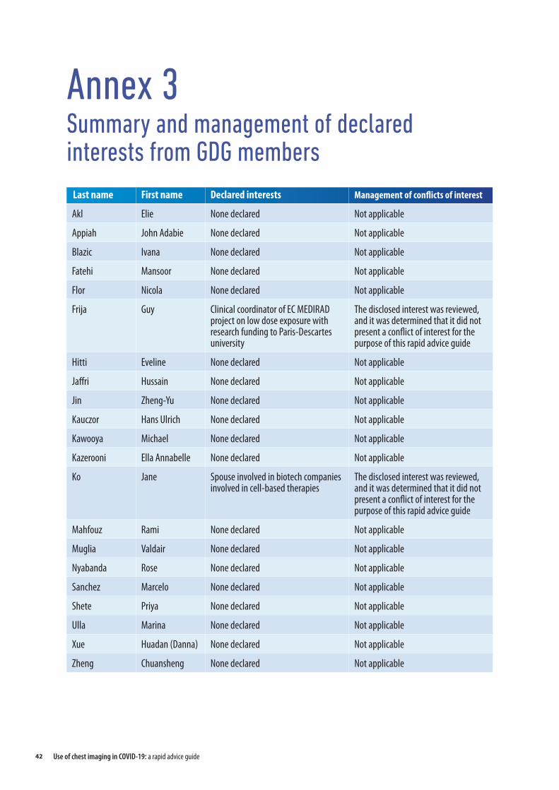
Annex 3Summary and management of declared interests from GDG members
Last name First name Declared interests Management of conflicts of interest
Akl Elie None declared Not applicable
Appiah John Adabie None declared Not applicable
Blazic Ivana None declared Not applicable
Fatehi Mansoor None declared Not applicable
Flor Nicola None declared Not applicable
Frija Guy Clinical coordinator of EC MEDIRAD project on low dose exposure with research funding to Paris-Descartes university
The disclosed interest was reviewed, and it was determined that it did not present a conflict of interest for the purpose of this rapid advice guide
Hitti Eveline None declared Not applicable
Jaffri Hussain None declared Not applicable
Jin Zheng-Yu None declared Not applicable
Kauczor Hans Ulrich None declared Not applicable
Kawooya Michael None declared Not applicable
Kazerooni Ella Annabelle None declared Not applicable
Ko Jane Spouse involved in biotech companies involved in cell-based therapies
The disclosed interest was reviewed, and it was determined that it did not present a conflict of interest for the purpose of this rapid advice guide
Mahfouz Rami None declared Not applicable
Muglia Valdair None declared Not applicable
Nyabanda Rose None declared Not applicable
Sanchez Marcelo None declared Not applicable
Shete Priya None declared Not applicable
Ulla Marina None declared Not applicable
Xue Huadan (Danna) None declared Not applicable
Zheng Chuansheng None declared Not applicable
Use of chest imaging in COVID-19: a rapid advice guide42


