
USA Department of Psychology and Neuroscience … · Department of Psychology and Neuroscience...
17
Parenting and plasticity Benedetta Leuner, Erica R. Glasper, and Elizabeth Gould Department of Psychology and Neuroscience Institute, Princeton University, Princeton NJ 08544, USA Abstract As any new parent knows, having a baby provides opportunities for enrichment, learning and stress –experiences known to change the adult brain. Yet surprisingly little is known about the effects of maternal experience, and even less about the effects of paternal experience, on neural circuitry not directly involved in parenting. Here we discuss how caregiving and the accompanying experiential and hormonal changes influence the hippocampus and prefrontal cortex, brain regions involved in cognition and mood regulation. A better understanding of how parenting impacts the brain is likely to help in devising strategies for treating parental depression, a condition that can have serious cognitive and mental health consequences for children. Introduction For all mammalian species, becoming a mother involves remarkable behavioral change driven by a combination of neuroendocrine and experiential factors. Considerable research has been devoted to understanding the neural mechanisms of maternal care in rodents and primates. For a small minority of mammalian species (~6%), including humans, fathers play a significant role in rearing young [1,2]. Less is known about the neural and hormonal mechanisms of paternal care but the limited available evidence suggests that mothers and fathers might recruit similar neural circuitry, hormones and neuromodulators in the service of parenting behavior (Box 1). Box 1 Mothers and fathers – are they really so different? Although significantly less is known about the neural mechanisms of parenting behavior in fathers than in mothers, evidence suggests striking similarities between the sexes. Such similarities are particularly surprising because maternal behavior has been linked, in part, to hormonal changes that occur with pregnancy, parturition and lactation, experiences that are not available to fathers. From studies of biparental rodents and primate species that engage in cooperative breeding, we know that caregiving behavior in fathers is similar to that in mothers, suggesting that the same neural pathways might be involved. Lesion studies, as well as immediate early gene and neuropeptide distribution studies, in rodents have identified similar brain regions involved in maternal and paternal behavior, including the olfactory bulb, medial preoptic area, lateral septum, bed nucleus of the stria terminalis, amygdala and PFC [84–88]. Although less is known about the neural circuitry of parenting in humans, exposure to crying infants activates the amygdala and PFC in fathers and mothers, but not in non-parents [62,63]. Given the hedonic aspects of parenting, it is not surprising that maternal and paternal behavior in rodents involves Corresponding author: Gould, E. ([email protected]). NIH Public Access Author Manuscript Trends Neurosci. Author manuscript; available in PMC 2011 April 13. Published in final edited form as: Trends Neurosci. 2010 October ; 33(10): 465–473. doi:10.1016/j.tins.2010.07.003. NIH-PA Author Manuscript NIH-PA Author Manuscript NIH-PA Author Manuscript
Transcript of USA Department of Psychology and Neuroscience … · Department of Psychology and Neuroscience...

Parenting and plasticity
Benedetta Leuner, Erica R. Glasper, and Elizabeth GouldDepartment of Psychology and Neuroscience Institute, Princeton University, Princeton NJ 08544,USA
AbstractAs any new parent knows, having a baby provides opportunities for enrichment, learning andstress –experiences known to change the adult brain. Yet surprisingly little is known about theeffects of maternal experience, and even less about the effects of paternal experience, on neuralcircuitry not directly involved in parenting. Here we discuss how caregiving and theaccompanying experiential and hormonal changes influence the hippocampus and prefrontalcortex, brain regions involved in cognition and mood regulation. A better understanding of howparenting impacts the brain is likely to help in devising strategies for treating parental depression,a condition that can have serious cognitive and mental health consequences for children.
IntroductionFor all mammalian species, becoming a mother involves remarkable behavioral changedriven by a combination of neuroendocrine and experiential factors. Considerable researchhas been devoted to understanding the neural mechanisms of maternal care in rodents andprimates. For a small minority of mammalian species (~6%), including humans, fathers playa significant role in rearing young [1,2]. Less is known about the neural and hormonalmechanisms of paternal care but the limited available evidence suggests that mothers andfathers might recruit similar neural circuitry, hormones and neuromodulators in the serviceof parenting behavior (Box 1).
Box 1
Mothers and fathers – are they really so different?
Although significantly less is known about the neural mechanisms of parenting behaviorin fathers than in mothers, evidence suggests striking similarities between the sexes. Suchsimilarities are particularly surprising because maternal behavior has been linked, in part,to hormonal changes that occur with pregnancy, parturition and lactation, experiencesthat are not available to fathers. From studies of biparental rodents and primate speciesthat engage in cooperative breeding, we know that caregiving behavior in fathers issimilar to that in mothers, suggesting that the same neural pathways might be involved.
Lesion studies, as well as immediate early gene and neuropeptide distribution studies, inrodents have identified similar brain regions involved in maternal and paternal behavior,including the olfactory bulb, medial preoptic area, lateral septum, bed nucleus of the striaterminalis, amygdala and PFC [84–88]. Although less is known about the neural circuitryof parenting in humans, exposure to crying infants activates the amygdala and PFC infathers and mothers, but not in non-parents [62,63]. Given the hedonic aspects ofparenting, it is not surprising that maternal and paternal behavior in rodents involves
Corresponding author: Gould, E. ([email protected]).
NIH Public AccessAuthor ManuscriptTrends Neurosci. Author manuscript; available in PMC 2011 April 13.
Published in final edited form as:Trends Neurosci. 2010 October ; 33(10): 465–473. doi:10.1016/j.tins.2010.07.003.
NIH
-PA Author Manuscript
NIH
-PA Author Manuscript
NIH
-PA Author Manuscript

reward circuitry, namely dopaminergic afferents [89], suggesting neurochemical andneuroanatomical overlap between the sexes. Similar involvement of reward-related brainregions has been observed using neuroimaging in human mothers exposed tophotographs of their own happy infants [90], although no study has yet investigated thisissue in fathers.
What role do hormones, with their obvious differences between mothers and fathers,play? Here the answer is complex. Mothers exhibit drastic alterations in hormonesincluding decreased estrogen and increased oxytocin and prolactin [11]. These changesare driven by pregnancy, parturition and lactation, as well as by infant contact, and areimportant for maternal behavior. Glucocorticoid levels also increase and althoughimportant for lactation, are not essential for other aspects of maternal behavior [25,91].Similar but not identical alterations in these hormones are detectable in fathers; theseinclude increased estrogen, oxytocin, prolactin and glucocorticoids [11,33]. Hormonalchanges in fathers are induced by contact with the mother and the offspring. For instance,oxytocin levels in human fathers are positively related to the amount of affection thefather displays toward his infant [92]. Variations in testosterone levels have also beenassociated with paternal care, although studies suggest species differences. In primatespecies, including humans, paternal behavior is related to reduced levels of testosterone[33,93]. The opposite is the case in some rodents for which paternal care is associatedwith elevated levels of testosterone [94–96]. Reduced testosterone, along with elevatedestrogen and oxytocin levels, might facilitate affiliative behavior in primate fathers,whereas increased testosterone levels in rodents seem to support parental aggressiontoward nest intruders. It remains unclear whether differential hormone responses cause,or are caused by, variation in paternal care, because a definitive link between specifichormones and paternal behavior remains uncertain [97]. Nonetheless, infant contact itselfseems to modulate endocrine systems and activate neural circuitry in fathers in a mannerthat is strikingly similar to that in mothers.
There is an increasing body of literature showing that structural, electrophysiological andmolecular changes occur in the maternal and paternal brain. These include modifications notonly in brain areas known to be involved in the control of parental behavior, but also inregions not traditionally associated with parenting, but that are instead more widely knownto be involved in cognition and mood regulation. Here we highlight maternal and paternalinfluences on neuroplasticity in two such brain regions, the hippocampus and prefrontalcortex (PFC). We also consider the complex role that hormones and environment play inmediating the impact of parenting on these brain regions and discuss the potential functionalconsequences of these changes.
Identifying how the brain changes in response to parenthood could be important to betterunderstand mood disturbances, including depression, which are known to be prevalent innew mothers and, as suggested by recent studies, in fathers as well (Box 2). Postpartummental illness is often associated with inadequate child care, which has been linked toimpaired cognitive and emotional function in offspring [3–5], including increased likelihoodof developing anxiety and depression later in life [6]. Elucidating the influence of parentingon the brain could also be critical for ensuring the long-term health of offspring.
Box 2
Depression in new parents
In the US, an estimated 10–15% of new mothers experience postpartum depression [98];a slightly lower percentage of men (6–12%) also experience depression shortly after the
Leuner et al. Page 2
Trends Neurosci. Author manuscript; available in PMC 2011 April 13.
NIH
-PA Author Manuscript
NIH
-PA Author Manuscript
NIH
-PA Author Manuscript

birth of a child [99,100]. Among men whose partners have postpartum depression, therate is even higher, suggesting a link between maternal and paternal depression [101].
Early parental depression is a significant problem, not just for the afflicted, but also forthose in their care. Depression in mothers has been related to persistent disturbances incognitive and emotional function in offspring [3–5], including increased likelihood ofdeveloping anxiety and depression later in life [6]. Depression in fathers can alsonegatively affect infant development; it has been associated with greater risk of lateremotional, behavioral and cognitive difficulties [100,102,103].
Depressed parents seem to provide inadequate care to their offspring, which in turnimpairs emotional and cognitive development [104]. Parents with depression are lessemotionally attached to their infants, display less affection and interact less overall[3,105], qualities that infants seem to be able to detect. For example, babies becomenoticeably distressed when an adult (mother, father or unrelated caregiver) maintains anon-interactive still face [106,107]. Lack of social interaction during early life probablycontributes heavily to emotional problems that emerge later in the children of depressedparents. Moreover, depressed parents read less often to their children than non-depressedparents, a difference that leads to smaller vocabularies in youngsters [100,103].
The neural mechanisms by which inadequate parental care contributes to negativeemotional and cognitive outcomes in offspring are incompletely understood, but animalstudies have provided some suggestions. Early postnatal maternal separation is a widelyused paradigm to study maternal influence over offspring development in rats. Inadulthood, rat pups subjected to maternal separation exhibit increased anxiety-likebehavior, impaired cognitive capabilities and dysregulation of the hypothalamus–pituitary–adrenal axis [108]. These effects stem not from maternal separation itself, butindirectly from abnormal parenting behavior exhibited by mothers on return to their pups[108]. Similar outcomes have been reported in the offspring of mother rats that naturallydisplay low levels of maternal care [109].
Maternal separation is associated with diminished structural plasticity in brain regionslinked to cognition and mood regulation, including the hippocampus and PFC. In thehippocampus, maternal separation and low levels of maternal care lead to suppressedpostnatal neurogenesis [110,111], reduced dendritic spine density [109,112], and reducedhippocampal BDNF expression and cholinergic innervation [109]. In the PFC, maternalseparation, as well as paternal separation (in biparental species), is associated withchanges in dendritic spines and synapses [113–115]. If structural changes contribute tobehavioral problems associated with inadequate parenting, then the potential to repairthese abnormalities by modulating ongoing plastic processes, including adultneurogenesis and dendritic remodeling, might exist. Indeed, studies have shown thatenvironmental enrichment, an experience that enhances structural plasticity in adulthood[28,29,47], restores many of the behavioral abnormalities arising from maternalseparation and low maternal care during early life [116,117]. Future studies will benecessary, however, to forge the translational link between animal models of earlyinadequate parenting and depressed human parents.
Behavioral changes that define parentingThe postpartum period is a time of dramatic behavioral changes for all mammalian species.In rodents, females that were previously unresponsive or aggressive toward pups engage inan elaborate repertoire of caregiving activities at birth and postpartum that includes cleaningthe pups, eating the debris of birth, nursing, nest building, licking and grooming, pupretrieval, assuming a nursing (i.e. arched back) posture over the pups and increased
Leuner et al. Page 3
Trends Neurosci. Author manuscript; available in PMC 2011 April 13.
NIH
-PA Author Manuscript
NIH
-PA Author Manuscript
NIH
-PA Author Manuscript

aggression toward intruders. In nonhuman primates, maternal behavior includes nursing,infant carrying, grooming and defending the infant from danger. The emergence of maternalbehaviors is stimulated by contact with offspring and a complex array of endocrine changes(Box 1). Maternal experience and the accompanying hormonal alterations affect variousmeasures of neural plasticity in numerous brain regions such as the hypothalamus [7,8],amygdala [9] and olfactory bulb [10], areas that are necessary for the full expression ofmaternal behaviors [11].
For most mammalian species, raising the young is accomplished exclusively by the mother,but for a small minority fathers play a role. Among species in which paternal care occurs(Box 3), there are two general social strategies – a biparental strategy in which the mother–father dyad care for the young and a cooperative breeding strategy in which caregiving isshared by the mother, father, older siblings and sometimes unrelated adults. Regardless ofwhether the species engages in biparental or cooperative breeding, paternal care typicallyinvolves the same behaviors as maternal care, with the exception of nursing. Evidence todate suggests that the neural circuitry underlying paternal behavior is similar to that formaternal behavior (Box 1) in that the same brain regions seem to be activated when fathersand mothers have contact with infants. Moreover, many of the same hormonal changes thataccompany the postpartum period also occur in fathers that display parenting behavior (Box1).
Box 3
Some mammalian species that engage in paternal care
Maternal and paternal experiences also influence brain regions that are known to be eitherunnecessary for the behaviors directly associated with parenting or only involved in amodulatory way. Among these are effects on brain regions associated with cognition andmood, including the hippocampus and PFC (Figure 1).
Parenting and the hippocampusLesions of the hippocampus have minimal effects on maternal behavior [12,13], but thisbrain region is nonetheless affected by parenting. The hippocampus plays an important rolein certain types of learning and memory, anxiety regulation and feedback of the stressresponse. In adult mammals, the hippocampus also exhibits a capacity for dramaticstructural reorganization in the form of adult neurogenesis, dendritic remodeling, and theformation and elimination of dendritic spines and synapses (Figure 1). In virgin animals
Leuner et al. Page 4
Trends Neurosci. Author manuscript; available in PMC 2011 April 13.
NIH
-PA Author Manuscript
NIH
-PA Author Manuscript
NIH
-PA Author Manuscript

without reproductive or maternal experience, these forms of structural plasticity aremodulated by hormones, including estrogen and glucocorticoids [14–19], levels of which arealtered in mothers and fathers (Box 1). Moreover, several experiences that seem intrinsic toparenting, such as stress, learning and environmental enrichment, also influencehippocampal structure in virgins [19–21]. Perhaps it is not surprising that parentalexperience alters structural plasticity in the hippocampus, although the manner in which thisoccurs seems to be complex, most probably reflecting the multitude of hormonal andexperiential changes associated with parenting.
Adult neurogenesisThe dentate gyrus of the hippocampus continues to produce several thousand new neuronseach day in young adulthood [22]. Adult neurogenesis in the hippocampus has beendocumented in many mammalian species, including humans [23]. The regulation of adultneurogenesis by hormones and experience has been the subject of intensive investigation[19]. Ovarian steroids enhance [15], whereas adrenal steroids suppress [14,18], cellproliferation in the dentate gyrus of virgin rats. Postpartum female rats exhibit decreasedestrogen levels [24] and increased glucocorticoid levels [24,25], suggesting that new neuronproduction might be impaired in mothers. The postpartum period is associated not just withhormonal changes, but also with experiential changes that might impact adult neurogenesis.Stress is known to inhibit, whereas learning and environmental enrichment are known toenhance, adult neurogenesis [19–21]. Despite the fact that some of the stimuli inherent toparenting seem to enhance adult neurogenesis, available evidence suggests that maternalexperience is associated with suppression of cell proliferation during the postpartum period[24–26].
In mother rats, decreased cell proliferation is evident as early as 1 day after parturition andoccurs in both first-time mothers (primiparous) (Figure 1) and females that have producedmore than one litter (multiparous) [24–26]. By the time of weaning and beyond, cellproliferation is restored in postpartum females [25]. Thus, maternal experience has asuppressive, albeit temporary, effect on cell production in the hippocampus that does notseem to be modulated by repeated reproductive experience. The suppressive effect ofmaternal experience on adult neurogenesis seems to be linked to both hormonal changes andinteraction with offspring. Reduced cell proliferation during the postpartum period isdependent on elevated basal glucocorticoid levels associated with lactation; removal ofnursing pups reduces corticosterone levels and prevents the decrease in number ofproliferating cells [25]. Moreover, prevention of increased basal corticosterone levels inpostpartum mothers by means of adrenalectomy and low-dose corticosterone replacementeliminates the reduction in cell proliferation. Thus, it seems that in postpartum females,offspring interactions inhibit hippocampal cell production through changes in adrenalsteroids (Figure 2). It should be noted that elevated adrenal steroid levels might not be thesole factor involved and that decreased estradiol levels, which are known to reduce adultneurogenesis, might participate as well [24,27].
Postpartum-induced suppression of adult neurogenesis is linked to glucocorticoid elevationsthat occur along with lactation, so it seems likely that this effect might be specific tolactating mothers. The glucocorticoid-induced decrease in cell proliferation in lactatingfemales suggests that other aspects of maternal experience, such as enriching effects, whichwould be expected to enhance adult neurogenesis, are secondary to glucocorticoid changes.Indeed, pup exposure in virgin female rats stimulates adult neurogenesis, an effect thatmight reflect environmental enrichment in the absence of hormonal changes associated withpregnancy, parturition and lactation [26]. Studies have shown that environmental enrichmentstimulates new neuron production in the dentate gyrus of virgin male and female rodents[28,29]. Males do not lactate, so it seems reasonable to predict that fathers would display
Leuner et al. Page 5
Trends Neurosci. Author manuscript; available in PMC 2011 April 13.
NIH
-PA Author Manuscript
NIH
-PA Author Manuscript
NIH
-PA Author Manuscript
adult neurogenesis changes that are more akin to enriched environment effects, such as thoseobserved in virgin females. Consistent with this, brief exposure to pups increases cellproliferation in the dentate gyrus of virgin male prairie voles [30]. A similar stimulatoryeffect of pup exposure on adult neurogenesis has been observed in laboratory mouse fathers,which do not normally engage in parental behavior, but do so after repeated interaction withtheir offspring [31]. By contrast, in California mice fathers that naturally demonstratepaternal care behaviors, a reduction in new cell production, similar to that detected inmother rats, has been observed [32]. This effect is probably linked to hormonal changes thatoccur in fathers. For example, fathers exhibit elevated glucocorticoids [33], although thishormonal change is not associated with lactation.
Many of the behavioral and neuroendocrine adaptations of the postpartum period are alsoevident during late pregnancy [34]. Thus, it seems plausible that suppression of theproduction of new neurons would emerge before parturition. Decreased hippocampalvolume has been observed in pregnant rats and humans [35,36], suggesting that reducedadult neurogenesis contributes to these volumetric changes. Cell proliferation in thehippocampus does not seem to be altered during pregnancy in rats or mice [10,37–39] andthe current evidence on pregnancy-induced alterations in cell survival is mixed, with somework suggesting a possible enhancement [37] and other studies showing a decrease [40] orno effect [39]. It is possible that these inconsistencies are related to the different gestationaltimepoints examined and could suggest that neurogenesis is altered, but only at specifictimes during pregnancy.
Despite the unresolved effects of pregnancy, some evidence suggests that maternalexperience has a lasting effect on progenitor cells in the dentate gyrus that extends intoaging. Whereas middle-aged virgin female rats exhibit no change in cell proliferation withestrogen treatment, middle-aged mothers exhibit an increase in cell proliferation, an effectsimilar to that observed in young adults [41]. This suggests that maternal experience slowsthe aging process, at least insofar as estrogen sensitivity of progenitor cells is concerned.The duration of this effect remains unknown. Long-term benefits of being a mother on age-related cognitive decline and other measures of brain aging have also been reported [42].
It is important to note that maternal experience also has effects on neurogenesis in thesubventricular zone (SVZ), an area that gives rise to neurons of the olfactory bulb, a brainregion important for maternal care [43]. Increased neurogenesis has been reported in miceand rats during pregnancy and the early postpartum period [10,38]. Pup exposure in virginfemale rats also stimulates neurogenesis in the SVZ [44]. In contrast to the involvement ofglucocorticoids in the postpartum reduction in neurogenesis in the hippocampus, increasedneurogenesis in the SVZ seems to be mediated by prolactin [10]. Thus, maternal experiencecan have opposite effects on the same form of structural plasticity in two different brainregions through distinct mechanisms.
Dendritic and synaptic remodelingThe effects of parenting on hippocampal plasticity are not limited to alterations in cellproliferation and neurogenesis, but also extend to dendritic spines and architecture. Like theeffects of maternal experience on adult neurogenesis, changes in these morphologicalmeasures are complex (Figure 2).
Increased spine densities on apical dendrites of hippocampal CA1 pyramidal neurons havebeen reported in late pregnant and postpartum (primiparous) rats [45,118]. The postpartumperiod is also associated with increases in spine density on dendrites of dentate gyrusgranule cells [118]. Hippocampal dendritic spine density is controlled by circulating levelsof ovarian hormones [17], so enhanced spine density in late pregnancy might be related to
Leuner et al. Page 6
Trends Neurosci. Author manuscript; available in PMC 2011 April 13.
NIH
-PA Author Manuscript
NIH
-PA Author Manuscript
NIH
-PA Author Manuscript

elevated estrogen levels that occur at this time. In support of this, virgin females treated witha pregnancy-like regimen of estradiol and progesterone exhibit similar spine enhancements[45]. Estrogen levels are low after birth, so it is likely that estrogen does not mediate theenhanced spine density postpartum. It is possible that the spines generated during latepregnancy are maintained postpartum or that the latter diminish and new spines aregenerated [46]. The specific hormonal or environmental stimuli that maintain or inducespine growth postpartum have yet to be determined. One possibility is that postpartumincreases in spine density result from the enriching effects of maternal experience (Figure2). In addition to its positive effect on the production of new neurons, environmentalenrichment is known to have a beneficial effect on dendritic spines in the hippocampus[19,47].
To date, no time-course studies have examined the duration of dendritic spine changesthroughout the post-partum period. It does not seem that spine density remains elevated inprimiparous females at the time of weaning, indicating that enhanced dendritic spine densitymight be transient and dependent on the presence of offspring [48]. Another possibility isthat the persistence of dendritic spines may be modulated by reproductive experience.Consistent with this are data showing that unlike primiparous females, multiparous femalesat weaning have more hippocampal dendritic spines compared to virgins, although thiseffect is specific to CA1 basal dendrites [48,49].
Dendritic architecture in the hippocampus is also influenced by maternal experience.Primiparous rats exhibit decreased dendritic length and fewer dendritic branch points onpyramidal neurons in both the CA1 and CA3 regions of the hippocampus relative to virginand multiparous females [48]. It is important to note that these results were observed inpostpartum females after weaning, leaving open the possibility that alterations in dendriticarchitecture emerge earlier in the postpartum period when pups are present. The factorsresponsible for dendritic tree shrinkage have yet to be determined. High levels of circulatingglucocorticoids have been associated with dendritic atrophy of pyramidal cells in thehippocampus [16,50], but did not correlate with hippocampal dendritic remodeling aroundthe postpartum period [48]. A role for glucocorticoids cannot be ruled out because levelswere measured at the time of weaning and not during pregnancy or postpartum, whenhormonal changes are likely to be more evident and contribute to dendritic remodeling.
Synaptic plasticity and gene expressionIn addition to structural changes, alterations in hippocampal synaptic plasticity also occur inthe postpartum period. Compared to virgin females, long-term potentiation (LTP) isenhanced in hippocampal slices from multiparous mice during the early postpartum periodof their second litter [51]. Oxytocin might be an important mediator of this effect, becauseLTP was also enhanced in hippocampal slices of virgins treated with this neuropeptide [51].Similarly, an enhancement in LTP is evident in primiparous females after weaning [52].These effects are surprising because other hormonal changes associated with the post-partum period, such as reduced estrogen and increased glucocorticoid levels, have beenlinked to impaired LTP [53,54]. Taken together, the results suggest that factors in thematernal brain might buffer against some of the negative influences associated withhormonal changes.
Maternal experience is also associated with changes in the expression of genes, such asthose encoding GABAA receptor subunits and neuropeptides, in the hippocampus and otherbrain regions that are likely to influence synaptic plasticity [55–57]. Whether parenting-induced changes have a positive, neutral or negative effect on behavior remains largelyundetermined, although a recent study has demonstrated abnormal postpartum behaviors inmice with deficiencies in δ-subunit-containing GABAA receptors [58].
Leuner et al. Page 7
Trends Neurosci. Author manuscript; available in PMC 2011 April 13.
NIH
-PA Author Manuscript
NIH
-PA Author Manuscript
NIH
-PA Author Manuscript

Parenting and the prefrontal cortexThe PFC is activated by parenting behavior in both rodents and humans [59–63]. Lesions ofthe PFC do not have a profound effect on the initiation of parenting behavior, although someevidence in rodents suggest that pup retrieval can be affected [64]. The PFC has beenimplicated in many functions that might indirectly influence parenting, including workingmemory, cognitive flexibility and mood regulation [65,66].
Structural reorganization has been observed in the PFC of rodents and primates. As in thehippocampus, these changes can be induced by alterations in hormones and experience.Manipulation of estrogen and glucocorticoids affects dendritic architecture and dendriticspine density on pyramidal neurons of the medial PFC [67–70]. Moreover, experiences thatseem to be related to parenting, such as stress and enriched environment living, also alterdendritic complexity and spine density in this brain region [47,71,72].
Structural reorganization occurs in the PFC of marmoset fathers. Marmoset fathers engagein extensive caregiving behavior by carrying, protecting and feeding the young (Box 3) [73].Marmoset fathers, regardless of whether they are first-time or experienced, have enhanceddendritic spine density on pyramidal neurons of the PFC [73]. A possible involvement ofvasopressin, a neuropeptide implicated in paternal care [33], was also suggested by thedemonstration that parallel increases in the abundance of vasopressin V1a receptors, and theproportion of dendritic spines that were labeled for this receptor, occurred in the PFC offathers. The abundance of V1a receptors correlated negatively with offspring age, so theseeffects might not be permanent, but instead might be driven by father–offspring interactions,which lessen as offspring mature. As with some of the effects of maternal experience onhippocampal structure, hormones and offspring probably work together in some way to alterthe PFC of fathers. In this regard, it is worth noting that living in an enriched environment,in the absence of caregiving experience, is sufficient to induce similar changes in dendriticspine density of pyramidal neurons in the PFC of marmosets [47]. These findings suggestthat the enriching aspect of parenting might be important for driving changes in the brains ofmarmoset fathers (Figure 2).
Female rats also exhibit increased dendritic spine density on pyramidal neurons of themedial PFC during the postpartum period [118]. In addition, glial changes have beenreported in specific PFC regions of postpartum rats [74]. The extent to which these structuralchanges directly influence cognitive function remains unknown, but recent evidenceindicates that mother rats perform better than virgins on attentional set shifting, a cognitivebehavioral task that requires the PFC [118].
Functional consequences of parenting-induced neuroplasticityThe fact that maternal and paternal experience can remodel neural systems not directlyrelated to parental care suggests that functions mediated by these brain regions might also bealtered. Numerous studies in non-parents have linked adult neurogenesis and dendriticremodeling to hippocampal functions, including learning and memory, anxiety regulationand stress responses [19,21,75,76]. An increasing body of evidence suggests that maternalexperience alters behaviors associated with the hippocampus, including enhanced spatialnavigation learning and reduced anxiety-like behaviors [24,52,77–82]. The extent to whichmaternal experience-induced changes in hippocampal structure contribute to changes inhippocampal function, however, remains unexplored. Likewise, the influence of paternalexperience on hippocampal function has been almost completely ignored, but given thesimilarities observed between mothers and fathers, it seems likely that changes inhippocampal function occur. Recent evidence suggests that changes in adult neurogenesis infathers might be linked to kin recognition, at least in some species [31]. It is also likely that
Leuner et al. Page 8
Trends Neurosci. Author manuscript; available in PMC 2011 April 13.
NIH
-PA Author Manuscript
NIH
-PA Author Manuscript
NIH
-PA Author Manuscript

functions mediated by the PFC are altered by parenting, because structural plasticity in thisbrain region has been linked to alterations in PFC-dependent cognitive behavior [72,118].Finally, given the connections between the hippocampus and the PFC, as well as theinvolvement of this pathway in the regulation of anxiety-like behavior [83], it is possiblethat the combined action of parenting on structural plasticity in both the hippocampus andthe PFC could mediate modulation of mood in mothers and fathers.
ConclusionAlthough it has long been recognized that parental care can have a profound influence onoffspring wellbeing, only recently has there been an appreciation of the impact of offspringon parents. The brains of parents are clearly different from those of non-parents, havingbeen changed by the presence of offspring and corresponding hormonal fluctuations.Available evidence suggests that structural reorganization occurs in the hippocampus andPFC of mothers and fathers, but these studies are incomplete and the link between changesin brain structure and function remains unexplored (Box 4). Future studies focused onidentifying brain changes, as well as the related behaviors, that are influenced in bothmothers and fathers will fill the gaps in our knowledge of how the brain is influenced bychildrearing.
Box 4
Unanswered questions
• What are the hormonal and neural mechanisms that underlie reduced anxietyand improved cognition in parenting?
• How do changes in structural plasticity influence brain function in parents?
• Are there fundamental differences in the brains of mothers and fathers and, if so,to what extent are these differences driven by experience and hormones?
• Do experienced parents undergo additional modifications in brain structure orare parenting-related brain changes specific to first-time parents?
• How does the parenting brain cope with high levels of glucocorticoids?
• Are parenting changes induced primarily by newborn cues or do older offspringelicit similar changes?
• Do changes in brain structure contribute to mood disorders in parents?
AcknowledgmentsThis work was supported by grants from the National Institute of Mental Health (MH54970 to E.G. and MH084148to B.L), a NARSAD Young Investigator Award (B.L.) and a Ruth L. Kirschstein postdoctoral NRSA fellowshipfrom the National Institute on Aging (E.R.G.).
References1. Lonstein JS, De Vries GJ. Sex differences in the parental behavior of rodents. Neurosci Biobehav
Rev. 2000; 24:669–686. [PubMed: 10940441]2. Fernandez-Duque E, et al. The biology of paternal care in human and non-human primates. Annu
Rev Anthropol. 2009; 38:115–130.3. Nadel J, et al. Two-month-old infants of depressed mothers show mild, delayed and persistent
change in emotional state after non-contingent interaction. Infant Behav Dev. 2005; 28:418–425.
Leuner et al. Page 9
Trends Neurosci. Author manuscript; available in PMC 2011 April 13.
NIH
-PA Author Manuscript
NIH
-PA Author Manuscript
NIH
-PA Author Manuscript

4. Hay DF, et al. Antepartum and postpartum exposure to maternal depression: different effects ondifferent adolescent outcomes. J Child Psychol Psychiatry. 2008; 49:1079–1088. [PubMed:19017024]
5. Fihrer I, et al. The impact of postnatal and concurrent maternal depression on child behaviour duringthe early school years. J Affect Disord. 2009; 119:116–123. [PubMed: 19342104]
6. Halligan SL, et al. Maternal depression and psychiatric outcomes in adolescent offspring: a 13-yearlongitudinal study. J Affect Disord. 2007; 97:145–154. [PubMed: 16863660]
7. Featherstone RE, et al. Plasticity in the maternal circuit: effects of experience and partum conditionon brain astrocyte number in female rats. Behav Neurosci. 2000; 114:158–172. [PubMed:10718271]
8. Keyser-Marcus L, et al. Alterations of medial preoptic area neurons following pregnancy andpregnancy-like steroidal treatment in the rat. Brain Res Bull. 2001; 55:737–745. [PubMed:11595357]
9. Rasia-Filho AA, et al. Influence of sex, estrous cycle and motherhood on dendritic spine density inthe rat medial amygdala revealed by the Golgi method. Neuroscience. 2004; 126:839–847.[PubMed: 15207319]
10. Shingo T, et al. Pregnancy-stimulated neurogenesis in the adult female forebrain mediated byprolactin. Science. 2003; 299:117–120. [PubMed: 12511652]
11. Numan, M.; Insel, T. The Neurobiology of Parental Behavior. Springer-Verlag; 2003.12. Kimble DP, et al. Hippocampal lesions disrupt maternal, not sexual, behavior in the albino rat. J
Comp Physiol Psychol. 1967; 63:401–407. [PubMed: 6064383]13. Terlecki LJ, Sainsbury RS. Effects of fimbria lesions on maternal behavior in the rat. Physiol
Behav. 1978; 21:89–97. [PubMed: 567818]14. Cameron HA, Gould E. Adult neurogenesis is regulated by adrenal steroids in the dentate gyrus.
Neuroscience. 1994; 61:203–209. [PubMed: 7969902]15. Tanapat P, et al. Ovarian steroids influence cell proliferation in the dentate gyrus of the adult
female rat in a dose- and time-dependent manner. J Comp Neurol. 2005; 481:252–265. [PubMed:15593136]
16. Woolley CS, et al. Exposure to excess glucocorticoids alters dendritic morphology of adulthippocampal pyramidal neurons. Brain Res. 1990; 531:225–231. [PubMed: 1705153]
17. Gould E, et al. Gonadal steroids regulate dendritic spine density in hippocampal pyramidal cells inadulthood. J Neurosci. 1990; 10:1286–1291. [PubMed: 2329377]
18. Gould E, et al. Adrenal hormones suppress cell division in the adult rat dentate gyrus. J Neurosci.1992; 12:3642–3650. [PubMed: 1527603]
19. Leuner B, Gould E. Structural plasticity and hippocampal function. Annu Rev Psychol. 2010;61:111–140. [PubMed: 19575621]
20. Mirescu C, Gould E. Stress and adult neurogenesis. Hippocampus. 2006; 16:233–238. [PubMed:16411244]
21. Leuner B, et al. Is there a link between adult neurogenesis and learning? Hippocampus. 2006;16:216–224. [PubMed: 16421862]
22. Cameron HA, McKay RD. Adult neurogenesis produces a large pool of new granule cells in thedentate gyrus. J Comp Neurol. 2001; 435:406–417. [PubMed: 11406822]
23. Eriksson PS, et al. Neurogenesis in the adult human hippocampus. Nat Med. 1998; 4:1313–1317.[PubMed: 9809557]
24. Darnaudery M, et al. Early motherhood in rats is associated with a modification of hippocampalfunction. Psychoneuroendocrinology. 2007; 32:803–812. [PubMed: 17640823]
25. Leuner B, et al. Maternal experience inhibits the production of immature neurons in thehippocampus during the postpartum period through elevations in adrenal steroids. Hippocampus.2007; 17:434–442. [PubMed: 17397044]
26. Pawluski JL, Galea LA. Reproductive experience alters hippocampal neurogenesis during thepostpartum period in the dam. Neuroscience. 2007; 149:53–67. [PubMed: 17869008]
27. Green AD, Galea LA. Adult hippocampal cell proliferation is suppressed with estrogen withdrawalafter a hormone-simulated pregnancy. Horm Behav. 2008; 54:203–211. [PubMed: 18423635]
Leuner et al. Page 10
Trends Neurosci. Author manuscript; available in PMC 2011 April 13.
NIH
-PA Author Manuscript
NIH
-PA Author Manuscript
NIH
-PA Author Manuscript

28. Kempermann G, et al. More hippocampal neurons in adult mice living in an enriched environment.Nature. 1997; 386:493–495. [PubMed: 9087407]
29. Tashiro A, et al. Experience-specific functional modification of the dentate gyrus through adultneurogenesis: a critical period during an immature stage. J Neurosci. 2007; 27:3252–3259.[PubMed: 17376985]
30. Ruscio MG, et al. Pup exposure elicits hippocampal cell proliferation in the prairie vole. BehavBrain Res. 2008; 187:9–16. [PubMed: 17913255]
31. Mak GK, Weiss S. Paternal recognition of adult offspring mediated by newly generated CNSneurons. Nat Neurosci. 2010; 13:753–758. [PubMed: 20453850]
32. Kozorovitskiy, Y., et al. Fatherhood influences neurogenesis in the hippocampus of Californiamice. Proceedings of the 2007 Neuroscience Meeting, Abstract 626.21, Society for Neuroscience;2007.
33. Wynne-Edwards KE. Hormonal changes in mammalian fathers. Horm Behav. 2001; 40:139–145.[PubMed: 11534974]
34. Brunton PJ, Russell JA. The expectant brain: adapting for motherhood. Nat Rev Neurosci. 2008;9:11–25. [PubMed: 18073776]
35. Galea LA, et al. Spatial working memory and hippocampal size across pregnancy in rats. HormBehav. 2000; 37:86–95. [PubMed: 10712861]
36. Oatridge A, et al. Change in brain size during and after pregnancy: study in healthy women andwomen with preeclampsia. Am J Neuroradiol. 2002; 23:19–26. [PubMed: 11827871]
37. Banasr M, et al. Serotonin mediates oestrogen stimulation of cell proliferation in the adult dentategyrus. Eur J Neurosci. 2001; 14:1417–1424. [PubMed: 11722603]
38. Furuta M, Bridges RS. Gestation-induced cell proliferation in the rat brain. Brain Res Dev BrainRes. 2005; 156:61–66.
39. Pawluski JL, et al. Pregnancy decreases ERα-expression and pyknosis, but not cell proliferation orsurvival, in the hippocampus. J Neuroendocrinol. 2010; 22:248–257. [PubMed: 20136685]
40. Rolls A, et al. Decrease in hippocampal neurogenesis during pregnancy: a link to immunity. MolPsychiatry. 2008; 13:468–469. [PubMed: 18421294]
41. Barha CK, Galea LA. Motherhood alters the cellular response to estrogens in the hippocampuslater in life. Neurobiol Aging. 200910.1016/j.neurobiolaging.2009.12.004
42. Gatewood JD, et al. Motherhood mitigates aging-related decrements in learning and memory andpositively affects brain aging in the rat. Brain Res Bull. 2005; 66:91–98. [PubMed: 15982524]
43. Levy F, Keller M. Olfactory mediation of maternal behavior in selected mammalian species. BehavBrain Res. 2009; 200:336–345. [PubMed: 19146885]
44. Furuta M, Bridges RS. Effects of maternal behavior induction and pup exposure on neurogenesis inadult, virgin female rats. Brain Res Bull. 2009; 80:408–413. [PubMed: 19712726]
45. Kinsley CH, et al. Motherhood and the hormones of pregnancy modify concentrations ofhippocampal neuronal dendritic spines. Horm Behav. 2006; 49:131–142. [PubMed: 16005000]
46. Woodside B. Morphological plasticity in the maternal brain: comment on Kinsley et al.motherhood and the hormones of pregnancy modify concentrations of hippocampal neuronaldendritic spines. Horm Behav. 2006; 49:129–130. [PubMed: 16226755]
47. Kozorovitskiy Y, et al. Experience induces structural and biochemical changes in the adult primatebrain. Proc Natl Acad Sci U S A. 2005; 102:17478–17482. [PubMed: 16299105]
48. Pawluski JL, Galea LA. Hippocampal morphology is differentially affected by reproductiveexperience in the mother. J Neurobiol. 2006; 66:71–81. [PubMed: 16216005]
49. Brusco J, et al. Plasma hormonal profiles and dendritic spine density and morphology in thehippocampal CA1 stratum radiatum, evidenced by light microscopy, of virgin and postpartumfemale rats. Neurosci Lett. 2008; 438:346–350. [PubMed: 18486341]
50. Magarinos AM, McEwen BS. Stress-induced atrophy of apical dendrites of hippocampal CA3cneurons: involvement of glucocorticoid secretion and excitatory amino acid receptors.Neuroscience. 1995; 69:89–98. [PubMed: 8637636]
51. Tomizawa K, et al. Oxytocin improves long-lasting spatial memory during motherhood throughMAP kinase cascade. Nat Neurosci. 2003; 6:384–390. [PubMed: 12598900]
Leuner et al. Page 11
Trends Neurosci. Author manuscript; available in PMC 2011 April 13.
NIH
-PA Author Manuscript
NIH
-PA Author Manuscript
NIH
-PA Author Manuscript

52. Lemaire V, et al. Motherhood-induced memory improvement persists across lifespan in rats but isabolished by a gestational stress. Eur J Neurosci. 2006; 23:3368–3374. [PubMed: 16820026]
53. Warren SG, et al. LTP varies across the estrous cycle: enhanced synaptic plasticity in proestrusrats. Brain Res. 1995; 703:26–30. [PubMed: 8719612]
54. Maggio N, Segal M. Striking variations in corticosteroid modulation of long-term potentiationalong the septotemporal axis of the hippocampus. J Neurosci. 2007; 27:5757–5765. [PubMed:17522319]
55. Byrnes EM, et al. Alterations in GABAA receptor α2 subunit mRNA expression followingreproductive experience in rats. Neuroendocrinology. 2007; 85:148–156. [PubMed: 17483577]
56. Nephew BC, et al. Enhanced maternal aggression and associated changes in neuropeptide geneexpression in multiparous rats. Behav Neurosci. 2009; 123:949–957. [PubMed: 19824761]
57. Sanna E, et al. Changes in expression and function of extrasynaptic GABAA receptors in the rathippocampus during pregnancy and after delivery. J Neurosci. 2009; 29:1755–1765. [PubMed:19211882]
58. Maguire J, Mody I. GABAAR plasticity during pregnancy: relevance to postpartum depression.Neuron. 2008; 59:207–213. [PubMed: 18667149]
59. Fleming AS, Korsmit M. Plasticity in the maternal circuit: effects of maternal experience on Fos-Lir in hypothalamic, limbic, and cortical structures in the postpartum rat. Behav Neurosci. 1996;110:567–582. [PubMed: 8889002]
60. Febo M, et al. Functional magnetic resonance imaging shows oxytocin activates brain regionsassociated with mother-pup bonding during suckling. J Neurosci. 2005; 25:11637–11644.[PubMed: 16354922]
61. Hernandez-Gonzalez M, et al. Electrical activity of prefrontal cortex and ventral tegmental areaduring rat maternal behavior. Behav Processes. 2005; 70:132–143. [PubMed: 16024182]
62. Seifritz E, et al. Differential sex-independent amygdala response to infant crying and laughing inparents versus nonparents. Biol Psychiatry. 2003; 54:1367–1375. [PubMed: 14675800]
63. Swain JE, et al. Brain basis of early parent-infant interactions: psychology, physiology, and in vivofunctional neuroimaging studies. J Child Psychol Psychiatry. 2007; 48:262–287. [PubMed:17355399]
64. Afonso VM, et al. Medial prefrontal cortex lesions in the female rat affect sexual and maternalbehavior and their sequential organization. Behav Neurosci. 2007; 121:515–526. [PubMed:17592942]
65. Dalley JW, et al. Prefrontal executive and cognitive functions in rodents: neural and neurochemicalsubstrates. Neurosci Biobehav Rev. 2004; 28:771–784. [PubMed: 15555683]
66. Price JL, Drevets WC. Neurocircuitry of mood disorders. Neuropsychopharmacology. 2010;35:192–216. [PubMed: 19693001]
67. Wellman CL. Dendritic reorganization in pyramidal neurons in medial prefrontal cortex afterchronic corticosterone administration. J Neurobiol. 2001; 49:245–253. [PubMed: 11745662]
68. Seib LM, Wellman CL. Daily injections alter spine density in rat medial prefrontal cortex.Neurosci Lett. 2003; 337:29–32. [PubMed: 12524164]
69. Hao J, et al. Estrogen alters spine number and morphology in prefrontal cortex of aged femalerhesus monkeys. J Neurosci. 2006; 26:2571–2578. [PubMed: 16510735]
70. Wallace M, et al. Ovariectomized rats show decreased recognition memory and spine density in thehippocampus and prefrontal cortex. Brain Res. 2006; 1126:176–182. [PubMed: 16934233]
71. Radley JJ, et al. Repeated stress induces dendritic spine loss in the rat medial prefrontal cortex.Cereb Cortex. 2006; 16:313–320. [PubMed: 15901656]
72. Liston C, et al. Stress-induced alterations in prefrontal cortical dendritic morphology predictselective impairments in perceptual attentional set-shifting. J Neurosci. 2006; 26:7870–7874.[PubMed: 16870732]
73. Kozorovitskiy Y, et al. Fatherhood affects dendritic spines and vasopressin V1a receptors in theprimate prefrontal cortex. Nat Neurosci. 2006; 9:1094–1095. [PubMed: 16921371]
74. Salmaso N, et al. Steroid hormones and maternal experience interact to induce glial plasticity in thecingulate cortex. Eur J Neurosci. 2009; 29:786–794. [PubMed: 19200063]
Leuner et al. Page 12
Trends Neurosci. Author manuscript; available in PMC 2011 April 13.
NIH
-PA Author Manuscript
NIH
-PA Author Manuscript
NIH
-PA Author Manuscript
75. Revest JM, et al. Adult hippocampal neurogenesis is involved in anxiety-related behaviors. MolPsychiatry. 2009; 14:959–967. [PubMed: 19255582]
76. Schloesser RJ, et al. Suppression of adult neurogenesis leads to an increased hypothalamo–pituitary–adrenal axis response. Neuroreport. 2009; 20:553–557. [PubMed: 19322118]
77. Pawluski JL, et al. First reproductive experience persistently affects spatial reference and workingmemory in the mother and these effects are not due to pregnancy or ‘mothering’ alone. BehavBrain Res. 2006; 175:157–165. [PubMed: 17011053]
78. Macbeth AH, Luine VN. Changes in anxiety and cognition due to reproductive experience: areview of data from rodent and human mothers. Neurosci Biobehav Rev. 2009; 34:452–467.[PubMed: 19761791]
79. Kinsley CH, et al. Motherhood improves learning and memory. Nature. 1999; 402:137–138.[PubMed: 10647003]
80. Pawluski JL, et al. Reproductive experience differentially affects spatial reference and workingmemory performance in the mother. Horm Behav. 2006; 49:143–149. [PubMed: 15992800]
81. Macbeth AH, et al. Pregnant rats show enhanced spatial memory, decreased anxiety, and alteredlevels of monoaminergic neurotransmitters. Brain Res. 2008; 1241:136–147. [PubMed: 18823955]
82. Macbeth AH, et al. Effects of multiparity on recognition memory, monoaminergicneurotransmitters, and brain-derived neurotrophic factor (BDNF). Horm Behav. 2008; 54:7–17.[PubMed: 17927990]
83. Adhikari A, et al. Synchronized activity between the ventral hippocampus and the medialprefrontal cortex during anxiety. Neuron. 2010; 65:257–269. [PubMed: 20152131]
84. Kirkpatrick B, et al. Limbic system fos expression associated with paternal behavior. Brain Res.1994; 658:112–118. [PubMed: 7834331]
85. Parker KJ, et al. Paternal behavior is associated with central neurohormone receptor bindingpatterns in meadow voles (Microtus pennsylvanicus). Behav Neurosci. 2001; 115:1341–1348.[PubMed: 11770064]
86. Wang ZX, et al. Hypothalamic vasopressin gene expression increases in both males and femalespostpartum in a biparental rodent. J Neuroendocrinol. 2000; 12:111–120. [PubMed: 10718906]
87. de Jong TR, et al. From here to paternity: neural correlates of the onset of paternal behavior inCalifornia mice (Peromyscus californicus). Horm Behav. 2009; 56:220–231. [PubMed: 19433091]
88. Lee AW, Brown RE. Comparison of medial preoptic, amygdala, and nucleus accumbens lesions onparental behavior in California mice (Peromyscus californicus). Physiol Behav. 2007; 92:617–628.[PubMed: 17610916]
89. Lonstein JS. Effects of dopamine receptor antagonism with haloperidol on nurturing behavior inthe biparental prairie vole. Pharmacol Biochem Behav. 2002; 74:11–19. [PubMed: 12376148]
90. Strathearn L, et al. What’s in a smile? Maternal brain responses to infant facial cues. Pediatrics.2008; 122:40–51. [PubMed: 18595985]
91. Rees SL, et al. The effects of adrenalectomy and corticosterone replacement on induction ofmaternal behavior in the virgin female rat. Horm Behav. 2006; 49:337–345. [PubMed: 16297919]
92. Feldman R, et al. Natural variations in maternal and paternal care are associated with systematicchanges in oxytocin following parent–infant contact. Psychoneuroendocrinology. 2010; 35:1133–1141. [PubMed: 20153585]
93. Fleming AS, et al. Testosterone and prolactin are associated with emotional responses to infantcries in new fathers. Horm Behav. 2002; 42:399–413. [PubMed: 12488107]
94. Brown RE, et al. Hormonal responses of male gerbils to stimuli from their mate and pups. HormBehav. 1995; 29:474–491. [PubMed: 8748509]
95. Reburn CJ, Wynne-Edwards KE. Hormonal changes in males of a naturally biparental and auniparental mammal. Horm Behav. 1999; 35:163–176. [PubMed: 10202124]
96. Trainor BC, Marler CA. Testosterone promotes paternal behaviour in a monogamous mammal viaconversion to oestrogen. Proc Biol Sci. 2002; 269:823–829. [PubMed: 11958714]
97. Wynne-Edwards KE, Timonin ME. Paternal care in rodents: weakening support for hormonalregulation of the transition to behavioral fatherhood in rodent animal models of biparental care.Horm Behav. 2007; 52:114–121. [PubMed: 17482188]
Leuner et al. Page 13
Trends Neurosci. Author manuscript; available in PMC 2011 April 13.
NIH
-PA Author Manuscript
NIH
-PA Author Manuscript
NIH
-PA Author Manuscript

98. O’Hara MW. Postpartum depression: what we know. J Clin Psychol. 2009; 65:1258–1269.[PubMed: 19827112]
99. Schumacher M, et al. Bringing birth-related paternal depression to the fore. Women Birth. 2008;21:65–70. [PubMed: 18479990]
100. Paulson JF, et al. Early parental depression and child language development. J Child PsycholPsychiatry. 2009; 50:254–262. [PubMed: 19175819]
101. Goodman JH. Paternal postpartum depression, its relationship to maternal postpartum depression,and implications for family health. J Adv Nurs. 2004; 45:26–35. [PubMed: 14675298]
102. Ramchandani P, et al. Paternal depression in the postnatal period and child development: aprospective population study. Lancet. 2005; 365:2201–2205. [PubMed: 15978928]
103. Stein A, et al. The influence of maternal depression, caregiving, and socioeconomic status in thepost-natal year on children’s language development. Child Care Health Dev. 2008; 34:603–612.[PubMed: 18549438]
104. Jung V, et al. Interventions with depressed mothers and their infants: modifying interactivebehaviours. J Affect Disord. 2007; 98:199–205. [PubMed: 16962666]
105. Paulson JF, et al. Individual and combined effects of postpartum depression in mothers andfathers on parenting behavior. Pediatrics. 2006; 118:659–668. [PubMed: 16882821]
106. Tronick E, et al. The infant’s response to entrapment between contradictory messages in face-to-face interaction. J Am Acad Child Psychiatry. 1978; 17:1–13. [PubMed: 632477]
107. Adamson LB, Frick JE. The still face: a history of a shared experimental paradigm. Infancy.2003; 4:451–473.
108. Huot RL, et al. Foster litters prevent hypothalamic–pituitary–adrenal axis sensitization mediatedby neonatal maternal separation. Psychoneuroendocrinology. 2004; 29:279–289. [PubMed:14604606]
109. Liu D, et al. Maternal care, hippocampal synaptogenesis and cognitive development in rats. NatNeurosci. 2000; 3:799–806. [PubMed: 10903573]
110. Bredy TW, et al. Maternal care influences neuronal survival in the hippocampus of the rat. Eur JNeurosci. 2003; 18:2903–2909. [PubMed: 14656341]
111. Mirescu C, et al. Early life experience alters response of adult neurogenesis to stress. NatNeurosci. 2004; 7:841–846. [PubMed: 15273691]
112. Gos T, et al. Stress-induced synaptic changes in the rat anterior cingulate cortex are dependent onendocrine developmental time windows. Synapse. 2008; 62:229–232. [PubMed: 18088062]
113. Bredy TW, et al. Effect of resource availability on biparental care, and offspring neural andbehavioral development in the California mouse (Peromyscus californicus). Eur J Neurosci.2007; 25:567–575. [PubMed: 17284199]
114. Helmeke C, et al. Paternal deprivation during infancy results in dendrite- and time-specificchanges of dendritic development and spine formation in the orbitofrontal cortex of thebiparental rodent Octodon degus. Neuroscience. 2009; 163:790–798. [PubMed: 19591905]
115. Ovtscharoff W Jr, et al. Lack of paternal care affects synaptic development in the anteriorcingulate cortex. Brain Res. 2006; 1116:58–63. [PubMed: 16945352]
116. Francis DD, et al. Environmental enrichment reverses the effects of maternal separation on stressreactivity. J Neurosci. 2002; 22:7840–7843. [PubMed: 12223535]
117. Bredy TW, et al. Partial reversal of the effect of maternal care on cognitive function throughenvironmental enrichment. Neuroscience. 2003; 118:571–576. [PubMed: 12699791]
118. Leuner B, et al. Dendritic growth in medial prefrontal cortex and cognitive flexibility areenhanced during the postpartum period. J Neurosci. (in press).
Leuner et al. Page 14
Trends Neurosci. Author manuscript; available in PMC 2011 April 13.
NIH
-PA Author Manuscript
NIH
-PA Author Manuscript
NIH
-PA Author Manuscript
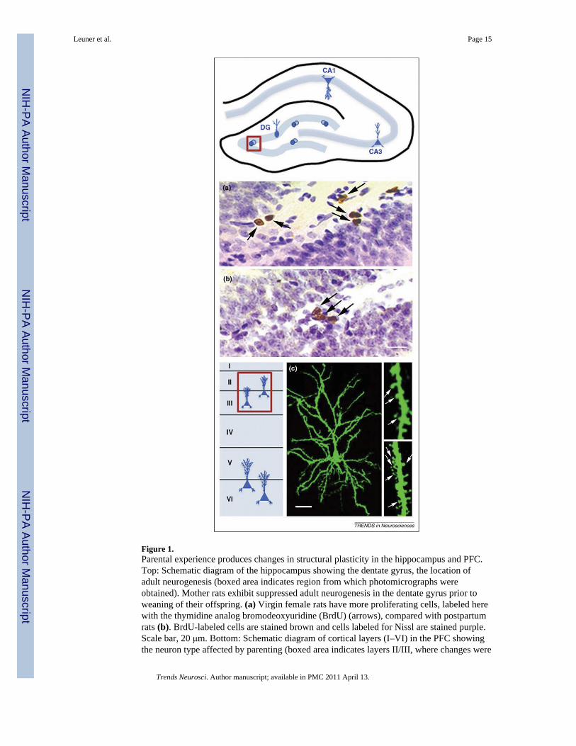
Figure 1.Parental experience produces changes in structural plasticity in the hippocampus and PFC.Top: Schematic diagram of the hippocampus showing the dentate gyrus, the location ofadult neurogenesis (boxed area indicates region from which photomicrographs wereobtained). Mother rats exhibit suppressed adult neurogenesis in the dentate gyrus prior toweaning of their offspring. (a) Virgin female rats have more proliferating cells, labeled herewith the thymidine analog bromodeoxyuridine (BrdU) (arrows), compared with postpartumrats (b). BrdU-labeled cells are stained brown and cells labeled for Nissl are stained purple.Scale bar, 20 μm. Bottom: Schematic diagram of cortical layers (I–VI) in the PFC showingthe neuron type affected by parenting (boxed area indicates layers II/III, where changes were
Leuner et al. Page 15
Trends Neurosci. Author manuscript; available in PMC 2011 April 13.
NIH
-PA Author Manuscript
NIH
-PA Author Manuscript
NIH
-PA Author Manuscript

detected in pyramidal neurons). Father marmosets exhibit enhanced dendritic spine densityon pyramidal neurons of layer II/III PFC compared to non-fathers. (c) Layer II/III PFCpyramidal cell of a marmoset father labeled with the lipophilic tracer DiI (fluorescentgreen). Magnified views of DiI labeled dendritic segments showing dendritic spines(arrows) in a control (right upper) and a father (right lower). Scale bar, 30 μm for cell, 5 μmfor dendritic segments. Adapted, with permission, from Ref. [25,73].
Leuner et al. Page 16
Trends Neurosci. Author manuscript; available in PMC 2011 April 13.
NIH
-PA Author Manuscript
NIH
-PA Author Manuscript
NIH
-PA Author Manuscript

Figure 2.Parenting can alter anxiety and cognition by inducing structural changes through potentiallydifferent mechanisms. This model diagram illustrates some of the structural changes thatoccur with parenting, including suppressed neurogenesis in the dentate gyrus, reduceddendritic complexity in the CA3 pyramidal cell population of the hippocampus, andenhanced dendritic spine density in pyramidal cells of the CA1 hippocampal region andlayer 2–3 of the PFC. Parenting-induced elevated glucocorticoid levels might underliechanges in the dentate gyrus and CA3 regions of the hippocampus, whereas the enrichingaspects of infant contact might produce changes in the CA1 region and PFC. Changes in thestructure of the hippocampus and PFC might be responsible for parenting-inducedalterations in behaviors associated with these brain regions, including reduced anxiety-likebehavior and enhanced cognition. Photo credit: J. Alberts (University of Indiana).
Leuner et al. Page 17
Trends Neurosci. Author manuscript; available in PMC 2011 April 13.
NIH
-PA Author Manuscript
NIH
-PA Author Manuscript
NIH
-PA Author Manuscript