US of the chest in pediatric patients - replacing the ... fileChest wall sonography Infection...
43
US of the chest in pediatric patients - replacing the chest x ray? Anat Ilivitzki Head Pediatric Imaging Unit Ruth Children Hospital Rambam Health Care Campus
Transcript of US of the chest in pediatric patients - replacing the ... fileChest wall sonography Infection...

US of the chest in pediatric
patients - replacing the
chest x ray?
Anat Ilivitzki
Head Pediatric Imaging Unit
Ruth Children Hospital
Rambam Health Care Campus
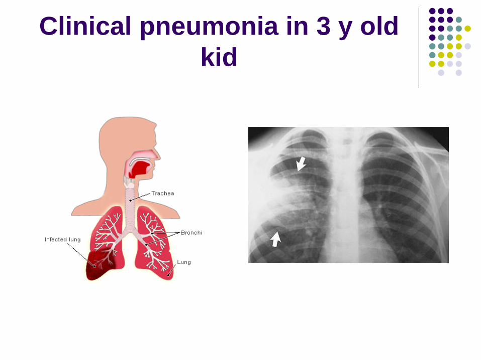
Clinical pneumonia in 3 y old
kid


Chest wall sonography
Infection – cellulitis, sternal osteomyelitis
Inflammation – SELSTOC
Trauma- rib fracture
Tumor - PNET

sternal osteomyelitis

SELSTOC
self limiting sternal tumor of childhood
Sonographic gray scale image of a dumb bell lesion is seen between the
segments of the sternum (white arrows), bulges anterior and posterior to
the sternum (stars) .The lesion is hypoechogenic, homogenic. No invasion
is seen to the bone or lung.

Fractured rib
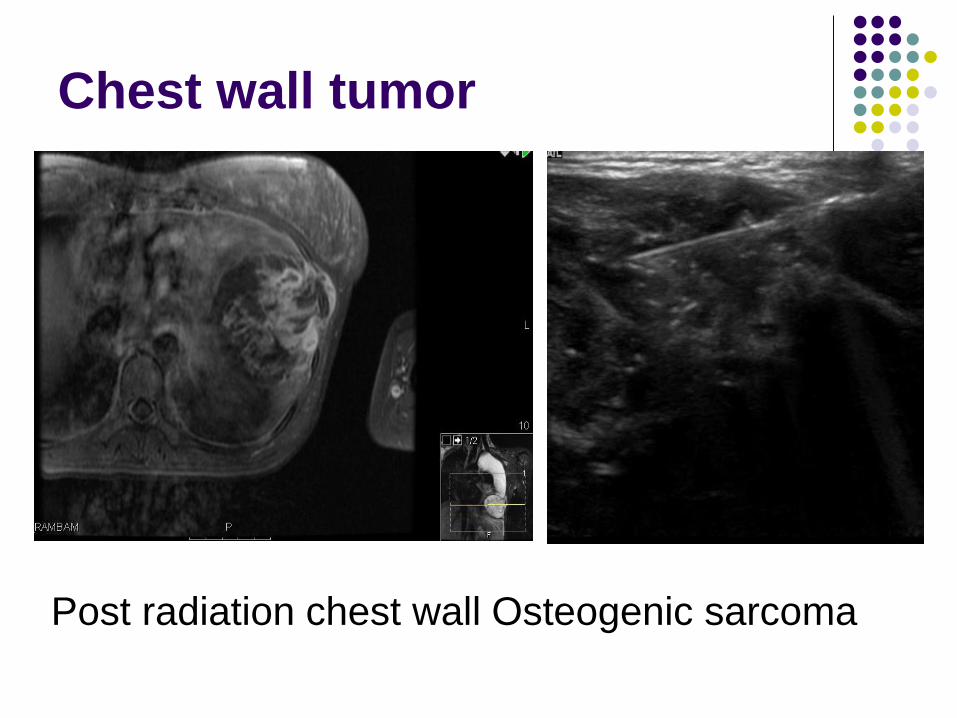
Chest wall tumor
Post radiation chest wall Osteogenic sarcoma
A bit deeper..
Thymus and thymic lesions
Anterior mediastinum tumors
Pleura: Pneumothorax
Effusion
Pericardial fluid
Diaphragm

Normal Thymus

HD in anterior mediastinum
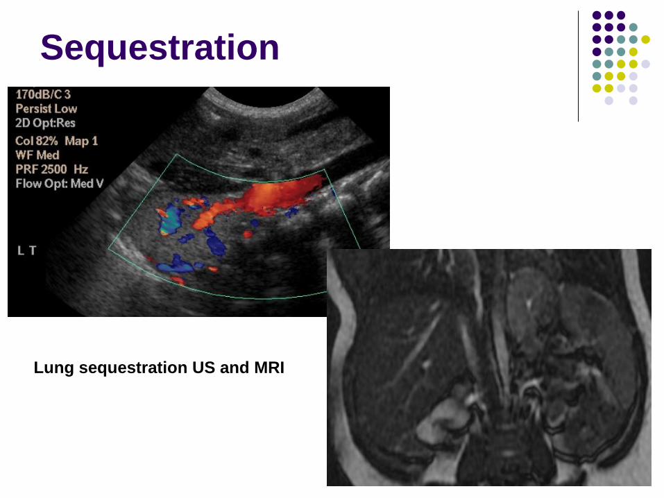
Sequestration
Lung sequestration US and MRI
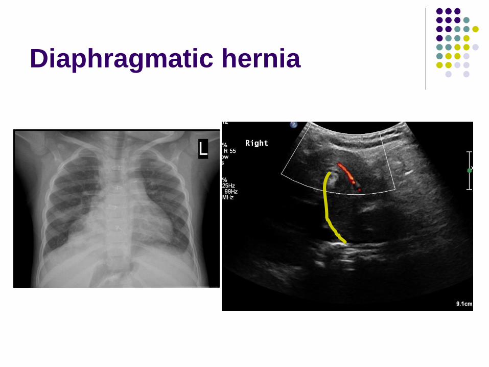
Diaphragmatic hernia
LUS for CAP in children?
Diagnosis of CAP in children is clinical
>80% of suspected CAP cannot be confirmed radiologically
Hazir T et al.Chest radiography in children aged 2-59m diagnosed with non – severe pneumonia as defined by WHO:descriptive multicenter study in Pakistan . BMJ2006:333
Murphy CG et al.Clinical predictors of occult pneumonia in febrile child. Acad Emerg Med2007:14;243-249.
LUS in adults showed good accuracy in diagnosing CAP, at least as good as X ray.

Sonography of the lung
Normally impossible:
“Because ultrasound energy is rapidly
dissipated by air, ultrasound imaging is not
useful for the evaluation of the pulmonary
parenchyma“Longo D, Fauci A, Kasper D, Hauser S, Jameson J, Loscalzo J: Harrison’s
Principles of Internal Medicine. 2008.

Physics
Air determines a high acoustic mismatch with
the surrounding tissues, causing a complete
reflection of the ultrasound beam, preventing
the creation of direct imaging of the
pulmonary parenchyma
In a normally aerated lung, the only
detectable structure is the pleura, visualized
as a hyperechoic horizontal line.

Anatomy of the normal lung
Arrows indicate A-lines. Above A-lines the pleural line is visible with
its horizontal movement, the lung sliding.

Anatomy of the normal lung
B lines

Physics
When the air content decreases and lung density increases
(exudate, transudate, collagen, blood, etc. ) - the
acoustic mismatch between the lung and the
surrounding tissues is lowered, and the ultrasound beam
can be partly reflected at deeper zones and repeatedly.
This phenomenon creates some vertical reverberation
artefacts known as B-lines
B-lines belong to the family of the comet-tail artifacts, well
known in the setting of abdominal ultrasound

Artifacts of the pathologic lung
B lines


Black to White ( 0-10 B lines)Extent of extravascular fluid in lung
Volpicelli G, Mussa A, Garofalo G, Cardinale L, Casoli G, Perotto F, Fava C, Frascisco M:
Bedside lung ultrasound in the assessment of alveolarinterstitial syndrome .Am J Emerg Med 2006 ,24:689–96

Technique of LUS

Signs of lung consolidation
VascularPleuralParenchymal
Enhanced
vascular tree like
Pleural line
attenuation
Echopoor area
Pulmonary and
bronchial a.
Localized pleural
effusion
Air bronchogram
CEUS – early
enhancement
Basal pleural
effusion
Fluid bronchogram
Necrotic area
within pneumonia
Superficial fluid
alveologram

Signs of lung consolidation
Most important parenchimal sign:
air bronchogram in echo poor area – 70-97%
of cases.
Most important pleural sign: basal effusion
34-61%
Vascular criteria needed only for D.D.

Echopoor area with air bronchogram

Air Bronchogram

Fluid Bronchogram

Lung Hepatization
Liver
*

Necrotic area within pneumonia


Pleural Effusion

Lung sonography in children

LUS in CAP
Prospective study
Clinial pediatric pneumonia(1m-14y).
Chest X ray (2 ) & LUS
48 cases of pneumonia by X ray
LUS compared to the X ray:
Sens 97.9%, spec 94.5%, PPV 94%, NPV 98.1%.
Concordance of the two methods in identifying
the type of CAP was poor.

Usefulness of LUS in the diagnosis of CAP
in children- Meng- Chieh et al.Pediatrics and Neonatology 2015
Retrospective
Up to 2 days between X ray and LUS.
Diagnosis of pneumonia was clinical.
LUS compared to X ray.
Cliniacl CAP in163 patients:
X ray 152 patients, LUS 159 .
Conclusion: LUS is a complementary tool to
chest radiography in the diagnosis of
pneumonia in children

Meta – analysis ( at last..)

Author (et al) Year Origin Prospective Sample size
Reali 2014 + 180
Liu 2014 + 107
Esposito 2014 + 103
Shah 2013 + 191
Caiulo 2013 + 102
Seif El Dien 2013 + 95
Iuri 2009 + 28
Copetti and
Cattarossi
2008 + 79
Altogether 765 kids

Results:
Overall sensitivity 96%, specificity 93%.
In another meta analysis by the same group
in adults: sensitivity 94%, specificity 96%.
FP: atelectases, infarct
FN: small consolidation which doesn’t reach
pleura
( In Shah et al sens. 86%, spec. 89% <1 cm,
spec. 97% when > 1 cm)

Results:
All studies used x ray for diagnosis of CAP
3 x ray alone
5 x ray + clinical diagnosis
When using X ray alone for diagnosis of
CAP, sensitivity of LUS stays the same but
specificity declines to 84%, meaning that x
ray alone is not sufficient for diagnosis of
pneumonia

Results:
Specificity
LUS
Sensitivity
LUS
92%96%6 studies kids
100%96%2 studies neonates
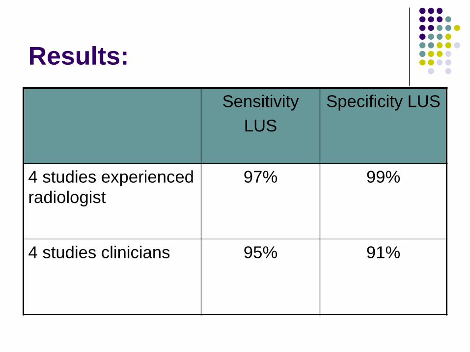
Results:
Specificity LUSSensitivity
LUS
99%97%4 studies experienced
radiologist
91%95%4 studies clinicians
Take home massages
It seems that LUS for CAP is as good as or
maybe better than x ray.
A bit better when done by radiologist
There are FP and FN
Still small numbers in the literature, but the
results are promising

Should we try it??