Uropathology 1 kidney
54
Pathology Specimens for Urology Residents Dr Prashant Bansal
-
Upload
prashant-bansal -
Category
Health & Medicine
-
view
78 -
download
2
Transcript of Uropathology 1 kidney

Pathology Specimens for Urology Residents
Dr Prashant Bansal

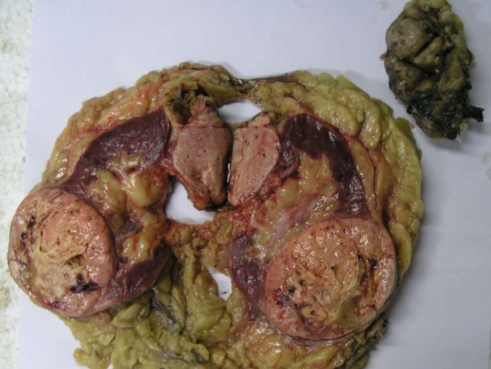
SPECIMEN NO 1


• Bilateral nephrectomy specimens.
• The external surface of both kidneys have a
bosselated appearance which is produced by
numerous cysts of varying sizes and
replacing the entire renal parenchyma.
• Cysts are thin walled and translucent and
filled with clear fluid.
• Adult (autosomal dominant) polycystic
kidney disease

• Cut open specimen of kidney. • The kidney is enlarged and replaced by numerous cysts
of varying sizes arising at all levels in the cortex and medulla.
• The cysts are unilocular and are filled with clear serous to hemorrhagic fluid.
• They range from a few millimeters to several centimeters.
• Normal renal parenchyma is not identified. • No mass or papillary lesions seen. • ADPKD

CASE NO 2


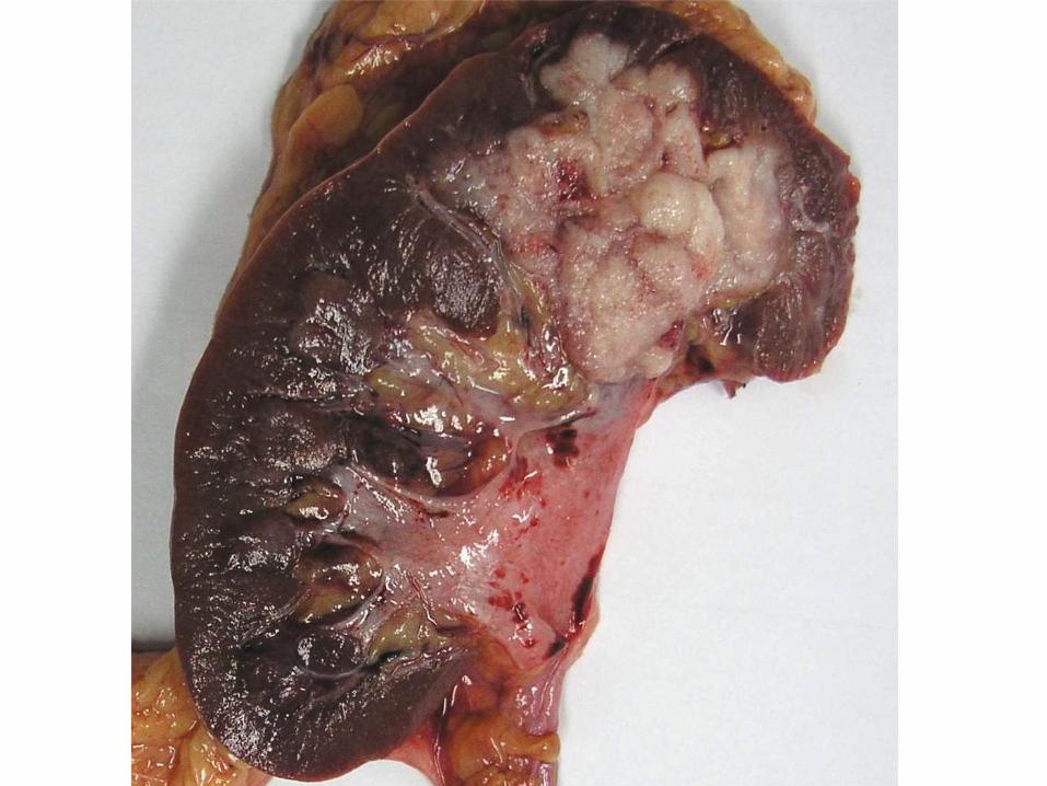
• A solitary poorly circumscribed solid grey white tumour is seen involving the upper calyceal system and infiltrating into the renal parenchyma.
• Foci of haemorrhage are identified in the pelvic mucosa.
• The rest of the renal parenchyma appears unremarkable.
• Urothelial carcinoma

CASE NO 4


• Hydatid Cyst Kidney

CASE NO 5

• Grossly hydronephrotic kidney with thinned out parenchyma

CASE NO 6


• Nephroureterectomy specimen
• Small sized kidney
• Grossly dilated and tortuous ureter
• s/o reflux nephropathy

CASE NO 7

• Nephrectomy specimen
• Upper pole mass
• S/o RCC

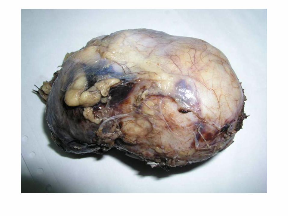
CASE NO 9


• Solitary well circumscribed exophytic tumour arising from the mid pole of kidney.
• Tumour is tan in colour, homogenous with a central scar.
• Rest of the renal parenchyma shows no significant pathology

Case 4


• Solitary well circumscribed tumour in the upper pole of kidney.
• Tumour is mahogany brown and homogenous.
• No cystic, necrotic or haemorrhagic areas seen.
• Rest of the renal parenchyma shows no significant pathology
• HPE was Chromophobe RCC (not to be commented in exam – JUST say RCC)

Case 6

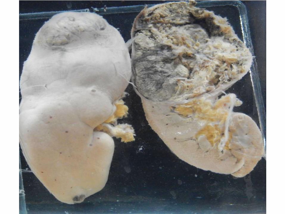
• Cut open specimen of kidney with dilated pelvicalyceal system.
• The calyceal mucosa has plaques of golden yellow tissue which focally invades the renal parenchyma and extends into the perirenal fat.
• Xanthogranulomatous pyelonephritis

Case 7

Case 9
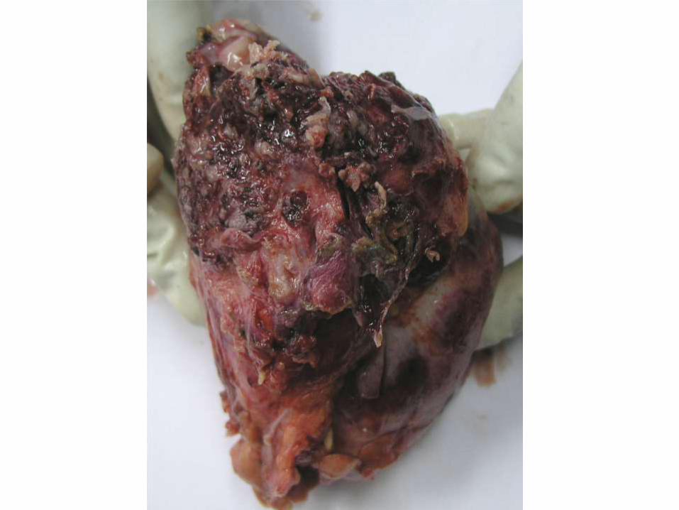

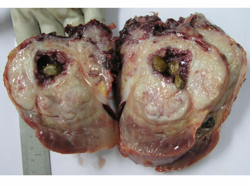
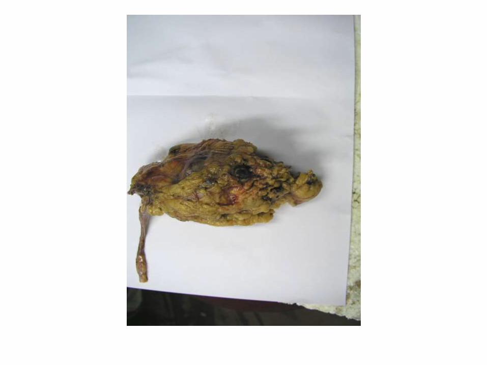
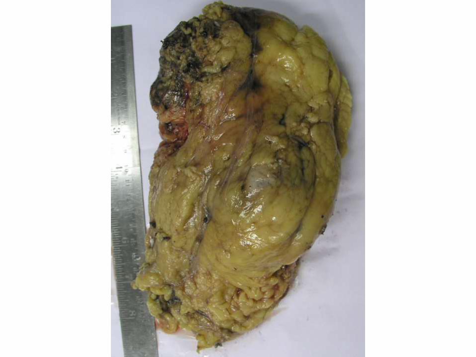
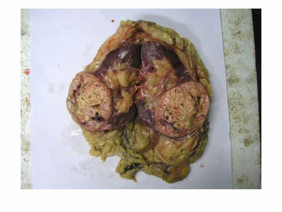
• Specimen of kidney.
• There is a solid white tumour involving the upper pole of the kidney.
• The tumour extends beyond the renal capsule into the perirenal fat.
• Calculi are embedded within the tumour.
• Squamous cell carcinoma with nephrolithiasis, kidney


Case 10


Case 11

• Renal tumour arising from the lower pole of the kidney. • Tumour is well circumscribed, solid with areas of necrosis. • The ipsilateral adrenal gland shows a tumour with the
same gross appearance as the renal tumour.• The renal parenchyma between the renal tumour and the
adrenal tumour is unremarkable.• The makes the adrenal mass a metastatic lesion and not an
extension from the primary tumour.• Also included in this image is the contra lateral adrenal
gland which appears grossly enlarged, suggesting metastatic involvement.
• (Clear cell RCC with bilateral adrenal metastasis)