Urological Divisions 1 、 Pediatric urology , 2 、 Urologic Oncology 3 、 Renal Transplantation...
51
Urological Divisions 1 、 Pediatric urology , 2 、 Urologic Oncolo gy 3 、 Renal Transplantation 4 、 Male Infertility 5 、 Urinary Tract Stones 6 、 Endourology 7 、 Female Urology 8 、 Urinary Incont inence 9 、 Genitourinary Trauma 10 、 Erectile Dysfuncti on 11 、 Genitourinary Reconstruction ) 12 、 Voiding Disorders
-
Upload
aldous-pierce -
Category
Documents
-
view
224 -
download
0
Transcript of Urological Divisions 1 、 Pediatric urology , 2 、 Urologic Oncology 3 、 Renal Transplantation...

Urological Divisions1 、 Pediatric urology , 2 、 Urologic Oncology
3 、 Renal Transplantation 4 、 Male Infertility
5 、 Urinary Tract Stones 6 、 Endourology
7 、 Female Urology 8 、 Urinary Incontinence
9 、 Genitourinary Trauma 10 、 Erectile Dysfunction
11 、 Genitourinary Reconstruction )12 、 Voiding Disorders
TUMORS OF GENITOURINA
RY TRACT
Yiran Huang
Department of Urology,
Renji Hospital, SSMU,

Renal cell carcinoma
Renal cell carcinoma (RCC) is the most
common type of kidney cancer. These
tumors occur twice as often in men as in
women and usually occur in adults between
the ages of 50 and 70. Between 25 and 30%
of patients have metastases at the time of
diagnosis.

Environmental risk factors include
smoking , Phencetin-containing painki
llers abused or over-used, some heavy
metals exposure ( lead and cadmium).
Other risk factors include long-term
dialysis, overweight, a high fat diet.

There are also hereditary risk factors.
There are some genes mutations in rar
e syndromes like tuberous sclerosis an
d von Hippel Lindau Disease that are
associated with an increased risk of de
veloping kidney tumors.

Von Hipple-Lindau (VHL) disease is an aut
osomal dominant disorder in which affected
individuals are at risk for retinal angiomas,
central nervous system hemangioblastomas,
renal cysts and carcinomas, pancreatic cysts
and tumor, pheochromocytomas,endolymph
atic sac tumors, and/or epididymal cystaden
omas.

Types of renal cell carcinoma:
Clear cell RCC (60 to 75 percent), papillary
RCC (15 percent), chromophobe RCC (5 p
ercent), collecting duct carcinoma (less tha
n I percent), and unclassified carcinoma (u
p to 5 percent).

Clinical Presentation
1. Renal cell carcinoma can become quite lar
ge without causing any symptoms. Many t
umors are found incidentally.
2. Hematuria is the most common symptom.
Other signs and symptoms include low back
pain, a mass in the abdomen,

3. General symptoms. fatigue, weight loss, fever, anemia, swelling of the legs and night sweats
4. Paraneoplastic syndromes. Hypercalcemia , Stauffer's syndrome protein-wasting enteropathy, erythrocytosis, neuromyopathy, and gonadotropin production. Amyloidosis is present in approximately 2 percent of patients.

There are multiple imaging studies, incl
uding intravenous pyelogram (IVP), ult
ra-sonography, computerized tomograp
hic (CT) scans, magnetic resonance ima
ging (MRI) which can produce images o
f the kidney.

Arteriography and angiography can
demostrate blood vessels and image th
e kidney. A chest x-ray is used to evalu
ate the lungs metastases. A bone scan c
an identify cancer in bone.

•Staging of Renal Cell Carcinoma
Stage Ⅰ cancer is confined to the kidney, Stage Ⅱ means the cancer has broken through the kidney capsule and has spread into adjacent tissue, Stage Ⅲ indicates that it may have spread further into lymph nodes or blood vessels, Stage Ⅳ indicates that it is more wide-spread, particularly in other organs

TreatmentRadical nephrectomy is the gold standard t
reatment for localized RCC. Components o
f a radical nephrectomy include early vascu
lar ligation and en bloc removal of the kidn
ey, Gerota's fascia, ipsilateral adrenal, upp
er ureter, and, for some, lymph nodes from
the crus of the diaphragm to the aortic bifu
rcation.

Laparoscopic radical nephrectomy
Nephron sparing surgery
Absolute indications: include a solitary kidney, bilateral tumors, poor bilateral or contralateral renal function.
Relative indications: smaller tumors (4 cm or smaller) with a normal contralateral kidney.
Approaches include segmental polar nephrectomy, wedge resection, or tumor enucleation
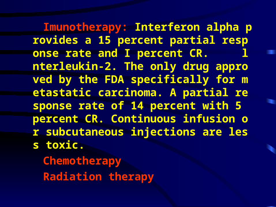
Imunotherapy: Interferon alpha provides a 15 percent partial response rate and I percent CR. lnterleukin-2. The only drug approved by the FDA specifically for metastatic carcinoma. A partial response rate of 14 percent with 5 percent CR. Continuous infusion or subcutaneous injections are less toxic.
Chemotherapy
Radiation therapy

NEPHROBLASTOMA ( Wilms’ Tumor )

Wilms’ tumor, is exclusively a disease of children under age 4 years. It is a highly malignant mixed tumor consisting of tissues of connective tissue origin and epithelial structures.

It is usually discovered as a palpable mass by the mother or by an examining physician. Treatment of the primary tumor is radical nephrectomy. Adjunctive therapy with irradiation and chemotherapy is important in improving survival.

TUMOR OF THE RENAL PELVIS

The transitional epithelium of the renal pelvis may arise to malignant tumors that are similar to lesion that occur in the bladder. Hematuria is the most commonly the presenting complain. Intravenous urography and retrograde urography can show the filling defect.

Treatment
Nephroureterectomy
The classic therapy for upper tract TCC is nephroureterectomy with excision of a bladder cuff.
Laparoscopic nephrourectomy

BLADDER CANCER

Bladder cancer is the most common urologi
c malignancy The most common histologic
diagnosis is transitional cell carcinoma (TC
C). Sixty to 75 percent of these lesions are n
oninvasive, superficial tumors, but 10 to 20
percent of these tumors will progress to mus
cle-invasive disease, especially patients wit
h high-grade disease and transitional cell ca
rcinoma in situ.

Several factors are associated with the development
of bladder cancer.
1.Occupational exposure to chemicals is thought to
cause the disease.
2.Smoking is also related to developing bladder can
cer. The risk of developing the disease in smoking i
s four times that in non-smoking.
3.Chronic cystitis and bladder stone can cause squa
mous cell carcinoma.

PathologyTypes of bladder tumor: transitional cell carcinoma, squamous cell carcinoma, adenocarcinoma, and others. More than 95% primary bladder carcinomas are transitional cell carcinoma. Squamous cell carcinoma and adenocarcinoma account for approximately 2-3% respectively. Adenocarcinoma may arise from an urachal remnant at the dome of the bladder or from submucosal gland in the vicinity of the bladder neck.

GradeThe grade is based on the degree of cellular differentiationgradeⅠbeing well differentiation, and gradeⅡ being moderate differentiation
grade Ⅲ being poorly differentiation.

StageThe stage of Bladder carcinoma includes tumor in situ (Tis), papillary tumor confined to mucosa (Ta), invades submucosa only (T1), invades superficial muscle (T2), invades deep muscle (T3), spreads beyond the bladder, including into the prostate or other organs (T4).

Transitional cell carcinoma has thre
e features: multiple lesions in bladder,
multiple organs (pelvis, ureter, and po
st urethral can develop transitional cel
l carcinoma at same time.), recurrence
(more than 50% patient will recur afte
r the operations).

Location of Bladder TumorMost are located on lateral and posterior wall of bladder, then on the top and the triangle.

Transitional cell carcinoma of the bladder develops more frequently in men than in women, and its frequency increases with age. The Prognosis is poor for aging patient and female. Carcinoma of bladder is quite rare in children.

Clinical presentations
Hematuria. Either gross or microscopic hematuria is present in 85 percent of cases. The amount of hematuria is not necessarily proportional to the severity of the lesion, and intermittence is not a reason to exclude an evaluation. Hematuria in older patients may result in the diagnosis of a urologic malignancy in 10 percent of patients, with the majority of these lesions being TCC.
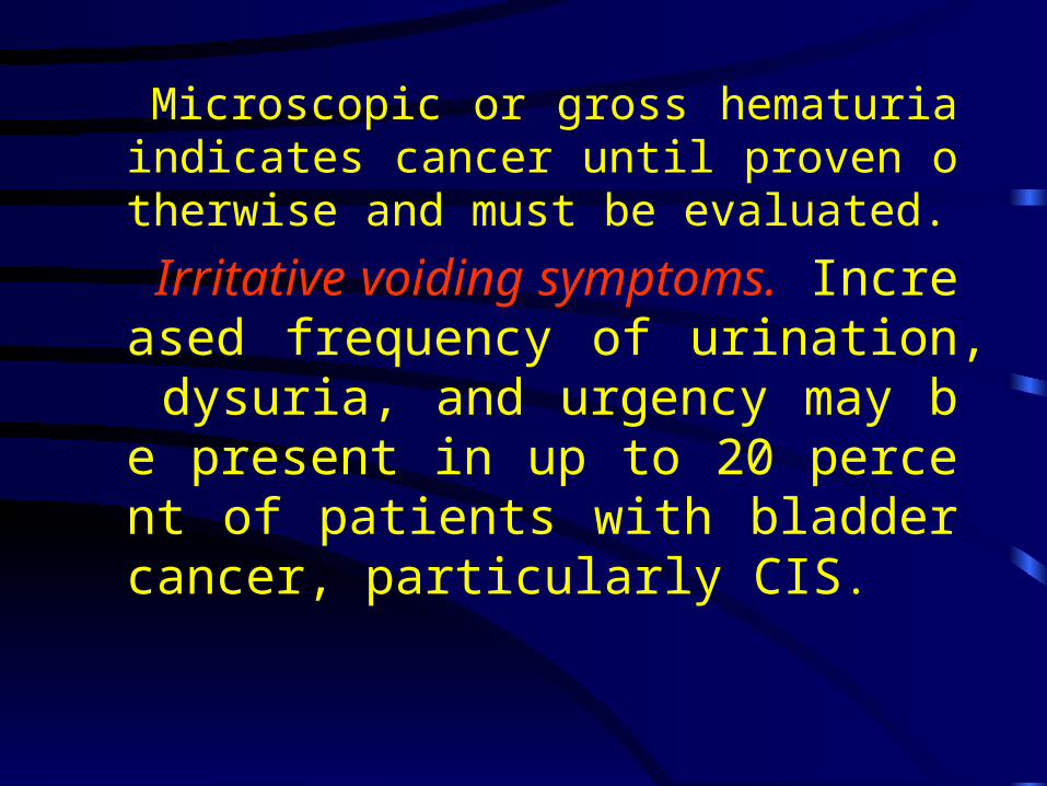
Microscopic or gross hematuria indicates cancer until proven otherwise and must be evaluated.
Irritative voiding symptoms. Increased frequency of urination, dysuria, and urgency may be present in up to 20 percent of patients with bladder cancer, particularly CIS.

Diagnosis1.The diagnosis is established by cystoscopy
and biopsy of the tumor.
2.Urinary cytological test may be used to fi
nd the cancer cell.
3.Intravenous urography is essential for the
bladder carcinoma.
4.Ultrasound, CT scan, and MRI detect the
tumor in bladder, especially for the tumor s
tage.

TreatmentSuperficial bladder tumors (Ta and T1) are often amenable to transurethral resection or transurethral coagulation of laser.Invasive bladder tumors (T2 and T3) may require partial cystotectomy, or radical cystotectomy.

Systemic chemotherapy with multiple
drug regimens incorporating cisplatin
has shown promise for the advanced d
isease.
Intravesical chemotherapy may be effe
ctive in controlling superficial disease
but is ineffective for treatment of invas
ive lesion.

•
The most effective agent is bacillus Calm
ette-Guerin (BCG) intravesical immunot
herapy, which decreases tumor recurrenc
e by 40 to 70 percent. In the case of transi
tional cell carcinoma in situ (CIS), it may
reduce tumor progression.

After treatment of the superficial tumor, pe
riodic cystoscopy and urinary cytological e
xamination is necessary for at least 5 years.
New tumors may also be well controlled by
transurethral means, but if they tend to rec
ur they apt to become progressively invasiv
e and of higher grade. Cystectomy must the
n be considered.

PROSTATE CARCINOMA

Prostate carcinoma is the most common
malignance in the West countries, and the
second death after lung cancer. Although
the incidence of prostate carcinoma is low in
China and eastern countries, remarkable
increase has been found recently.

It occurs predominantly in old men.
It has the capacity to grow and invad
e locally and to metastasize by blood
and lymph. There is a strong predile
ction for metastases to bone, and the
se metastases have the unique charac
teristic of being osteoblastic.

Prostate cancer has few dramatic prima
ry signs or symptoms. It can be associa
ted with urinary obstructive symptoms
or hematuria, although these findings a
re usually due to other causes. Bone pa
in can unfortunately be an initial sympt
om, but it represents very advanced dis
ease.

Diagnosis1. digital rectal examination, 2. prostate specific antigen,3. transrectal ultrasound exam, 4. biopsy,5. bone scan, and so on.

Androgen blockade is the best method for the treatment of advanced prostate carcinoma.Radical prostatectomy and Radiation can be used for early stage patient.

TESTICULAR TUMORS

Testis tumors most commonly occur in men between 18 and 35 years of age. Tumors arising from the Leydig’s cell and Sertoli’s cells are rarely seen. Most of the testis tumors arise from the germinal epithelium, and all of these tumors should be considered malignant except for teratomas that occur before puberty.

The histologic types of germinal tumors of the testis are seminoma, and non-seminoma (embryonal carcinoma, teratoma, and choriocarcinoma). The diagnosis is usually suspected because of discomfort and enlargement of the testis. а-FP and β-HCG are the tumor markers, and the later is a poor prognosis marker.

Radical orchetectomy is the initial treatment. Seminoma is quite radiosensitive, and metastatic disease may be definitively treated with radiotherapy. Non-seminoma is usually managed by a combination of retroperitoneal lymph node dissection and multiple drupe chemotherapy.

CARCINOMA OF THE PENIS

Squamous cell carcinoma of the penis is encountered most often in older men who have not been circumcised. It has a tendency to invade the erectile tissue of the penile shaft and to metastasize to inguinal lymph nodes. Penectomy is usually required.

Questions of the lessons
1. What paraneoplastic syndromes are associ
ated with renal cell carcinoma?
2. What are the indications for intravisical th
erapy for bladder carcinoma?
3. What are the indications for radical cystot
ectomy?
4. The diagnosis of prostate carcinoma.