Urinary system outflow obstruction and urinary system tumors

Module A
Urinary System
Martin ŠpačekHistology and Embryology
• Pictures from:• Junqueira et al.: Basic histology• Rarey, Romrell: Clinical human embryology• Sadler: Langman’s medical embryology• Young, Heath: Wheather’s functional histology

UrinarySystem
• Kidney• Ureter• Urinary
bladder• Urethra


Development of the Urinary System
• Development of the kidney– Pronephros– Mesonephros– Metanephros
• Development of excretory passages

Pronephros• Intermediate mesoderm of the cranial 12-13 somites• Rudimentary, nonfunctional

Mesonephros
• At the end of the 4th week• Functional for a short time• Renal corpuscle
– glomerulus– Bowman’s capsule
• Tubules enter collecting duct –mesonephric or Wolffian duct

Mesonephros

Metanephros• Permanent kidney• Development begins in the 5th week• Ureteric bud (an outgrowth of the mesonefric duct)
• Metanephric blastema

Metanephros• Ureteric bud → collecting tubules,
calyces, renal pelvis, ureter• Metanephric blastema → nephrons

Position of the Kidney• Ascent of the kidney
– initially located in the pelvic region– later more cranial position in the abdomen
Horseshoe kidney

Urogenital SinusUrorectal septum divides cloaca into:
anorectal canalurogenital sinus

Urinary BladderDevelops from the upper and largest part of the urogenital sinusContinuous with the allantois, but the lumen obliterates → urachus (median umbilical ligament)

Kidney
• Nephrons• Collecting tubules
& ducts• Juxtaglomerular
apparatus• Blood supply• Intersticium

Nephron
• Renal corpuscle– Glomerulus– Bowman’s capsule
• Proximal tubule• Henle’s loope• Distal tubule

Renal Corpuscle• Glomerulus
– fenestrated capillaries (no diaphragms)

Renal Corpuscle• Bowman’s capsule
– parietal layer (simple squamous epitelium)– visceral layer (podocytes with pedicels)


Renal Corpuscle• Filtration barrier
– basement membrane (collagen type IV and heparansulfate)

glomerular capillary – lumen urinary space
direction of flow of the plasma ultrafiltrate

Renal Corpuscle• Mesangial cells
– synthesize the extracellular matrix and collagen– macrophages (?)

Proximal Tubule
• Simple cuboidalepithelium– brush border– basolateral labyrinth– abundant
mitochondria• Reabsorption of NaCl
and water (80-95%)


Henle’s loope
• Squamous simple epithelium– ascending limb is
impermeable to water• Juxtamedullary
nephrons have long HL

Distal Tubule• Simple cuboidal
epithelium– cells are smaller than
those of the proximal tubule
– no brush border– basolateral labyrinth
• Absorption of sodium, secretion of potassium
• Macula densa – modified segment– chemoreceptors


Collecting Tubules & Ducts• Collecting tubules
– simple cuboidalepithelium
• Papillary ducts– simple columnar
epithelium• ADH-dependent
water reabsorption


Juxtaglomerular Apparatus
• Macula densa of the distal tubule– sensitive to changes in NaCl concentration
• Extraglomerular mesangial cells (laciscells)
• Juxtaglomerular cells of the afferent arteriole– produce renin


Blood supply
• renal artery• interlobar arteries• arcuate arteries• interlobular arteries• afferent arterioles• efferent arterioles• peritubular capillary
network or vasa recta

Renal Intersticium
• Intersticial cells in the spaces between tubules and vessels
• Non-cellular elements• proteoglycans, glycoproteins, interstitial fluid

Kidney HE (U1)Kidney HE (U1)

Kidney HE (U1)Kidney HE (U1)

Kidney Van Kidney Van GiesonGieson (U2)(U2)

Excretory Passages
• Calyces and renal pelvis
• Ureters• Urinary bladder• Urethra

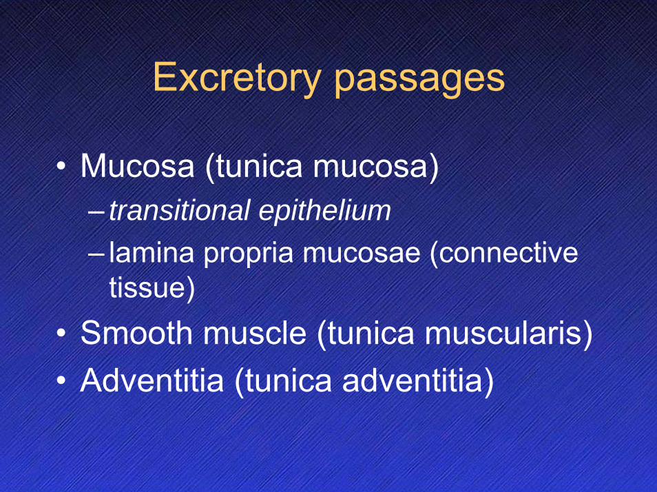
Excretory passages
• Mucosa (tunica mucosa)– transitional epithelium– lamina propria mucosae (connective
tissue)• Smooth muscle (tunica muscularis)• Adventitia (tunica adventitia)

Ureter
• Folded mucosa• Smooth muscle
– inner layer longitudinal
– outer layer circular

UreterUreter HE (U3)HE (U3)

UreterUreter HE (U3)HE (U3)

Urinary bladder
• Mucosa folded• Smooth muscle layer
– mixture of randomly arranged fibers (m. detrusor)
– at the neck the fibers form a three layer sphincter (inner longitudinal, middle circular, outer longitudinal)

Urinary bladder HE (U4)

Urinary bladder HE (U4)

Urinary bladder Van Urinary bladder Van GiesonGieson (U5)(U5)