Urinary System Ji-Cheng Li Urinary System Ji-Cheng Li.
36
Urinary Sys Urinary Sys tem tem Ji-Cheng Li Ji-Cheng Li
-
Upload
randolf-bishop -
Category
Documents
-
view
257 -
download
1
Transcript of Urinary System Ji-Cheng Li Urinary System Ji-Cheng Li.
Urinary SystemUrinary System
Ji-Cheng LiJi-Cheng Li
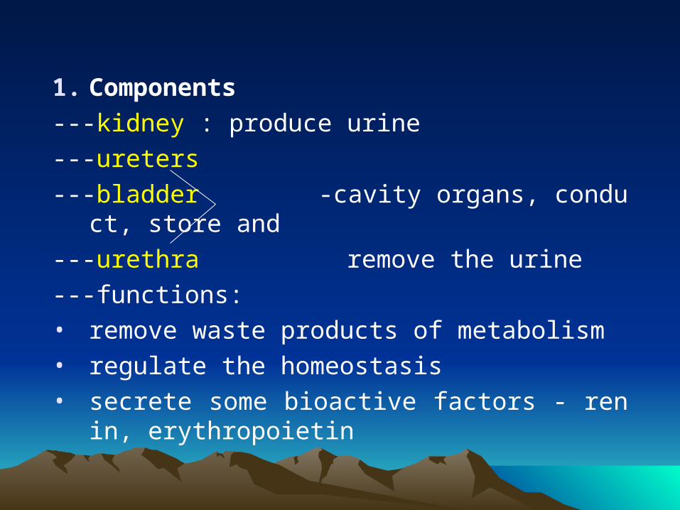
1. Components
---kidney : produce urine
---ureters
---bladder -cavity organs, conduct, store and
---urethra remove the urine
---functions: • remove waste products of metabolism• regulate the homeostasis• secrete some bioactive factors - renin, erythr
opoietin
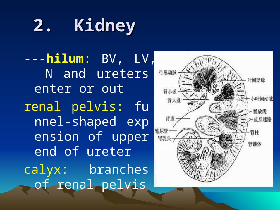
2. Kidney2. Kidney
---hilum: BV, LV, N and ureters enter or out
renal pelvis: funnel-shaped expension of upper end of ureter
calyx: branches of renal pelvis
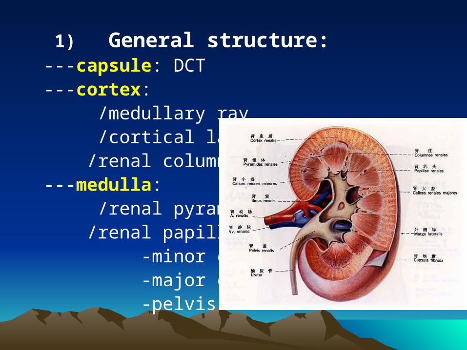
1) General structure: ---capsule: DCT---cortex: /medullary ray /cortical labyrinth /renal columns---medulla: /renal pyramid /renal papillae: -minor calyx -major calyx -pelvis
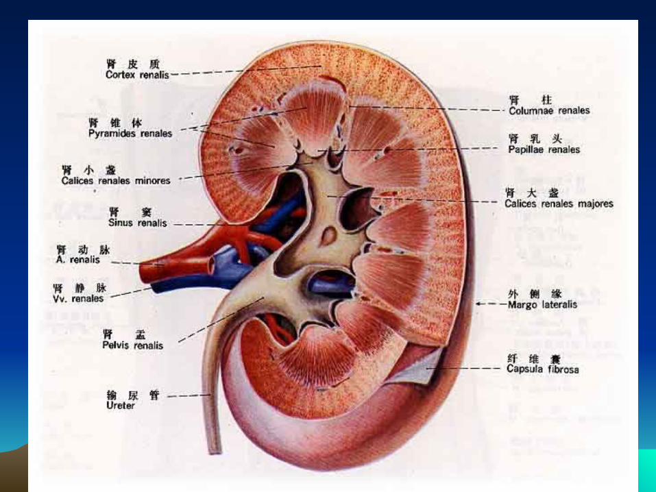
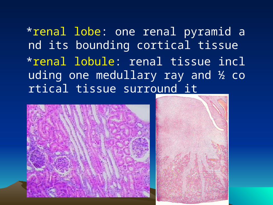
*renal lobe: one renal pyramid and its bounding cortical tissue
*renal lobule: renal tissue including one medullary ray and ½ cortical tissue surround it
According to function, renal parenchyma is mainly consists of uriniferous tubules
---parenchyma:
/uriniferous tubules= renal tubule+ collecting tubules
/renal corpuscle = glomerulus + renal capsule (beginning part of renal tubule)
/nephron = renal corpuscle + renal tubule
---interstitium: CT, BV, N
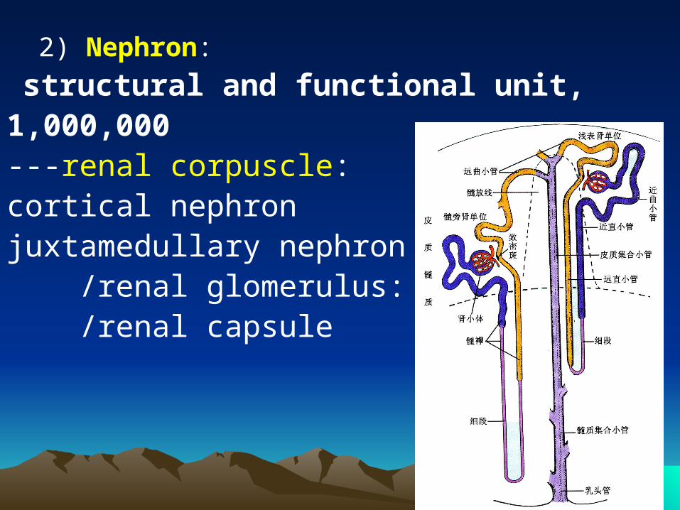
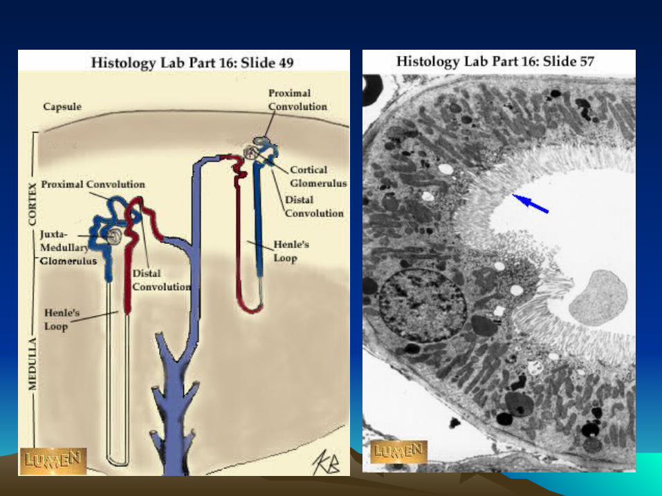
2) Nephron:
structural and functional unit, 1,000,000---renal corpuscle: cortical nephronjuxtamedullary nephron /renal glomerulus: /renal capsule
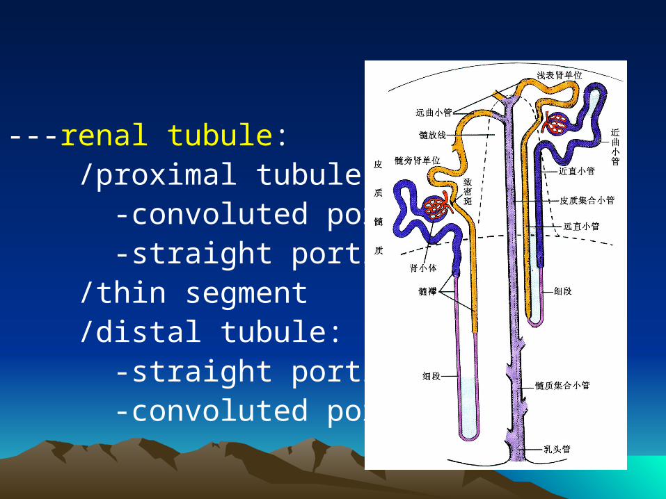
---renal tubule: /proximal tubule: -convoluted portion -straight portion /thin segment /distal tubule: -straight portion -convoluted portion
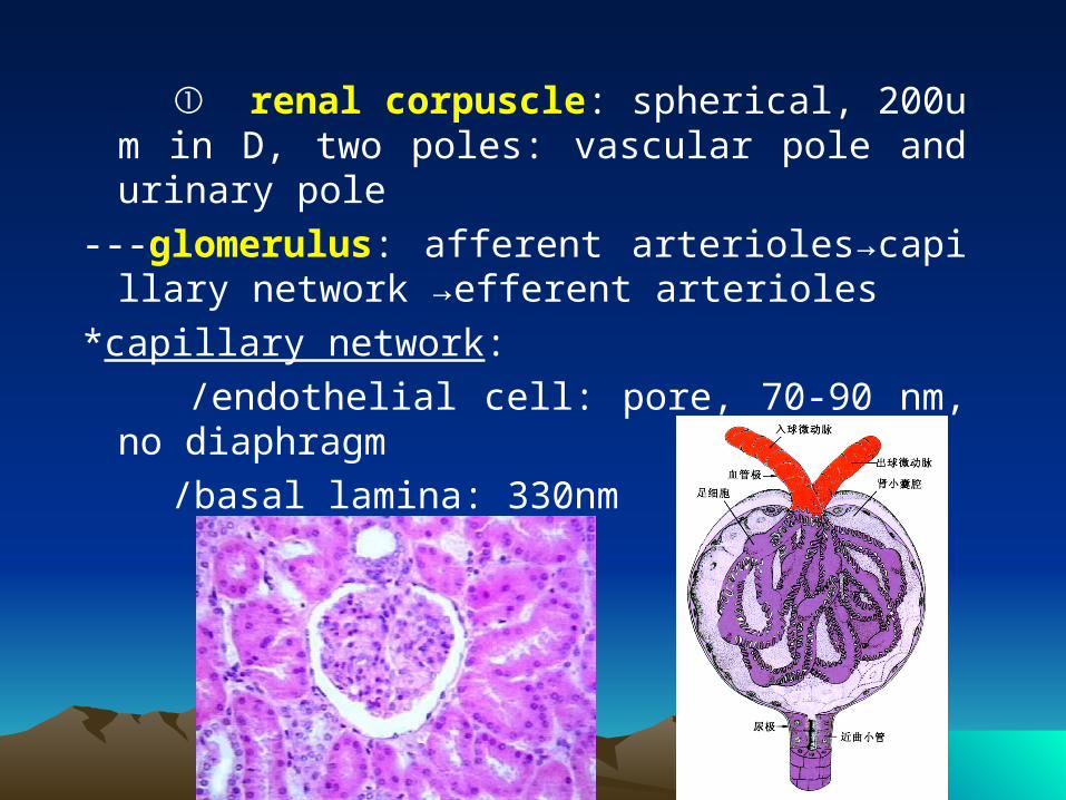
① renal corpuscle: spherical, 200um in D, two poles: vascular pole and urinary pole
---glomerulus: afferent arterioles→capillary network →efferent arterioles
*capillary network:
/endothelial cell: pore, 70-90 nm, no diaphragm
/basal lamina: 330nm
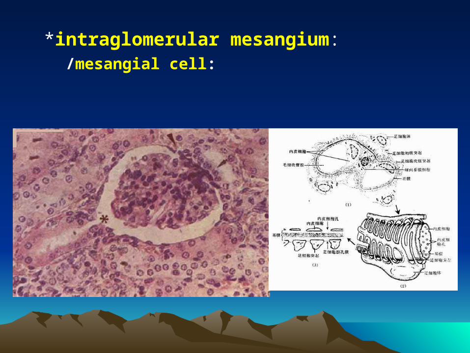
*intraglomerular mesangium: /mesangial cell:
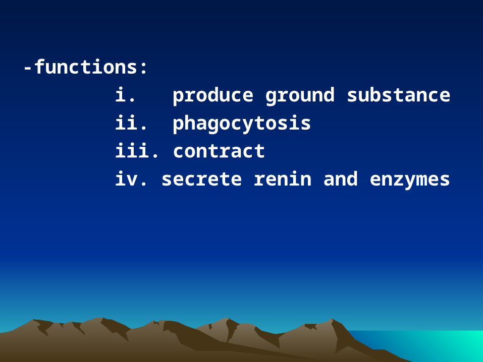
-functions:
i. produce ground substance
ii. phagocytosis
iii. contract
iv. secrete renin and enzymes
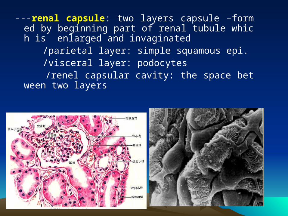
---renal capsule: two layers capsule –formed by beginning part of renal tubule which is enlarged and invaginated
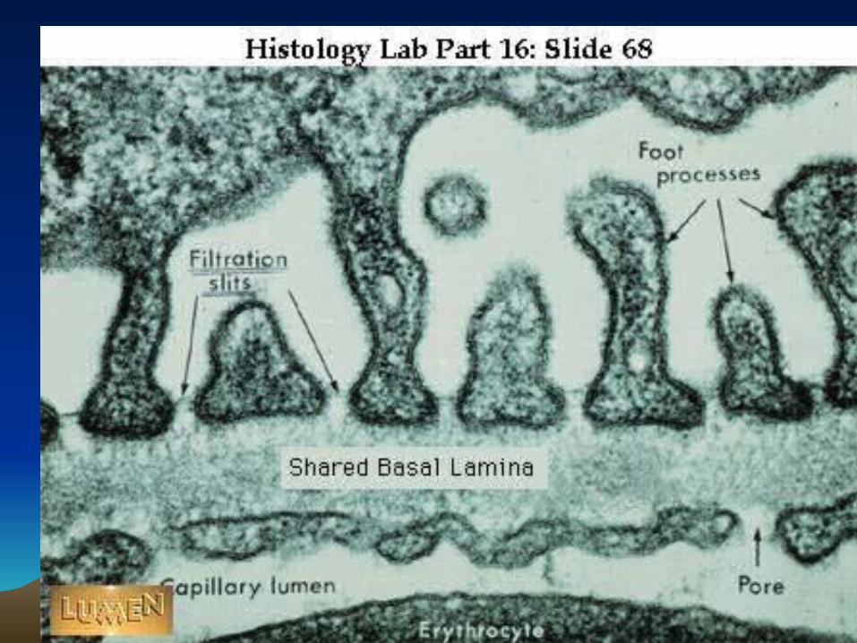
/parietal layer: simple squamous epi. /visceral layer: podocytes /renel capsular cavity: the space between two layer
s
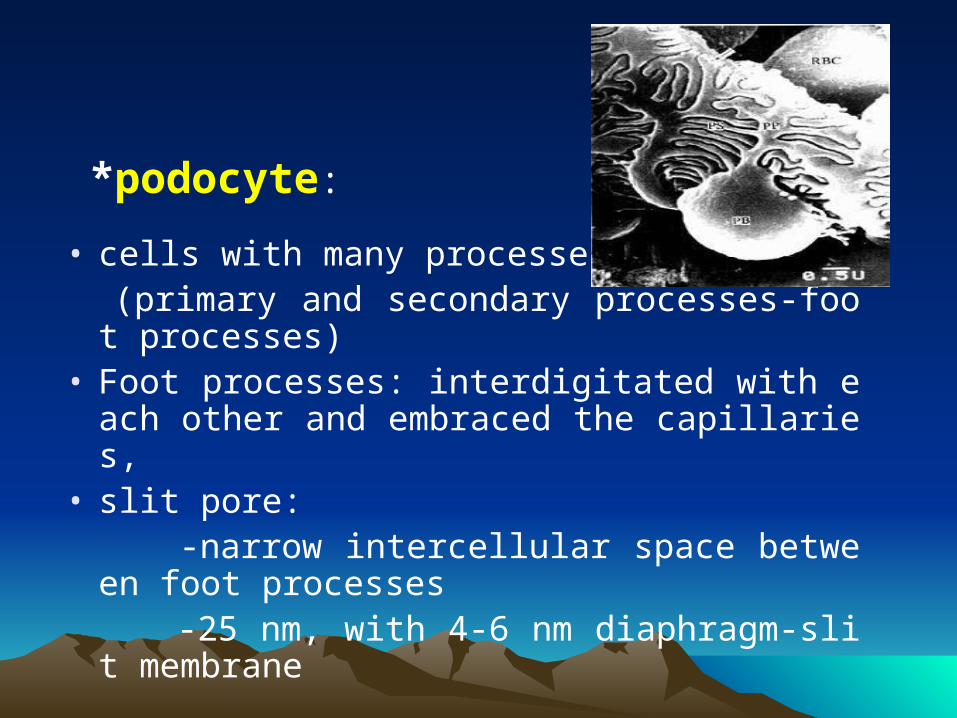
• cells with many processes (primary and secondary processes-foot proces
ses)• Foot processes: interdigitated with each other
and embraced the capillaries, • slit pore: -narrow intercellular space between foot pro
cesses -25 nm, with 4-6 nm diaphragm-slit membran
e
*podocyte:
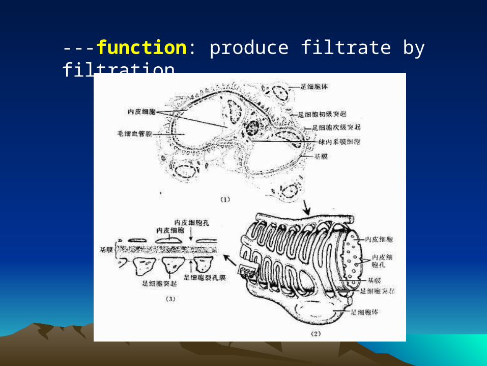
---function: produce filtrate by filtration
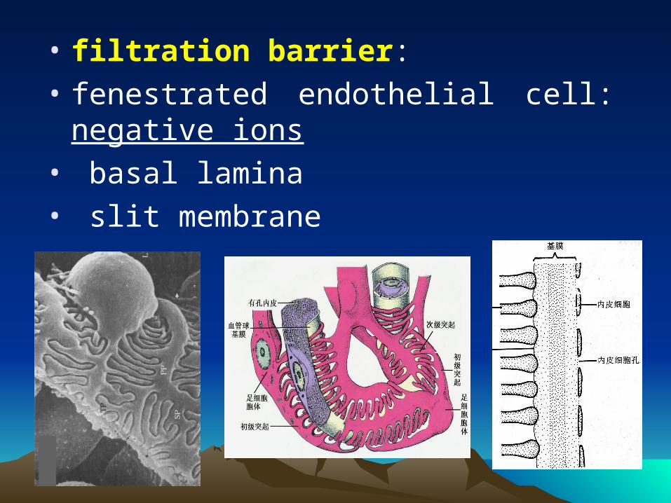
• filtration barrier:
• fenestrated endothelial cell: negative ions
• basal lamina
• slit membrane
② renal tubule:a. proximal tubule: 50-60um in D, 14 mm long---structure LM: EM:• pyramidal cuboidal• eosinophilic• round N• brush-liked border - microvilli• longitudinal striation- plasma membrane infolding• no clear boundary - lateral extension (rich in Na+ K+ ATPase)
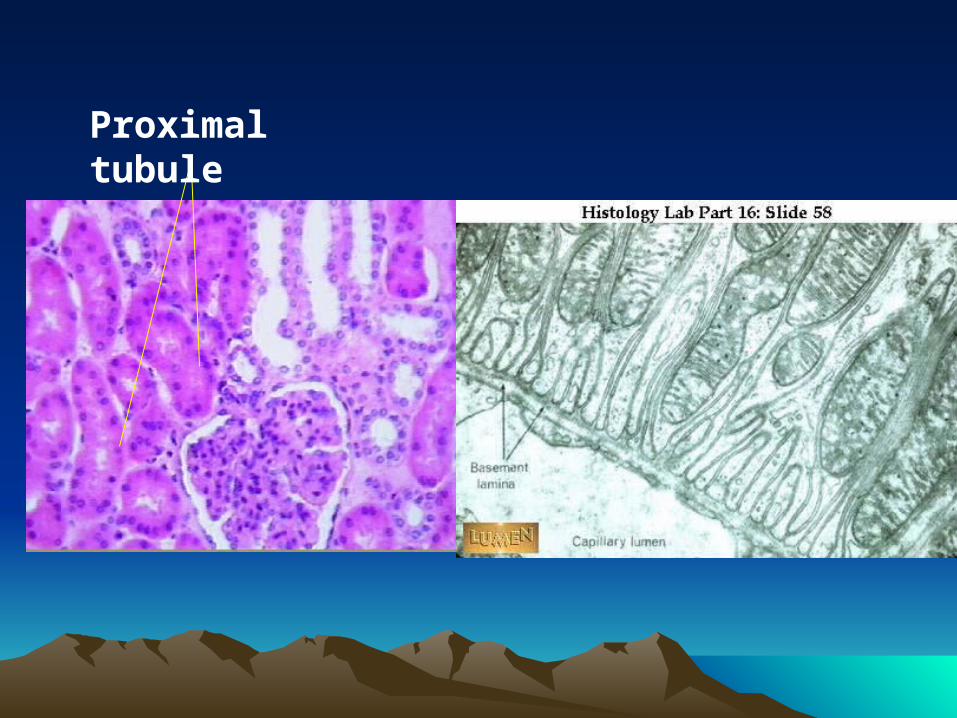
Proximal tubule
---Function:
i. reabsorption :
-85% Na+ ions and water
-All of glucose, aminoacid, polypeptide, proteins and vitamin
ii. secrete H+, NH3, hippuric acid and creatinine
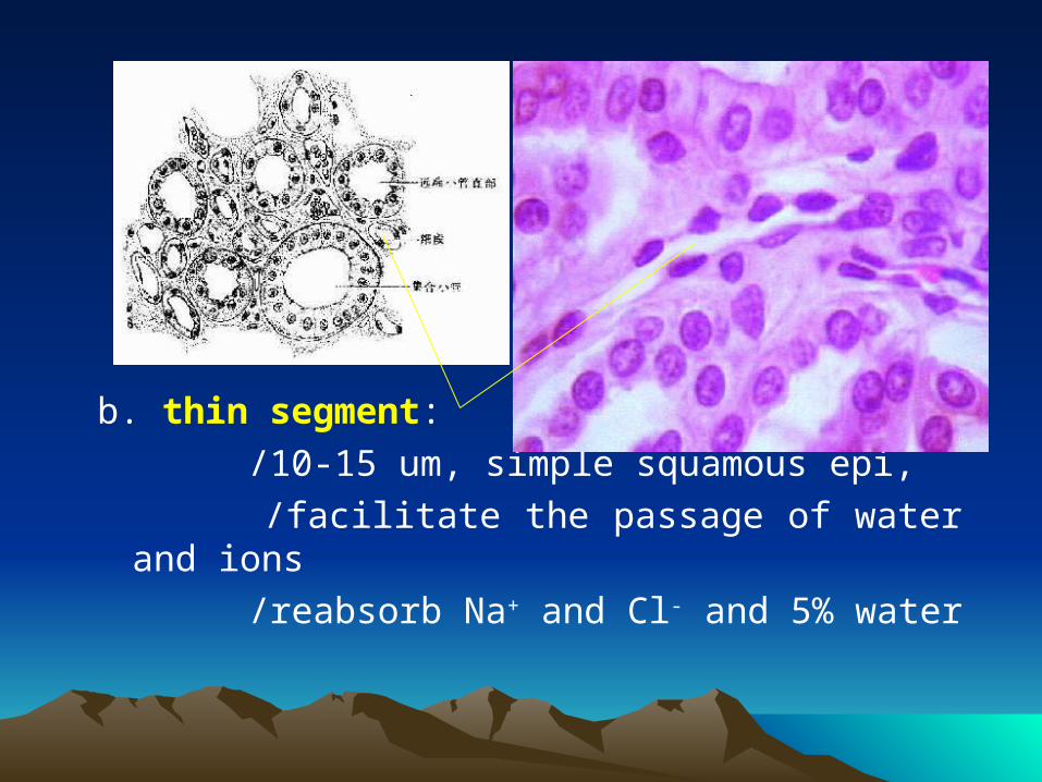
b. thin segment:
/10-15 um, simple squamous epi,
/facilitate the passage of water and ions
/reabsorb Na+ and Cl- and 5% water
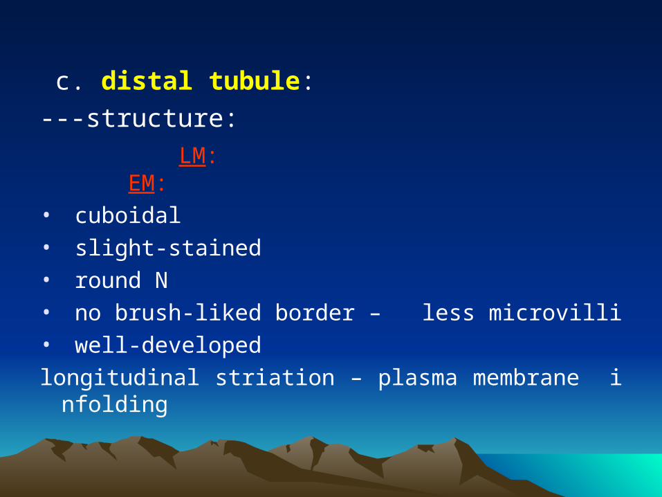
c. distal tubule:
---structure:
LM: EM:
• cuboidal• slight-stained• round N• no brush-liked border – less microvilli• well-developed
longitudinal striation – plasma membrane infolding

Distal tubule Plasma membrane infolding
---function:
i. reabsorption of 8% water, Na+ ions
ii. excretion of K+, H+,NH3
iii. regulated by aldosterone (adrenal gland) and antidiuretic hormone (vasopressin) (pituitary gland)
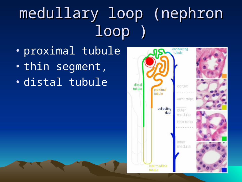
medullary loop (nephron loop )medullary loop (nephron loop )
• proximal tubule ,
• thin segment,
• distal tubule
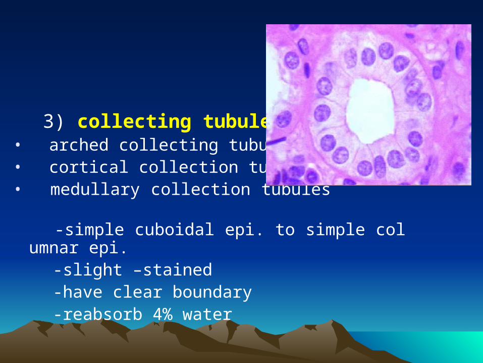
3) collecting tubule:• arched collecting tubules• cortical collection tubules • medullary collection tubules -simple cuboidal epi. to simple columnar epi. -slight –stained -have clear boundary -reabsorb 4% water
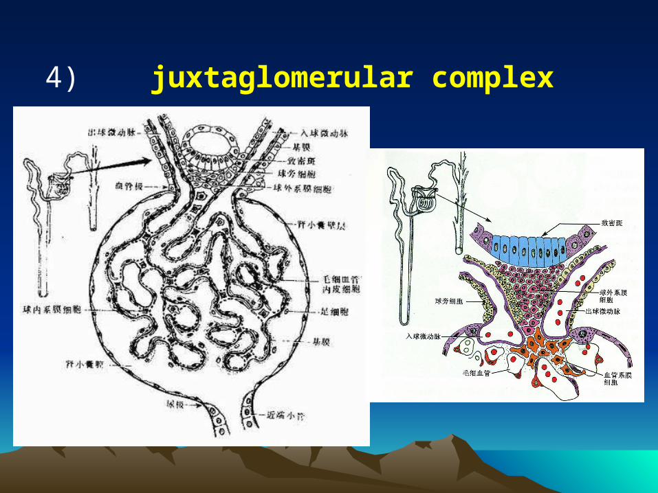
4) juxtaglomerular complex
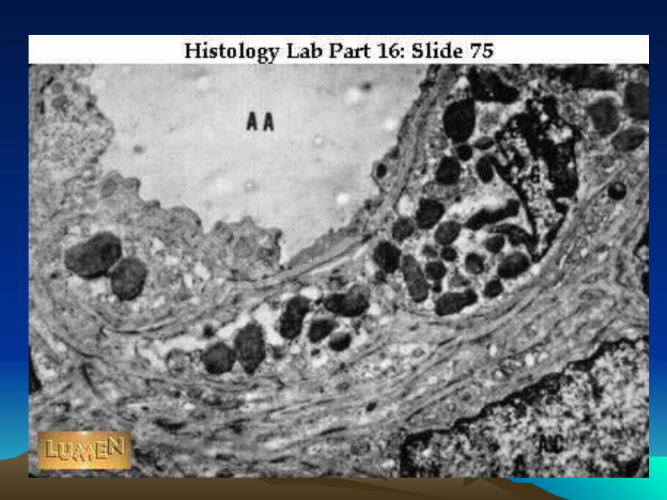
a. juxtaglomerular cell:
---a groups of modified SM cell of afferent arterioles
---structure:
-larger, cuboidal in shaped, with round N
-contain secretory granules
---function:
i.secrete renin→adrenal gland→aldosterone→blood pressure↑
↑
angiotensinogen→angiotensin I→angiotensin II→contraction of
SM of BV
ii. secrete erythropoietin to promote erythropoiesis
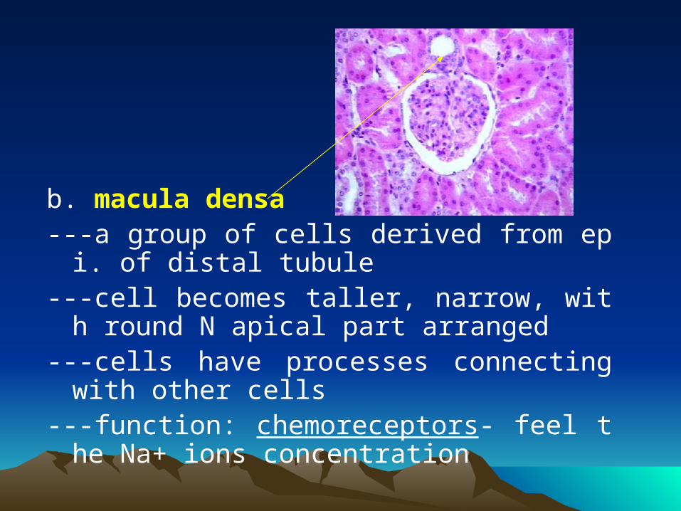
b. macula densa---a group of cells derived from epi. of distal tub
ule---cell becomes taller, narrow, with round N apic
al part arranged---cells have processes connecting with other c
ells---function: chemoreceptors- feel the Na+ ions c
oncentration
c. extraglomerular mesangial cell (polar cushion cell)
---similar to intraglomerular mesangial cell---transfer the informationd. peripolar cell---structure: EM: -microvilli -junctional complexes -RER, Golgi, and granules---function: regulate the reabsorption and secret
ion of renal tubule
5) renal interstitial: CT---fibers: type I,III,IV collagen---matrix---cell: • fibroblast• macrophage• lipid-laden interstitial cell: -stellate cell with processes -osmiophilic lipid droplets: -function: i. involve in formation of F and matrix ii. secret prostaglandin
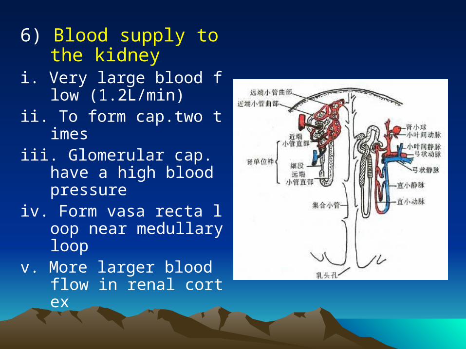
6) Blood supply to the kidney
i. Very large blood flow (1.2L/min)
ii. To form cap.two timesiii. Glomerular cap. have
a high blood pressure
iv. Form vasa recta loop near medullary loop
v. More larger blood flow in renal cortex
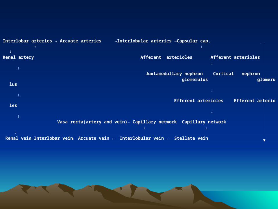
Interlobar arteries → Arcuate arteries →Interlobular arteries →Capsular cap. ↑ ↓ ↓Renal artery Afferent arterioles Afferent arterioles ↓ ↓ Juxtamedullary nephron Cortical nephron glomerulus glomerulus ↓ ↓ Efferent arterioles Efferent arterioles ↓ ↓ Vasa recta(artery and vein)← Capillary network Capillary network ↓ ↓ ↓ Renal vein←Interlobar vein← Arcuate vein ← Interlobular vein ← Stellate vein

Thanks!Thanks!