Urinalysis - Functional medicine · 2015-10-05 · Mod 1 * Lesson 8: Urinalysis ...
23
Functional Medicine University’s Functional Diagnostic Medicine Training Program Module 1 * Lesson 8 Urinalysis By Wayne L. Sodano, D.C., D.A.B.C.I., & Ron Grisanti, D.C., D.A.B.C.O., M.S. http://www.FunctionalMedicineUniversity.com Limits of Liability & Disclaimer of Warranty We have designed this book to provide information in regard to the subject matter covered. It is made available with the understanding that the authors are not liable for the misconceptions or misuse of information provided. The purpose of this book is to educate. It is not meant to be a comprehensive source for the topic covered, and is not intended as a substitute for medical diagnosis or treatment, or intended as a substitute for medical counseling. Information contained in this book should not be construed as a claim or representation that any treatment, process or interpretation mentioned constitutes a cure, palliative, or ameliorative. The information covered is intended to supplement the practitioner’s knowledge of their patient. It should be considered as adjunctive and support to other diagnostic medical procedures. This material contains elements protected under International and Federal Copyright laws and treaties. Any unauthorized reprint or use of this material is prohibited. Functional Medicine University; Functional Diagnostic Medicine Training Program/Insider’s Guide Mod 1 * Lesson 8 : Urinalysis Copyright © 2010 Functional Medicine University, All Rights Reserved
Transcript of Urinalysis - Functional medicine · 2015-10-05 · Mod 1 * Lesson 8: Urinalysis ...

Functional Medicine University’s Functional Diagnostic Medicine
Training Program
Module 1 * Lesson 8
Urinalysis
By Wayne L. Sodano, D.C., D.A.B.C.I., & Ron Grisanti, D.C., D.A.B.C.O., M.S. http://www.FunctionalMedicineUniversity.com
Limits of Liability & Disclaimer of Warranty
We have designed this book to provide information in regard to the subject matter covered. It is made available with the understanding that the authors are not liable for the misconceptions or misuse of information provided. The purpose of this book is to educate. It is not meant to be a comprehensive source for the topic covered, and is not intended as a substitute for medical diagnosis or treatment, or intended as a substitute for medical counseling. Information contained in this book should not be construed as a claim or representation that any treatment, process or interpretation mentioned constitutes a cure, palliative, or ameliorative. The information covered is intended to supplement the practitioner’s knowledge of their patient. It should be considered as adjunctive and support to other diagnostic medical procedures. This material contains elements protected under International and Federal Copyright laws and treaties. Any unauthorized reprint or use of this material is prohibited.
Functional Medicine University; Functional Diagnostic Medicine Training Program/Insider’s Guide
Mod 1 * Lesson 8 : Urinalysis Copyright © 2010 Functional Medicine University, All Rights Reserved

Functional Medicine University’s
Functional Diagnostic Medicine Training Program
Mod 1 * Lesson 8: Urinalysis
By Wayne L. Sodano, D.C., D.A.B.C.I., & Ron Grisanti, D.C., D.A.B.C.O., M.S.
http://www.FunctionalMedicineUniversity.com
1
Contents
End Stage Renal Disease (ESRD): 2005 Statistics 2
Anatomy & Physiology of the Kidney & Urinary System 2
Urinalysis 3
Types of Urine Specimens 3
Urinary Collection Methods 4
Urinalysis: Normal Findings 4
Chemical Testing of Urine 8
Some Causes of Proteinuria 11
Microalbumin 13
Blood 13
Bilirubin 15
Urobilinogen 15
Nitrite 16
Leukocyte Esterase 16
Microscopic examination of Urine 17
Kidney Stones 17
Urinary Tract Infections: Integrative Therapy 20
References 22
Functional Medicine University’s
Functional Diagnostic Medicine Training Program
Mod 1 * Lesson 8: Urinalysis
By Wayne L. Sodano, D.C., D.A.B.C.I., & Ron Grisanti, D.C., D.A.B.C.O., M.S.
http://www.FunctionalMedicineUniversity.com
2
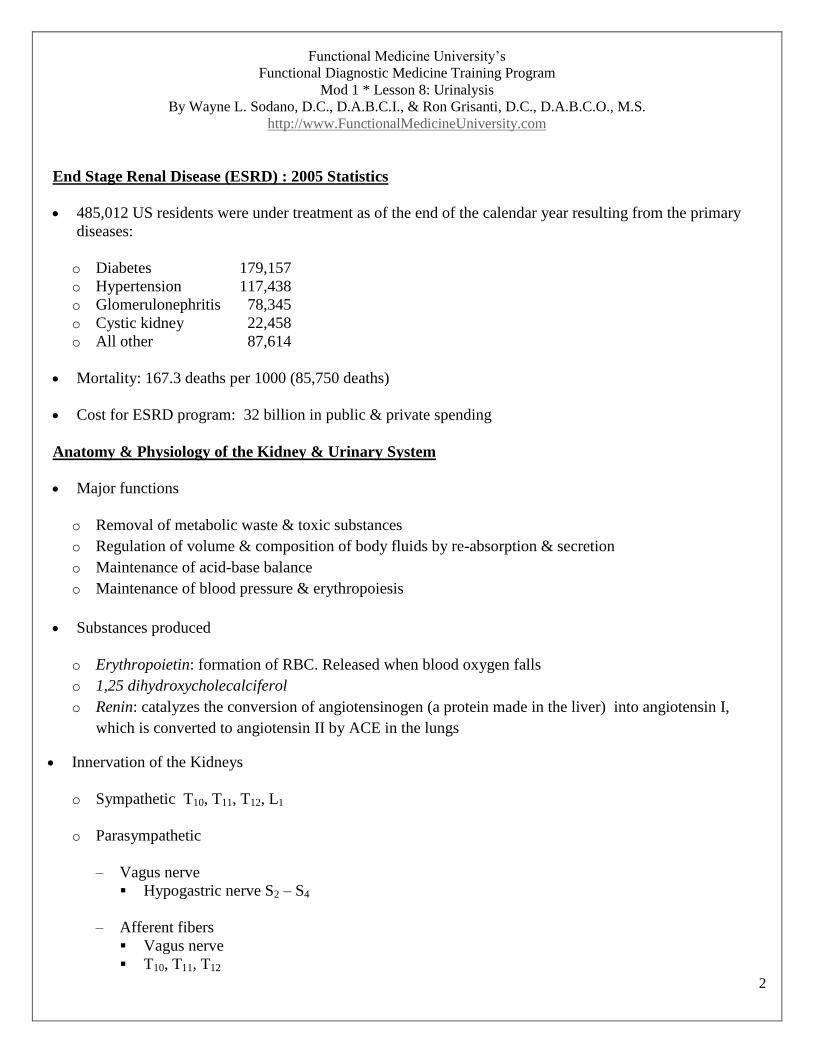
End Stage Renal Disease (ESRD) : 2005 Statistics
485,012 US residents were under treatment as of the end of the calendar year resulting from the primary
diseases:
o Diabetes 179,157
o Hypertension 117,438
o Glomerulonephritis 78,345
o Cystic kidney 22,458
o All other 87,614
Mortality: 167.3 deaths per 1000 (85,750 deaths)
Cost for ESRD program: 32 billion in public & private spending
Anatomy & Physiology of the Kidney & Urinary System
Major functions
o Removal of metabolic waste & toxic substances
o Regulation of volume & composition of body fluids by re-absorption & secretion
o Maintenance of acid-base balance
o Maintenance of blood pressure & erythropoiesis
Substances produced
o Erythropoietin: formation of RBC. Released when blood oxygen falls
o 1,25 dihydroxycholecalciferol
o Renin: catalyzes the conversion of angiotensinogen (a protein made in the liver) into angiotensin I,
which is converted to angiotensin II by ACE in the lungs
Innervation of the Kidneys
o Sympathetic T10, T11, T12, L1
o Parasympathetic
– Vagus nerve
Hypogastric nerve S2 – S4
– Afferent fibers
Vagus nerve
T10, T11, T12
Functional Medicine University’s
Functional Diagnostic Medicine Training Program
Mod 1 * Lesson 8: Urinalysis
By Wayne L. Sodano, D.C., D.A.B.C.I., & Ron Grisanti, D.C., D.A.B.C.O., M.S.
http://www.FunctionalMedicineUniversity.com
3
Urinalysis
• Provides information about
o Major metabolic functions
o Renal disease
o First indication of asymptomatic diabetes mellitus and some liver diseases
Types of Urine Specimens
First morning urine specimen
o A.M. Urine is concentrated – testing is more apt to detect positive finding.(proteins, nitrites)
o Empty bladder before bed the night before.
o Increases accuracy of patient’s 24-hours urine
o If specimen will not arrive at the lab within 2 hours of collection, it must be preserved (refrigerated).
Random Urine Specimen
o Used in routine screening (i.e. drug testing)
o Substance tested has no diurnal variation
Timed Urine Collection Specimen
o Used for substances with variable excretions over 24 hour period ( e.g. hormones, proteins &
electrolytes)
• Double Voided Specimen
o Used to monitor urine glucose and ketones
o Bladder is emptied; second void is collected shortly thereafter

Functional Medicine University’s
Functional Diagnostic Medicine Training Program
Mod 1 * Lesson 8: Urinalysis
By Wayne L. Sodano, D.C., D.A.B.C.I., & Ron Grisanti, D.C., D.A.B.C.O., M.S.
http://www.FunctionalMedicineUniversity.com
4
Urinary Collection Methods
o Routine void specimen
o No preparation necessary
o Collection time can be random or first morning urine
o Mid-stream and clean-catch specimen for culture & sensitivity
o Clean urinary meatus
o Midstream collection in sterile container
o 24 Hour Urine Collection
o Discard 1st urine specimen and note the time
o Collect urine for 24 hours
Note: It is important to check lab manuals (Quest, LabCorp, etc) for proper collection procedure and type of
urine specimen for details on collection methods. And…be sure that you are CLIA compliant!
Urinalysis: Normal Findings
• Appearance: clear
• Color: Amber yellow
• Odor: aromatic
• pH: 4.6 – 8.0 average 6.0
• Protein: 0-8 mg/dL 50-80 mg/24 at rest <250 mg/24 (during exercise)
• Specific gravity: 1.005-1.080 (usually 1.010-1.025)
• Leukocyte esterase: negative
• Nitrites: none
• Ketones: none
• Bilirubin: none
• Urobilinogen: 0.01-1 Erlich unit/ml
• Crystals: none
• Casts: none
• Glucose: none

Functional Medicine University’s
Functional Diagnostic Medicine Training Program
Mod 1 * Lesson 8: Urinalysis
By Wayne L. Sodano, D.C., D.A.B.C.I., & Ron Grisanti, D.C., D.A.B.C.O., M.S.
http://www.FunctionalMedicineUniversity.com
5
Urinalysis: Normal Findings (con’t)
• White blood cells: 0-4 per high-power field
• WBC casts: none
• RBC’s: ≤ 2 hpf
• RBC casts: none
Physical exam of urine
• Appearance
o Normal : Clear to somewhat hazy
o Hazy or cloudy
– Amorphous, phosphates, or urates
– WBC’s RBC’s, epithelial cells, bacteria
– Fat (milky appearance)
– Mucous
• Color
o Normal: pale yellow to amber due to the pigment urochrome (product of bilirubin metabolism)
o Abnormal:
– Pathological conditions
– Ingestion of certain foods (B-vitamins, beets) *
– Medicines
* Because urine color may be affected by the use of various types of drugs, (i.e., antibiotics, laxatives, muscle
relaxants, etc.), and some vitamins (vitamin B) and supplements, it is important to determine if the patient is
taking any of these when performing urine analysis.
o Colorless:
– Large fluid intake
– Diabetes insipidus
– Untreated diabetes mellitus

Functional Medicine University’s
Functional Diagnostic Medicine Training Program
Mod 1 * Lesson 8: Urinalysis
By Wayne L. Sodano, D.C., D.A.B.C.I., & Ron Grisanti, D.C., D.A.B.C.O., M.S.
http://www.FunctionalMedicineUniversity.com
6
Physical exam of urine – Color (con’t)
o Orange:
– Drugs
– Bile
– Diet
– Uric acid crystals
o Brownish/greenish yellow
– Bilirubin
– Drugs
– Indican
– Pseudomonas infections
o Red:
– Blood
– Porphyria
– Drugs
– Diet (beets)
o Brown:
– Blood
– Bilirubin
– Urobilinogen
– Indican
– Diet (rhubarb)
o Greenish:
– Pseudomonas infection
– Food dyes
– Medications

Functional Medicine University’s
Functional Diagnostic Medicine Training Program
Mod 1 * Lesson 8: Urinalysis
By Wayne L. Sodano, D.C., D.A.B.C.I., & Ron Grisanti, D.C., D.A.B.C.O., M.S.
http://www.FunctionalMedicineUniversity.com
7
• Volume
o Influencing factors
– Fluid intake
– Nonrenal fluid loss
– Variation in ADH secretions
– The body’s necessity to excrete large amounts of dissolved solids (salts, glucose)
o Polyuria: increased urine volume
– Diabetes mellitus
– Diabetes insipidus (depressed ADH)
o Oliguria: decreased volume
– Dehydration, vomiting, severe burns
– Normal volume for 24-hr urine sample: 800-2000 ml
o Clinical Significance
– Low volume
Dehydration
Renal disease
Urinary tract obstruction
– High volume
Diuretics
Diabetes mellitus
Diabetes insipidus

Functional Medicine University’s
Functional Diagnostic Medicine Training Program
Mod 1 * Lesson 8: Urinalysis
By Wayne L. Sodano, D.C., D.A.B.C.I., & Ron Grisanti, D.C., D.A.B.C.O., M.S.
http://www.FunctionalMedicineUniversity.com
8
o Urine Odor
– Normal:
aromatic odor
– Sweet:
diabetic ketoacidosis
– Foul smelling:
UTI
– Fecal Odor:
enterovesical fistula
Chemical Testing of Urine
Specific Gravity – density of urine
o Measures the weight of urine compared to that of water (SG of water is 1.000)
o Matter in the urine give it weight
o Used to evaluate the concentrating and excretory effectiveness of the kidneys
o Must be interpreted in light of the presence or absence of glycosuria or proteinuria, and hydration status
o Usual normal range: 1.010-1.025
o Optimal range: 1.015 o Clinical Significance
– Low
Diabetes insipidus (decreased production ADH)
Glomerulonephritis (inability to conc. urine)
Pyelonephritis
– High
Adrenal insufficiency
Hepatic disease
CHF
Dehydration
Diabetes mellitus

Functional Medicine University’s
Functional Diagnostic Medicine Training Program
Mod 1 * Lesson 8: Urinalysis
By Wayne L. Sodano, D.C., D.A.B.C.I., & Ron Grisanti, D.C., D.A.B.C.O., M.S.
http://www.FunctionalMedicineUniversity.com
9
• pH
o Measure of hydrogen ion concentration
o Indicates the acid-base balance
o Reflects the work of the kidneys in maintaining normal pH homeostasis
o Range variable: 4.5 – 8
• Alkaline pH
o Alkemia
o UTI
o Diet high in citrus or vegetables
• Acid pH
o Diabetes mellitus
o Metabolic or respiratory acidosis
o Starvation
o Dehydration
o Diet high in meat products or cranberries
o COPD
Urine pH is useful in identifying crystals and determining the predisposition to form a given type of stone.
• Acid urine
o Xanthine, cysteine, uric acid and calcium oxalate- to treat or prevent, keep the urine alkaline
• Alkaline urine
o Calcium carbonate, calcium phosphate, magnesium phosphate – to treat or prevent, keep urine acidic.
• Glucose (Glycosuria)
o Normal value: negative
o Occurs when blood glucose level exceeds the re-absorption capacity of the renal tubules. Glucose is not
excreted by the kidney unless blood levels exceed 180 mg/dL.

Functional Medicine University’s
Functional Diagnostic Medicine Training Program
Mod 1 * Lesson 8: Urinalysis
By Wayne L. Sodano, D.C., D.A.B.C.I., & Ron Grisanti, D.C., D.A.B.C.O., M.S.
http://www.FunctionalMedicineUniversity.com
10
o Glucose (Glycosuria) (con’t)
o Not always indicative of diabetes. May also occur in diseases that affect the renal tubules or in genetic
disorders.
o Glycosuria with high blood sugar
– Diabetes mellitus (chief cause of glycosuria)
– Infections
– Obesity
o Glycosuria without a high blood sugar
– Renal glycosuria
– Fanconi syndrome (transport defect of proximal tubules) genetic defect
– Inflammatory renal disease
– Nephrotoxic chemicals (mercury, lead, cadmium)
– Pregnancy
o Ketones
o Normal value: negative
– Intermediary products of fat metabolism
– Inadequacy of carbohydrates – indicative of problem with metabolism or malabsorption which
causes an increase in fat metabolism.
– Usually associated with poorly controlled diabetes
– Associated with alcoholism, fasting, starvation, high protein diets.
– Indicated in acute febrile illnesses, particularly infants and children
o Protein
o Reference range: negative to trace
– random sample: 0 -8 mg/dL
o 24 hour sample may contain up to 35 mg of albumin and up to 50 mg of other proteins (i.e.
immunoglobulin, globulins, Tamms-Horsfall protein)

Functional Medicine University’s
Functional Diagnostic Medicine Training Program
Mod 1 * Lesson 8: Urinalysis
By Wayne L. Sodano, D.C., D.A.B.C.I., & Ron Grisanti, D.C., D.A.B.C.O., M.S.
http://www.FunctionalMedicineUniversity.com
11
• Protein (con’t)
o Unless abnormally high, normal amounts will not be detected on the urine reagent strip in a random
specimen (for truer reading, a Microalbumin test or concentrated urine test is recommended)
o Tends to increase with age, exercise and standing posture.
o Proteinuria has been defined as 24 hour urine protein excretion >150 mg/24 hours. Infants and children
>100 mg/24 hours.
o Even with normal kidney function, some proteins will appear in the urine if the levels of protein in the
blood become high.
o Is a sensitive marker of kidney function and renal disease
o Forms the basic matrix of urinary casts.
o Functional proteinuria refers to protein excretion in association with fever, excessive exercise or
emotional stress.
Some Causes of Proteinuria
• Prerenal proteinuria
o Congestive heart failure
o Orthostatic proteinuria
o Transient, associated with febrile illness, surgery, anemia, hyperthyroidism, stroke, exercise, seizures
o Bence Jones proteinuria associated with myeloma, Waldenstrom macroglobulinemia, amyloidosis (light
chain proteinuria)

Functional Medicine University’s
Functional Diagnostic Medicine Training Program
Mod 1 * Lesson 8: Urinalysis
By Wayne L. Sodano, D.C., D.A.B.C.I., & Ron Grisanti, D.C., D.A.B.C.O., M.S.
http://www.FunctionalMedicineUniversity.com
12
• Renal proteinuria
o Renovascular hypertension
o Malignant hypertension of any cause
Membranous nephropathy & proliferative glomerulonephritis
Chronic pyelonephritis
Polycystic disease
Diabetic nephropathy
Amyloidosis
Lupus erythematosus (SLE)
Goodpasture’s syndrome
Renal vein thrombosis
Minimal change nephropathy
HIV nephropathy
Alport syndrome
Preeclampsia
o Fanconi Syndrome
o Wilson disease
o Heavy metal poisoning:
lead
mercury
cadmium
o Galactosemia
o Bacterial pyelonephritis
o Uric acid, urate, or calcium deposition
o Idiosyncratic drug reaction:
methicillin
phenindione
sulfonamides
phenytoin
others
o Interstitial diseases generally reflected as tubular defects or
mixed tubular interstitial
Glomerular Proteinuria
> 3.5 g/24 hr usually
Reflects a glomerular lesion
(in children > 1.0 g/m2/day)
Tubular
Usually <1 g/24h
Interstitial

Functional Medicine University’s
Functional Diagnostic Medicine Training Program
Mod 1 * Lesson 8: Urinalysis
By Wayne L. Sodano, D.C., D.A.B.C.I., & Ron Grisanti, D.C., D.A.B.C.O., M.S.
http://www.FunctionalMedicineUniversity.com
13
• Postrenal proteinuria
o Tumors of the bladder or renal pelvis
o < 1 g/24 h, IgM excretion significant marker, amount of proteinuria related to size and spread of tumor
o Cystitis, severe
• Interfering factors
o Drugs can increase protein in the urine.
o Radiopaque contrast media administered within 3 days may cause false positive.
o Urine contaminated with prostate or vaginal secretions
o High protein diet
o Severe emotional stress
Note: Increased protein and leukocytes usually indicates a UTI. Microalbumin
• Normal Range: < 20 mg/L
o MA/Creatinine Ratio: 0 - 30 mg/g
• Microalbuminuria refers to an albumin concentration in the urine that is greater than normal, but is not
detectable with routine protein testing.
• MA is the earliest indicator for development of diabetic complications (nephropathy, CVD, and
hypertension). Can identify diabetic nephropathy 5 years before routine protein urine tests.
• Micral urine test strip – best performed with 1st morning urine void.
Blood
• Reference Range: Negative
• Microscopy: up to 2-3 RBC/hpf
• Positive dipstick indicates hematuria, hemoglobinuria, or myoglobinuria.
• If positive in dipstick, microscopy is indicated.
• 20% of patients older than 40 years with asymptomatic hematuria have significant urologic lesions, of
which half are malignant.
• Blood may be present as intact cells (non-hemolyzed) or as free hemoglobin (hemolyzed or ruptured RBC).
• Hemoglobinuria without hematuria implies intravascular hemolysis, while hematuria suggests a bleeding
source in the urinary tract.

Functional Medicine University’s
Functional Diagnostic Medicine Training Program
Mod 1 * Lesson 8: Urinalysis
By Wayne L. Sodano, D.C., D.A.B.C.I., & Ron Grisanti, D.C., D.A.B.C.O., M.S.
http://www.FunctionalMedicineUniversity.com
14
Causes of hemoglobinuria
o Hemoglobinuria with intact red cells in sediment. No casts present.
– Possible causes:
Menstruation
Vigorous exercise
Trauma to urinary tract
Cystitis
Calculi
Kidney tumors
Malignant hypertension
Sickle cell (disease or trait)
o Hemoglobinuria with intact red cells in sediment. Red cell or granular casts noted. Proteinuria present.
– Possible causes:
Acute glomerulonephritis
Chronic glomerulonephritis
Lupus nephritis
Polyarteritis
Goodpasture’s syndrome
Allergic nephropathy
• Hemoglobinuria with no intact red cells in sediment.
– Possible causes:
Delayed examination (esp. if dilute urine)
Hemolytic disorders (immune and non-immune)
Presence of myoglobin
Serum creatinine kinase levels high
Oxidative stress
Functional Medicine University’s
Functional Diagnostic Medicine Training Program
Mod 1 * Lesson 8: Urinalysis
By Wayne L. Sodano, D.C., D.A.B.C.I., & Ron Grisanti, D.C., D.A.B.C.O., M.S.
http://www.FunctionalMedicineUniversity.com
15
Bilirubin
Normal values: negative
Is a major constituent of bile.
If excretion is inhibited, conjugated (direct) hyperbilirubinemia will result.
Bilirubin in the urine suggests disease affecting bilirubin metabolism after conjugation or defects in
excretion.
Only conjugated bilirubin is passed into the urine, thus a positive reaction is evidence of hepatocellular or
biliary disease.
Clinical implications of elevated urine bilirubin
o Extrahepatic duct obstruction (gallstones, inflammation, tumor)
o Infections or hepatotoxic agents (liver cells are unable to excrete all of the direct bilirubin)
Note: Bilirubin is very light sensitive. It will disappear from urine on standing; specimen should be
processed immediately.
Urobilinogen
• Normal value: trace or < 2 Ehrlich units/dL (1 EU = 1 mg of urobilinogen)
• One of the earliest signs of acute liver damage
• Direct bilirubin is excreted from liver cells into the bile, then into the intestinal tract through the bile duct. It
is converted by bacterial action to urobilinogen.
• About 50% of the urobilinogen is reabsorbed into the portal circulation.
• Urobilinogen is increased by any condition that causes an increase in the production of bilirubin and by any
disease that prevents the liver from normally removing the reabsorbed urobilinogen from portal circulation.
• Urobilinogen is decreased or absent when normal amounts of bilirubin are not excreted in the intestinal
tract.

Functional Medicine University’s
Functional Diagnostic Medicine Training Program
Mod 1 * Lesson 8: Urinalysis
By Wayne L. Sodano, D.C., D.A.B.C.I., & Ron Grisanti, D.C., D.A.B.C.O., M.S.
http://www.FunctionalMedicineUniversity.com
16
Nitrite
• Normal Value: negative
• Screening test for UTI’s (some UTI’s are asymptomatic)
• Many bacteria produce an enzyme called Reductase, which can reduce urinary nitrates to nitrites.
• A positive result indicates the need for urine culture
Leukocyte Esterase
• Normal Value: negative
• Indirect test for the detection of bacteremia
• Will detect either intact or lysed WBC
• Leukocyte esterase is unacceptable as a screen unless combined with nitrite testing.
• Leukocyte esterase, even combined with nitrite should not replace microscopy and culture in symptomatic
patients.
Differential Diagnosis Using Urine Bilirubin and Urobilinogen Tests
Type of JaundiceUrine
Bilirubin
Urine Urobilinogen
Normal 0 0 - trace
Hepatocellular jaundice
(eg, hepatitis, chemical, or drug injury)
Biliary obstruction (extrahepatic& intraphepatic); obstructive jaundice
0
Hemolytic jaundice 0
0 = absent; present

Functional Medicine University’s
Functional Diagnostic Medicine Training Program
Mod 1 * Lesson 8: Urinalysis
By Wayne L. Sodano, D.C., D.A.B.C.I., & Ron Grisanti, D.C., D.A.B.C.O., M.S.
http://www.FunctionalMedicineUniversity.com
17
Microscopic examination of Urine
• Types of sediment
o As one author puts it:
– Cells
– Casts
– Crystals
– Critters
o Organized biological part
– RBC
– WBC
– Casts
– Epithelial cells
– Bacteria, parasites, yeast & fungi
o Unorganized
– Crystals
– Amorphous crystalline matter
Kidney Stones
Calcium Containing Stones: Causes
• SAD (Standard American Diet)
Low in fiber
High in refined carbohydrates, alcohol consumption, animal protein, fat
High intake of high-calcium, low-magnesium, vitamin D-enriched milk products.
• Obesity and insulin insensitivity
• Sugar: increase in urinary calcium oxalate
Functional Medicine University’s
Functional Diagnostic Medicine Training Program
Mod 1 * Lesson 8: Urinalysis
By Wayne L. Sodano, D.C., D.A.B.C.I., & Ron Grisanti, D.C., D.A.B.C.O., M.S.
http://www.FunctionalMedicineUniversity.com
18
Kidney Stones (Calcium Containing Stones: Causes) con’t
• Magnesium deficiency
o Magnesium increases the solubility of calcium oxalate and inhibits both calcium phosphate and calcium
oxalate stone formation. Supplementing magnesium shown to prevent recurrences of kidney stones.
o Vitamin B6 deficiency
Vitamin B6 reduces the production of excretion of oxalates.
Kidney Stones : Preventive Measures
• For all types of kidney stones
o Maintain ideal weight
o Drink at least 3L of non-alcoholic, (preferably purified water) fluid daily
o Exercise regularly
o Minimize intake of sugar, refined carbohydrates, and alcohol
o Consume a diet in green leafy vegetables, nutrient-rich, whole foods
• Kidney Stones : Treatment
o Calcium Stones
– Diet
Increase intake of fiber, complex carbohydrates, and green leafy vegetables.
Increase intake of high magnesium-to-calcium ratio foods
– barley, bran, corn, buckwheat, rye, soy, oats, brown rice, avocado, banana, cashew, coconut,
peanuts, sesame seeds, lima beans, potato
If stones are oxalate, reduce oxalate-containing foods
– black tea, cocoa, spinach, beet leaves, rhubarb, parsley, cranberries, and nuts
Limit intake of products made with milk

Functional Medicine University’s
Functional Diagnostic Medicine Training Program
Mod 1 * Lesson 8: Urinalysis
By Wayne L. Sodano, D.C., D.A.B.C.I., & Ron Grisanti, D.C., D.A.B.C.O., M.S.
http://www.FunctionalMedicineUniversity.com
19
• Kidney Stones : Treatment (con’t)
o Uric Acid Stones
– Diet
Decrease consumption of purine-rich foods
– Organ meats, meats, shellfish, yeast (brewer’s and bakers), herring, sardines, mackerel, and
anchovies
Decrease consumption of foods with moderate levels of purines
– Dried legumes, spinach, asparagus, fish, poultry, and mushrooms
• Nutritional Supplements
o Calcium Stones
– Vitamin B6: 25 mg q.d.
– Magnesium 600 mg q.d.
increases the solubility of calcium oxalate
– Calcium citrate or malate: 300-1000 mg q.d.
– Citrate bound to magnesium or potassium: 500-1000 mg q.d.
citrate has the ability to reduce urinary saturation of calcium oxalate and calcium phosphate
o Uric Acid Stones
– Folic acid 5 mg q.d.
folic acid inhibits xanthine oxidase, the enzyme responsible for the production of uric acid

Functional Medicine University’s
Functional Diagnostic Medicine Training Program
Mod 1 * Lesson 8: Urinalysis
By Wayne L. Sodano, D.C., D.A.B.C.I., & Ron Grisanti, D.C., D.A.B.C.O., M.S.
http://www.FunctionalMedicineUniversity.com
20
Urinary Tract Infections : Integrative Therapy
• Nutrition
o Removal of Bladder Irritants
– Dietary bladder irritants such as caffeine, refined sugar, white flour, alcohol, and nicotine should be
avoided.
o Increase consumption of
– Garlic
– Onion
– Fluids
– Flaxseed
Contains mucilage, which soothes the lining of the urinary tract.
Dosage: 1-3 Tablespoons of ground fresh flaxseed taken with food.
o Vitamin C
– Has an acidifying effect on the urine.
– Probiotics
Taken after meals to promote digestive tract health
o Botanicals
– Cranberry
Recommended for its anti-adherence properties
– Dosage: sixteen ounces of unsweetened pure (not ‘cocktail’) juice a day, diluted with water,
1 - 2 capsules of dried cranberry powder, 2 to 4 times per day.
Functional Medicine University’s
Functional Diagnostic Medicine Training Program
Mod 1 * Lesson 8: Urinalysis
By Wayne L. Sodano, D.C., D.A.B.C.I., & Ron Grisanti, D.C., D.A.B.C.O., M.S.
http://www.FunctionalMedicineUniversity.com
21
Urinary Tract Infections : Integrative Therapy (con’t)
– Uva Ursi
Key constituents are arbutin and hydroquinone. Arbutin has demonstrated antimicrobial activity
against gram-positive and gram-negative bacteria, including E.coli and is absorbed from the
gastrointestinal tract and hydrolyzed to hydroquinone in alkaline urine.
– Dosage: 3 g of the dried herb or 400 – 800 mg of hydroquinone derivatives 4 times a day.
– Contraindications – This herb should not be used during pregnancy and lactation or in
children younger than 12 years. Because of the large amount of tannins present in this
herb, it is not recommended for use beyond a 1-week period and not more than five times
a year. Prolonged tannin exposure may lead to gastrointestinal intolerance and
hepatotoxicity.
– Goldenseal
Berberine is one of the alkaloids in goldenseal and is believed to have antibacterial properties
against gram-positive and gram-negative bacteria, including E. coli.
– Dosage: recommend a standardized extract with berberine as the main component, 2 – 4 g of
dried root or solid extract (4:1, or 8% - 12% alkaloid content) 250 – 500 mg daily.
– Precautions: avoid goldenseal in pregnancy
Note: It is highly recommended that you obtain PDR (Physician Desk Reference) Guides for Herbal Medicines,
Prescription and Non-Prescription Drugs, and Dietary Supplements.

Functional Medicine University’s
Functional Diagnostic Medicine Training Program
Mod 1 * Lesson 8: Urinalysis
By Wayne L. Sodano, D.C., D.A.B.C.I., & Ron Grisanti, D.C., D.A.B.C.O., M.S.
http://www.FunctionalMedicineUniversity.com
22
References
1. Laboratory Test Handbook; 5th
Edition; David S. Jacobs, M.D., Wayne R. DeMott, M.D., Dwight K.
Oxley, M.D.
2. Integrative Medicine; 1st Edition; David Rakel, M.D.
3. Mosby’s Manual of Diagnostic and Laboratory Tests; 3rd
Edition; Pagana/Pagana
4. Atlas of Human Anatomy; 2nd
Edition; Frank H. Netter, M.D.
5. Laboratory Evaluations for Integrative & Functional Medicine; 2nd
Edition; Richard S. Lord, Ph.D, J.
Alexander Bralley, Ph.D.
6. Natural Medicine Instructions for Patients; 1st Edition; Lara U. Pizzorno, MA(Div), MA(Lit), LMT,
Joseph E. Pizzorno, Jr., ND, Michael T. Murray, ND
7. CMDT 2006; Current Medical Diagnosis & Treatment; 45th
Edition; Lawrence M. Tierney, Jr., M.D.,
Stephen J. McPhee, M.D., Maxine A Papadakis, M.D.
8. NIH; National Institutes of Health; www.nih.gov; Kidney and Urologic Disease Statistics for the United
States