UPPER LIMB/HIGH YIELD/AXILLA UPPER LIMB/HIGH YIELD ... · UPPER LIMB/HIGH YIELD/AXILLA Quadrangular...
37
UPPER LIMB/HIGH YIELD/AXILLA BORDER Trapezoid Apex 1 st rib, clavicle, acromion Medial Serratus Ant, Ribs 1-4, Lateral Intertubercular Groove 1 Anterior P Major 2 , P Minor, Subclavius Posterior Subscapularis, T Major 3 , Lat Dorsi 3 Base Skin 1 Coracobrachialis and short head of biceps within axilla 2 Anterior Axillary Fold 3 Posterior Axillary Fold CONTENTS Vessels: Axillary artery & vein Nerves: Brachial plexus cords, Long thoracic, Intercostobrachial Lymphatics: 5 groups of nodes Subcut: Fat, areolar tissue UPPER LIMB/HIGH YIELD/AXILLARY SHAPES Shape Content Quadrangular Space Axillary Nerve Post Cx Humeral a. Triangular Interval Radial Nerve Deep artery of arm Triangular Space Cx scapular a.
Transcript of UPPER LIMB/HIGH YIELD/AXILLA UPPER LIMB/HIGH YIELD ... · UPPER LIMB/HIGH YIELD/AXILLA Quadrangular...
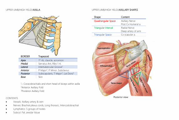
UPPER LIMB/HIGH YIELD/AXILLA
BORDER Trapezoid
Apex 1st rib, clavicle, acromion
Medial Serratus Ant, Ribs 1-4,
Lateral Intertubercular Groove1
Anterior P Major2, P Minor, Subclavius
Posterior Subscapularis, T Major3, Lat Dorsi3
Base Skin
1Coracobrachialis and short head of biceps within axilla 2Anterior Axillary Fold 3Posterior Axillary Fold
CONTENTS
Vessels: Axillary artery & vein
Nerves: Brachial plexus cords, Long thoracic, Intercostobrachial
Lymphatics: 5 groups of nodes
Subcut: Fat, areolar tissue
UPPER LIMB/HIGH YIELD/AXILLARY SHAPES
Shape Content
Quadrangular Space Axillary Nerve
Post Cx Humeral a.
Triangular Interval Radial Nerve
Deep artery of arm
Triangular Space Cx scapular a.

UPPER LIMB/HIGH YIELD/ANTECUBITAL FOSSA
BORDER Inverted Triangle
Superior Epicondyles
Medial PT (lat border)
Lateral Brachioradialis (med border)
Apex Meeting btwn Brachioradialis & PT
Floor Brachialis & Supinator
Roof Skin
Superficial Fascia
incl median cubital vein, med/lat cut n of forearm)
Deep Fascia
incl bicipital aponeurosis
CONTENTS (Really Need Booze To Be At My Nicest – lat to med)
Radial Nerve
Biceps Tendon
Brachial Artery
Median Nerve
NB veins are superficial to true “cubital fossa” (lat to med)
Cephalic Vein
Median Cubital Vein
Cephalic Vein
Basilic Vein
Other Superficial Structures
Lateral cutaneous nerve to forearm
UPPER LIMB/HIGH YIELD/HUMERAL FRACTURES
Head: Axillary Nerve
Shaft: Radial Nerve
Distal Medial: Ulnar Nerve
Distal Lateral: Median Nerve

UPPER LIMB/HIGH YIELD/CARPAL TUNNEL
BORDER
Roof Flexor Retinaculum
Lateral Scaphoid & Trapezium
Medial Pisiform & Hamate
Floor Carpal bones
Contents
Tendons of FDS/FDP in uncontinuous sheath
FPL in own sheath
Median Nerve
FCR which penetrates mid way
Other relations
Med/Sup: Ulnar artery & nerve
Lat/Sup: Palmar cut branch of median & Palmar branch radial a.
UPPER LIMB/HIGH YIELD/PALMAR SPACES & COMPARTMENTS
HypothenarC
CentralC
Flexor tendons, lumbricals, Sup
Palmar Arch
ThenarC
ThenarS
AdductorC MidpalmarS, 1
InterosseousS
5th MC 3rd MC
CCompartment SSPACE 1Continuous with ant compartment & flexor sheath
UPPER LIMB/HIGH YIELD/AUSCULATORY WINDOW
Trapezius
Scapula
Lat Dorsi
Sp
ine
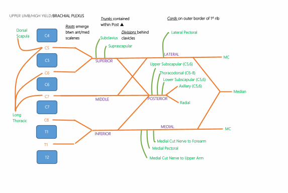
UPPER LIMB/HIGH YIELD/BRACHIAL PLEXUS
C4
C5
C
C6
C
C7
C
C
C
T1
C
T2
C
C5
C
C6
C
C7
C
C8
C
T1
C
MC
Median
MC
Axillary (C5,6)
Radial
Lateral Pectoral
Subclavius
Suprascapular
Upper Subscapular (C5,6)
Thoracodorsal (C6-8)
Lower Subscapular (C5,6)
Medial Cut Nerve to Forearm
Medial Pectoral
Medial Cut Nerve to Upper Arm
Dorsal
Scapula
Long
Thoracic
SUPERIOR
MIDDLE
SUPERI
INFERIOR
LATERAL
POSTERIOR
MEDIAL
Roots emerge
btwn ant/med
scalenes
Trunks contained
within Post
Divisions behind
clavicles
Cords on outer border of 1st rib
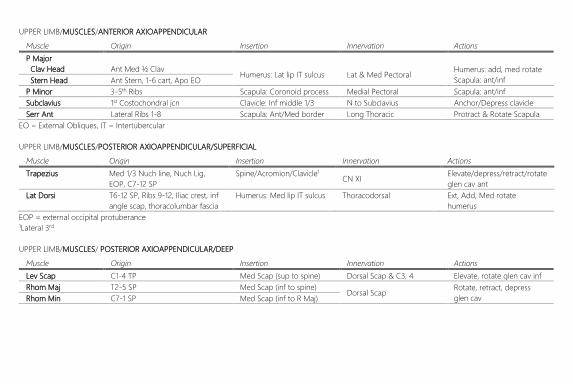
UPPER LIMB/MUSCLES/ANTERIOR AXIOAPPENDICULAR
Muscle Origin Insertion Innervation Actions
P Major
Clav Head Ant Med ½ Clav Humerus: Lat lip IT sulcus Lat & Med Pectoral
Humerus: add, med rotate
Scapula: ant/inf Stern Head Ant Stern, 1-6 cart, Apo EO
P Minor 3-5th Ribs Scapula: Coronoid process Medial Pectoral Scapula: ant/inf
Subclavius 1st Costochondral jcn Clavicle: Inf middle 1/3 N to Subclavius Anchor/Depress clavicle
Serr Ant Lateral Ribs 1-8 Scapula: Ant/Med border Long Thoracic Protract & Rotate Scapula
EO = External Obliques, IT = Intertubercular
UPPER LIMB/MUSCLES/POSTERIOR AXIOAPPENDICULAR/SUPERFICIAL
Muscle Origin Insertion Innervation Actions
Trapezius Med 1/3 Nuch line, Nuch Lig,
EOP, C7-12 SP
Spine/Acromion/Clavicle1 CN XI
Elevate/depress/retract/rotate
glen cav ant
Lat Dorsi T6-12 SP, Ribs 9-12, Iliac crest, inf
angle scap, thoracolumbar fascia
Humerus: Med lip IT sulcus Thoracodorsal Ext, Add, Med rotate
humerus
EOP = external occipital protuberance 1Lateral 3rd
UPPER LIMB/MUSCLES/ POSTERIOR AXIOAPPENDICULAR/DEEP
Muscle Origin Insertion Innervation Actions
Lev Scap C1-4 TP Med Scap (sup to spine) Dorsal Scap & C3, 4 Elevate, rotate glen cav inf
Rhom Maj T2-5 SP Med Scap (inf to spine) Dorsal Scap
Rotate, retract, depress
glen cav Rhom Min C7-1 SP Med Scap (inf to R Maj)

UPPER LIMB/MUSCLES/SCAPULOHUMERAL/ROTATOR CUFF
Muscle Origin Insertion Innervation Actions
Supraspinatous Supraspinous fossa Sup facet2 Suprascapular
Abduction1
Infraspinatous Infraspinous fossa Middle facet2 Lateral Rotation
Teres Minor Sup to T major Inf facet2 Axillary
Subscapularis Subscapular fossa Lesser tubercle Upper/Lower Subscapular Medial Rotation 1Supraspinatous does 1st 15 2Of Greater Tubercle
UPPER LIMB/MUSCLES/SCAPULOHUMERAL/OTHER
Muscle Origin Insertion Innervation Actions
Deltoid Inf margin of trap insertion Deltoid tuberosity Axillary Abduction1
Teres Major Scapula: Post/Lat/Inf border Post to Lat dorsi Lower subscapular Adduct, medially rotate

UPPER LIMB/MUSCLES/ARM/ANTERIOR COMPARTMENT (FLEXORS)
Muscle Origin Insertion Innervation Additional Actions
Biceps Brachii
Short Coracoid Process Radial tub & Biceps apo
MC
Supinate, flex, resist
shoulder disloc1 Long Supraglenoid tubercle
Brachialis Distal ½ and surface humerus Ulna tub & coronoid proc Main flexor
Coracobrachialis Coracoid process Middle 3rd med humerus Flex, Adduct, resist disloc 1Short head mostly
UPPER LIMB/MUSCLES/ARM/POSTERIOR COMPARTMENT (EXTENSORS)
Muscle Origin Insertion Innervation Additional Actions
Triceps Brachii
Long Infraglenoid tubercle
Proximal olecranon Radial
Extend, resist disloc2 Lateral Post surf, sup to radial groove
Medial Post surf, inf to radial groove
Anconeous Lat epicondyle & lat edge olec Ulna: Prox/Med border Stabilise elbow 2Long head only

UPPER LIMB/MUSCLES/FOREARM/ANTERIOR (FLEXORS)/SUPERFICIAL
Muscle Origin Insertion Innervation Additional Actions
Pronator Teres
Ulnar Head Coronoid process Radius: Lat Mid1
Median
Pronate2 Hum Head
Medial Epicondyle – ANTERIOR
SURFACE
Flex Carpi Rad MC: Base 2nd Abduct hand
Palm Longus Dist ½ flex ret & palm apo Tense Palm Apo
Flex Carpi Uln
Hum Head Pisiform, hook ham, Base
5th MC Ulnar Adduct Hand
Ulnar Head Medial Epicondyle – post surface
UPPER LIMB/MUSCLES/FOREARM/ ANTERIOR (FLEXORS)/INTERMEDIATE
Muscle Origin Insertion Innervation Additional Actions
Flex Dig Sup
Humeroulnar Medial Epi & Coronoid process 2-5 MP: Shaft Median Flex @ 2-5 PIJ & MCPJ
Radial Lat spiralling, ending mid lat bord
UPPER LIMB/MUSCLES/FOREARM/FLEXORS/DEEP
Muscle Origin Insertion Innervation Additional Actions
Flex Dig Prof
Medial part Ulnar: Prox ¾ surface
4-5th DP base Ulnar Flex @ 2-5 DIPJ
Lateral part 2-3rd DP base
Median (ant int branch) Flex Poll Long Rad: Mid ½ surface 1st DP base Flex @ 1st DIPJ, MCPJ
Pronator Quad Ulnar: distal slither Radius: Dist ¼ surface Pronate2
1Cont of FDS insertion site 2Pronator Quadratus stronger

UPPER LIMB/MUSCLES/FOREARM/POSTERIOR (EXTENSORS)/SUPERFICIAL
Muscle Origin Insertion Innervation Additional Actions
Brachioradialis Supraepicondylar ridge1
Prox to radial styloid Radial
Flexion (in pronation, weak)
Ext Carp Rad Longus 2nd MC: Lat base Ext & ABduct, Clenching
Ext Carpi Rad Brevis
Lateral epicondyle2
2nd MC: Med base
Deep Branch of Radial
Ext Digitorum 2-5th Ext expansion Ext 2-4th MCPJ
Ext Digiti Minimi 5th Ext expansion Ext 5th MCPJ
Ext Carp Ulnaris
Humeral head 5th MC base Ext & ADduct, Clenching
Ulnar head Ulnar: mid shaft btwn FDP & FCU
UPPER LIMB/MUSCLES/FOREARM/ POSTERIOR (EXTENSORS)/DEEP
Muscle Origin Insertion Innervation Additional Actions
Supinator Ulnar: opp radial tub3 Radius: Lat Prox ½
Deep Branch of Radial
Supinate
Ext Indicis Rad & IOM: distal-most 2nd Ext Expansions Ext 2nd DIPJ & PIPJ
Abd Poll Longus Rad, Uln, IOM: Mid ½ 1st MC base Abduct, Ext 1st CMJ
Ext Poll Longus Rad & IOM (inf to APL) 1st DP base Ext 1st DIPJ
Ext Poll Brevis IOM & Uln (inf to EPL) 1st PP base Ext 1st MPJ
1Brachioradialas sup to ECRL 2As a common extensor tendon 3± lat epicondyle below Anconeous
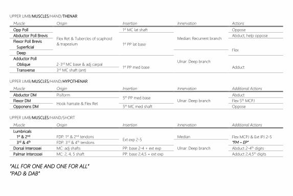
UPPER LIMB/MUSCLES/HAND/THENAR
Muscle Origin Insertion Innervation Actions
Opp Poll
Flex Ret & Tubercles of scaphoid
& trapezium
1st MC lat shaft
Median: Recurrent branch
Oppose
Abductor Poll Brevis
1st PP lat base
Abduct, help oppose
Flexor Poll Brevis
Superficial Flex
Deep
Ulnar: Deep branch Adductor Poll
Oblique 2-3rd MC base & adj carpal 1st PP med base Adduct
Transverse 3rd MC shaft (ant)
UPPER LIMB/MUSCLES/HAND/HYPOTHENAR
Muscle Origin Insertion Innervation Additional Actions
Abductor DM Pisiform 5th PP med base
Ulnar: Deep branch
Abduct
Flexor DM Hook hamate & Flex Ret
Flex 5th MCPJ
Opponens DM 5th MC med shaft Oppose
UPPER LIMB/MUSCLES/HAND/SHORT
Muscle Origin Insertion Innervation Additional Actions
Lumbricals
1st & 2nd FDP: 1st & 2nd tendons Ext exp 2-5
Median Flex MCPJ & Ext IPJ 2-5
“FM – EP” 3rd & 4th FDP: 3rd & 4th tendons
Ulnar: Deep branch Dorsal Interossei MC: adj shafts PP: base 2-4 + ext exp Abduct 2-4th digits
Palmar Interossei MC: 2, 4, 5 shaft PP: base 2,4,5 + ext exp Adduct 2,4,5th digits
“ALL FOR ONE AND ONE FOR ALL”
“PAD & DAB”
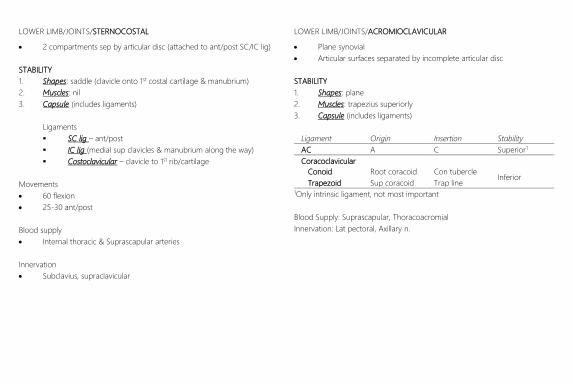
LOWER LIMB/JOINTS/STERNOCOSTAL
2 compartments sep by articular disc (attached to ant/post SC/IC lig)
STABILITY
1. Shapes: saddle (clavicle onto 1st costal cartilage & manubrium)
2. Muscles: nil
3. Capsule (includes ligaments)
Ligaments
SC lig – ant/post
IC lig (medial sup clavicles & manubrium along the way)
Costoclavicular – clavicle to 1st rib/cartilage
Movements
60 flexion
25-30 ant/post
Blood supply
Internal thoracic & Suprascapular arteries
Innervation
Subclavius, supraclavicular
LOWER LIMB/JOINTS/ACROMIOCLAVICULAR
Plane synovial
Articular surfaces separated by incomplete articular disc
STABILITY
1. Shapes: plane
2. Muscles: trapezius superiorly
3. Capsule (includes ligaments)
Ligament Origin Insertion Stability
AC A C Superior1
Coracoclavicular
Conoid Root coracoid Con tubercle Inferior
Trapezoid Sup coracoid Trap line 1Only intrinsic ligament, not most important
Blood Supply: Suprascapular, Thoracoacromial
Innervation: Lat pectoral, Axillary n.

LOWER LIMB/JOINTS/GLENOHUMERAL
Ball & socket synovial
STABILITY
1. Shapes: large head, small cavity (+ glen labrum) = 30% contact
2. Muscles: Rotator Cuff especially
3. Capsule: 3 intrinsic, 1 extrinsic
Muscles
Supraspinatous Superior
ie no reinforcement inferiorly Infraspinatous Posterior
Teres minor Posterior
Subscapularis Ant
Capsule
Openings
1. Intertubercular groove for long head biceps tendon
2. Subscapular bursa (ant/inferior)
Intrinsic Lig Origin Insertion Stability
Glenohumeral Glen labrum Neck Anterior
Coracohumeral Base coracoid Ant > Tub Superior
Transv humeral > Tub < Tub None
Extrinsic Lig Origin Insertion Stability
Coracoacromial Distal coracoid Head acrom Superior
Blood Supply: ant/post Cx, Suprascapular
Innervation: Suprascapular, Axillary, Lat pec
Important Bursa (that comm with joint)
Subscapular: Subscapularis tendon under coracoid process
Subacromial: btwn acromion/deltoid/ca lig Supraspinatous tendon
LOWER LIMB/JOINTS/ELBOW
Hinge synovial
STABILITY
1. Shapes: trochlea trochlear notch, capitulum head of radius
2. Muscles: Biceps/Brachioradialis, Triceps
3. Capsule: collaterals only
Intrinsic Lig Origin Insertion Stability
Radial Collat Lat epi Annular lig Lateral
Ulnar Collat
Ant Med epi Coronoid Strong, cord
Post Olecranon Fan
Oblique Olecranon Coronoid Deepens trochlea
Movements: Max 170 = carrying angle
Bloods Supply: Elbow anastomosis
Innervation: MC, Radial, Ulnar

LOWER LIMB/JOINTS/PROXIMAL RADIOULNAR
Pivot synovial
STABILITY
1. Shapes: radial head in radial notch & annular ligament
2. Muscles: sup (supinator, biceps), pro (pro quad, pro teres)
3. Capsule: annular ligament only (ant uln radial notch)
Blood Supply: radial part of peri articular anastomosis of elbow
Innervation: MC, median, radial (ie NOT ulnar)
LOWER L
IMB/JOINTS/DISTAL RADIOULNAR
Pivot synovial
STABILITY
1. Shapes: round head of ulnar into ulnar notch
2. Muscles:
3. Capsule: exposed proximally
Ligaments
Articular Disc
Medial edge ulnar notch lat base ulnar styloid
Prox surface articulates with ulna head
**Separates ulna from wrist joint
Other
Ant/post radio-ulnar - weak
Blood Supply: ant/post Interosseous
Innervation: Ant/post Interosseous
LOWER LIMB/JOINTS/WRIST
Condyloid synovial
Surface anatomy: at level of PROXIMAL wrist crease
Most stable position = slightly extended
STABILITY
1. Shapes: radius/articular disc with scaphoid, lunate, Triquetrium
2. Muscles: Carpi muscles (NB FCU 5th MC via pisohamate ligament)
3. Capsule: radiocarpal and collateral ligaments
Intrinsic Lig Origin Insertion Stability
Radiocarpal
Palmar Radius 2 rows carp
Support supination
Dorsal Support pronation
Collateral
Radial Radial styloid Scaphoid Lateral
Ulnar Ulnar styloid Triquetrium Medial
NB Pisiform only articulates with Triquetrium, is a sesamoid for FCU
Movements: ADD > AB, Flex > Ext
Blood Supply: Dorsal & Palmar carpal arches
Innervation: Median, Radial, Ulnar

LOWER LIMB/JOINTS/INTERCARPAL
Plane synovial
Multiple subunits in common joint cavity (not 1st CMCJ)
Joints include
Intercarpal (proximal row or distal row)
Prox to Distal rows (Midcarpal joint)
Pisotriquetral (since pisiform doesn’t articulate with other bones)
Ligaments
Anterior – Posterior – Interosseous
Movements
Flex/Ext @ Midcarpal joint
Ext & Abduction occur @ Midcarpal as well (vs Flex/Add @ wrist)
Proximal row more mobile
Blood supply/Innervation same as wrist
LOWER LIMB/JOINTS/CARPOMETACARPAL & INTERMETACARPAL
Plane synovial (except 1st CMCJ – saddle)
1st CMCJ (trapezium + 1st MC) has on articular capsule
Ligaments
CMC, IM & Interosseous ligaments
Transverse (sup & deep) MC ligaments
Movements
1st CMC = angular movement in any plane
2-3rd CMC = minimal
4-5th = mild/mod mobility
Blood Supply: dorsal/palmar arch, deep palmar, MC arteries
Innervation: Median, Radial, Ulnar
LOWER LIMB/JOINTS/METACARPOPHALANGEAL & INTERPHALANGEAL
MCP: Condyloid synovial ie 2 planes
IP: hinge synovial
Intrinsic Lig Origin Insertion Stability
Collateral
Cord-like Dorsal head Palmar base strong
Fan-like Dorsal head Palmar lig Support palmar lig
Palmar Digital sheath Retinac for flexor
Deep Transverse MC Ligament attaches to/btwn palmar MCPJ 2-4 = one unit
UPPER LIMB/NERVES
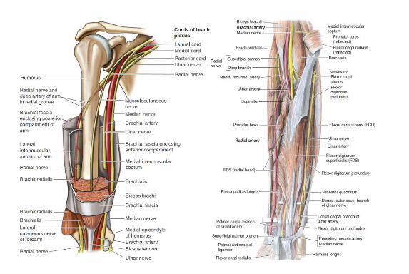
RADIAL (Branch of posterior cord: C5 – T1)
Course
2 branches before exiting axilla (4 branches above ACF in total)
Enters posterior compartment though triangular interval
Descends oblique/lateral on medial head of triceps (2 more branches)
Lies in radial groove in distal 3rd of upper arm
Pierces lateral IM septum to enter anterior compartment
Descends between Brachialis & Brachioradialis
Descends anterior to lateral epicondyle
Superficial branch cont under brachialis, pierces deep fascia @
wrist level
Deep branch pierces supinator to become Posterior
Interosseous Nerve (PIN)
Innervation
Post Comp Arm (Innervates long head triceps before entering arm)
Post Comp Forearm (Brachioradialis & ECRL, all else by PIN)
Sensory Post Upper Limb, & Dorsal aspect Lat Hand/2½ Digits (to PIPJ)
Associated Pathology
Crutch palsy
Humeral shaft fracture
Saturday night palsy
MUSCULOCUTANEOUS (Branch of lateral cord: C6,7)
Course
Pierces coracobrachialis to enter anterior compartment
Descends laterally btwn biceps/brachialis
Pierces deep fascia lateral to biceps tendon
Cont in forearm as lateral cutaneous nerve of forearm
Innervation
Anterior compartment of arm
Humerus via nutrient foramen
Cutaneous to lateral forearm
Associated pathology
Biceps hypertrophy eg weightlifting
Brachial plexus injury eg Erb’s palsy or heavy backpacks
Sx: loss of biceps reflex, sensation to radial forearm
UPPER LIMB/NERVES
MEDIAN (Branch of Lateral & Medial Cords: C6-T1)
Course
Axillary artery btwn convergence of 2 roots (medial cord anterior)
Enters ant compartment under T major
Descends ant to brachial a. (medially btwn biceps & brachialis)
Starts lat to brachial a. then crosses ant/med @ mid-upper arm
Descends from ACF btwn 2 heads of PT
Descends btwn FDS/FDP and reappears btwn FDS/FPL tendons
Branches in forearm
Anterior Interosseous: deep flexors (not ulnar ½ FDP)
Palmar cutaneous: distal branch, over flex ret, thenar skin only
Enters carpal tunnel medial to FDS/FDP tendons
Branches in hand
Recurrent median: thenar muscles “million dollar nerve”
Cutaneous: palmar lat 3½ digits, dorsal tips of same digits
Unnamed branches to 1st & 2nd lumbricals
Other Innervation
Unbranched nerve: superficial & intermediate group (not FCU)
Associated Pathology
Above elbow: loss of pronation, flexion
In elbow: “the blessed hand”
Ant Interosseous syndrome
Carpal tunnel syndrome
ULNAR (Branch of Medial cord: C8,T1)
Course
Descends posterior to brachial a.
Pierces medial IM septum ½ way down
Descends Posterior to medial epicondyle
Enters ant compartment of forearm btwn humeral/ulnar heads of FCU
Descends medial to ulnar a. deep to FCU (btwn FDS/FDP)
Branches in forearm
Muscular: FCU, Ulnar ½ FDP
Palmar/Dorsal: Cut to Med 1½ digits/hand (palmar does nails)
Enters hand through Guyon’s canal (above flex ret)
Branches in hand (over pisiform)
Superficial: Palmaris Brevis
Deep: btwn flex/add DM, on hook, in concavity of deep palmar
arch to supply Hypothenar muscles, lumbricals 3,4, Interossei,
add pollucis

UPPER LIMB/VESSELS/ARTERY/SUBCLAVIAN
Left from Ao, R from brachiocephalic trunk
Courses behind Ant Scalenes/In front of Med Scalenes
1st, 2nd, 3rd part defined in relation to Anterior Scalenes
Cont as axillary artery over 1st rib
Branches (VITamin C & D)
1 Vertebral
Internal Thoracic
Thyrocervical Trunk Suprascapular
Inferior Thyroid
Transverse cervical
2 Costocervical Trunk 1st IC
Deep cervical
3 Dorsal Scapular
UPPER LIMB/VESSELS/ARTERY/AXILLARY
Cont of Subclavian over 1st rib
Courses behind Pec Minor (defines parts of axillary a.)
Cont as brachial artery @ inferior border Teres Major
Branches (She Tastes Like Sweet Apple Pie)
1 Superior Thoracic
2 Thoracoacromial Trunk Clavicular
Pectoral
Acromial
Deltoid
Lateral Thoracic
3 Subscapular Cx Scapular
(largest branch) Thoracodorsal
Ant/Post Cx Humeral
NB collaterals exist if axillary artery clamped proximal to part 3
UPPER LIMB/VESSELS/ARTERY/BRACHIAL
Cont of axillary a. below inf border teres major
Course
Descends in groove btwn medial biceps/coracobrachialis
Deep artery of arm branches through interval (with rad n.)
Medial to Biceps tendon @ ACF
Bifurcation is variable but usually at head level of radial head
Relevant Relations
Median nerve starts ant/lat and midway crosses medially
Other Branches
Sup/Inf ulnar collaterals (in UPPER arm)
Elbow collaterals
UPPER LIMB/VESSELS/ARTERY/ULNAR
Branch of brachial a. in ACF (variable)
Course
Median nerve crosses laterally just after bifurcation
Courses under pronator teres for prox ½
Descends btwn FDS/FDP for distal ½ (with nerve medial)
Emerges proximal to wrist btwn FDS/FCU
Over flex ret and bifurcates into superficial/deep and respective arches
UPPER LIMB/VESSELS/ARTERY/RADIAL
Branch of brachial a. in ACF (variable)
Course
Desc under Brachioradialis
Emerges btwn tendons of Brachioradialis & FCR
Wraps around scaphoid & trapezium to enter snuff box
Descends btwn 2 heads of 1st lumbricals then heads of Add Poll to
enter palmar space and form deep palmar arch
Significant Branches
Princeps Pollicis (supplies thumb)
Radialis Indicis (supplies index)
Superficial (forms superficial palmar arch)
Scaphoid Supply
From dorsal and volar branches of radial artery
Enter distally (no vessels enter proximally
Middle # most common (60%), Prox # rarest (20%)
UPPER LIMB/VESSELS/VEINS/SUPERFICIAL
CEPHALIC
Starts in dorsal network
Ascends on lateral forearm
Communicates with basilic via median cubital vein
Cont along lateral aspect of distal upper arm then in deltopectoral
groove
Travels deep through costocoracoid membrane to join start of axillary
vein
BASILIC
Starts in dorsal network
Ascends medial forearm
Communicates with cephalic in ACF via median cubital vein
In upper arm penetrates biceps fascia to travel deep
Receives from Ant/Post humeral Cx then joins brachial vein
UPPER LIMB/VESSELS/VEINS/DEEP
BRACHIAL
Runs with arteries starting at superficial & deep palmar arches
Radial & Ulnar veins
Join to become brachial vein
Ascends in similar path to basilic but LATERAL to

LOWER LIMB/HIGH YIELD/FEMORAL
BORDER
Superior Inguinal Ligament
Lateral Sartorius
Medial Adductor Longus (Medial Border)
Floor Psoas, Pectineus, Adductor Longus, Obt Internus n.
Roof Fascia Lata
LOWER LIMB/HIGH YIELD/ADDUCTOR CANAL
BORDER
Posterior V. Medius
Anterior Sartorius
Medial Adductor Longus
Superior Apex of Femoral
Inferior Adductor Hiatus (of Add magnus)

LOWER LIMB/HIGH YIELD/POPLITEAL FOSSA
BORDER
Upper Medial Biceps Femoris
Upper Lateral Semimembranosus/Semitendinosus
Lower Medial Medial Head Gastrocnemius
Lower Lateral Lateral Head Gastrocnemius & Popliteus
Floor Popliteus, Capsule, Femur
Roof Fascia lata, Short saphenous & sural nerves
NB Tibial artery starts medial to nerve but ends lateral to nerve
LOWER LIMB/HIGH YIELD/FLEXOR RETINACULUM
Tip of medial mall calcaneal process/plantar aponeurosis
Contents (superior to inferior)
“Tom, Dick ANd Harry”
Tibialis posterior
Flexor Digitorum Longus
Posterior Tibial vein & Artery
Posterior Tibial nerve
Flexor Hallucis Longus
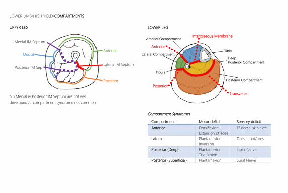
LOWER LIMB/HIGH YIELD/COMPARTMENTS
UPPER LEG
NB Medial & Posterior IM Septum are not well
developed compartment syndrome not common
LOWER LEG
Compartment Syndromes
Compartment Motor deficit Sensory deficit
Anterior Dorsiflexion
Extension of Toes
1st dorsal skin cleft
Lateral Plantarflexion
Inversion
Dorsal foot/toes
Posterior (Deep) Plantarflexion
Toe flexion
Tibial Nerve
Posterior (Superficial) Plantarflexion Sural Nerve
Anterior Medial
Posterior
Lateral IM Septum
Medial IM Septum
Posterior IM Sep
Anterior
Interosseous Membrane
Posterior
Transverse
LOWER LIMB/MUSCLES/THIGH/ANTERIOR/HIP FLEXORS
Muscle Origin Insertion Innervation Additional Actions
PECTINEUS Sup Pubic Ramus Pectineal Line Femoral
Adduct, Medial Rotate
SARTORIUS ASIS Pes Anserinus1 Stabilise Hip
ILIOSPOAS
PSOAS MINOR T12-L1: Body, Disc, TP Lesser Trochanter
L1-2 Ant Ramus Stabilise Hip
ILIACUS Iliac Crest, Fossa, Ala Scarum Femoral
PSOAS MAJOR T12-L4: Body, Disc, TP Iliopubic eminence2 L1-3 Ant Ramus Flex L/S-spine
1Conjoint tendon of Sartorius, Gracilis, Semitendinous. SGT FOT = Femoral, Obturator, Tibial innervation 2Sometimes to inguinal ligament, absent in 50% of population
LOWER LIMB/MUSCLES/THIGH/ANTERIOR/KNEE EXTENDERS (QUADRICEPS)
Muscle Origin Insertion Innervation Additional Actions
RECTUS FEMORIS AIIS, Sup lip acetabulum Tibial Tuberosity via
Quad tendon
(Patella syndesmosis)
Femoral
Flex hip
V. LATERALIS Femur: Inf to > trochanter & Post1
Nil V. INTERMEDIUS Femur: Sup 2/3 Ant + Lat Post
V. MEDIALIS Femur: Inf to < trochanter & Post2
1Lateral to lateral lip linea aspera 2Medial to medial lip linea aspera

LOWER LIMB/MUSCLES/THIGH/MEDIAL (HIP ADDUCTORS)
Muscle Origin Insertion Innervation Additional Actions
A. LONGUS Pubic Body: Ant/Sup Medial Linea Aspera Inf Obturator
A. BREVIS Pubic Body: Inf to Longus Medial Linea Aspera Sup Minimal flex
A. MAGNUS
HAMSTRING Inferior Ramus
Common Linea Aspera Tibial branch of sciatic Extend
ADDUCTOR Adductor tubercle1
Obturator
Flex
GRACILIS Pubic Body/Inf Ramus Pes Anserinus2 Flex, medial rotate
OBTURATOR EXT Inf ½ ant obt foramen Trochanteric Fossa Lateral rotate
1On medial femoral epicondyle, two heads form adductor canal 2Conjoint tendon of Sartorius, Gracilis, Semitendinous. SGT FOT = Femoral, Obturator, Tibial innervation
LOWER LIMB/MUSCLES/THIGH/POSTERIOR (HIP EXTENDERS)
Muscle Origin Insertion Innervation Additional Actions
SEMITENDINOUS Ischial Tub: Medial1 Pes Anserinus3 Tibial branch of Sciatic Leg flex & MED rotate4
SEMIMEMBRANOUS Ischial Tub: Lateral Tibia: Prox, Med, Post
BICEPS FEMORIS
LONG HEAD Ischial Tub: Medial1 Lateral head of Fibula
Tibial branch of Sciatic Leg flex & LAT rotate4
SHORT HEAD Inf Linea Aspera2 Common Fib branch of Sci
1With long head of biceps femoris 2Along same line and Inf to glut max insertion 3Conjoint tendon of Sartorius, Gracilis, Semitendinous. SGT FOT = Femoral, Obturator, Tibial innervation 4When knee flexed

LOWER LIMB/MUSCLES/THIGH/GLUTEAL/SUPERFICIAL
Muscle Origin Insertion Innervation Actions
G. MAXIMUS Sacrum, Coccyx, ST ligament1 Gluteal tuberosity2 & TFL Inf Gluteal (L5-S2) Ext, Lat Rotate, Sit Stand
G. MEDIUS Btwn Sup & Ant G. Line > Trochanter (Lat/Post)
Sup Gluteal (L5-S1) ABd, Med Rotate G. MINIMUS Btwn Ant & Inf G. Line > Trochanter (Lat/Ant)
TENSOR FASCIA ASIS & Ant Iliac Crest Gerdy’s Tubercle3
1Posterior to superior gluteal line 2Superior continuation of lateral lip of linea aspera 3Proximal lateral tibia
LOWER LIMB/MUSCLES/THIGH/POSTERIOR/GLUTEAL/DEEP
Muscle Origin Insertion Innervation Actions
PIRIFORMIS Ant surface sacrum
> Trochanter (Med/Ant)
S1-2 Ant Rami
Lat Rotate, Stabilise Hip,
Abduct (when flexed)
OBTURATOR INT All post obt foramen N to Obturator Internus
(L5-S1) SUP GEMELLI Ischial spine
INF GEMELLI Ischial tub1 N to Quadratus (L5-S1)
QUAD FEMORIS Ischial body2 Intertrochanteric crest
1Edge of lesser sciatic notch 2Inferior to inf lip of acetabulum

LOWER LIMB/MUSCLES/LEG/ANTERIOR COMPARTMENT (DORSIFLEXORS)
Muscle Origin Insertion Innervation Additional Actions
TIB ANT Sup lat ½ Tib2 & Med ½ Int Plantar Med Cu, Base 1st MT3
Deep Fibular
Invert4
EXT DIG LONG Lat Condyle, Med fib1, Int Dorsal base 2-5th MP & DP Extend 2-5th digits
EXT HAL LONG Mid-Med Fib1 & Int Dorsal base 1st DP Extend 1st digits
FIB TERT Just inf to EDL1 Dorsal prox shaft 5th PP Evert
Int = Interosseous, Cu = Cuneiform 1Medial to Interosseous border of fibula 2Incl Lat Condyle 3Medial Side 4With TP
LOWER LIMB/MUSCLES/LEG/LATERAL COMPARTMENT (EVERTORS)
Muscle Origin Insertion Innervation Additional Actions
FIB LONG Fib: Sup Lat1 Med Cuneiform, Base 1st MT Sup Fibular Plantarflexion (weak)
FIB BREV Fib: Inf Lat1 post inf Dorsal base 5th PP 1Lateral to Interosseous border of fibula
LOWER LIMB/MUSCLES/LEG/POSTERIOR COMPARTMENT/SUPERFICIAL (PLANTARFLEXORS)
Muscle Origin Insertion Innervation Additional Actions
GASTROCNEMIUS
LAT HEAD Lat Fem condyle
Calcaneal tendon
Calcaneal tuberosity Tibial
Flex leg
MED HEAD Med femoral condyle Nil
SOLEOUS Tib: Soleal line1 Fib: Sup/Lat
PLANTARIS Lat Fem condyle (sup to lat gas) Weak plantarflexor 1Line from Prox tib/fib jt mid medial tibia
LOWER LIMB/MUSCLES/LEG/POSTERIOR COMPARTMENT/DEEP (PLANATARFELXORS)
Muscle Origin Insertion Innervation Additional Actions
POPLITEUS Lat Fem condyle (inf to lat gas) Prox Med Tib (to Sol)1
Tibial
Support PCL, Med Rotate
FLEX HAL LONG Mid post fibula (inf to sol) Plantar base 1st DP Flex 1st digit
FLEX DIG LONG Mid post tibia Plantar base 2-5th DP3 Flex 2-5th digits
TIB POST Sup ½ Tib, Fib, Int Plantar Nav, Mid & Lat Cu,
base 2-4 PP3 Inversion2
Nav = Navicular, Cu = Cuneiform 1Attaches to medial meniscus along course 2With TA 3Forms groove just sup to medial malleolus
LOWER LIMB/MUSCLES/FOOT/LAYER 4 (INTEROSSEI)
Muscle Origin Insertion Innervation Actions
EXT DIG BREVIS Dorsal Lat Calc & Ext Ret
2-4th EDL tendons Deep Fibular
Helps EDL
EXT HAL BREVIS Lat Base 1st PP Helps EHL

LOWER LIMB/MUSCLES/FOOT/LAYER 1
Muscle Origin Insertion Innervation Actions
AB HAL Med process of Calc Tub
Med Base 1st PP1
Medial Plantar
ABduct, Flex 1st Digit
FLEX DIG BREVIS Bilat Base 2-5th MP Flex 2-5th digit
AB DIG MINI Lat process of Calc Tub Lat Base 5th PP ABduct, Flex 5th Digit
LOWER LIMB/MUSCLES/FOOT/LAYER 2
Muscle Origin Insertion Innervation Actions
QUAD PLANTAE Calc Tub (both processes) FDL tendon Lat Plantar Helps FDL
LUMBRICALS FDL tendon 2-5th Med Flexor expansion 1,2 Med 3,4 Lat Plantar Flex MP, Ext DP
LOWER LIMB/MUSCLES/FOOT/LAYER 3 (ADductors & Flexors)
Muscle Origin Insertion Innervation Actions
FLEX HAL BREVIS Cuboid & Lat Cuni Bilat base 1st PP1 Medial Plantar Flex 1st PP
ADD HAL
OBLIQUE Base 2-4th MT Lat base 1st PP1
Lateral Plantar (deep) Adduct 1st digit
TRANSVERSE Plantar lig of 5th MTPJ Lateral Plantar (sup)
FLEX DIG MINI Base 5th MT Base of 5th PP Flex 5th PP
LOWER LIMB/MUSCLES/FOOT/LAYER 4 (INTEROSSEI)
Muscle Origin Insertion Innervation Actions
PLANTAR Base, Medial shaft 3-5th MT Medial Base of 3-5th PP Lateral Plantar PAD
DORSAL Adj sides of 1-5th MT2 Lateral Base 2-4th PP3 Lateral Plantar DAB
1FHB shares insertion with Abductor HB on medial and adductor HB on lateral base 1st PP 21st Lumbrical attaches to lat base of 1st MT, all others attach to shaft 31st Lumbrical inserts into base of 2nd PP, all others insert into medial PP
LOWER LIMB/JOINTS/HIP
STABILITY
1. Shapes: Lunate surface & Acetabular labrum = 75% sphere
2. Muscles: Medial & Lateral rotators
3. Capsule (includes ligaments)
Intracapsular Ligaments
Ligament of head of femur – no structure, originates in notch
Transverse acetabular ligament – bridges acetabular notch
Capsule
Proximal: Just distal to acetabular rim
Distally: intertrochanteric line & root of > trochanter
(Doesn’t attach post)
Capsular Ligaments
Extension tightening of ligaments (restricting to 10-20)
Ligament Origin Insertion Stability
Iliofemoral AIIS, Sup rim IT line Strongest
Pubofemoral Obt crest IT line Ext/Abduction
Ischiofemoral Post rim Fem neck Weak
Balance
Strong ligaments ant = weaker muscles (med rot) ant
Weak ligaments post = strong muscles (lat rot) post
Blood Supply
Retinacular arteries (branch of Cx which are branches of deep fem)
NB Medial Cx Post (free capsular edge) = contributes more
Artery to head of femur (branch of Obturator a.)
Nerve Supply
Anterior: Femoral (hip flexors/knee extensors)
Inferior: Obt Int (lat rotators)
Posterior: N to quad femoris
Superior: Sup gluteal nerve (adductors)
LOWER LIMB/JOINTS/KNEE 1 of 2
Includes femoro-tibial & femoro-patellar (ie NOT fibula)
STABILITY
1. Shapes: poorly stable “2 balls on a warped table top”
2. Muscles: Flexors & Extensors – esp v. med & lat
3. Capsule (includes ligaments)
Extracapsular Ligaments
Ligament Origin Insertion Notes
Patellar Patella Tib Tuberosity
Fib Collat Lateral1 Lat head fibula Cord like
Tib Collat2 Medial1 Med tibial Condyle Med Meniscus
Obliq Pop Semimemb3 Lat post capsule
Arcuate Pop Fib Head Blends with post cap Over pop tend 1 Femoral epicondyle 2Weaker injury = tib collat + med meniscus 3Recurrent
Capsule
Proximal: Just proximal to articular surface
Distal: Just distal to articular surface
Incomplete edges: post lat tib (for popliteus to exit capsule)
Cont with quadriceps tendon & Suprapatella bursa
Patellar retinacular (made of deep fascia) join patella lig and form walls
of capsule – fcn to align patella – Q angle = lateral dispalcement
Intracapsular Ligaments
1. Anterior Cruciate
Ant-Med Intercondylar tib Post-Lat Fem condyle
In capsule not in synovial cavity
Weaker
Limits posterior rolling and displacement of femur on tibia
Prevents hyperextension
2. Posterior Cruciate
Post-Lat Intercondylar tib Ant-Med Fem condyle
Medial to ACL at crux
Prevents downhill slippage
3. Medial Meniscus
Transverse ligament of knee attached ant med to lat meniscus
C shaped (broader post)
Less mobile due to medial & lateral attachments:
Ant attached to intercondylar area of tibia (ant to ACL)
Post attached to intercondylar area of tibia (ant to PCL)
Adherent to TCL
4. Lateral Meniscus
Circular, smaller, mobile
Medial head of popliteus attaches
Post meniscofemoral ligament attaches lat men to PCL

LOWER LIMB/JOINTS/KNEE 2 of 2
Movements
Fully extended = locked = fem condyle med rotated on tib
(Screw-home mechanism)
Unlock: popliteus contraction = femur rotates 5 laterally
Bloods Supply
10 Genicular branches of femoral, popliteal, ant tibial (ant/post
recurrent branches), Cx fibular
Middle Genicular branches of popliteal supply Intracapsular structures
Innervation
Anterior: Femoral
Posterior: Tibial
Lateral: Common fibular
Medial: Obturator, Saphenous
Bursae
12 in total (4 communicate with synovial cavity)
1. Suprapatella
2. Popliteus
3. Anserine (deep to pan anserine)
4. Gastrocnemius
TIBIOFIBULAR Joints
TF = superior TiFJ
TF syndesmosis = inferior TiFJ
Interosseous membrane attaches tib-fib in sup-inf direction
Fib unable to move inf
TiFJ
Plane synovial
Tense capsule reinforced by ant/post lig of fib head
TiF Syndesmosis
Compound fibrous joint
IOM + Tibiofibular ligaments (ant, post, Interosseous)
Interosseous part: strongest, ext of IOM
Post TiFL cont inf as Inf transverse lig (forms post wall of ankle)

LOWER LIMB/JOINTS/ANKLE
STABILITY
1. Shapes: Trochlea wider anteriorly Dorsiflexion most stable position
Forces trochlea backwards into mortise
2. Muscles
3. Capsule (includes ligaments)
Capsule
Reinforced with medial & lateral collateral ligaments ie weak ant/post
Lateral Collateral
ATFL: lat mal neck talus, weak – most prone to damage
PTFL: lat mal lat tubercle talus
CFL: lat mal lat calcaneus
Medial Collateral (Deltoid) – actually one ligament with 4 parts
Tibio-Navicular
Tibio-Calcaneal
Tibio-Talar – Anterior & Posterior
Movements
Dorsiflexion & Plantarflexion (NB Inversion & Eversion is Subtalar jt)
Some Ab, Ad, In, Ev when ankle in extreme plantarflexion
Blood Supply & Innervation
Mall branches of fib, ant/post tib aa
Tibial & Deep Fibular (branch of common fib)
LOWER LIMB/JOINTS/FOOT
Important Joints
Subtalar: talo-calcaneal (NB Surgical STJ = TCJ + TCNJ)
Talocalcaneal ligaments support (weakly): med, lat, post, Interosseous
Transverse Tarsal: calcaneo-cuboid & talo-navicular
Augments inversion/eversion by rotating AP btwn hind and midfoot
Major Ligaments
Plantar calcaneonavicular ligament (spring) - Supports talus, long arch
Long plantar (calcaneus cuboid), 2 distal heads form arch for Fib L
Plantar Calcaneocuboid
Arches
Longitudinal Arch
MEDIAL: Calcaneus, Talus, Navicular, 3 Cuneiforms, 3 MT
Talar head is key
TA supports through attachments to 1st MAT & Medial Cuneiform
Higher & more important than lateral
LATERAL: Contents: Calcaneus, Cuboid, Lat 2 MT
Transverse Arch: Cuboid, Cuneiforms, Base of MT
Long arch helps support
FL & TP tendons cross under sole to support
Passive Factors: shape of bones, 4 layers of fibrous tissue (plant apo, long
plantar lig, plantar Calcaneocuboid lig, plantar calcaneonavicular lig)
Dynamic Factors: intrinsic muscles, other muscles (FHL & DL = long arch) (FL,
TP = transverse arch)

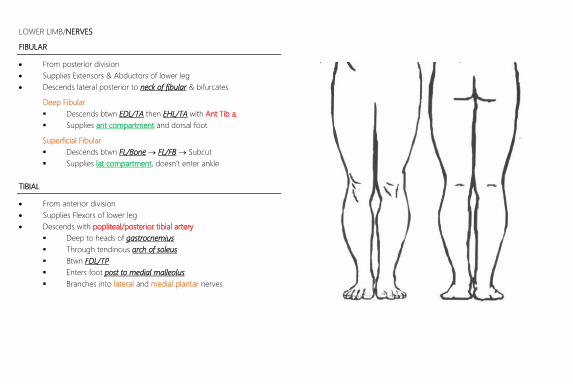
LOWER LIMB/NERVES
FEMORAL (ANTERIOR RAMI/POSTERIOR DIVISION L2,3,4)
Course
Descends behind Psoas Major emerging at lateral border
Descends between psoas & Iliacus into femoral (not within sheath)
Branches into anterior/posterior divisions as enters
Anterior Division
Anterior cutaneous thigh
Pectineus & Sartorius
Posterior Division
Descends in Adductor Canal & exits through Gracilis/Sartorius
to cont as Saphenous Nerve (sensory only)
Supplies quadriceps, knee, hip
SAPHENOUS NERVE
Branches in adductor canal
Descends btwn tendons of Sartorius & Gracilis to enter posterior
superficial compartment
Descends with Saphenous Vein
Supplies medial lower leg and foot
LOWER LIMB/NERVES
SCIATIC (ANT RAMI/ANT & POST DIVISION L4-S3)
Actually made up of Tibial & Fibular nerves distinctly but run together as
sciatic nerve in upper leg
Course
Roots converge on anterior surface of piriformis (as sacral plexus)
Enter sciatic foramen to enter lower limb
Ant to Piriformis
Post to Sup/Inf Gemellus & Obt Int
Runs deep to biceps femoris
Branches at femoral condyle
LOWER LIMB/NERVES
FIBULAR
From posterior division
Supplies Extensors & Abductors of lower leg
Descends lateral posterior to neck of fibular & bifurcates
Deep Fibular
Descends btwn EDL/TA then EHL/TA with Ant Tib a.
Supplies ant compartment and dorsal foot
Superficial Fibular
Descends btwn FL/Bone FL/FB Subcut
Supplies lat compartment, doesn’t enter ankle
TIBIAL
From anterior division
Supplies Flexors of lower leg
Descends with popliteal/posterior tibial artery
Deep to heads of gastrocnemius
Through tendinous arch of soleus
Btwn FDL/TP
Enters foot post to medial malleolus
Branches into lateral and medial plantar nerves

LOWER LIMB/VESSELS/ARTERY/PELVIC
COMMON ILIACS
Arise from bifurcation of Ao @ L4
Run along medial border of Psoas Major to Pelvic Brim
Bifurcate at Pelvic Brim/SIJ
Relevant Relations
Ureters cross antero-medial
Vein is post-lateral
EXTERNAL ILIAC
Bifurcation of common iliac @ pelvic brim/SIJ
Cont as Femoral artery @ Inguinal Ligament (1/3 lat to pubic tub)
Branches
Inferior epigastric (anastomoses sup with Sup epigastric)
Deep circumflex iliac (travels along iliac crest)
FEMORAL
Cont of external iliac @ Inguinal Ligament
Exits femoral triangle at Apex (Sartorius/Adductor Longus)
Descends in Adductor Canal
Cont as Popliteal artery as exits canal through Adductor Hiatus
Branches
Deep femoral branch just after exits
Btwn Pectineus & add longus posteriorly
Branches include medial/lateral Cx femoral head
Sup/Deep ext pudendal
Superficial epigastric/Cx Iliac
Descending Genicular
POPLITEAL
Cont of superficial femoral after exiting adductor hiatus
Descends medial-posterior through pop fossa
Bifurcates at level of fibular neck under popliteus into
Anterior Tibial
Posterior Tibial (fibular branches here)
Branches
Genicular: medial/lateral sup/inf & middle (ie 6 branches)
Sural artery
ANTERIOR TIBIAL
From bifurcation of popliteal a. under popliteus
Descends btwn TA/EDL
Cont as Dorsalis Pedis under dorsal retinaculum medially
POSTERIOR TIBIAL
Runs medial-post
Bifurcates into medial/lateral plantar after winding under medial mall
DORSALIS PEDIS
Btwn EHL/EDL
Cont as deep plantar & 1st MT artery
MEDIAL/LATERAL PLANTAR
Lateral much bigger & anastomoses with Dorsalis Pedis = plantar arch
FIBULAR
Branch of posterior tibial
Supplies lateral compartment only
UPPER LIMB/VESSELS/VEINS/SUPERFICIAL
GREAT SAPHENOUS
Formed by the union of 1st digit vein & Dorsal arch
Ascends ant to medial malleolus
Ascends post to medial epicondyle
Ascends medially to midline anterior
Joins femoral vein after penetrating saphenous opening
SMALL SAPHENOUS
Formed by union of 5th digit & Dorsal arch
Ascends post to lat malleolus
Ascends midline in lower leg then deep to head of gastroc
Empties into popliteal vein
LOWER LIMB/VESSELS/VEINS/DEEP
Plantar Drainage
Med/Lat plantar post tibial popliteal
Dorsal Drainage
Dorsal arch ant tibial popliteal
Popliteal vein femoral ext iliac