UPLC-QTOF/MS for a Rapid Characterisation of Phenolic Compounds from Leaves of Myrtus...
8
UPLC–QTOF/MS for a Rapid Characterisation of Phenolic Compounds from Leaves of Myrtus communis L. Amani Taamalli, a Ihsan Iswaldi, b David Arráez-Román, c,d Antonio Segura-Carretero, c,d * Alberto Fernández-Gutiérrez c,d and Mokhtar Zarrouk a ABSTRACT: Introduction – Although continuous research has been conducted on the biological activities of myrtle and the characterisa- tion of its essential oil, few studies have focused on its phenolic composition despite major beneficial properties. Objective – To carry out a comprehensive characterisation of infusion and methanolic extract from myrtle leaves by UPLC–QTOF/MS. Methods – Myrtle-leaf infusions, prepared using deionised water, and the methanolic extracts were analysed by reversed- phase ultra-performance liquid chromatography (UPLC) coupled to electrospray ionisation quadrupole time-of-flight mass spectrometry (ESI/QTOF/MS). The MS and MS/MS experiments were conducted using the negative-ionisation mode, in order to provide molecular-mass information and production spectra of the compounds for structural elucidation. Results – The analytical method applied enabled the characterisation of several compounds such as gallic acid and galloyl deriv- atives, ellagic acid and derivatives, hexahydroxydiphenolyl and derivatives, flavonoids, lignans and gallomyrtucommulones. Flavonoids, ellagic acid and its derivatives and gallic acid and its derivatives formed the major fractions. Conclusion – UPLC combined with QTOF/MS is a powerful analytical method for characterising infusions and alcoholic extracts from myrtle leaves. Copyright © 2013 John Wiley & Sons, Ltd. Supporting information can be found in the online version of this article. Keywords: UPLC–QTOF/MS/MS; phenolic compounds; myrtle. Introduction Myrtus communis L. (Myrtaceae family) is an evergreen shrub typical of the Mediterranean maquis and which grows spontane- ously in many countries (Gardeli et al., 2008). From 1–3 m tall, it has bright green leaves and white flowers from June to July. Different parts of the plant have various uses in the food indus- try, such as for flavouring meat and sauces and in the cosmetic industry (Chalchat et al., 1998), and have long been used in folk medicine. Leaves have been used as a folk remedy to treat hypertension, hyperglycaemia, haemorrhoids, cold, diaorrhea, internal diseases, rheumatic pain, to pass kidney stones and wounds (Mine et al., 2009), among others. In recent years this plant has received increasing attention for its biological proper- ties, such as anti-oxidant (Romani et al., 2004; Aidi Wannes et al., 2010), anti-nociceptive, anti-inflammatory (Hosseinzadeh et al., 2011), cytotoxic and anti-bacterial (Cottiglia et al., 2012; Messaoud et al., 2012) activities. The berries have also been stud- ied for the characterisation of the phenolic composition (Barboni et al., 2010), stability and anti-oxidant activity (Montoro et al., 2006). The individual polyphenols in myrtle-leaf tissues were first quantified using HPLC/DAD (Romani et al., 1999). Myrtle leaves contain different types of secondary metabolites, including phenolic compounds, which are derivatives of the pentose phosphate, shikimate and phenylpropanoid pathways in plants (Randhir et al., 2004). These compounds, ubiquitous in the plant kingdom, are one of the most widely occurring groups of phytochemicals and of considerable physiological and morpho- logical importance in plants (Balasundram et al., 2006), and are also extremely important components of the human diet because of their health benefits (Martin and Appel, 2010). Phenolic compounds extracted from myrtle leaves have proved to exert anti- hyperglycaemic effects (Benkhayal et al., 2009), while myricetin-3- O-galactoside and myricetin-3-O-rhamnoside isolated from the leaves have shown anti-mutagenic activity (Hayder et al., 2008). Also, flavonoids and anthocyanins in myrtle-berry extract have anti-oxidant activity, as demonstrated by trolox equivalent * Correspondence to: A. Segura-Carretero, Department of Analytical Chemistry, University of Granada, Avda. Fuentenueva s/n, 18071 Granada, Spain. E-mail: [email protected] a Laboratoire de Biotechnologie de l’olivier, Centre de Biotechnologie de Borj Cedria, BP 901, 2050 Hammam-Lif, Tunisia b Department of Chemistry, Faculty of Mathematics and Natural Sciences, University of Andalas, Kampus Limau Manis, Padang 25163, Indonesia c Department of Analytical Chemistry, University of Granada, Avda. Fuentenueva s/n, 18071 Granada, Spain d Functional Food Research and Development Centre (CIDAF), Health-Science Technological Park, Avda. Del Conocimiento s/n, 18100 Granada, Spain Phytochem. Anal. 2013 Copyright © 2013 John Wiley & Sons, Ltd. Research Article Received: 28 April 2013, Revised: 2 August 2013, Accepted: 12 August 2013 Published online in Wiley Online Library (wileyonlinelibrary.com) DOI 10.1002/pca.2475
Transcript of UPLC-QTOF/MS for a Rapid Characterisation of Phenolic Compounds from Leaves of Myrtus...

Research Article
Received: 28 April 2013, Revised: 2 August 2013, Accepted: 12 August 2013 Published online in Wiley Online Library
(wileyonlinelibrary.com) DOI 10.1002/pca.2475
UPLC–QTOF/MS for a Rapid Characterisation ofPhenolic Compounds from Leaves of Myrtuscommunis L.Amani Taamalli,a Ihsan Iswaldi,b David Arráez-Román,c,d
Antonio Segura-Carretero,c,d* Alberto Fernández-Gutiérrezc,d
and Mokhtar Zarrouka
ABSTRACT:Introduction – Although continuous research has been conducted on the biological activities of myrtle and the characterisa-tion of its essential oil, few studies have focused on its phenolic composition despite major beneficial properties.Objective – To carry out a comprehensive characterisation of infusion and methanolic extract from myrtle leaves byUPLC–QTOF/MS.Methods – Myrtle-leaf infusions, prepared using deionised water, and the methanolic extracts were analysed by reversed-phase ultra-performance liquid chromatography (UPLC) coupled to electrospray ionisation quadrupole time-of-flight massspectrometry (ESI/QTOF/MS). The MS and MS/MS experiments were conducted using the negative-ionisation mode, in orderto provide molecular-mass information and production spectra of the compounds for structural elucidation.Results – The analytical method applied enabled the characterisation of several compounds such as gallic acid and galloyl deriv-atives, ellagic acid and derivatives, hexahydroxydiphenolyl and derivatives, flavonoids, lignans and gallomyrtucommulones.Flavonoids, ellagic acid and its derivatives and gallic acid and its derivatives formed the major fractions.Conclusion – UPLC combined with QTOF/MS is a powerful analytical method for characterising infusions and alcoholicextracts from myrtle leaves. Copyright © 2013 John Wiley & Sons, Ltd.
Supporting information can be found in the online version of this article.
Keywords: UPLC–QTOF/MS/MS; phenolic compounds; myrtle.
* Correspondence to: A. Segura-Carretero, Department of Analytical Chemistry,University of Granada, Avda. Fuentenueva s/n, 18071 Granada, Spain.E-mail: [email protected]
a Laboratoire de Biotechnologie de l’olivier, Centre de Biotechnologie de BorjCedria, BP 901, 2050 Hammam-Lif, Tunisia
b Department of Chemistry, Faculty of Mathematics and Natural Sciences,University of Andalas, Kampus Limau Manis, Padang 25163, Indonesia
c Department of Analytical Chemistry, University of Granada, Avda.Fuentenueva s/n, 18071 Granada, Spain
d Functional Food Research and Development Centre (CIDAF), Health-ScienceTechnological Park, Avda. Del Conocimiento s/n, 18100 Granada, Spain
IntroductionMyrtus communis L. (Myrtaceae family) is an evergreen shrubtypical of the Mediterranean maquis and which grows spontane-ously in many countries (Gardeli et al., 2008). From 1–3m tall, ithas bright green leaves and white flowers from June to July.Different parts of the plant have various uses in the food indus-try, such as for flavouring meat and sauces and in the cosmeticindustry (Chalchat et al., 1998), and have long been used in folkmedicine. Leaves have been used as a folk remedy to treathypertension, hyperglycaemia, haemorrhoids, cold, diaorrhea,internal diseases, rheumatic pain, to pass kidney stones andwounds (Mine et al., 2009), among others. In recent years thisplant has received increasing attention for its biological proper-ties, such as anti-oxidant (Romani et al., 2004; Aidi Wannes et al.,2010), anti-nociceptive, anti-inflammatory (Hosseinzadeh et al.,2011), cytotoxic and anti-bacterial (Cottiglia et al., 2012;Messaoud et al., 2012) activities. The berries have also been stud-ied for the characterisation of the phenolic composition (Barboniet al., 2010), stability and anti-oxidant activity (Montoro et al.,2006). The individual polyphenols in myrtle-leaf tissues were firstquantified using HPLC/DAD (Romani et al., 1999).
Myrtle leaves contain different types of secondary metabolites,including phenolic compounds, which are derivatives of thepentose phosphate, shikimate and phenylpropanoid pathways inplants (Randhir et al., 2004). These compounds, ubiquitous in the
Phytochem. Anal. 2013 Copyright © 2013 John
plant kingdom, are one of the most widely occurring groups ofphytochemicals and of considerable physiological and morpho-logical importance in plants (Balasundram et al., 2006), and are alsoextremely important components of the human diet because oftheir health benefits (Martin and Appel, 2010). Phenolic compoundsextracted from myrtle leaves have proved to exert anti-hyperglycaemic effects (Benkhayal et al., 2009), while myricetin-3-O-galactoside and myricetin-3-O-rhamnoside isolated from theleaves have shown anti-mutagenic activity (Hayder et al., 2008).Also, flavonoids and anthocyanins in myrtle-berry extract haveanti-oxidant activity, as demonstrated by trolox equivalent
Wiley & Sons, Ltd.
A. Taamalli et al.
antioxidant capacity (TEAC) assay and free-radical activity(Montoro et al., 2006). Nevertheless, knowing the compositionand occurrence of bioactive compounds is crucial for investigat-ing the bioactivity of any dietary phytochemical in a particularplant species. Thus the characterisation of bioactive compoundsfrom M. communis leaf extracts would have relevance fordeveloping nutraceuticals and dietary supplements.
Polyphenols constitute a complex group of compounds with adiverse chemical nature of varying polarity and size. In recentyears, ultra-performance liquid chromatography (UPLC) hasopened new possibilities for improving the analytical methodsfor complex sample matrices. The development of ultra-high-pressure pump systems and small-size packing materials hasresulted improved resolution, higher peak efficiency, fasterseparations and reduced solvent consumption compared withconventional HPLC (Motilva et al., 2013). In this sense, thecombination of UPLC with mass spectrometry permits a betterseparation and characterisation of phenolic compounds. Thus,the mass analyser quadrupole-time-of-flight mass spectrometry(QTOF/MS) combines the high performance of time-of-flightanalysis in both MS and tandem MS (MS/MS) modes, providinghigh sensitivity and high mass accuracy for both precursor andproduct ions.
As the characterisation of phenolic compounds in myrtleleaves appears to be biologically and ecophysiologically useful,the objective of the present study was to gain insight of thephenolic composition of the leaves of M. communis L. usingthe advanced analytical platform UPLC–QTOF/MS/MS.
Experimental
Chemicals
All chemicals were of analytical reagent grade and used as received. Aceticacid was purchased from Sigma-Aldrich (Steinheim, Germany) andacetonitrile and methanol from Lab-Scan (Gliwice, Sowinskiego, Poland).Water was purified by a Milli-Q system from Millipore (Bedford, MA, USA).
Plant samples and extraction
The aerial parts were collected from myrtle (M. communis L.) shrubs fromthe mountain Jbel Eddis (Nefza region, Béja Province, northwesternTunisia). A voucher specimen (ID number: M0013) was deposited at ourlaboratory for future reference. The leaves were removed by hand inthe laboratory, dried at ambient temperature till reaching a constantweight (300 g), milled to a fine powder using a Jata ML-469 blender,and then used for both modes of extractions. Infusions were preparedaccording to a standard protocol. Thus, water was heated till reaching98°C, then 3 g of plant material were dissolved in 200mL of the heatedwater and infusions were left to steep at room temperature withoutadditional heating for an infusion time of 30min (Katalinic et al., 2006).As for the methanolic extracts, 1 g of ground dried leaves was extractedby stirring with 10mL of pure methanol for 30min. After centrifugationat 5000 × g, extracts were filtered through a 0.45μm syringe filter priorto analysis. The extractions and the analyses were performed in triplicatefor each extraction mode.
UPLC conditions
The UPLC analyses were made using a Waters Acquity UPLC system(Waters Corporation, Milford, MA, USA). Separations were carried out witha Zorbax Eclipse Plus C18 analytical column (4.6×150mm, 1.8μm particlesize). The mobile phases were acidified water (0.5% acetic acid, v/v) andacetonitrile as eluents A and B, respectively. The chromatographic method
Copyright © 2013 Johnwileyonlinelibrary.com/journal/pca
consisted of the following multistep linear gradient: 0min, 0% B; 10min,20% B; 15min, 30% B; 20min, 50% B; 25min, 75% B; 30min, 100% B;32min 0% B; and finally a 3min post-run after each analysis. The injectionvolumewas 1μL and the column temperature wasmaintained at 25°C. Theflow rate was set at 0.8mL/min throughout the gradient.
ESI/QTOF/MS conditions
The UPLC system was coupled to a Quadrupole-Time-of-FlightmicrOTOF-Q (Bruker Daltonik GmbH, Bremen, Germany), an orthogonalaccelerated QTOF mass spectrometer, equipped with an ESI. Analysisparameters were set using a negative ion mode with spectra acquiredover a mass range from m/z 50 to 1100. The optimum values of theESI/MS parameters were: capillary voltage, +4.0 kV; drying gas tempera-ture, 190°C; drying gas flow, 9.0 L/min; nebulising gas pressure, 29 psi;collision RF, 150 Vpp; transfer time 70μs; and pre-pulse storage, 5μs.Moreover, automatic MS/MS experiments were performed adjustingthe collision-energy values as follows: m/z 100, 20 eV; m/z 500, 30 eV;and m/z 1000, 35 eV; and by using nitrogen as the collision gas. TheMS data were processed through Data Analysis 4.0 software (BrukerDaltonics, Bermen, Germany), which provided a list of possible elementalformulae by using the Generate Molecular Formula editor. The editoruses a CHNO algorithm, which provides standard functionalities suchas minimum/maximum elemental range, electron configuration and ringplus double-bond equivalents, as well as a sophisticated comparison ofthe theoretical with the measured isotope pattern (msigma value) forincreasing the confidence in the suggested molecular formula. Thewidely accepted accuracy for confirmation of elemental compositionshas been established to be 5 ppm.
During the UPLC method development, an external instrumentcalibration was performed using a Cole Palmer syringe pump (VernonHills, Illinois, USA) directly connected to the interface, passing a solutionof sodium formate cluster containing 5mM sodium hydroxide and 0.2%formic acid in water:isopropanol 1:1 (v/v). Using this method an exactcalibration curve based on numerous cluster masses, each differing by68Da (NaCHO2), was plotted. Due to the compensation of temperaturedrift in the QTOF, this external calibration provided accurate mass valuesfor a complete run without the need for a dual sprayer set-up for internalmass calibration.
In the present study, the quantification of the characterised compoundswas based on the peak-area calculation. The results are expressed aspercentage of the total area of identified compounds.
Results and discussion
Chromatographic profile and compound characterisation
The base-peak chromatograms (BPCs) of the infusion andmethanolic extracts of myrtle leaves are presented in Fig. 1(A) and(B), respectively. The compounds characterised are summarisedin Table 1 (with numbers from 1 to 42 indicating the elutionorder), including retention time, experimental and calculatedm/z, molecular formula, andmsigma and error values provided bythe software and QTOF/MS/MS fragments. All the compoundswere characterised by the interpretation of their mass spectradetermined by the QTOF/MS and also taking into account thedata provided by the literature and databases. In the presentstudy, the compounds characterised are classified into sevengroups: gallic acid and galloyl derivatives; ellagic acid andderivatives; hexahydroxydiphenolyl derivatives; flavonoids; lignans;gallomyrtucommulones; and other compounds. TheMS/MS spectraof the identified compounds are presented as supplementary data.
Gallic acid and galloyl derivatives. The Smart Formula Editorprovided an identical molecular formula for the peaks 3, 6 and 9,which yielded a parent ion [M�H]� at m/z 343 in the ESI/MS
Phytochem. Anal. 2013Wiley & Sons, Ltd.

Figure 1. Base peak chromatograms of (A) infusion and (B) methanolic extract from myrtle leaves.
Phenolic Compounds in Myrtle Leaves
spectra. The fragmentation pattern shows the presence of ionsat m/z 191.0558, 169.0144 and 125.0257. The first fragment ioncorresponds to the loss of the gallic acid molecule from theprecursor ion. The second is due to the loss of the quinic acidmolecule, which in turn yielded an ion at m/z 125 after a lossof CO2 moiety (44Da), suggesting the presence of carboxylicacid in the parent ion. Thus these compounds were identifiedas galloyl quinic acid isomers. This fragmentation is consistentwith that reported by Romani et al. (2012), who also found twoisomers of galloyl quinic acid in leaf extract of myrtle from Italy.This compound was also identified in Pistacia lentiscus leafextract (Rodríguez Pérez et al., 2013).
Peaks number 4 and 5 with [M�H]� at m/z 331 yielded afragmentation pattern composed of ions at m/z 271.0453,169.0137 and 125.0240. The ion at m/z 271 is due to a cross-ringfragmentation of a glucose molecule [M�H� 60]�. The ion atm/z 169.0137 corresponds to the aglycon form due to the lossof a hexosyl moiety (162Da). The fragment at m/z 125.0240 isassigned as a decarboxylation of the gallic acid moiety [galloylCO2]
�. This fragmentation pattern agrees with data reported
Phytochem. Anal. 2013 Copyright © 2013 John
elsewhere (Meyers et al., 2006; Romani et al., 2012) and thusthese compounds were identified as galloyl glucose isomers.Peak number 8 was identified as gallic acid, which showed a
base peak at m/z 169.0163 corresponding to [M�H]� and afragment ion at m/z 125.0132 ([M�H� 44]�) correspondingto the loss of CO2. Gallic acid was identified in myrtle-berry(Tuberoso et al., 2007) and myrtle-leaf extracts (Aidi Wanneset al., 2010; Romani et al., 2012).Peak 18, which showed an m/z of 495.0766, was regarded as
digalloyl quinic acid based on the presence of two mainfragments at m/z 343.0672, which resulted from a loss of galloylquinic acid molecule, and 169.0141, which corresponds to agalloyl moiety (Clifford et al., 2007).Digalloyl-β-d-glucopyranose was assigned to peaks 11 and 20
based on the MS (molecular ion at m/z 483.0768 with molecularformula C20H19O14) and MS/MS data, which provided fragmentions at m/z 331.0965 and 169.0143 in agreement with the datareported by Meyers et al. (2006) and Fernández-Arroyo et al.(2010). Thus, the fragment ion at m/z 331.0965 corresponds toa galloyl glucose molecule (after a loss of gallic acid moiety from
Wiley & Sons, Ltd. wileyonlinelibrary.com/journal/pca

Table
1.Prop
osed
compo
unds
detected
inmyrtle
aque
ousan
dmetha
nolic
extracts
byUPLC-QTO
F/MS
Peak
t R(m
in)
Measured
m/z
Form
ula
Calculated
m/z
Error
(ppm
)msigm
aMS/MSfrag
men
tsProp
osed
compo
und
11.92
191.05
70C7H11O6
191.05
61�4
.75.0
127.03
99Quinicacid
isom
er1
22.50
191.02
07C7H11O6
191.01
97�5
.02.3
127.03
99Quinicacid
isom
er2
32.77
343.06
71C14H15O10
343.06
710.0
2.3
191.
0516
,169
.014
4,12
5.02
57Galloyl
quinicacid
isom
er1
43.18
331.06
76C13H15O10
331.06
71�1
.62.8
271.04
53,1
69.013
7,12
5.02
40Galloyl
glucoseisom
er1
53.44
331.06
81C13H15O10
331.06
71�3
.19.0
169.01
38,1
25.024
3Galloyl
glucoseisom
er2
63.85
343.06
70C14H15O10
343.06
710.2
2.7
169.01
44,1
25.026
7Galloyl
quinicacid
isom
er1
74.05
633.07
38C27H21O18
633.07
33�0
.71.3
300.99
2Strictin
inisom
er1
84.18
169.01
44C7H5O5
169.01
42�0
.61.4
125.02
47Gallic
acid
94.44
343.06
75C14H15O10
343.06
71�1
.33.9
169.01
63,1
25.025
8Galloyl
quinicacid
isom
er3
105.06
633.07
35C27H21O18
633.07
33�0
.24.9
300.99
2,16
9.01
37Strictin
inisom
er2
115.46
483.07
69C20H19O14
483.07
802.3
2.8
331.09
65,1
69.014
3Digalloyl-d-glucopy
rano
seisom
er1
126.38
305.06
79C15H13O7
305.06
67�4
.14.9
261.07
60,1
65.019
8,12
5.02
49(�
)-Ep
igallocatechin
136.79
361.08
30C14H17O11
361.07
76�1
4.8
91.9
249.06
15,1
11.008
2Not
iden
tified
147.26
633.07
29C27H21O18
633.07
330.7
8.0
300.99
2Strictin
inisom
er3
157.58
793.06
47C28H25O27
793.05
89�7
.416
.763
3.07
26,3
00.998
7Not
iden
tified
167.98
787.09
27C23H31O30
787.09
06�2
.759
.060
1.04
83,4
31.025
8,30
0.99
85,1
69.014
5Not
iden
tified
178.25
443.19
21C21H31O10
443.19
230.4
9.9
Not
frag
men
ted
Not
iden
tified
188.45
495.07
76C21H19O14
495.07
800.8
5.0
343.06
72,1
69.014
1Digalloylqu
inicacid
199.12
783.06
65C34H23O22
783.06
862.7
38.0
765.05
76,6
13.046
0,30
0.99
81,1
69.013
8Cornu
siin
Cisom
er1
209.83
483.07
74C20H19O14
483.07
801.3
5.3
331.06
76,1
69.013
0Digalloyl-d-glucopy
rano
seisom
er2
2110
.23
783.07
667
C34H23O22
783.07
22�2
.716
.976
5.05
48,6
13.051
0,30
0.99
74,1
69.014
7Cornu
siin
Cisom
er2
2210
.90
785.07
79C16H33O35
785.08
083.6
11.9
Not
frag
men
ted
Not
iden
tified
2311
.24
785.08
30C34H25O22
785.08
433.9
3.6
633.07
03,4
83.079
3,30
0.99
78Digalloyl-hexah
ydroxydiph
enoy
l-glucose
isom
er1
2411
.57
635.08
90C27H23O18
635.08
850.8
11.1
465.06
61,3
13.074
9,16
9.01
36Trigalloyl-glucose
2511
.77
463.05
20C20H15O13
463.05
18�0
.511
.530
0.99
79Ellagicacid-hexoside
2612
.44
457.07
84C22H17O11
457.07
76�1
.62.7
305.06
68,1
69.014
2,12
5.04
8Ep
igallocatechin
gallate
2712
.65
631.09
47C28H23O17
631.09
41�1
.03.3
479.08
63,3
16.022
4Myricetin
galloyl
hexoside
2813
.45
785.08
15C34H25O22
785.08
43�0
.17.2
483.07
99,3
00.998
6Digalloyl-hexah
ydrodiph
enoy
l-glucose
isom
er2
2913
.83
479.08
37C21H19O13
479.08
31�1
.24.2
316.02
21Myricetin
galactosideisom
er1
3014
.11
479.08
31C21H19O13
479.08
31�0
.03.0
316
Myricetin
galactosideisom
er2
3114
.90
615.09
88C28H23O16
615.09
920.5
5.1
463.08
53Myricetin
galloyl
rham
nopy
rano
side
3215
.63
449.07
26C20H17O12
449.07
25�0
.02.9
316.02
21Myricetin-3-O-arabino
side
3316
.09
463.08
91C21H19O12
463.08
82�2
.01.8
316.02
29Myricetin-3-O-rha
mno
side
3416
.31
300.99
93C14H5O8
300.99
90�1
.22.8
Not
frag
men
ted
Ellagicacid
3516
.64
599.10
44C28H23O15
599.10
42�0
.35.9
447.09
33,3
13.058
6Ka
empferol-O-galloyl-hexoside
3617
.23
523.21
87C26H35O11
523.21
85�0
.48.2
361.16
60Secoisolaricire
sino
lhexoside
3718
.14
431.22
90C21H35O9
431.22
87�0
.73.7
371.21
01,1
61.047
5Neo
rehm
annioside
3818
.47
447.09
20C21H19O11
447.09
332.9
1.0
301.03
51Que
rcetin
rham
noside
3919
.71
569.22
36C27H37O13
569.22
400.7
2.9
371.37
9Not
iden
tified
4022
.11
583.20
24C27H35O14
583.20
321.3
11.3
539.21
21,3
31.066
9,27
1.04
59,1
66.998
2Not
iden
tified
4122
.43
567.20
84C27H35O13
567.20
83�0
.29.9
331.07
19,2
71.046
6,31
3.05
49,1
69.012
7Gallomyrtucommulon
eC
4222
.82
569.22
31C27H37O13
569.22
401.6
4.3
331.
0509
,313
.053
6Gallomyrtucommulon
eA
A. Taamalli et al.
Phytochem. Anal. 2013Copyright © 2013 John Wiley & Sons, Ltd.wileyonlinelibrary.com/journal/pca

Phenolic Compounds in Myrtle Leaves
the precursor ion), which loses the glucose moiety to give theion at m/z 169.0143.
Finally, peak 24 with m/z 635.0890 was characterised astrigalloyl glucose. The fragment spectra showed ions at m/z465.0863 and 313.0749 that could be assigned to the sequentialloss of gallic acid [M�H� 169]� and [M�H� 321]�. Thefragment ion at m/z 169.0136 corresponds to a gallic acid mole-cule. This compounds was also identified in tanoak (LithocarpusDensiflorus) acorn extract (Meyers et al., 2006).
Ellagic acid and derivatives. Peaks 19 and 21 exhibited thesame molecular ion at m/z 783.0640 in the MS spectra. Theexploration of their corresponding MS/MS fragmentationpatterns permitted four main fragment ions to be distinguishedat m/z 765.0576, 613.0460, 300.9981, and 169.0138 for bothpeaks (Fig. 2). The first ion fragment is due to dehydration ([MH�H2O]
�) whereas the fragment at m/z 613.0460 is due tothe loss of a gallate molecule, which was confirmed by theobserved fragment at m/z 169.0138. Also, the fragment at m/z300.9981 corresponds to an ellagic acid molecule. Thus, thesecompounds were characterised as cornusiin C isomers.
Peak 25 showed an ion at m/z 463.05 with a formulaC20H15O13. The MS/MS spectra of this ion exhibited a fragmentat m/z 300.9979, which corresponds to ellagic acid and couldbe the result of a loss of a hexosyl moiety (162Da). Thus, basedon these data, this compound was characterised as ellagic acid-hexoside (Hollebeeck et al., 2012).
Finally, peak 34 displayed an ion at m/z 300.9992. The MS/MSfragmentation revealed two main fragments atm/z 257 and 229,which could be the result of a loss of a CO2 group and the
Figure 2. Fragmentation patterns for strictinin, cornusiin C
Phytochem. Anal. 2013 Copyright © 2013 John
loss of a CO group from the fragment ion at m/z 257, respec-tively. Thus, this compound was identified as ellagic acid(Romani et al., 2012).
Hexahydroxydiphenoyl derivatives. Peaks 7, 10 and 14,which exhibited a [M�H]� at m/z 633, are likely to behexahydroxydiphenoyl (HHDP)-galloylglucose, showing twomain fragment ions at m/z 300.992, which corresponds to theloss of a galloyl glucose unit (332Da) from the precursor ion,and at m/z 169.137, corresponding to gallic acid (see Fig. 2).Thus, these compounds were characterised as tannin strictininisomers, in agreement with the data reported in the literature(Hollebeeck et al., 2012; Romani et al., 2012).Finally, peaks 23 and 28 showed a molecular ion at m/z
785.0830 and their MS/MS spectra gave a fragment at m/z633.0703, after the loss of gallic acid, a fragment at m/z 483.0793,after the loss of HHDP, which was confirmed by the presence ofthe fragment at m/z 300.9978 (Fig. 2). Thus, based on these data,this compound was characterised as digalloyl-HHDP-glucose. Thisfragmentation is consistent with that reported in a study ontanoak acorns (Meyers et al., 2006).
Gallomyrtucommulones. Regarding gallomyrtucommulones,peak 41, eluted at 22.43min, revealed an ion at m/z 567 with amolecular formula C27H35O13. Its fragmentation pattern (Fig. 2)yielded an ion at m/z 331.0719 after a loss of a molecule witha molecular formula C14H20O3 (236Da). This indicated thepresence of galloyl glucose, which in turn lost the glucosylmoiety to yield the fragment at m/z 169.0127 (gallic acidmolecule). The fragment ion at m/z 271.0466 is due to thecross-ring fragmentation of the fragment at m/z 331.0719
, digalloyl-HHDP-glucose and gallomyrtucommulone C.
Wiley & Sons, Ltd. wileyonlinelibrary.com/journal/pca

A. Taamalli et al.
([galloyl glucose�C2H4O2]�). Thus, based on these data, this
compound was considered to be gallomyrtucommulone C.Finally, peak number 42 exhibited an ion at m/z 569.2231 and
was characterised as gallomyrtucommulone A. The MS/MSspectra provided two main fragment ions at m/z 331.0509,indicating the galloyl glucose moiety at m/z 313.0536, whichcould be due to the loss of H2O molecule from the latter.
Flavonoids. Peak 12, with an ion m/z 305.0674, wascharacterised as (�)-epigallocatechin according to the MS andMS/MS data. Thus, the fragment ion at m/z 261.760 was due tothe loss of CO2 (44Da), whereas the fragment ions at m/z165.0198 and 125.0249 were the result of a cleavage of the C-ringand a loss of C6H5O3 from the precursor ion, respectively. Thisflavanol has also been identified previously in myrtle berries(Barboni et al., 2010).
On other hand, peak 26 showed a molecular ion at m/z457.0783 and was identified as (�)-epigallocatechin-3-O-gallate.The proposed identification was confirmed by the presence offragments at m/z 305.0668, 169.0136 and 125.0260 (Fig. 3).Thus, the fragment at m/z 305.0668 was the result of a lossof galloyl moiety, the observed m/z 169 corresponds to thegallate molecule, which gives rise to the fragment ion at m/z125.0260, corresponding to the loss of a CO2 molecule fromthe latter.
The analysis of peak 27 (m/z 631.0932) revealed the presenceof different fragments at m/z 479.0863 after a loss of a galloylmoiety, and at m/z 316.0224, which could be a result of the lossof a hexosyl moiety from the molecule at m/z 479 andcorresponds to myricetin. Thus, this compound was tentativelyconsidered myricetin galloyl hexoside.
Myricetin galactoside isomers were assigned to peaks 29 and30. Their MS/MS fragmentation patterns revealed the presence
Figure 3. Fragmentation patterns for (�)-epigallocatechin 3-O-gallate, ka
Copyright © 2013 Johnwileyonlinelibrary.com/journal/pca
of a fragment at m/z 316.0229, which indicates the loss of agalactosyl moiety from the precursor ion at m/z 479 (Romaniet al., 1999, 2012).
Peak 32 (m/z 449.0726) was tentatively proposed as myricetinarabinoside based on its MS/MS fragmentation pattern, whichyielded fragment ions at m/z 316.0221 after a loss of thearabinosyl moiety. This compound has also previously beendetected in myrtle berries (Barboni et al., 2010) and leaves(Messaoud et al., 2012).
At 16.09min peak 33 appeared, showing a molecular ion atm/z 463.0891. The exploration of its MS/MS fragmentationpattern permitted the detection of an intense ion at m/z316.0229, which corresponds to the aglycon form myricetinafter a loss of a rhamnosyl moiety from the precursor ion.Thus, this compound was considered to be myricetin 3-O-rhamnoside, which is consistent with fragmentation patternreported in the literature (Romani et al., 2012; Rodríguez-Pérezet al., 2013).
Peak 31, which eluted at 14.90min and showed m/z at615.0988, was characterised as myricetin galloyl rhamno-pyranoside according to the MS and MS/MS data and theliterature (Rodríguez-Pérez et al., 2013). The presence ofthe fragment ion at m/z 463.0853 indicates the loss of agalloyl moiety and the presence of a myricetin rhamnoside(Fig. 3).
Peak 35 was detected at 16.64min with m/z at 599.1040.The MS/MS spectrum of this molecular ion presented twomain fragment ions at m/z 447.0933 and 313.0586. Theformer could be the result of a loss of 152 Da [gallic acidOH]� and the fragment at m/z 313.0586 is due to the lossof 286 Da, which corresponds to kaempferol. Thus, thiscompound was identified as the flavanol kaempferol-O-galloyl-hexoside (Fig. 3).
empferol-O-galloyl-hexoside and myricetin galloyl rhamnopyranoside.
Phytochem. Anal. 2013Wiley & Sons, Ltd.
Phenolic Compounds in Myrtle Leaves
Finally, peak 38 with a molecular formula C21H19O11 wasdesignated as quercetin 3-O-rhamnoside based on the MS andMS/MS, permitting the distinction of a fragment at m/z301.0351 with the formula C15H9O7, which corresponds to theaglycon flavonol quercetin. On the other hand, this compoundwas also reported to be present in berries (Tuberoso et al.,2007), leaf and stem (Aidi Wannes et al., 2010) of myrtle.
Lignans. One lignan was characterised (peak 36), whichshowed a molecular ion atm/z 523.2187 with a molecular formulaC26H35O11. This compound was considered a secoisolariciresinolhexoside according to the MS/MS fragment at m/z 361(C20H25O6), which corresponds to the aglycon form after a loss ofa hexosyl moiety.
Other compounds. Other compounds belonging to organicacids and carotenoids were also detected in the extracts. Thus,peaks 1 and 2 yielded the same [M�H]� at m/z 191.0570 andthe GMF provided the formula C7H11O6. These compounds werecharacterised as organic acid quinic acid isomers, which wereconfirmed by the presence of the fragment ion at m/z127.0399 ([M�H�CO� 2H2O]
�).Regarding carotenoids, it was proposed that peak 37 (m/z
431.2290) was the carotenoid glycoside neorehmannioside(Rodríguez-Pérez et al., 2013). Its MS/MS spectra showed frag-ments at m/z 371 and 161, which correspond to the loss of fourmethyl groups followed by a cleavage of the glycosidic bond.
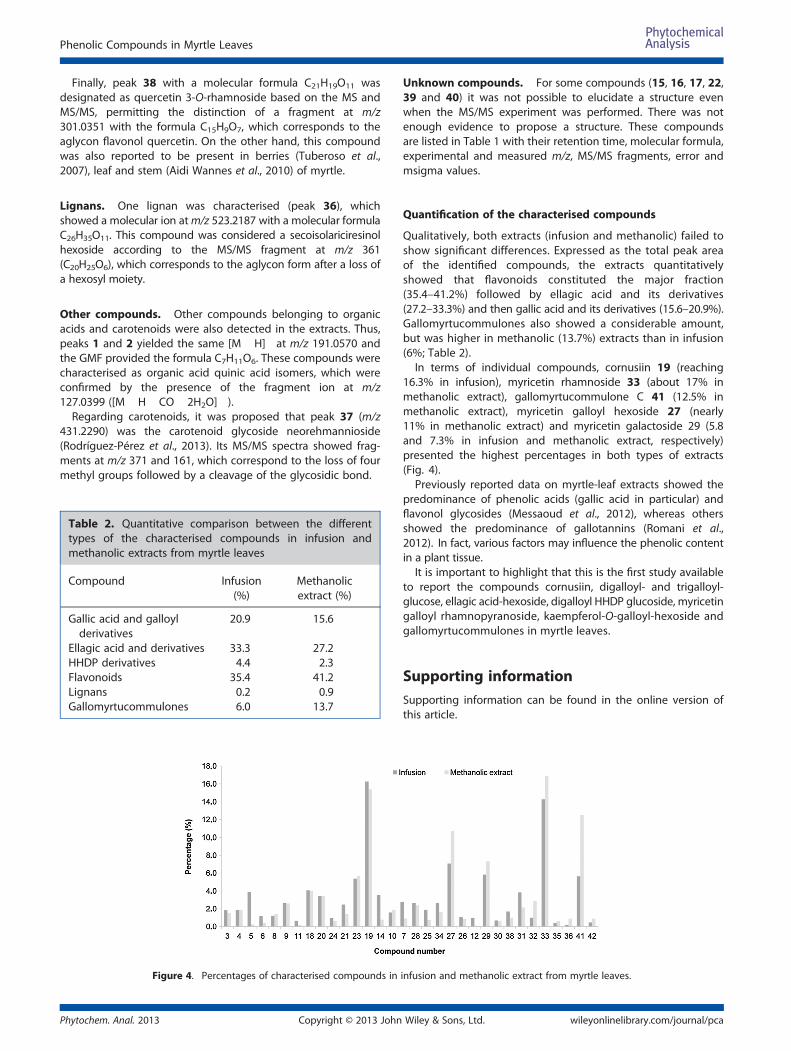
Table 2. Quantitative comparison between the differenttypes of the characterised compounds in infusion andmethanolic extracts from myrtle leaves
Compound Infusion(%)
Methanolicextract (%)
Gallic acid and galloylderivatives
20.9 15.6
Ellagic acid and derivatives 33.3 27.2HHDP derivatives 4.4 2.3Flavonoids 35.4 41.2Lignans 0.2 0.9Gallomyrtucommulones 6.0 13.7
Figure 4. Percentages of characterised compounds in
Phytochem. Anal. 2013 Copyright © 2013 John
Unknown compounds. For some compounds (15, 16, 17, 22,39 and 40) it was not possible to elucidate a structure evenwhen the MS/MS experiment was performed. There was notenough evidence to propose a structure. These compoundsare listed in Table 1 with their retention time, molecular formula,experimental and measured m/z, MS/MS fragments, error andmsigma values.
Quantification of the characterised compounds
Qualitatively, both extracts (infusion and methanolic) failed toshow significant differences. Expressed as the total peak areaof the identified compounds, the extracts quantitativelyshowed that flavonoids constituted the major fraction(35.4–41.2%) followed by ellagic acid and its derivatives(27.2–33.3%) and then gallic acid and its derivatives (15.6–20.9%).Gallomyrtucommulones also showed a considerable amount,but was higher in methanolic (13.7%) extracts than in infusion(6%; Table 2).In terms of individual compounds, cornusiin 19 (reaching
16.3% in infusion), myricetin rhamnoside 33 (about 17% inmethanolic extract), gallomyrtucommulone C 41 (12.5% inmethanolic extract), myricetin galloyl hexoside 27 (nearly11% in methanolic extract) and myricetin galactoside 29 (5.8and 7.3% in infusion and methanolic extract, respectively)presented the highest percentages in both types of extracts(Fig. 4).Previously reported data on myrtle-leaf extracts showed the
predominance of phenolic acids (gallic acid in particular) andflavonol glycosides (Messaoud et al., 2012), whereas othersshowed the predominance of gallotannins (Romani et al.,2012). In fact, various factors may influence the phenolic contentin a plant tissue.It is important to highlight that this is the first study available
to report the compounds cornusiin, digalloyl- and trigalloyl-glucose, ellagic acid-hexoside, digalloyl HHDP glucoside, myricetingalloyl rhamnopyranoside, kaempferol-O-galloyl-hexoside andgallomyrtucommulones in myrtle leaves.
Supporting informationSupporting information can be found in the online version ofthis article.
infusion and methanolic extract from myrtle leaves.
Wiley & Sons, Ltd. wileyonlinelibrary.com/journal/pca

A. Taamalli et al.
Acknowledgements
The authors are grateful to the Tunisian Ministry of HigherEducation and Scientific Research, the Spanish Ministry ofEducation and Science for the projects AGL 2011-29857-C03-02and also to Andalusian Regional Government for the excellenceprojects P09-CTS-4564, P10-FQM-6563 and P11-CTS-7625. Theauthors are also grateful to D. Nesbitt for the English revision.
ReferencesAidi Wannes W, Mhamdi B, Sriti J, Ben Jemia M, Ouchikh O, Hamdaoui G,
Kchouk ME, Marzouk B. 2010. Antioxidant activities of the essentialoils and methanolextracts from myrtle (Myrtus Communis). FoodChem Toxicol 48: 1362–1370.
Balasundram N, Kalyana S, Samir S. 2006. Phenolic compounds in plantsand agri-industrial by-products: antioxidant activity, occurrence, andpotential uses. Food Chem 99: 191–203.
Barboni T, Cannac M, Massi L, Perez-Ramirez Y, Chiaramonti N. 2010.Variability of polyphenol compounds in Myrtus communis L.(Myrtaceae) berries from Corsica. Molecules 15: 7849–7860.
Benkhayal F, El-ghaithi Musbah A, Ramesh S, Dhayabaran D. 2009. Bio-chemical studies on the effect of phenolic compounds extractedfrom Myrtus communis in diabetic rats. Tamil J Vet Anim Sci 5: 87–93.
Chalchat JC, Garry RP, Michet A. 1998. Essential oils of myrtle (Myrtuscommunis L.) of the Mediterranean Littoral. J Essent Oil Res 10: 613–617.
Clifford MN, Stoupi S, Kuhnert N. 2007. Profiling and characterization byLC–MSn of the galloylquinic acids of green tea, tara tannin, and tannicacid. J Agric Food Chem 55: 2797–2807.
Cottiglia F, Casu L, Leonti M, Caboni P, Floris C, Busonera B, Farci P,Ouhtit A, Sanna G. 2012. Cytotoxic phloroglucinols from the leavesof Myrtus communis. J Nat Prod 75: 225–229.
Fernández-Arroyo S, Barrajón-Catalán E, Micol V, Segura-Carretero A,Fernández-Gutiérrez A. 2010. High-performance liquid chromatographywith diode array detection coupled to electrospray time-of-flight andion-trap tandem mass spectrometry to identify phenolic compoundsfrom a Cistus ladanifer aqueous extract. Phytochem Anal 21: 307–313.
Gardeli C, Vassiliki P, Athanasios M, Kibouris T, Komaitis M. 2008. Essentialoil composition of Pistacia lentiscus L. and Myrtus communis L.: evalu-ation of antioxidant capacity of methanolic extracts. Food Chem 107:1120–1130.
Hayder N, Bouhlel I, Skandrani I, Kadri M, Steiman R, Guiraud P, MariotteAM, Ghedira K, Dijoux-Franca MG, Chekir-Ghedira L. 2008. In vitro an-tioxidant and antigenotoxic potentials of myricetin-3-O-galactosideand myricetin-3-O-rhamnoside from Myrtus communis: modulationof expression of genes involved in cell defence system using cDNAmicroarray. Toxicol In Vitro 22: 567–581.
Copyright © 2013 Johnwileyonlinelibrary.com/journal/pca
Hollebeeck S, Winand J, Hérent MF, During A, Leclercq J, Larondelle Y,Schneider YJ. 2012. Anti-inflammatory effects of pomegranate(Punica granatum L.) husk ellagitannins in Caco-2 cells, an in vitromodel of human intestine. Food Funct 3: 875–885.
Hosseinzadeh H, Khoshdel M, Ghorbani M. 2011. Antinociceptive, anti-inflammatory effects and acute toxicity of aqueous and ethanolicextracts of Myrtus communis L. Aerial parts in mice. J AcupunctMeridian Stud 4: 242–247.
Katalinic V, Milos M, Kulisic T, Jukic M. 2006. Screening of 70 medicinalplant extracts for antioxidant capacity and total phenols. Food Chem94: 550–557.
Martin K, Appel C. 2010. Polyphenols as dietary supplements: a double-edged sword. Nutr Diet Suppl 2: 1–12.
Messaoud C, Laabidi A, Boussaid M. 2012. Myrtus communis L. infusions:The effect of infusion time on phytochemical composition, antioxi-dant, and antimicrobial activities. J Food Sci 77: C941–C947.
Meyers KJ, Swiecki TJ, Mitchell AE. 2006. Understanding the nativeCalifornian diet: Identification of condensed and hydrolyzabletannins in tanoak acorns (Lithocarpus densiflorus). J Agric Food Chem54: 7686–7691.
Mine A, Özkan G, Güray ÇG. 2009. A Mediterranean Myrtus communis L.(Myrtle). In: Plants and Culture: Seeds of the Cultural Heritage of Europe,Morel JP and Mercuri AM (ed.). EDIPUGLIA Bari; 159–168.
Montoro P, Tuberoso CI, Piacente S, Perrone A, De Feo V, Cabras P, PizzaC. 2006. Stability and antioxidant activity of polyphenols in extracts ofMyrtus communis L. berries used for the preparation of myrtle liqueur.J Pharm Biomed Anal 41: 1614–1619.
Motilva MJ, Serra A, Macià A. 2013. Analysis of food polyphenols by ultrahigh-performance liquid chromatography coupled to mass spec-trometry: an overview. J Chromatogr A 1292: 66–82.
Randhir R, Lin Y, Shetty K. 2004. Phenolics, their antioxidant and antimi-crobial activity in dark germinated fenugreek sprouts in response topeptide and phytochemical elicitors. Asia Pac J Clin Nutr 13: 295–307.
Rodríguez-Pérez C, Quirantes-Piné R, Amessis-Ouchemoukh N, Madani K,Segura-Carretero A, Fernández-Gutierrez A. 2013. A metabolite-profiling approach allows the identification of new compounds fromPistacia lentiscus leaves. J Pharm Biomed Anal 77: 167–174.
Romani, A, Pinelli P, Mulinacci N, Vincieri FF, Tattini M. 1999. Identificationand quantitation of polyphenols in leaves of Myrtus communis L.Chromatographia 49: 17–20.
Romani A, Coinu R, Carta S, Pinelli P, Galardi C, Vincieri FF, Franconi F.2004. Evaluation of antioxidant effect of different extracts of Myrtuscommunis L. Free Radical Res 38: 97–103.
Romani A, Campo M, Pinelli P. 2012. HPLC/DAD/ESI-MS analyses and anti-radical activity of hydrolyzable tannins from different vegetal species.Food Chem 130: 214–221.
Tuberoso CIG, Melis M, Angioni A, Pala M, Cabras P. 2007. Myrtlehydroalcoholic extracts obtained from different selections of Myrtuscommunis L. Food Chem 101: 806–811.
Phytochem. Anal. 2013Wiley & Sons, Ltd.