Update on Primary Cicatricial Alopecias CME July 2005dermpathmd.com/Clinical Dermatology/Cicatricial...
61
Transcript of Update on Primary Cicatricial Alopecias CME July 2005dermpathmd.com/Clinical Dermatology/Cicatricial...

Overview Classification Epidemiology Pathophysiology General Approaches Subtypes
Lymphocytic, Neutrophilic, Mixed

Hair Loss Categories Non-scarring
Androgenetic Male Female
Effluviums Telogen Anagen
Alopecia Areata Traumatic
Trichotillomania Traction alopecia
Drug/toxin induced
Scarring Pseudopelade of Brocq Central centrifugal
Follicular Degeneration Folliculitis Decalvans Tufted Folliculitis
Alopecia Mucinosa Lichen Planopilaris
Graham Little Syndrome Frontal Fibrosing Alopecia
Acne Keloidalis Dissecting Cellulitis DLE
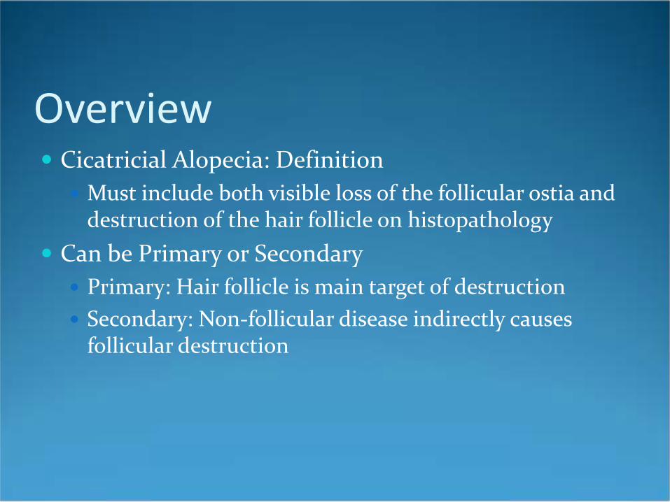
Overview Cicatricial Alopecia: Definition
Must include both visible loss of the follicular ostia and destruction of the hair follicle on histopathology
Can be Primary or Secondary Primary: Hair follicle is main target of destruction Secondary: Non-follicular disease indirectly causes
follicular destruction
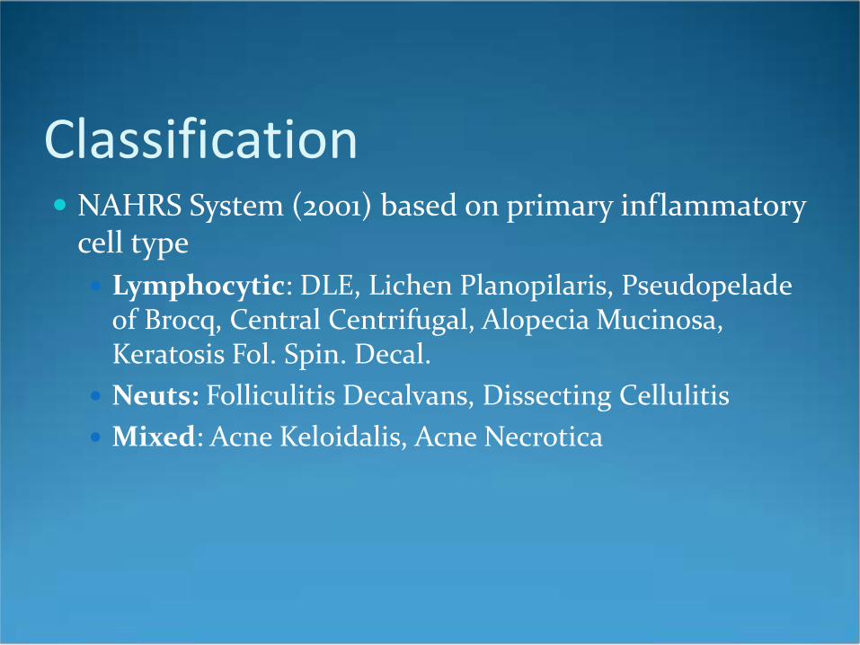
Classification NAHRS System (2001) based on primary inflammatory
cell type Lymphocytic: DLE, Lichen Planopilaris, Pseudopelade
of Brocq, Central Centrifugal, Alopecia Mucinosa, Keratosis Fol. Spin. Decal.
Neuts: Folliculitis Decalvans, Dissecting Cellulitis Mixed: Acne Keloidalis, Acne Necrotica

Epidemiology Retrospective Studies:
Cicatricial alopecia in 7.3% (n=427) Primary cicatricial in 3.2% (n=112)
F:M 2.6:1 Average age
Women: 43 Men: 36
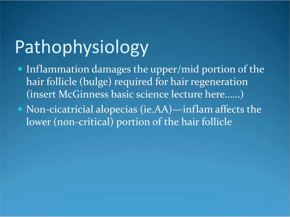
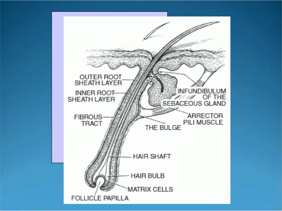
Pathophysiology Inflammation damages the upper/mid portion of the
hair follicle (bulge) required for hair regeneration (insert McGinness basic science lecture here……)
Non-cicatricial alopecias (ie.AA)—inflam affects the lower (non-critical) portion of the hair follicle
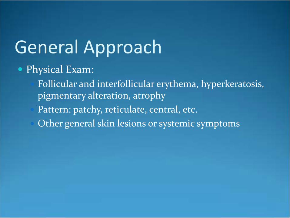
General Approach Physical Exam:
Follicular and interfollicular erythema, hyperkeratosis, pigmentary alteration, atrophy
Pattern: patchy, reticulate, central, etc. Other general skin lesions or systemic symptoms

General Approach Scalp Biopsy
Biopsy clinically active areas, not “burnt-out” Obtain 2, 4mm punch biopsies
Send one for horizontal sectioning Send the other for vertical only or cut in half and send half for
H&E and the other for DIF

Lymphocytic Cicatricial Alopecias DLE LPP Frontal Fibrosing Graham-Little Syndrome Lupus/LPP overlap Pseudopelade of Brocq Central Centrifugal Alopecia Mucinosa Keratosis Follicularis Spinulosa Decalvans
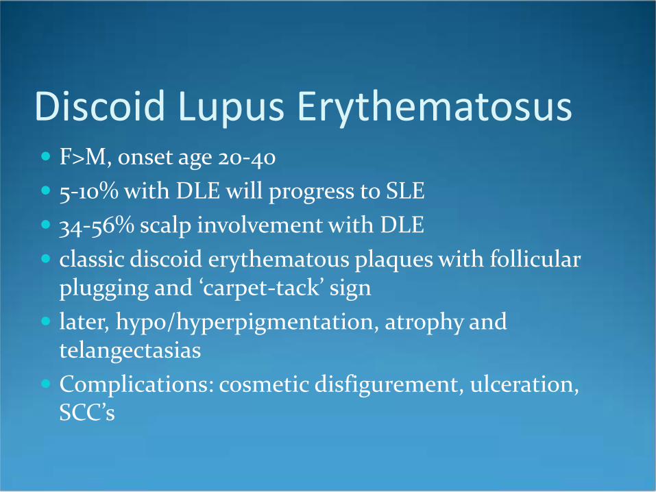
Discoid Lupus Erythematosus F>M, onset age 20-40 5-10% with DLE will progress to SLE 34-56% scalp involvement with DLE classic discoid erythematous plaques with follicular
plugging and ‘carpet-tack’ sign later, hypo/hyperpigmentation, atrophy and
telangectasias Complications: cosmetic disfigurement, ulceration,
SCC’s
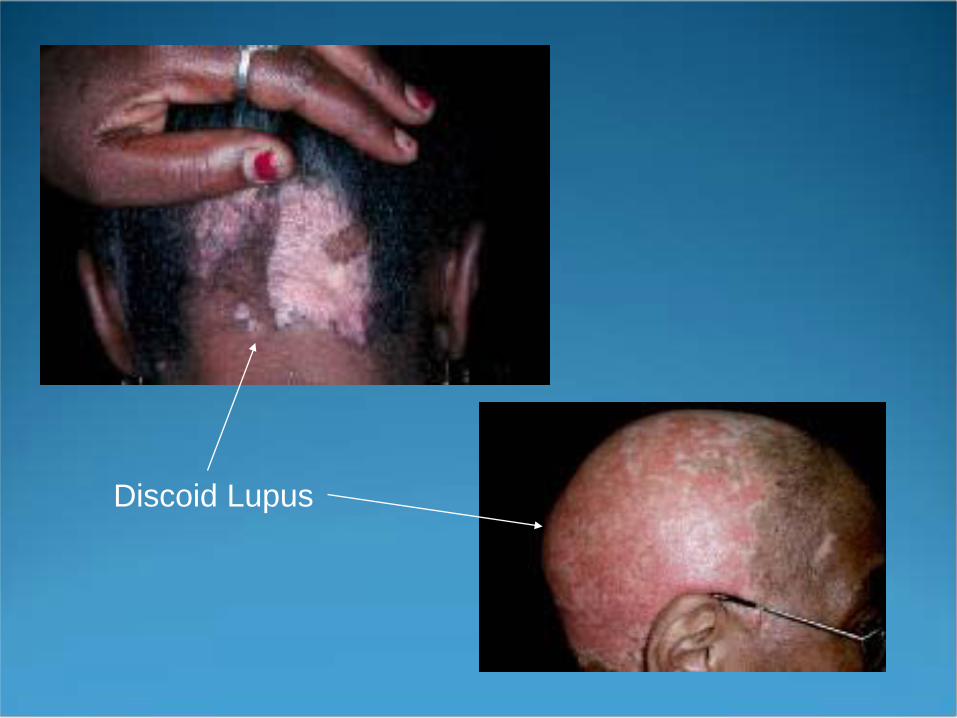
Discoid Lupus
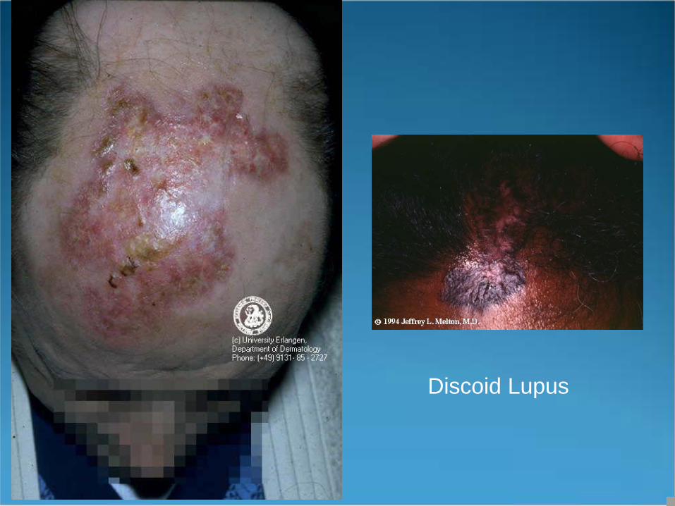
Discoid Lupus
DLE Histopath
Vacuolar interface change of the follicular epithelium Scattered dyskeratotic keratinocytes Periadnexal, perifollicular and interstitial lymphocytic
infiltrate with dermal mucin Follicular plugging DIF often positive
Treatment of DLE alopecia ROS focused on sx of SLE ANA and U/A Limited, active disease
Class I or II topical steroids BID Intra-lesional Kenalog 3-10mg/cc Q4-6 weeks
Rapidly progressive or extensive disease Plaquenil 200 mg BID +/- oral prednisone for the first 8
weeks Accutane (2nd line) 1mg/kg/day
Sun protection and avoidance of trauma
Lichen Planopilaris A follicular variant of lichen planus 3 forms
Classic, Graham-Little, frontal fibrosing Thought to be secondary to an antigenic trigger or
related to certain medications (similar to classic LP) Gold, quinacrine, atabrine, hep B vaccination, hepatitis
C infection
LPP Seen in adults, usually arising in middle-aged females Extracranial LP present in 17-50% of pts. Sx at presentation: shedding, hair loss, pruritus PE: perifollicular erythematous papules and spinous
follicular hyperkeratosis. Can see unaffected hairs in scarred areas
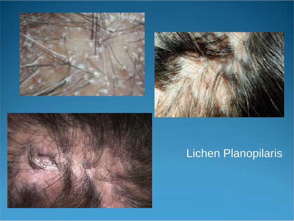
Lichen Planopilaris

LPP DDx includes other cicatricial alopecias
Activity limited to hair-bearing periphery (unlike DLE and alopecia mucinosa)
No pustules (unlike folliculitis decalvans) Histopath
Lichenoid infiltrate Lymphs at upper portion of follicle DIF may reveal patchy deposition of fibrinogen and IgM
along the BMZ
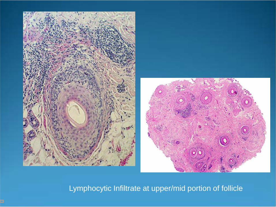
Lymphocytic Infiltrate at upper/mid portion of follicle
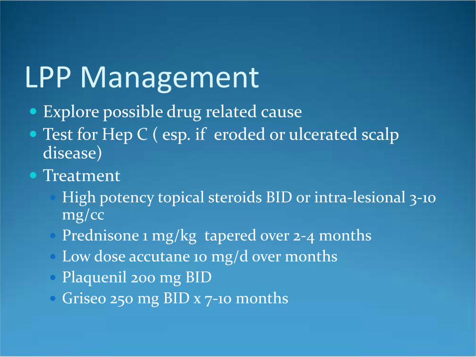
LPP Management Explore possible drug related cause Test for Hep C ( esp. if eroded or ulcerated scalp
disease) Treatment
High potency topical steroids BID or intra-lesional 3-10 mg/cc
Prednisone 1 mg/kg tapered over 2-4 months Low dose accutane 10 mg/d over months Plaquenil 200 mg BID Griseo 250 mg BID x 7-10 months
Frontal Fibrosing Alopecia Also called postmenopausal FFA Frontotemporal hairline scarring pattern mainly
affecting postmenopausal women Shiny, pale, bandlike zone; active areas reveal hairs
with perifollicular erythema and hyperkeratosis Absent or thinned eyebrows May have axillary and extremity hair loss May also have classic LPP or LP

Frontal Fibrosing Alopecia DDx: ophiasis, female pattern recession, traction
alopecia Histopath:
Features indistinguishable from LPP Lichenoid infiltrate Upper follicular inflamm
Management Attempt to stabilize disease with topical mid-potency
steroids BID Po prednisone or plaquenil may slow progression Other case reports: intralesional steroids, accutane,
soriatane, griseo, minoxidil, etc. are mostly ineffective
Graham-Little Syndrome Aka Graham-Little-Piccardi-Lassueur Considered a variant of LPP Uncommon alopecia seen in adults Patchy cicatricial scalp alopecia, nonscarring alopecia
of the axillary and pubic areas, and grouped follicular papules on the trunk/extremities resembling lichen spinulosus or KP
Graham-Little Syndrome Histopath:
similar to LPP Treatment:
High potency topical steroids alone or in combo with intralesional steroids (10mg/cc)
PO steroids Cyclosporine 4mg/kg/d x 3 months

Pseudopelade of Brocq Distinct entity or common endpoint? Onset in adulthood Atrophic, oval to round, white to ivory scarred plaques
of alopecia, vertex almost always involved No clinical evidence of inflammation Slowly progressive

Pseudopelade of Brocq

Pseudopelade of Brocq Histopath: (none classic)
Early- perifollicular lymphocytic infiltrate Late-concentric lamellar fibrosis of the hair follicle
Treatment: Mainly none Many topical and oral therapies have been tried with
little success
Central Centrifugal Cicatricial Alopecia New term coined to encompass hot comb alopecia and
follicular degeneration syndrome Most commonly in African-American women Presents with flesh-colored, non-inflammatory
cicatricial alopecia of the central scalp that enlarges over time
inherited follicular defect vs. exogenous trauma?
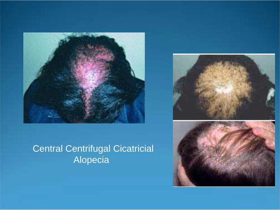
Central Centrifugal CicatricialAlopecia
CCC Alopecia Histopath:
Characteristic premature desquamation of the inner root sheath
Perifollicular lymph infiltrate surrounding the upper portion of the follicle
Management Cessation of traumatic hair practices Topical steroids Tetracycline 500 mg BID
Alopecia Mucinosa Characterized by intrafollicular deposition of mucin
(follicular mucinosus) Idiopathic and lymphoma related types All ages affected Lesions are often pruritic, dysesthetic, and/or
anhidrotic Other body locations besides the scalp can be involved

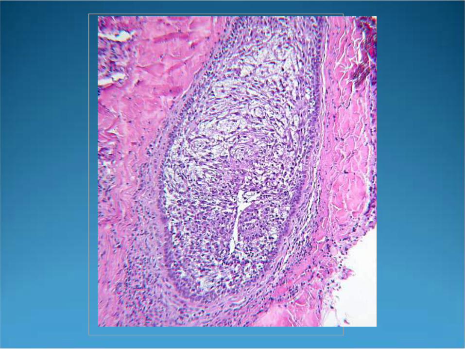
AlopeciaMucinosa

Alopecia Mucinosa Presentation of scalp disease is highly variable In adults, MF is associated 9-60% of the time In children, Hodgkin’s lymphoma is the most
commonly associated malignancy Alopecia mucinosa can present as a paraneoplastic
phenomenon
Alopecia Mucinosa There are no reliable clinical or histo criteria to
distinguish benign from malignant cases Histopath:
Intrafollicular mucin Perifollicular lymphocytic infiltrate No lamellar fibrosis

Management Close follow up and serial biopsies if progression of
disease Lymph node examination at all visits Topical or intra-lesional steroids Minocycline 100 BID for 5-8 weeks Accutane 0.5-1.0 mg/kg for 4-5 months If colonized with Staph, treating with Abx may clear
disease
Keratosis Follicularis Spinulosa Decalvans Aka keratosis pilaris decalvans related to KP atrophicans faciei and atrophoderma
vermiculata X-linked or sporadic Follicular hyperkeratosis beginning on the face
and spreading to involve other body areas, eventually leading to punctate atrophy
Begins in infancy or childhood Also have photophobia and scarring alopecia

Management Intervene early in childhood when the disease is active Treatment studies are limited High potency topical or intralesional steroids Accutane 0.5 mg/kg for 3 months Baseline and routine ophtho exams recommended

Neutrophilic Cicatricial Alopecias
Folliculitis Decalvans Dissecting Cellulitis
Folliculitis Decalvans Common form of primary cicatricial alopecia Is a destructive suppurative folliculitis seen in young
and middle aged adults Staph aureus is thought the be the inciting factor Begin as grouped follicular pustules which evolve into
abscesses and eventually scarring Often see tufted folliculitis

FolliculitisDecalvans
Folliculitis Decalvans Histopath:
Upper and mid follicular neutrophilic infiltrate Late disease: granulomatous inflamm and perifollicular
fibrosis
Management Culture pustules Abx with anti-Staph coverage Rifampin 300 mg BID in combination with
Clindamycin 300 mg BID for 10 weeks New combo of Rifampin, fusidic acid (not available in
US) and zinc has shown good success Eliminate Staph carrier state with mupirocin

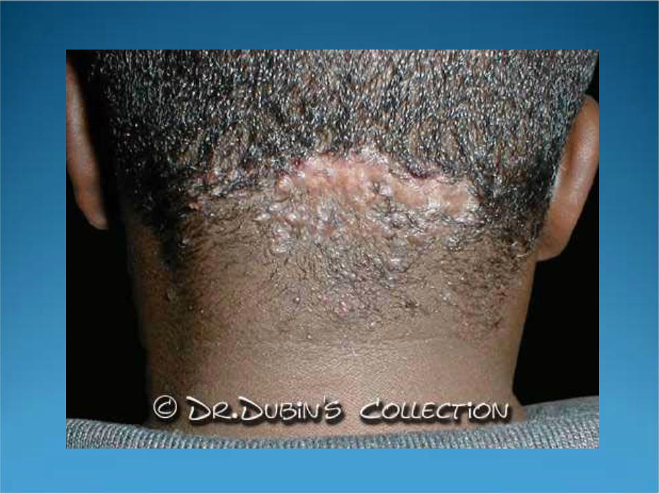
Dissecting Cellulitis Aka perifolliculitis capitis abscedens et suffodiens Part of the follicular occlusion triad (tetrad) Abnormal follicular keratinization leads to
obstruction, secondary infection, and follicular destruction
>80% of patients are black men ages 18-40
Dissecting Cellulitis Initial lesions are pustules often beginning on occipital
or vertex scalp Later large,fluctuant nodules that coalesce and form
tracts Coexisting acne conglobata or HS is a risk factor for
development of spondyloarthropathy. Peripheral and axial joints may be involved

Dissecting Cellulitis
Dissecting Cellulitis Histopath:
Intra and perifollicular neuts Abscesses in mid to deep dermis Late: sinus tracts lined with squamous cells and fibrosis

Management Accutane now considered first line
1 mg/kg/d for at least 4 months followed by 0.75-1 mg/kg/d for another 5-7 mo. if needed
Other opitions Topical clindamycin gel Oral TCN Dapsone or colchicine CO2 laser and surgical excision

Mixed Cicatrical Alopecias
Acne Keloidalis Acne Necrotica Erosive pustular dermatosis

Acne Keloidalis
Mostly seen in AA males Thought to be secondary to mechanical traumas,
seborrhea, or infections Present as follicular papules or keloidal plaques on the
occiput and nape of neck
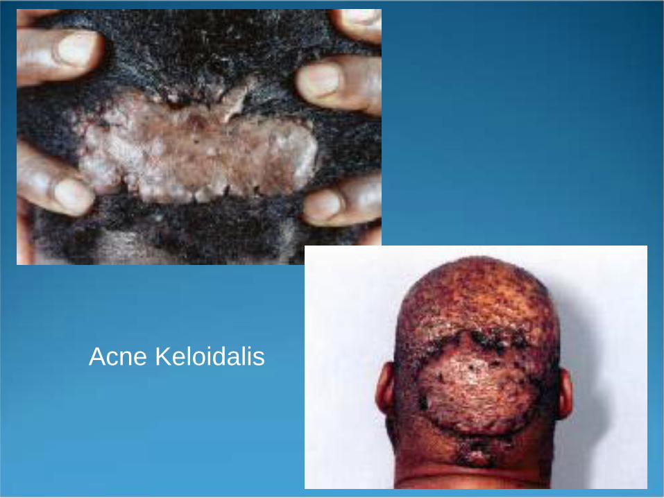
Acne Keloidalis
Management Early, limited disease
Avoid mechanical traumas Topical high potency steroids in combo with topical
clinda Intralesional kenalog (10mg/cc) Oral TCN
More extensive disease Surgical excision

Acne Necrotica Aka Greer’s disease Chronic, relapsing d/o of crops of pruritic, small
pustules that undergo central necrosis and crusting Management
Cx pustules and tx accordingly Antibacterial shampoos Topical antibiotics Topical steroids
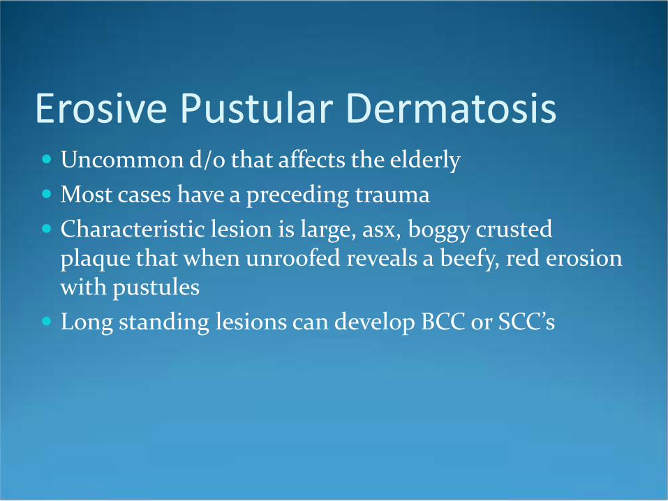
Erosive Pustular Dermatosis Uncommon d/o that affects the elderly Most cases have a preceding trauma Characteristic lesion is large, asx, boggy crusted
plaque that when unroofed reveals a beefy, red erosion with pustules
Long standing lesions can develop BCC or SCC’s
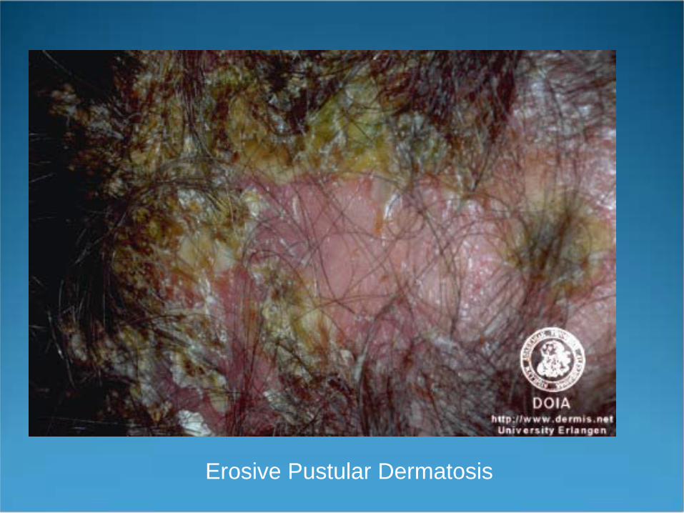
Erosive Pustular Dermatosis
Management
Topical high potency steroids BID Dovonex cream BID Oral or topical abx prn
Adjunctive Therapies for all Types Hair pieces/wigs Hair color matched powders (Toppik, Spencer Forrest,
Westport) Hair transplantation Scalp reduction


![Of ck15, s100 - termedia.pl (lichen planopilaris – LPP), LP pigmentosus and LP pigmentosus-inversus forms [2, 3]. Lichen planus is a common dermatosis characterized by pruritic,](https://static.fdocuments.us/doc/165x107/6082dd23409de75ded015edc/of-ck15-s100-lichen-planopilaris-a-lpp-lp-pigmentosus-and-lp-pigmentosus-inversus.jpg)