Up-regulation of autophagy by low concentration of ...
25
Up-regulation of autophagy by low concentration of salicylic acid delays methyl jasmonate-induced leaf senescence Runzhu Yin South China Normal University Xueyan Liu Jinan University Jingfang Yu South China Normal University Yingbin Ji South China Normal University Jian Liu Fujian Agriculture and Forestry University Lixin Cheng Jinan University Jun Zhou ( [email protected] ) South China Normal University https://orcid.org/0000-0001-9655-6588 Research article Keywords: autophagy, gene modules, jasmonic acid, leaf senescence, RNA-Seq, salicylic acid Posted Date: December 13th, 2019 DOI: https://doi.org/10.21203/rs.2.18798/v1 License: This work is licensed under a Creative Commons Attribution 4.0 International License. Read Full License Version of Record: A version of this preprint was published at Scientiヲc Reports on July 10th, 2020. See the published version at https://doi.org/10.1038/s41598-020-68484-3.
Transcript of Up-regulation of autophagy by low concentration of ...

Up-regulation of autophagy by low concentration ofsalicylic acid delays methyl jasmonate-induced leafsenescenceRunzhu Yin
South China Normal UniversityXueyan Liu
Jinan UniversityJingfang Yu
South China Normal UniversityYingbin Ji
South China Normal UniversityJian Liu
Fujian Agriculture and Forestry UniversityLixin Cheng
Jinan UniversityJun Zhou ( [email protected] )
South China Normal University https://orcid.org/0000-0001-9655-6588
Research article
Keywords: autophagy, gene modules, jasmonic acid, leaf senescence, RNA-Seq, salicylic acid
Posted Date: December 13th, 2019
DOI: https://doi.org/10.21203/rs.2.18798/v1
License: This work is licensed under a Creative Commons Attribution 4.0 International License. Read Full License
Version of Record: A version of this preprint was published at Scienti�c Reports on July 10th, 2020. Seethe published version at https://doi.org/10.1038/s41598-020-68484-3.

1
Up-regulation of autophagy by low concentration of salicylic acid delays 1
methyl jasmonate-induced leaf senescence 2
Runzhu Yin1#, Xueyan Liu2#, Jingfang Yu1, Yingbin Ji1,3, Jian Liu4, Lixin Cheng2*, Jun 3
Zhou1* 4
1 MOE Key Laboratory of Laser Life Science & Guangdong Provincial Key Laboratory of 5
Laser Life Science, College of Biophotonics, South China Normal University, Guangzhou 6
510631, China 7
2 Department of Critical Care Medicine, Shenzhen People's Hospital, The Second Clinical 8
Medicine College of Jinan University, Shenzhen 518020, China 9
3 Luoyang Tmaxtree Biotechnology Co., Ltd, Luoyang 471023, China 10
4 Fujian Provincial Key Laboratory of Plant Functional Biology, College of Life Sciences, 11
Fujian Agriculture and Forestry University, Fuzhou 350002, China 12
*Correspondence authors: Jun Zhou, [email protected]; Lixin Cheng, 13
# These authors contributed equally to this work. 15
Running title: Role of autophagy in LCSA-delayed leaf senescence. 16
17

2
ABSTRACT 18
Crosstalk between salicylic acid (SA) and jasmonic acid (JA) signaling plays an important 19
role in molecular regulation of plant senescence. Our previous works found that SA could 20
delay methyl jasmonate (MeJA)-induced leaf senescence in a concentration-dependent 21
manner. Here, the effect of low concentration of SA (LCSA) application on MeJA-induced 22
leaf senescence was further assessed. High-throughput sequencing (RNA-Seq) results 23
showed that LCSA did not have dominant effects on the genetic regulatory pathways of 24
basal metabolism like nitrogen metabolism, photosynthesis and glycolysis. The 25
ClusterONE was applied to identify discrete gene modules based on protein-protein 26
interaction (PPI) network. Interestingly, an autophagy-related (ATG) module was identified 27
in the differentially expressed genes (DEGs) that exclusively induced by MeJA together 28
with LCSA. RT-qPCR confirmed that the expression of most of the determined ATG genes 29
were upregulated by LCSA. Remarkably, in contrast to wild type (Col-0), LCSA cannot 30
alleviate the leaf yellowing phenotype in autophagy defective mutants (atg5-1 and atg7-2) 31
upon MeJA treatment. Confocal results showed that LCSA increased the number of 32
autophagic bodies accumulated in the vacuole during MeJA-induced leaf senescence. 33
Collectively, our work revealed up-regulation of autophagy by LCSA as a key regulator to 34
alleviate MeJA-induced leaf senescence. 35
Key Words: autophagy; gene modules; jasmonic acid; leaf senescence; RNA-Seq; 36
salicylic acid 37
38

3
INTRODUCTION 39
Senescence in green plants is a complex and orderly regulated process that is crucial for 40
transiting from nutrient assimilation to nutrient remobilization (Masclaux et al., 2000; 41
Quirino et al., 2000; Lim et al., 2003; Yoshida, 2003; Schippers, 2015). During senescence, 42
the most visible characteristic is leaf yellowing, which is the consequence of a succession 43
of changes in cellular physiology including chlorophyll degradation and photosynthetic 44
activity reduction (Lim et al., 2003; Yoshida, 2003). Chloroplast as an early senescence 45
signaling response organelle, its dismantling plays an important role in the major nitrogen 46
source recycling and remobilization (Avila-Ospina et al., 2014). The progression of leaf 47
senescence can be prematurely induced by multiple environmental and endogenous 48
factors, such as temperature, light, humidity and phytohormones (Lim et al., 2007). 49
Hormone signaling pathways play roles at all the stages of leaf senescence, including the 50
initiation, progression, and the terminal phases of senescence (Lim et al., 2007). Recent 51
progresses show that senescence can be coordinately regulated by several 52
phytohormones like cytokinins, ethylene, abscisic acid, salicylic acid (SA), and jasmonic 53
acid (JA) (Gan and Amasino, 1995; van der Graaff et al., 2006; Hung and Kao, 2004; He 54
et al., 2002; Morris et al., 2000). However, the detailed molecular mechanisms for these 55
phytohormone signals in plant senescence remain poorly understood. 56
JA has been known as a key plant hormone for promoting senescence, based on the 57
findings that exogenously applied methyl jasmonate (MeJA, methyl ester of JA) leads to a 58
rapid loss of chlorophyll content and accompany with reduction of photochemical efficiency 59
(Yue et al., 2012; Ji et al., 2016). Studies with JA-insensitive mutant coronatine insensitive 60
1 (coi1) that exhibited defective senescence response to MeJA treatment (He et al., 2002), 61
supporting the notion that JA signaling pathway is crucial for leaf senescence. Some other 62
evidences indicate that SA is also involved in plant senescence (Morris et al., 2000; Chai 63
et al., 2014). The concentration of endogenous SA increases to upregulate several 64
senescence-associated genes during leaf senescence (Morris et al., 2000; Yoshimoto et 65
al., 2009). However, such genetic regulatory mechanisms are abolished in plants defective 66
in the SA signaling or biosynthetic pathway (npr1 and pad4 mutants, and NahG transgenic 67
plants) (Morris et al., 2000). Crosstalk between MeJA and SA has been broadly 68
documented in plant defense response, which commonly manifests as a reciprocal 69
antagonism pattern (Thaler et al., 2012). Evidence suggests that antagonistic interactions 70
between SA and MeJA modulate the expression of a senescence-specific transcription 71
factor WRKY53, showing induced by SA, but repressed by MeJA (Miao and Zentgraf, 72

4
2007). Overall, mechanisms determining the specificity and coordination between SA and 73
JA still need to be further explored. 74
Most of phytohormones have both stimulatory and inhibitory effects on the growth and 75
metabolism of higher plants in a dose dependent manner. It seems that SA functions in the 76
same way on the physiological and biochemical processes of plants (Ji et al., 2016; 77
Pasternak et al., 2019). Low-concentration SA (hereafter as LCSA) at below 50 micromole 78
(µM) promotes adventitious roots and altered architecture of the root apical meristem, 79
whereas high-concentration SA (greater than 50 µM) inhibits root growth (Pasternak et al., 80
2019). Interestingly, we previously demonstrated that MeJA-induced leaf senescence 81
could be delayed by LCSA (1-50 μM), but accelerated when the concentration higher than 82
100 µM (Ji et al., 2016). Our other related works have verified such high dose of SA greatly 83
activates NPR1 (nonexpressor of pathogenesis-related genes 1) translocation into nucleus, 84
thereby promoting leaf senescence (Chai et al., 2014). Based on the dose dependent effect 85
of SA, Pasternak et al. (2019) proposes that at low levels it acts as a developmental 86
regulator and at high levels it acts as a stress hormone. 87
Autophagy is associated with plant senescence as defective mutants display early and 88
strong yellowing leaf symptoms (Hanaoka et al., 2002; Xiong et al., 2005; Avila-Ospina et 89
al., 2014; Li et al., 2014). Autophagy negatively regulates cell death by controlling NPR1-90
dependent SA signaling during senescence in Arabidopsis (Yoshimoto et al., 2009). The 91
senescence process always accompanies with the equilibrium between oxidative and 92
antioxidative capacities of the plant, which creates a characteristic oxidative environment 93
resulting in the production of reactive oxygen species (ROS) and more toxic derivatives 94
(Bhattacharjee, 2005). Moreover, autophagy is involved in the degradation of oxidized 95
proteins under oxidative stress conditions in Arabidopsis (Xiong et al., 2007). Actually, there 96
is a complicated interplay between ROS and autophagy, i.e., ROS can induce autophagy 97
while autophagy be able to reduce ROS production (Signorelli et al., 2009). Our previous 98
studies showed that LCSA application delays senescence by enhancing the activities of 99
antioxidant enzymes and restricting reactive oxygen species (ROS) accumulation in MeJA-100
treated leaves (Ji et al., 2016). However, it is still unclear whether autophagy pathway is 101
implicated in the LCSA-alleviated leaf senescence. 102
Here, the interactions between SA and MeJA in plant senescence were further 103
investigated. By applying transcriptome and interaction network analysis, we identified 104
autophagy-related (ATG) gene module. In contrast to wild type (Col-0), LCSA cannot 105
alleviate the leaf yellowing phenotype in autophagy defective mutants upon MeJA 106

5
treatment. Further results revealed that LCSA increased the number of autophagic bodies 107
during MeJA-induced leaf senescence. Collectively, our work provides new insight that up-108
regulation of autophagy by LCSA is a key regulator to alleviate MeJA-induced leaf 109
senescence. 110
MATERIALS AND METHODS 111
Plant materials and chemical treatments 112
Arabidopsis plants of wild-type (WT, Col-0), atg5-1 (SAIL_129_B07), atg7-2 (GK-655B06) 113
and eYFP-ATG8e (Zhuang et al., 2013) were grown in a greenhouse at 22 °C with 16 h 114
light photoperiod (120 μmol quanta−2 m−2). Phytohormones treatment was performed as 115
described by Ji et al (2016). Briefly, the 3rd and 4th rosette leaves from four weeks of plants 116
were detached and incubated in 3 mM MES buffer (pH 5.8) containing 50 μM methyl 117
jasmonate (MeJA) and/or 10 μM salicylic acid (SA). MeJA was prepared from a 50 mM 118
stock solution in ethanol. Solutions without MeJA were supplemented with equal amounts 119
of ethanol. Concanamycin A (ConcA) was prepared as a 1 mM stock solution in DMSO and 120
used at final concentration 1 μM. 121
Photochemical efficiency and chlorophyll content measurements 122
The photochemical efficiency was measured with an Imaging-PAM Chlorophyll 123
Fluorometer (PAM-MINI, Walz, Germany) followed the procedure described previously 124
(Zhou et al., 2015). After dark-adapted for 1 h, parameters Fo (minimum fluorescence with 125
PSII reaction centers fully open) and Fm (maximum fluorescence after dark adaptation) 126
were acquired with a 0.8-s saturating pulse (4,000 μmol photons m-2 s-1). The value of 127
Fv/Fm was calculated by the formulas (Fm-Fo)/Fm. Total Chlorophyll was determined as 128
reported by Coombs et al. (1987). Chlorophyll was extracted by immersion in 90% ethanol 129
at 65 °C for 2 h. The absorbance at 664 nm and 647 nm were determined with a Lambda 130
35 UV/VIS Spectrometer (Perkin-Elmer) (Zeng et al., 2016). The concentration per fresh 131
weight of leaf tissue was calculated according to the formula: micromoles of chlorophyll 132
per milliliter per gram fresh weight = 7.93(A664) + 19.53(A647). The percentages of Fv/Fm 133
and chlorophyll content are calculated relative to the initial levels of samples before 134
treatment (time zero). 135
RNA-Seq analysis 136
Detached 3rd and 4th rosette leaves from 4-week old plants were immersed in 3 mM MES 137
buffer (pH 5.8) containing 10 μM SA, 50 μM MeJA, and MeJA together with SA for 24 h. 138

6
Total RNA for RNA-Seq was extracted from leaves using a Hipure plant RNA kit (Magen, 139
China). Purified RNA was analyzed either using a ND-1000 Nanodrop (Thermo Fisher, 140
USA), or by agarose gel electrophoresis to determine the RNA quantity. Those RNA 141
samples with no smear seen on agarose gels, a 260/280 ratio above 2.0, and RNA integrity 142
number greater than 8.0 were used. For RNA-Seq analysis, we mixed three replication 143
samples for each treatment into one, and total RNA samples were then sent to RiboBio 144
Co., Ltd (Guangzhou, China) for sequencing. The NEBNext Poly(A) mRNA Magnetic 145
Isolation Module (NEB, USA) was used for mRNA purification. The Ultra II RNA Library 146
Prep Kit for Illumina was used for RNA library construction. The libraries were sequenced 147
as 50-bp single end reads using Illumina Hiseq2500 according to the manufacturer's 148
instructions. 149
Differential Expression Analysis 150
Raw read count of each gene was generated using HTSeq with union-count mode (Love 151
et al., 2014). After normalization by Reads Per Kilobase per Million mapped reads (rpkm), 152
normalized read count table was used for determining differentially expressed genes 153
(DEGs) (Anders and Huber, 2010; Cheng et al., 2016a, 2016b), which were defined as 154
those with 2-fold changes. Fold change was calculated using log2 (normalized read 155
count+1). An R package clusterprofiler was used to perform the functional category 156
analysis to detect the significantly enriched Gene Ontology (GO) terms (Cheng and Leung, 157
2018a, and 2018b). Significantly enriched GO terms were selected by a threshold of p ≤ 158
0.05. Protein-protein interaction (PPI) data was obtained from the STRING database (v.10, 159
http://string-db.org) (Szklarczyk et al., 2014). To construct a high-confidence network, only 160
the PPIs with confidence scores larger than 0.7 were considered in this work. ClusterONE 161
was adopted for the identification of protein clusters or functional modules using default 162
parameters as described previously (Cheng et al., 2017; Cheng et al., 2019). The protein 163
modules including five or more than five members and having connection density over 0.5 164
are defined as modules. 165
RT-qPCR 166
Total RNA was isolated using Eastep Super RNA Kit (Promega, Shanghai, China) and 167
genomic DNA was removed using DNase I. 1 μg of RNA was used to make cDNA with the 168
GoScript™ Reverse Transcription System (Promega, Shanghai, China). For qPCR 10 μL 169
of Green-2-Go 2X qPCR-S Mastermix (Sangon, Shanghai, China) and 1 μL of cDNA (100 170
ng/μL) for a total of 20 μL was used in each well. Real-time PCR was done on a CFX 171

7
Connect Real-Time System (BioRad) at 95 °C for 2 mins, and 45 cycles of 95 °C for 15 s, 172
55 °C for 30s, and 72 °C for 30s followed by a melting curve analysis. For each sample 3 173
biological reps were used and repeated 3 times for technical replication. qPCR was 174
analyzed using the ΔΔCt method. Primers for qPCR were showed in Table S2. Statistical 175
significance was determined using Duncan’s multiple range test. 176
Confocal microscopy 177
Detached 3rd and 4th rosette leaves were immersed in 3 mM MES buffer (pH 5.8) 178
containing 50 μM MeJA and/or 10 μM SA for 24 h. ConcA (1 μM) was added at 6 h before 179
confocal imaging. Confocal images were captured with 63x (numerical aperture [NA], 1.4) 180
objective using an LSM 880 microscope (Zeiss). For quantification of autophagic puncta, 181
randomly selected 15 to 20 images for each three independent experiments were 182
quantified with ImageJ. All images were collected with the same settings determined prior 183
to the experiment to yield nonsaturating conditions. 184
RESULTS 185
LCSA delays MeJA-induced leaf senescence 186
Our previous results indicated that SA delays MeJA-induced leaf senescence in a 187
concentration-dependent manner, showing accelerated by high SA concentrations (greater 188
than 100 μM) but attenuated by low SA concentrations (1-50 μM) (Ji et al., 2016). On this 189
basis, 10 μM SA, the most effective concentration according to Ji et al., 2016, was selected 190
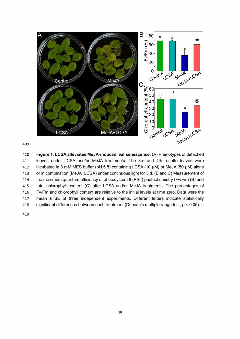
as low working solution to further confirm the effect of LCSA. As shown in Figure 1, in 191
contrast to control, LCSA did not appear to have a discernible effect on senescence. 192
Leaves incubated with MeJA (50 μM) were greatly turned yellow after 5 days treatment. 193
However, when MeJA worked together with LCSA (MeJA+LCSA), the leaf yellowing was 194
alleviated (Figure 1A). Consistent with the visible phenotype, the photochemical efficiency 195
Fv/Fm and loss of chlorophyll content in the leaves combined treatment with LCSA and 196
MeJA was less severe relative to that of the leaves treated with MeJA alone (Figure 1B 197
and 1C). These physiological and biochemical data is consistent with our previous finding 198
that LCSA provide protection against senescence caused by MeJA. 199
Expression patterns of genes in LCSA-induced delayed leaf senescence 200
To investigate the genome-wide effect of LCSA on MeJA-induced gene expression 201
changes, we performed RNA-sequencing experiments. Since gene transcription regulation 202
occurs prior to visible phenotype, leaves treatment with phytohormones at 1 d were 203

8
selected according to our previous study (Ji et al., 2016). Totally, 408, 2536 and 2800 genes 204
displayed at least 2-fold changes in the expression level of LCSA, MeJA, and MeJA+LCSA 205
-treated leaves, respectively, relative to control leaves (Figure 2A). Of these, the number 206
of differentially expressed genes (DEGs) of LCSA alone were greatly less than that in MeJA 207
or MeJA+LCSA treatment group, in consistent with the inconspicuous phenotype between 208
SA and control leaves (Figure 1). Therefore, our study is mainly concentrated on the 209
differential expression of genes between MeJA and MeJA+LCSA. 210
To interpret the up-regulated and down-regulated DEGs resulting from the MeJA and 211
MeJA+LCSA treatment, functional enrichment of Gene Ontology (GO) terms was 212
performed using the hypergeometric test (P-value < 0.05). The analysis of biological 213
process GO terms illustrated that most of the induced DEGs related to amino acid 214
(Glutathione, Cyanamino acid, arginine, proline, alanine, aspartate and glutamate) 215
metabolism, nitrogen metabolism, and flavonoid biosynthesis, whereas, the repressed 216
DEGs mainly related to carbon metabolism, photosynthesis, and glycolysis (Figure 2B). 217
These features of nitrogen and carbohydrate metabolism are consistent with the senescing 218
phenotype of leaves. In contrast to MeJA alone, unexpectedly, LCSA together with MeJA 219
treatment did not make much differences on the enriched biological processes (Figure 2C). 220
The heatmap illustrated the top 50 up-regulated and down-regulated DEGs, which also 221
revealed an extremely similar expression pattern between the DEGs of MeJA and 222
MeJA+LCSA treatment (Figure 2D). These results indicated that LCSA does not appear to 223
have dominant effects on the genetic regulatory network of basal metabolism like nitrogen 224
metabolism, photosynthesis, and glycolysis. 225
Network analysis identifies autophagy-related gene module 226
Since the enrichment analysis only provided undifferentiated biological processes about 227
basal metabolism, network analysis was conducted using DEGs that induced by MeJA and 228
MeJA+LCSA, respectively. The protein-protein interactions (PPI) were collected from the 229
STRING database, and only the PPIs with confidence scores higher than 0.7 were selected, 230
resulting in a high confidence network with 719964 interactions and 17372 proteins. 231
ClusterONE was used to identify functional protein modules, which were defined by the 232
protein clusters including five or more than five members and having connection density 233
over 0.5 (Cheng et al., 2019). According to such screening specifications, we identified 15 234
gene modules in MeJA treatment group and 16 gene modules in MeJA+LCSA group, 235
respectively (Figure S1 and S2). Of these, six gene modules were specially detected in the 236
MeJA treatment group (Figure S3). Interestingly, MeJA together with SA exclusively 237

9
induced seven gene modules, covering genes involved in autophagy-related pathway, 238
phytohormone response, ATP-binding cassette transporters, aquaporins, and flavonoid 239
biosynthesis (Figure 3A). In this context, autophagy is an essential intracellular degradation 240
system that plays important roles in nutrient remobilization during leaf senescence (Avila-241
Ospina et al., 2014). We found that the transcript abundance for ATG proteins (ATG4, ATG8, 242
ATG9, and ATG12) was differentially sensitive to the MeJA+LCSA treatment. From the 243
enriched biological processes and molecular functions, we observed that these ATGs are 244
the core components that contribute to autophagosome mature and biogenesis (Figure 3B 245
and 3C). Collectively, these results suggest a framework in which MeJA together with LCSA 246
regulates the abundance of specific gene network, such as the autophagy process. 247
We next investigated whether the autophagy pathway was involved in LCSA-delayed 248
leaf senescence. Ten ATG genes (ATG4A, ATG4B, ATG5, ATG6, ATG7, ATG8A, ATG8E, 249
ATG8H, ATG12A, and ATG12B) that implemented in autophagosome formation were 250
examined by RT-qPCR (Figure 4). In contrast to MeJA alone, most of these determined 251
ATG genes, except for ATG8A and ATG8E, were up-regulated by the combined treatment 252
group (MeJA+LCSA). The differential gene expression of ATG8 isoforms is possible due to 253
they have different expression pattern in distinct tissues (Hanaoka et al., 2002). 254
Interestingly, it should be mentioned that MeJA together with LCSA did not stimulate a 255
much more increase in gene expression compared with control, especially LCSA treatment 256
(Figure 4). These results indicate that restoration of ATG genes expression is closely 257
related to LCSA-delayed leaf senescence. 258
SA-delayed leaf senescence is dependent on a functional autophagy pathway 259
To further resolve whether autophagy pathway was crucial for LCSA-delayed leaf 260
senescence, two autophagy defective mutants (atg5-1 and atg7-2), that involved in ATG8 261
lipidation during phagophore elongation (Feng et al., 2014), were analyzed upon LCSA 262
and/or MeJA treatment. In contrast to wild type (Col-0), leaves from atg5-1 and atg7-2 263
mutants were showed much more yellowing after incubated with MeJA for 5 days (Figure 264
5A). As expect, the leaf yellowing phenotype was not alleviated when MeJA worked 265
together with LCSA (Figure 5A). Consistently, the photochemical efficiency Fv/Fm in the 266
atg5-1 and atg7-2 mutant leaves treated with MeJA+LCSA was not restored relative to that 267
of the leaves treated with MeJA (Figure 5B). Similarly, none of the two mutants had 268
recovered relative chlorophyll content as the Col-0 after combined treatment with MeJA 269
and LCSA (Figure 5C). These genetic results clearly illustrated that the protection against 270
MeJA-induced senescence by LCSA is dependent on a functional autophagy pathway. 271

10
SA increases autophagy activity upon MeJA-induced leaf senescence 272
Since autophagy pathway was verified involved in LCSA-delayed leaf senescence, we next 273
further determined the detailed autophagy activity. Wild-type Arabidopsis plants expressing 274
the eYFP-ATG8e fusion protein were subjected to LCSA and/or MeJA treatment, and the 275
effects of LCSA on autophagy activity were analyzed by confocal microscopy of the YFP 276
fluorescence. In control and LCSA treatment conditions, we observed a few fluorescent 277
punctate structures that were identified previously as ATG8-tagged autophagosomes (or 278
autophagic bodies) (Zhuang et al., 2013). Incubation of MeJA alone induced a slightly 279
increase in accumulation of autophagic bodies (Figure 6A and 6B). However, when the 280
detached leaves were subjected to combined treatment with MeJA and LCSA, there was 281
a greatly increase in the fluorescent vesicles (Figure 6A and 6B). The statistical results 282
showed that the number of autophagic bodies was more than 2-fold higher in MeJA+LCSA 283
group than that of treatment with MeJA alone (Figure 6C and 6D). Taken together, our 284
observations collectively suggest that LCSA activates the autophagy activity to delay 285
MeJA-induced leaf senescence. 286
DISCUSSION 287
As the final stage of leaf development, leaf senescence is a complex process that involves 288
thousands of genes and multiple layers of regulation. Mechanisms governing the specificity 289
regulation of phytohormones and output gene expression are therefore of great interest. 290
The primary objective of the work is to further explore the crosstalk between SA and JA 291
signaling in regulating plant leaf senescence. We have concentrated on examining the 292
mechanisms likely to underpin changes in the transcriptome in response to LCSA and/or 293
MeJA. Specifically, an autophagy module was identified from the DEGs that exclusively 294
induced by MeJA together with SA (Figure 3). Further results demonstrate that the 295
upregulation of autophagy by LCSA serves important function in alleviating MeJA-induced 296
leaf senescence (Figure 5 and 6). 297
Previously, we found that SA delays MeJA-induced leaf senescence in a concentration 298
dependent manner (Ji et al, 2016). The dosage-dependent effect of SA also has been 299
reported in plant root meristem regulation. SA at low levels (below 50 µM) promotes 300
adventitious roots and alters architecture of the root apical meristem, whereas high-301
concentration SA (>50 µM) inhibits root growth (Pasternak et al., 2019). Such 302
discrepancies are probably due to SA acts as a developmental regulator at low levels, but 303
acts as a stress hormone at high levels (Pasternak et al., 2019). Interestingly, RNA-Seq 304

11
results showed that the number of DEGs in LCSA alone treatment were less than MeJA or 305
LCSA and MeJA combined treatment group (Figure 2A), which consistent with LCSA itself 306
did not have a discernible effect on senescence, showing the inconspicuous phenotype 307
between LCSA and control leaves (Figure 1). Moreover, in contrast to MeJA alone, LCSA 308
together with MeJA treatment did not make much differences on the biological process of 309
GO terms (Figure 2C). These results indicated that LCSA at low level is more likely function 310
as a signaling regulator, which does not have a marked impact on the basal metabolism at 311
least at the genetic regulatory level. 312
Autophagy promotes cell survival by adapting cells to stress conditions both in plants 313
and mammals. Recent reverse-genetic studies have revealed that autophagy is closely 314
associated with plant senescence, and autophagy defective mutants like atg2, atg5 and 315
atg7 all showed early yellowing leaf symptoms (Doelling et al., 2002; Yoshimoto et al., 316
2009). SA is one of the most promising phytohormones that contribute to the induction of 317
autophagy under stress. It has previously been reported that autophagy negatively 318
regulates cell death by controlling NPR1-dependent SA signaling during senescence in 319
Arabidopsis (Yoshimoto et al., 2009). Here, the ClusterONE was applied to identify discrete 320
gene modules based on PPI network. We identified several modules including autophagy-321
related network in DEGs that exclusively induced by MeJA together with LCSA (Figure 3A). 322
Importantly, the protection against MeJA-induced senescence by LCSA was abolished in 323
autophagy defective mutant atg5-1 and atg7-2 (Figure 5). These data strongly suggest an 324
important role for autophagy in LCSA-alleviated leaf senescence. Notably, unlike the 325
greatly increase of autophagic bodies induced by MeJA+LCSA, autophagosomes under 326
LCSA alone treatment were not statistically significant when compared with control (Figure 327
6). Nevertheless, it is worth pointing out that SA at 100 µM, a high-concentration that could 328
promote leaf senescence based on our previous study (Chai et al., 2016), greatly induced 329
autophagic structures formation (Figure S4). In this context, we speculate that LCSA might 330
be function like a priming regulator, which could initiate signal amplification and lead to a 331
robust activation of stress response upon MeJA treatment. Actually, the priming induced 332
by some plant activators (e.g. β-aminobutyric acid, and thiamine) are dependent on SA 333
signaling (Ahn et al., 2005; Jung et al., 2009; Zhou et al., 2013). It would be interesting to 334
test the priming effect of LCSA on leaf senescence in future research. 335
In summary, this study further investigated the interactions between SA and MeJA in 336
plant senescence. Several modules including an autophagy-related (ATG) cluster were 337
identified by analyzing the transcriptome data and protein interaction networks. Further 338
12
results showed that LCSA could upregulate autophagy to alleviate MeJA-induced leaf 339
senescence. This was confirmed by founding that LCSA cannot alleviate the leaf yellowing 340
phenotype in autophagy defective mutants upon MeJA treatment. Collectively, our work 341
reveals LCSA tend to function as a signaling regulator to upregulate autophagy pathway, 342
which serves as an important cellular mechanism responsible for alleviation of MeJA-343
induced leaf senescence. 344
Data availability 345
RNA-seq data were deposited in the Sequence Read Archive (SRA) database 346
https://www.ncbi.nlm.nih.gov/sra with accession no. PRJNA578602. 347
AUTHOR CONTRIBUTIONS 348
JZ and LC designed the research. RY, JY, and YJ conducted the experiments. RY, XL, JL, 349
JZ and LC analyzed data. JZ and LC wrote the manuscript. All authors read and approved 350
the manuscript. 351
ACKNOWLEDGEMENTS 352
Thanks for Professor Liwen Jiang (the Chinese University of Hong Kong) for giving the 353
Arabidopsis seeds materials atg5-1, atg7-2 and eYFP-ATG8e. Thanks for Yang Lv 354
(Fengyuan biotechnology co. LTD, Shanghai, China) for the valuable suggestions for this 355
manuscript. This work was supported by National Science Foundation of China (NSFC) 356
(31600288); Guangdong Provincial Science and Technology Project (2016A020210127); 357
SCNU Youth Teacher Research and Development Fund Project (671075); Scientific 358
Research Projects of Guangzhou (201805010002). 359
CONFLICTS OF INTEREST 360
The authors declare no conflict of interest. 361

13
REFERENCES
Anders, S., Huber, W. (2010). Differential expression analysis for sequence count data. Genome Biol. 11(10), R106. doi: 10.1186/gb-2010-11-10-r106
Ahn, I. P., Kim, S., Lee, Y. H., Suh, S. C. (2007). Vitamin B1-induced priming is dependent on hydrogen peroxide and the NPR1 gene in Arabidopsis. Plant Physiol. 143(2), 838-848. doi: 10.1104/pp.106.092627
Avila-Ospina, L., Moison, M., Yoshimoto, K., Masclaux-Daubresse, C. (2014). Autophagy, plant senescence, and nutrient recycling. J. Exp. Bot. 65(14), 3799-3811. doi: 10.1093/jxb/eru039
Bhattacharjee, S. (2005). Reactive oxygen species and oxidative burst: roles in stress, senescence and signal transducation in plants. Curr. Sci. 89, 1113-1121.
Chai, J., Liu, J., Zhou, J., Xing, D. (2014). Mitogen-activated protein kinase 6 regulates NPR1 gene expression and activation during leaf senescence induced by salicylic acid. J. Exp. Bot. 65(22), 6513-6528. doi: 10.1093/jxb/eru369
Cheng, L., & Leung, K. S. (2018a). Identification and characterization of moonlighting long non-coding RNAs based on RNA and protein interactome. Bioinformatics, 34(20), 3519-3528. doi: 10.1093/bioinformatics/bty399
Cheng, L., & Leung, K. S. (2018b). Quantification of non-coding RNA target localization diversity and its application in cancers. J Mol. Cell Biol. 10(2), 130-138. doi: 10.1093/jmcb/mjy006
Cheng, L., Liu, P., Leung, K. S. (2017). SMILE: a novel procedure for subcellular module identification with localisation expansion. IET Syst. Biol. 12(2), 55-61. doi: 10.1049/iet-syb.2017.0085
Cheng, L., Liu, P., Wang, D., Leung, K. S. (2019). Exploiting locational and topological overlap model to identify modules in protein interaction networks. BMC bioinformatics, 20(1), 23. doi: 10.1186/s12859-019-2598-7
Cheng, L., Lo, L. Y., Tang, N. L., Wang, D., & Leung, K. S. (2016a). CrossNorm: a novel normalization strategy for microarray data in cancers. Sci. Rep. 6, 18898. doi: 10.1038/srep18898
Cheng, L., Wang, X., Wong, P. K., Lee, K. Y., Li, L., Xu, B., et al. (2016b). ICN: a normalization method for gene expression data considering the over-expression of informative genes. Mol. BioSyst. 12(10), 3057-3066. doi: 10.1039/c6mb00386a
Coombs. J., G. Hind, R. C. Leegood, L. L. Tieszen and A. Vonshak (1987). Analytical Techniques. In: Techniques in Bioproductivity and photosynthesis 2nd Edition. (Eds) J. Coombs, D. O. Hall, S. P. Long and J. M. O. Scurlock. 219-220. Pergamon Press.
Doelling, J. H., Walker, J. M., Friedman, E. M., Thompson, A. R., Vierstra, R. D. (2002). The APG8/12-activating enzyme APG7 is required for proper nutrient recycling and senescence in Arabidopsis thaliana. J. Biol. Chem. 277(36), 33105-33114.
Feng, Y., He, D., Yao, Z., & Klionsky, D. J. (2014). The machinery of macroautophagy. Cell Res. 24(1), 24-41. doi: 10.1038/cr.2013.168
Gan S, Amasino RM. 1995. Inhibition of leaf senescence by autoregulated production of cytokinin. Science 270:1986-1988. doi: 10.1126/science.270.5244.1986
Ji, Y., Liu, J., Xing, D. (2016). Low concentrations of salicylic acid delay methyl jasmonate-induced leaf senescence by up-regulating nitric oxide synthase activity. J.

14
Exp. Bot. 67(17), 5233-5245. doi: 10.1093/jxb/erw280
Jung, H. W., Tschaplinski, T. J., Wang, L., Glazebrook, J., Greenberg, J. T. (2009). Priming in systemic plant immunity. Science, 324(5923), 89-91. doi: 10.1126/science.1170025
Hanaoka, H., Noda, T., Shirano, Y., Kato, T., Hayashi, H., Shibata, D., et al. (2002). Leaf senescence and starvation-induced chlorosis are accelerated by the disruption of an Arabidopsis autophagy gene. Plant Physiol. 129(3), 1181-1193. doi: 10.1104/pp.011024
He, Y., Fukushige, H., Hildebrand, D.F., Gan, S. 2002. Evidence supporting a role of jasmonic acid in Arabidopsis leaf senescence. Plant Physiol. 128(3), 876-884. doi: 10.1104/pp.010843
Hung, K.T., Kao, C.H. 2004. Hydrogen peroxide is necessary for abscisic acid-induced senescence of rice leaves. J. Plant Physiol. 161(12), 1347-1357. doi: 10.1016/j.jplph.2004.05.011
Li, F., Chung, T., Vierstra, R. D. (2014). AUTOPHAGY-RELATED11 plays a critical role in general autophagy-and senescence-induced mitophagy in Arabidopsis. Plant Cell 26(2), 788-807. doi: 10.1105/tpc.113.120014
Love, M. I., Huber, W., Anders, S. (2014). Moderated estimation of fold change and dispersion for RNA-seq data with DESeq2. Genome Biol. 15(12), 550. doi: 10.1186/s13059-014-0550-8
Lim, P.O., Woo, H.R., & Nam, H.G. (2003). Molecular genetics of leaf senescence in Arabidopsis. Trends Plant Sci. 8(6), 272-278. doi: 10.1016/S1360-1385(03)00103-1
Lim, P.O., Kim, H.J., Nam, H.G. 2007. Leaf senescence. Annu. Rev. Plant Biol. 58, 115-136. doi: 10.1146/annurev.arplant.57.032905.105316
Masclaux, C., Valadier, M.H., Brugiere, N., Morot-Gaudry, J.F., Hirel, B. 2000. Characterization of the sink/source transition in tobacco (Nicotiana tabacum L.) shoots in relation to nitrogen management and leaf senescence. Planta 211, 510-518. doi: 10.1007/s004250000310
Miao Y., Zentgraf U. 2007. The antagonist function of Arabidopsis WRKY53 and ESR/ESP in leaf senescence is modulated by the jasmonic and salicylic acid equilibrium. Plant Cell 19, 819-830. doi: 10.1105/tpc.106.042705
Morris, K., Mackerness, S.A., Page, T., John, C.F., Murphy, A.M., et al. 2000. Salicylic acid has a role in regulating gene expression during senescence. Plant J. 23:677-685. doi: 10.1046/j.1365-313x.2000.00836.x
Pasternak, T., Groot, E. P., Kazantsev, F. V., Teale, W., Omelyanchuk, N., Kovrizhnykh, V., et al. (2019). Salicylic acid affects root meristem patterning via auxin distribution in a concentration-dependent manner. Plant Physiol. 180(3), 1725-1739. doi: 10.1104/pp.19.00130
Quirino, B.F., Noh, Y.S., Himelblau, E., Amasino, R.M. (2000). Molecular aspects of leaf senescence. Trends Plant Sci. 5(7), 278-282. doi: 10.1016/S1360-1385(00)01655-1
Schippers, J.H. (2015). Transcriptional networks in leaf senescence. Curr. Opin. Plant Biol. 27, 77-83. doi: 10.1016/j.pbi.2015.06.018
Szklarczyk, D., Franceschini, A., Wyder, S., Forslund, K., Heller, D., Huerta-Cepas, J., et al. (2014). STRING v10: protein–protein interaction networks, integrated over the

15
tree of life. Nucleic Acids Res. 43(1), 447-452. doi: 10.1093/nar/gku1003
Thaler, J. S., Humphrey, P. T., & Whiteman, N. K. (2012). Evolution of jasmonate and salicylate signal crosstalk. Trends Plant Sci. 17(5), 260-270. doi: 10.1016/j.tplants.2012.02.010
van der Graaff, E., Schwacke, R., Schneider, A., Desimone, M., Flugge, U.I., Kunze, R. 2006. Transcription analysis of Arabidopsis membrane transporters and hormone pathways during developmental and induced leaf senescence. Plant Physiol. 141:776-792. doi: 10.1104/pp.106.079293
Xiong, Y., Contento, A. L., Bassham, D. C. (2005). AtATG18a is required for the formation of autophagosomes during nutrient stress and senescence in Arabidopsis thaliana. Plant J. 42(4), 535-546. doi: 10.1111/j.1365-313X.2005.02397.x
Xiong, Y., Contento, A. L., Nguyen, P. Q., Bassham, D. C. (2007). Degradation of oxidized proteins by autophagy during oxidative stress in Arabidopsis. Plant Physiol. 143(1), 291-299. doi: 10.1104/pp.106.092106
Yoshida, S. (2003). Molecular regulation of leaf senescence. Curr. Opin. Plant Biol. 6(1), 79-84. doi: 10.1016/S1369526602000092
Yoshimoto, K., Jikumaru, Y., Kamiya, Y., Kusano, M., Consonni, C., Panstruga, R., et al. (2009). Autophagy negatively regulates cell death by controlling NPR1-dependent salicylic acid signaling during senescence and the innate immune response in Arabidopsis. Plant Cell, 21(9), 2914-2927. doi: 10.1105/tpc.109.068635
Yue, H., Nie, S., Xing, D. (2012). Over-expression of Arabidopsis Bax inhibitor-1 delays methyl jasmonate-induced leaf senescence by suppressing the activation of MAP kinase 6. J. Exp. Bot. 63(12), 4463-4474. doi: 10.1093/jxb/ers122
Zeng, L., Wang, Y., Zhou, J. (2016). Spectral analysis on origination of the bands at 437 nm and 475.5 nm of chlorophyll fluorescence excitation spectrum in Arabidopsis chloroplasts. Luminescence, 31(3), 769-774. doi: 10.1002/bio.3022
Zhou, J., Sun, A., Xing, D. (2013). Modulation of cellular redox status by thiamine-activated NADPH oxidase confers Arabidopsis resistance to Sclerotinia sclerotiorum. J. Exp. Bot. 64(11), 3261-3272. doi: 10.1093/jxb/ert166
Zhou, J., Zeng, L., Liu, J., Xing, D. (2015). Manipulation of the xanthophyll cycle increases plant susceptibility to Sclerotinia sclerotiorum. PLoS Pathog. 11(5), e1004878. doi: 10.1371/journal.ppat.1004878
Zhuang, X., Wang, H., Lam, S. K., Gao, C., Wang, X., Cai, Y., Jiang, L. (2013). A BAR-domain protein SH3P2, which binds to phosphatidylinositol 3-phosphate and ATG8, regulates autophagosome formation in Arabidopsis. Plant Cell, 25(11), 4596-4615. doi: 10.1105/tpc.113.118307

16
FIGURE LEGENDS 362
Figure 1. LCSA alleviates MeJA-induced leaf senescence. (A) Phenotypes of detached 363
leaves under LCSA and/or MeJA treatments. The 3rd and 4th rosette leaves were 364
incubated in 3 mM MES buffer (pH 5.8) containing LCSA (10 μM) or MeJA (50 μM) alone 365
or in combination (MeJA+LCSA) under continuous light for 5 d. (B and C) Measurement of 366
the maximum quantum efficiency of photosystem II (PSII) photochemistry (Fv/Fm) (B) and 367
total chlorophyll content (C) after LCSA and/or MeJA treatments. The percentages of 368
Fv/Fm and chlorophyll content are relative to the initial levels at time zero. Data were the 369
mean ± SE of three independent experiments. Different letters indicate statistically 370
significant differences between each treatment (Duncan’s multiple range test, p < 0.05). 371
Figure 2. RNA-Seq analyses of differentially expressed genes (DEGs) in samples 372
treated with LCSA, MeJA and LCSA+MeJA. (A) Venn diagram showing the overlap of 373
DEGs between LCSA, MeJA and LCSA+MeJA-treated samples. (B and C) The pathway 374
enrichment analysis of up or down -regulated DEGs induced by MeJA alone (B) or 375
LCSA+MeJA (C). (D) The heatmap showing expression of top 50 up-regulated and down-376
regulated DEGs between MeJA and MeJA+LCSA treatment group. 377
Figure 3. Network analysis identifies distinct signal modules in the DEGs exclusively 378
induced by LCSA+MeJA treatment. (A) Interconnected clusters enriched among the 889 379
genes and their interactions with neighboring genes. The autophagy specific module was 380
drawn in a red dotted line. Genes are colored in red if they are induced and in blue if they 381
are repressed. (B and C) Biological process (B) and molecular function (C) classification 382
in gene ontology analysis of the DEGs that identified in coexpression networks. 383
Figure 4. RT-qPCR confirmation of differentially expressed genes that involved in 384
regulation of autophagy. The relative mRNA expression levels were calculated using the 385
ΔΔCt method. The value of each ATG genes were relative to the initial levels at time zero 386
of treatment. Data were the mean ± SE of three independent experiments. Different letters 387
in each genes indicate statistically significant differences between the treatments 388
(Duncan’s multiple range test, p<0.05). 389
Figure 5. Defective in autophagy restrains the effect of SA on the senescence 390
symptoms. (A) Phenotypes of LCSA-alleviated senescence in Col-0 and autophagy 391
defective mutants (atg5-1 and atg7-2). Detached leaves form four-week-old Col-0, atg5-1, 392
and atg7-2 plants were transferred to MES buffer (pH 5.8) containing LCSA (10 μM) or 393
MeJA (50 μM) or both MeJA and LCSA under continuous light and photographs were taken 394
after 5 days of treatment. (B and C) Relative Fv/Fm (B) chlorophyll levels (C) in the leaves 395
of the Col-0, atg5-1, and atg7-2 described in (A). Data were the mean ± SE of three 396
independent experiments. Different letters indicate statistically significant differences 397
between the treatments (Duncan’s multiple range test, p<0.05). 398
Figure 6. LCSA enhances the formation of autophagosomes upon MeJA-induced 399
leaf senescence. (A) Microscopic analyses of autophagosome-related structures in the 400
mesophyll cells of eYFP-ATG8e plant under LCSA or MeJA or both MeJA and LCSA 401
treatment. (B) Examination of autophagic bodies accumulated in the vacuoles. ConcA, 402

17
concanamycin A. Bars, 20 μm. (C and D) Statistical analysis of the puncta numbers 403
displayed in (A) and (B), respectively. The number of puncta was calculated per 0.01 mm2 404
from at least 15 pictures. This experiment was repeated in triplicate with similar results. 405
Different letters indicate statistically significant differences between the treatments 406
(Duncan’s multiple range test, p<0.05). 407
408
18
409
Figure 1. LCSA alleviates MeJA-induced leaf senescence. (A) Phenotypes of detached 410
leaves under LCSA and/or MeJA treatments. The 3rd and 4th rosette leaves were 411
incubated in 3 mM MES buffer (pH 5.8) containing LCSA (10 μM) or MeJA (50 μM) alone 412
or in combination (MeJA+LCSA) under continuous light for 5 d. (B and C) Measurement of 413
the maximum quantum efficiency of photosystem II (PSII) photochemistry (Fv/Fm) (B) and 414
total chlorophyll content (C) after LCSA and/or MeJA treatments. The percentages of 415
Fv/Fm and chlorophyll content are relative to the initial levels at time zero. Data were the 416
mean ± SE of three independent experiments. Different letters indicate statistically 417
significant differences between each treatment (Duncan’s multiple range test, p < 0.05). 418
419

19
420
Figure 2. RNA-Seq analyses of differentially expressed genes (DEGs) in samples 421
treated with LCSA, MeJA and LCSA+MeJA. (A) Venn diagram showing the overlap of 422
DEGs between LCSA, MeJA and LCSA+MeJA-treated samples. (B and C) The pathway 423
enrichment analysis of up or down -regulated DEGs induced by MeJA alone (B) or 424
LCSA+MeJA (C). (D) The heatmap showing expression of top 50 up-regulated and down-425
regulated DEGs between MeJA and MeJA+LCSA treatment group. 426
427

20
428
Figure 3. Network analysis identifies distinct signal modules in the DEGs 429
exclusively induced by LCSA+MeJA treatment. (A) Interconnected clusters enriched 430
among the 889 genes and their interactions with neighboring genes. The autophagy 431
specific module was drawn in a red dotted line. Genes are colored in red if they are 432
induced and in blue if they are repressed. (B and C) Biological process (B) and molecular 433
function (C) classification in gene ontology analysis of the DEGs that identified in 434
coexpression networks. 435
436

21
437
Figure 4. RT-qPCR confirmation of differentially expressed genes that involved in 438
regulation of autophagy. The relative mRNA expression levels were calculated using 439
the ΔΔCt method. The value of each ATG genes were relative to the initial levels at time 440
zero of treatment. Data were the mean ± SE of three independent experiments. Different 441
letters in each genes indicate statistically significant differences between the treatments 442
(Duncan’s multiple range test, p<0.05). 443
444

22
445
Figure 5. Defective in autophagy restrains the effect of SA on the senescence 446
symptoms. (A) Phenotypes of LCSA-alleviated senescence in Col-0 and autophagy 447
defective mutants (atg5-1 and atg7-2). Detached leaves form four-week-old Col-0, atg5-1, 448
and atg7-2 plants were transferred to MES buffer (pH 5.8) containing LCSA (10 μM) or 449
MeJA (50 μM) or both MeJA and LCSA under continuous light and photographs were taken 450
after 5 days of treatment. (B and C) Relative Fv/Fm (B) chlorophyll levels (C) in the leaves 451
of the Col-0, atg5-1, and atg7-2 described in (A). Data were the mean ± SE of three 452
independent experiments. Different letters indicate statistically significant differences 453
between the treatments (Duncan’s multiple range test, p<0.05). 454
455

23
456
Figure 6. LCSA enhances the formation of autophagosomes upon MeJA-induced 457
leaf senescence. (A) Microscopic analyses of autophagosome-related structures in the 458
mesophyll cells of eYFP-ATG8e plant under LCSA or MeJA or both MeJA and LCSA 459
treatment. (B) Examination of autophagic bodies accumulated in the vacuoles. ConcA, 460
concanamycin A. Bars, 20 μm. (C and D) Statistical analysis of the puncta numbers 461
displayed in (A) and (B), respectively. The number of puncta was calculated per 0.01 mm2 462
from at least 15 pictures. This experiment was repeated in triplicate with similar results. 463
Different letters indicate statistically significant differences between the treatments 464
(Duncan’s multiple range test, p<0.05). 465
466

Supplementary Files
This is a list of supplementary �les associated with this preprint. Click to download.
SupplementaryMaterials.pdf